biomechanics in the intervertebral disc a literature revie · chapter 1 introduction low-back pain,...
TRANSCRIPT
Biomechanics in the intervertebral disc
A literature review
C.J.M. JongeneelenMarch 2006BMTE 06.18
Part I of MSc-thesis
Supervisors:prof. dr. ir. F.P.T. Baaijensdr. ir. J.M.R. Huyghe
Eindhoven University of TechnologyDepartment of Biomedical Engineering

Contents
1 Introduction 1
2 The Intervertebral disc 3
2.1 Anatomy and Physiology . . . . . . . . . . . . . . . . . . . . . . . . . . . . . 3
2.1.1 Nucleus pulposus . . . . . . . . . . . . . . . . . . . . . . . . . . . . . . 3
2.1.2 Annulus fibrosus . . . . . . . . . . . . . . . . . . . . . . . . . . . . . . 5
2.2 The intervertebral disc matrix . . . . . . . . . . . . . . . . . . . . . . . . . . . 5
2.2.1 Collagen . . . . . . . . . . . . . . . . . . . . . . . . . . . . . . . . . . . 5
2.2.2 Proteoglycan . . . . . . . . . . . . . . . . . . . . . . . . . . . . . . . . 6
2.2.3 Cells . . . . . . . . . . . . . . . . . . . . . . . . . . . . . . . . . . . . . 7
2.2.4 Pericellular microenvironment . . . . . . . . . . . . . . . . . . . . . . . 7
2.3 Intervertebral disc degeneration . . . . . . . . . . . . . . . . . . . . . . . . . . 8
3 Biomechanics of the intervertebral disc 11
3.1 Extracellular matrix level . . . . . . . . . . . . . . . . . . . . . . . . . . . . . 12
3.2 Cellular level . . . . . . . . . . . . . . . . . . . . . . . . . . . . . . . . . . . . 13
3.3 Pericellular matrix level . . . . . . . . . . . . . . . . . . . . . . . . . . . . . . 14
4 Conclusion 15
Bibliography 17
i

Contents
ii

Chapter 1
Introduction
Low-back pain, which is often caused by disc degeneration, is a major health problem inwestern society, but the etiology of intervertebral disc degeneration is still poorly understood.To understand the mechanism of low-back pain and disc degeneration a precise knowledge ofthe local mechanical and chemical environment around the disc cells in the annulus fibrosisis needed. This will also be helpful in the progress of disc tissue engineering. Different re-searches have suggested that the cause of disc degeneration can be both abnormal mechanicalas abnormal chemical factors within the intervertebral disc which are reflected in the disccomposition, structure and properties [34,55].The disc has a complex structure, it contains very few cells embedded in an extracellularmatrix. These cells have the essential function of maintaining and repairing the matrixby synthesising matrix macromolecules and synthesising proteinases for matrix breakdown.When this balance is in place, damaged discs can be restored by cellular repair responses.When there is an imbalance between matrix synthesis and matrix breakdown, the matrixcomposition and organisation alters and the cellular repair responses are inadequate. Thedegraded matrix can no longer carry load effectively. Some of the cells become necrotic. Theendplate of the disc calcifies and disc degeneration begins.Discs degenerate far more rapidly than other tissues. With prolonged disc degeneration, bloodvessels and nerves penetrate the previously avascular and aneural disc giving rise to discogenicpain. This, in turn, causes further disc degeneration, which alters the spinal mechanics andleads to painful and disabling conditions. With the problem of what causes degeneration oneof the most important questions will be what causes the imbalance between matrix synthesisand matrix breakdown. Therefore an understanding of cell-matrix interactions is needed.Information on the mechanisms that govern cell responses to mechanical stimuli in the in-tervertebral disc are just emerging. Studies must address determination of the factors thatcontrol micromechanical stimuli [6].
1
Chapter 1. Introduction
2
Chapter 2
The Intervertebral disc
2.1 Anatomy and Physiology
The human spine is build up out of 4 major zones starting in the neck with the cervical ver-tebrae (C1 to C7), followed by the thoracic vertebrae (T1 to T12), the lumber vertebrae (L1to L5) and ending with the sacral vertebrae as shown in figure 2.2. It consists of 24 motionsegments and one motion segment is composed of 2 vertebral bodies and the intervertebraldisc (IVD) in between. The disc is in contact with the vertebral bodies through the cartilagi-nous endplates.The intervertebral disc is a heterogeneous, cartilaginous structure that contributes to flex-ibility and load support in the spine. It is the largest avascular and anervous structure inthe human body. The disc consists of three anatomic zones: the outer annulus fibrosus, thecentral nucleus pulposus, and the intermediate transition zone. The outer annulus is suppliedby capillaries, which also feed the surrounding ligaments and soft tissue in this area. Theother areas in the disc depend on diffusion of nutrients from blood vessels in the vertebralbodies via the end plate to the cells in the disc center [16].The tissue resembles that of articular cartilage, however, the disc has its own unique structuraland metabolic properties. The cell density in the disc is even less than in articular cartilageand the disc shows degenerative and ageing changes earlier than does any other connectivetissue in the human body.
2.1.1 Nucleus pulposus
The nucleus pulposus is the central part of the disc. Its extracellular matrix is a gelatinous,isotropic tissue that consist of a proteoglycan-water gel enmeshed in a randomly orientednetwork of fine collagen fiber and non-collagenous proteins. The collagen network in thenucleus has a interconnection with the network of the inner annulus and some with the end-plate. There is a high concentration of proteoglycan which attracts water and gives thenucleus its swelling capacity [51]. Therefore the nucleus consists of water for 70 to 90 %.Cells of the nucleus pulposus are originally derived from the notochord and may contain large
3

Chapter 2. The Intervertebral disc 2.1. Anatomy and Physiology
Figure 2.1: Schematic view of spinal column and disc (adapted from Kramer 1981 and Jayson 1976)
vacuoles and prominent cytoskeletal elements in situ. The nucleus contains fibrocytes as wellas chondrocytes.
2.1.2 Annulus fibrosus
The Annulus fibrosus forms the outer boundary of the disc and is divided into two regions:the outer collagenous area, and the inner transitional area adjacent to the nucleus. There isa gradually changing of composition from its outer to inner regions.The extracellular matrix of the annulus fibrosus is cartilaginous tissues containing highly ori-ented collagen fibers that impart a high degree of material anisotropy. The collagen fibrilsof the annulus are arranged into parallel bundles making up concentric lamellae around thenucleus. The direction of the fibres alternates in neighbouring lamellae and they are at anangle varying between 40◦ and 70◦ to the vertical axis. They are attached to the cartilagi-nous endplates in the inner zone and to the vertebral bodies in the outer zone. Because ofthe dense collagen network the permeability in the outer annulus is less than in the innerannulus [24,50].Cells of the annulus fibrosus and transition zone originate from the mesenchyme and exhibitmany characteristics of fibroblasts and chondrocytes such as synthesis of types I and II col-lagen and aggregating proteoglycans. The cells are elongated and are generally aligned withthe oriented collagen fiber arrays in the extracellular matrix. Proteoglycan concentrationgradually increases from the outer region into the nucleus
4

2.2. The intervertebral disc matrix Chapter 2. The Intervertebral disc
2.2 The intervertebral disc matrix
The intervertebral disc matrix is made and maintained by the cells. It consists of 3 majortypes of macromolecules: collagen fibers, proteoglycans and glycoproteins, [15] and has a verylow cell density (about 9000 cells per mm3 in the annulus and about 4000 cells per mm3 in thenucleus [9]). Roberts et al. [45] defined the IVD matrix as shown in figure 2.3, were there is aInterterritorial and territorial extracellular matrix and a pericellular matrix directly aroundthe cell.First the matrix components will be discussed and then we will continue by going into thepericellular matrix also called pericellular microenvironment.
Figure 2.2: Zones within the matrix of the intervertebral disc adapted from Roberts et al. [45]
2.2.1 Collagen
The collagen network is similar to that in articular cartilage. The collagen content of the discincreases from the center of the nucleus pulposus to the outer annulus. The primary functionof the collagen fibres is to provide a tough, three-dimensional network to support the cellsand to provide a framework that will confine the proteoglycans [9].The collagen network is inflated by the hydrated proteoglycans, which are macromoleculesand therefor immobilized within the collagen network. Type I and II collagen constitute themajor part of the fibrous network, but also other types of collagen have been found withinthe disc. Within the annulus fibrosus type I, II, III, V, VI, IX, XI collagen have been found,whereas the nucleus contains type II, VI, IX and XI collagen [9].
5

Chapter 2. The Intervertebral disc 2.2. The intervertebral disc matrix
Proteoglycan
(a) The extracellular matrix structure adapted from A.
Maroudas
Proteoglycan
(b) Fixed charge density (FCD)
in the matrix adapted from Ur-
ban [54]
Figure 2.3: The intervertebral disc matrix
2.2.2 Proteoglycan
Proteoglycans are one of the major structural components of the intervertebral disc and theirconcentration and organization in the extracellular matrix have a considerable influence on itsmechanical properties. The hydrophilic properties of these molecules gives them the potentialfor influencing the organization of the fibrous element, i.e. collagen [9].Horner et al. [23] shows that bovine nucleus cells produce significantly more proteoglycansthan cells from other areas of the disc. This is what gives the nucleus its swelling capacity.The fixed negative charges on the proteoglycans are the main cause of this tissue swellingbecause they attract water molecules. This swelling behouviour can be explaned in terms ofDonnan osmotic pressure [28,33,51].The concentration of the proteoglycans is higher around the cells and in the extrafibrilarregions. Figure 2.4 shows the matrix composition and the proteoglycans around the cell.
2.2.3 Cells
Only 1% of the disc volume is formed by the cells, but their role is vital because they areresponsible for tissue composition and turnover. The disc shows at least three distinct cellpopulations. The chondrocyte-like cells in the nucleus, fibrocartilaginous cells in the innerannulus and fibroblast-like cells in the outer annulus [11].Horner et al. [23] shows the difference in morphology, amount and produced matrix type.Cell shape has been shown to influence the types of matrix components synthesized. Thecells of the disc may be considered a range of specialized chondrocytes modulated by shape,or more precisely the organization of their cytoskeleton, to produce all of the extracellularmatrix macromolecules found in the disc.Bruehlmann et al. [14] shows the variation of cell shape and alignment in different parts ofthe annulus fibrosus. Different stimulants of a disc cell are shown in Figure 2.6. The mostimportant stimulants are said to be the mechanical stress, the nutrient supply and the osmotic
6

2.2. The intervertebral disc matrix Chapter 2. The Intervertebral disc
and ionic environment. The cells depend on the capillaries at the margins of the annulus andon the blood supply beneath the hyaline cartilage end-plate for their supply of nutrients andclearance of their metabolic waste products. The activity of disc cells is regulated by growthfactors and cytokines. It is also strongly regulated by the physical extracellular environmentof these cells and is this strongly influenced by the avascular nature of the disc and by themechanical load applied to the disc during normal activities.Nutrient transport into and through the disc is largely due to diffusion. Diffusion coefficientsincrease with concentration of solids in the matrix and therefore are higher in the less highlyhydrated nucleus. The relative rates of transport of cations and anions by the two routesof nutrient supply will depend on the relative proteoglycan concentrations in the differentregions of the disc [53]. The high concentration of proteoglycan around the cells creates theosmotic and ionic environment.
Figure 2.4: Schematic representation of the stimulations of intervertebral disc cells adapted from Urban
2.2.4 Pericellular microenvironment
The mechanical environment of chondrocytes, in conjunction with other genetic and envi-ronmental factors, plays an important role in cartilage homeostasis and as a consequence,the health of the joint. The specific sequence of events through which chondrocytes convertbiomechanical signals to an intracellular biochemical responses is not fully understood.The cells are completely surrounded by a narrow region of tissue, termed the pericellularmatrix (PCM). The PCM is characterized by the presence of type VI and type III collagen,which is not found elsewhere in cartilage under normal circumstances [2, 20, 22, 41, 45, 49]. Italso contains aggrecan, hyaluronan, decorin, fibronectin and collagen type II and IX and ischaracterized by the presence of smaller collagen fibers and a high concentration of proteo-glycans relative to the other regions of the extracellular matrix.
7

Chapter 2. The Intervertebral disc 2.3. Intervertebral disc degeneration
It has been hypothesized that the chondron provides a protective effect for the chondrocytesduring loading through anadaptive water loss from PCM proteoglycans [42]. It is also sug-gested that the chondron serves as a mechanical transducer [40]. Increasing evidence pointsto the PCM as a distinct functional compartment in cartilage and serves to regulate themechanical environment of the chondrocytes. Their findings provide further support for thehypothesis that the chondron serves roles in protecting the chondrocyte, as well as potentiallyacting as a mechanotransducer by influencing the mechanical environment of the chondrocytein a site-specific manner.All of these experiments were done on isolated cells and chondrons, so the real mechanicalcell-matrix interaction is still questioned. Experiments on tissue samples are needed to tellsomething more about this possible protective function of the pericellular matrix
2.3 Intervertebral disc degeneration
Intervertebral disc degeneration is an important problem concerning low back pain. It canlead to seconday clinical problems including disc herniation, spinal stenosis and degenerativespondylolisthesis [25]. In the beginning the balance between catabolism and synthesis and/or
Figure 2.5: Normal disc and degenerated disc adapted from Maroudas
retention in the matrix is disturbed. Degenerative changes of the intervertebral disc occur asa natural part of aging. But also environmental and genetic factors contribute to degenerationof the disc and its ultimate mechanical failure [43, 52, 58]. These three factors influence eachother. Because of aging environmental and genetic failure will occur more often and becauseof the different changes by environmental and genetic factors the aging process will go faster.Figure 2.7 shows the aging process. Examples of extrinsic factors are heavy lifting, vibrations,immobilization and trauma and examples of intrinsic factors are smoking diabetes infectionand vascular disease. Collagen packing is shown to weaken with degeneration [46,57].The process of degeneration begins in the nucleus pulposus with cell loss and matrix alter-ation. The collagen composition of the nucleus extracellular matrix changes and there arealso significant changes in the chondrocytes pericellular matrix [38]. Progression of the de-generation causes the outer annulus fibrosus to lose its normal lamellar arrangement andcompromises the mechanical strength of the disc [10]. The collagen and elastin networks alsoappear to become more disorganised [52]. There is nerve and vessel ingrowth and cell deathand cell proliferation occurs.
8
2.3. Intervertebral disc degeneration Chapter 2. The Intervertebral disc
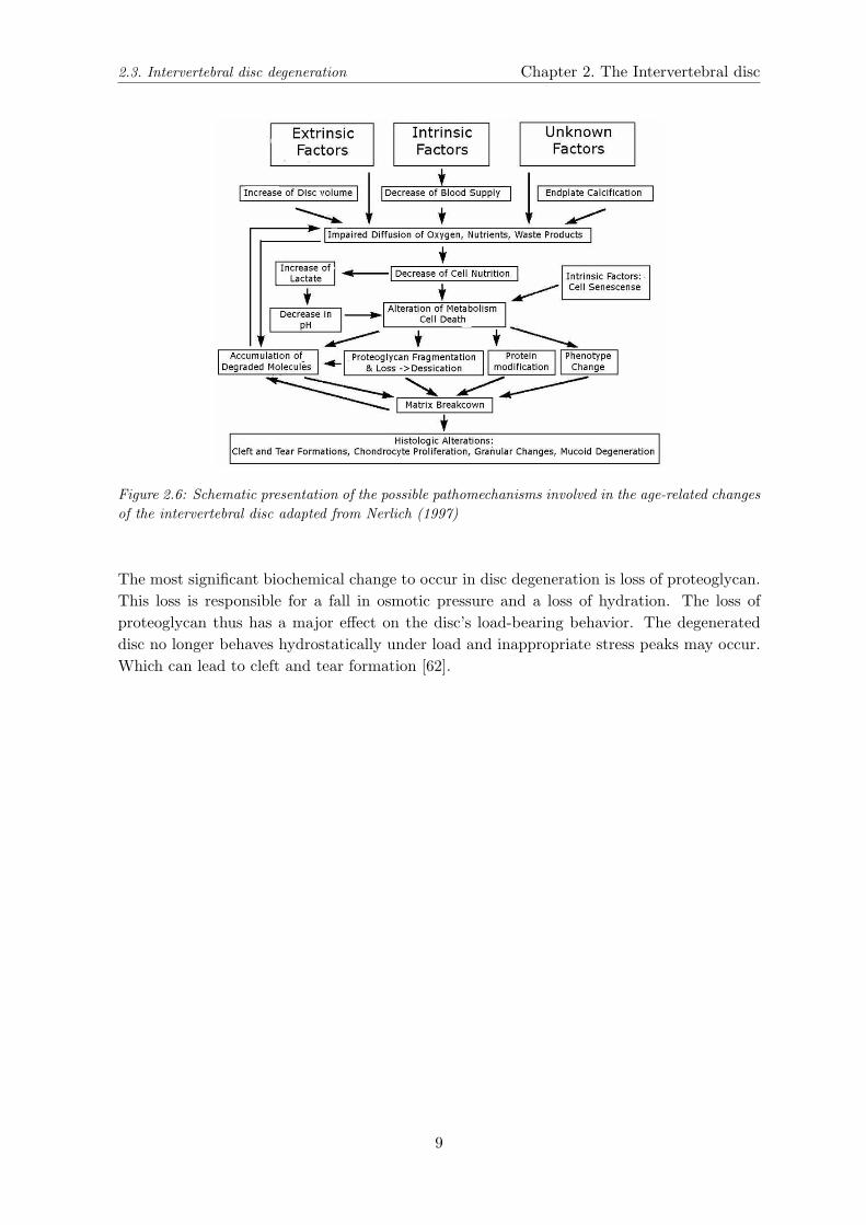
Figure 2.6: Schematic presentation of the possible pathomechanisms involved in the age-related changesof the intervertebral disc adapted from Nerlich (1997)
The most significant biochemical change to occur in disc degeneration is loss of proteoglycan.This loss is responsible for a fall in osmotic pressure and a loss of hydration. The loss ofproteoglycan thus has a major effect on the disc’s load-bearing behavior. The degenerateddisc no longer behaves hydrostatically under load and inappropriate stress peaks may occur.Which can lead to cleft and tear formation [62].
9

Chapter 2. The Intervertebral disc 2.3. Intervertebral disc degeneration
10

Chapter 3
Biomechanics of the intervertebral
disc
The disc task in the spine is to maintain flexibility and motion. Along with the facet joints,it is responsible for carrying all the compressive loading to which the trunk is subjected. Thisincludes different types of load and stresses, like dynamic loads, static loads, tensile stresses,torsional loads, shear stresses and a combination of tensile, compressive and shear stresses.The nucleus carries the compressive loads and the annulus the tensile stresses. This changeswith degeneration (Figure 3.1) when the hydration of the disc is less and the tensile stressesin the collagen fibers of the inner annulus become compressive stresses.
Figure 3.1: Disc under pressure adapted from Shirazi-Adl et al. [47]
(a) Normal disc under pressure (b) Degenerated disc under pressure
The stress distributions inside healthy and degenerated discs are investigated [1, 35, 59, 60]
11

Chapter 3. Biomechanics of the intervertebral disc 3.1. Extracellular matrix level
and have been shown to change with disc degeneration. Because the functional properties areeffected it significantly affects load-bearing and kinematic patterns on the lumbar spine [39].The range of pressures in the disc during daily activities is investigated and shown in differentresearches [37,59,60]. These researches show that abnormal mechanical loads may provide apathway to disc degeneration, and give an idea of the hight of these loads. Simple mechanicalstimulations cannot cause disc degeneration, for this a complex mechanical stimulation com-bining forward and lateral bending of the spine followed by violent compression is needed [44].Mechanical factors not only have a negative effect on the disc, evidence suggests that substan-tial biologic remodeling occurs in the intervertebral disc in response to mechanical stimuli.To know what the cells experience and what are the mechanisms that govern their responsesmore information is needed [6]. What do we know about the mechanics of the extracellularand pericellular matrix and what forces do cells experience?
3.1 Extracellular matrix level
The mechanical properties of the disc matrix are very complex and vary with region andstate of degeneration. The functional properties of the disc largely depend on the structureof its extracellular matrix and in particular on the organization and molecular interaction ofcollagen, proteoglycan, other non-collagenous proteins and water. The collagen fibers givethe tissue its tensile strength while proteoglycans give the tissue stiffness and resistance tocompression because they hold the fluid inside the disc (Figure 3.1). Because of the fluidin the matrix of the nucleus pulposus the disc is incompressible, and compressive forces areconverted to circumferential forces to the annulus.Global material properties have been tested with confined compression and under chemicalloading [17]. This showed that because of the different angles the lamellae give a cross-wovenand reinforced appearance to the IVD and produce anisotropic material behavior.The high osmotic pressure exerted by the proteoglycans of cartilaginous tissues accounts forthe ability of the latter to withstand mechanical pressure of several atmospheres without sig-nificant loss of hydration [51]. But the disc stays an osmotic system and application of loaddisturbs the osmotic balance. When applying a load on the disc for a longer time period,fluid is expressed to restore the equilibrium. This increases the concentration of proteoglycan(Figure 3.2).Collagen fibers within the annulus provide reinforcement during compression, bending andtorsion of the disc. They also provide tensile reinforcement and prevent tears spreading acrossthe ligaments [26]. The equilibrium moduli that are along the fiber direction are shown tobe one to two orders of magnitude higher as compared to the transverse or radial direc-tion [18,48].Bruehlmann et al. [12, 13] studies in situ matrix mechanics by looking at cellular displace-ments. The found strains were highly dependent on the structure and behavior of the extra-cellular matrix and did not correspond with the applied strains.Different experiments with compressive loading, hydrostatic loading, osmotic loading and ten-sile loading were done looking at the changes in cell expression and cell death [6].Besides the proteogylcans and collagen fibers Yu et al. [63] suggests that elastic fibres con-tribute to the mechanical functioning of the intervertebral disc, because the varying organi-
12

3.2. Cellular level Chapter 3. Biomechanics of the intervertebral disc
zation of the elastic fibres in the different regions of the disc is likely to relate to the differentregional loading patterns. So the mechanical properties of the disc in these different regionsmay also differ.
Figure 3.2: The effect of load on the chondrocyte’s environment. Load deforms the matrix and chon-drocyte(1), hydrostatic pressure rises (2), fluid is expressed increasing the extracellular concentrationof proteoglycans and hence of cations (3), adapted from Urban [54]
3.2 Cellular level
The biomechanical responses of the disc depend on its macromolecular composition whichis ultimately regulated by the activity of the discs cells [56]. The mechanical environmentof a cell in the IVD is expected to be highly heterogeneous due to the spatial variationsin cell morphology and phenotype and the significant differences in the material propertiesof the extracellular matrices among regions. The micromechanical environment of the cellvaries dramatically between the anatomic zones of the IVD and Baer et al. suggests thatcell geometry in situ is an adaptation to reduce cell strains during tissue loading [7] IVDcells experience a complex array of physical stimuli, including compressive, tensile and shearstresses and strains, fluid flows, hydrostatic and osmotic pressures, and electrokinetic effects.The cells response depends on their anatomic region of cell origin [32, 36, 55]. Cells in thenucleus pulposus are stiffer the the ones in the annulus fibrosus. Large hydrostatic pressures,but little volume change, are predicted to occur for cells of the nucleus pulposus in responseto compressive loading, and both compressive and tensile strains may be quite small. Incontrast, cells within the lamella or the anulus fibrosus are well adapted to experience mini-mal tensile strains and volumetric changes in response to tensile loading, but may experience
13

Chapter 3. Biomechanics of the intervertebral disc 3.3. Pericellular matrix level
compressive strains in the transverse directions that are substantially higher than the sur-roundings [13,19].Most of the material tests on cellular level were done with isolated cells [3]. Although thisgives us an idea of the material properties of the cells, it does not gives us information aboutthe cell-matrix interaction. Bruehlmann et al. [13] studied in situ cell mechanics with confo-cal microscopy to measure intercellular and collagen matrix mechanics in the inner and outeranulus fibrosus under flexion. She saw that both the extracellular matrix deflection and theintercellular displacements within the lamellar layers were nonuniform. It has been shownthat hydrostatic pressure affects the synthesis of collagen and proteoglycan [27, 30]. Cells inthe nucleus pulposus are stiffer the the ones in the annulus fibrosus.
3.3 Pericellular matrix level
The pericellular matrix is believed to form a bridge between the extracellular matrix forcesand the forces the cell is subjected to. We would expect the material parameters of thispericellular matrix to be different then the material parameters of the extracellular matrix.In articular cartilage the pericellular microenvironment was examined to have a stiffness thatwas considerably greater than that of the cell (PCM 65 kPa, cells 55 kPa) [3–5, 31]. Theseexperiments were done on isolated chondrons. Hing et al. [21] suggests with his experimentsthat the microenvironment functions in situ to mediate the chondrocyte response to physic-ochemical changes associated with joint loading.Alexopoulos et al. shows that the mechanical properties of the PCM, relative to those ofthe chondrocyte and ECM, can have a dramatic influence on the local stress-strain and fluidflow environment of the condrocyte in situ [3]. In the intervertebral disc, the PCM materialproperties are not tested yet. Different models have been made to predict effects in the mi-cromechanical environment of disc cells [8] which show that there may be an important role ofthe cellular mechanical environment in the inhomogeneous response to mechanical loading.
14
Chapter 4
Conclusion
The mechanics of load transfer in the spine and in the intervertebral disc in particular is animportant factor in understanding the patterns and mechanics of disc pain. Knowledge of themechanical factors that play part around the cell starts with knowledge and understandingof the regional, environmental and temporal variations in mechanics of the extracellular andpericellular matrix. Bruehlmann et al. did in vitro studies on tissue, all the other researcheson cell, peri- and extracellular interactions were on isolated cells or chondrons, or were pre-dictions based on computational studies.For this study we have the hypothesis that a pericellular microenvironment exists aroundthe cells of the intervertebral disc anulus fibrosus, as has been showed in several immuno-histochemistry studies. And that this pericellular microenvironment has different materialparameters comparing with the extracellular matrix, because it functions as a protective re-gion for the cells.The aim of this study will be the design of an experiment to measure the material proper-ties of intervertebral disc tissue in vitro in both the extracellular matrix and the pericellularmicroenvironment of the cells.
15

16

Bibliography
[1] A.M. Adams, D.S. McNally, and P. Dolan. ’stress’ distributions inside intervertebraldiscs, the effects of age and degeneration. Journal of Bone Joint Surgery, 78:B:965–972,1996.
[2] T. Aigner, L. Hambach, S. Sder, U. Schltzer-Schrehardt, and E. Pschl. The c5 domainof col6a3 is cleaved off from the col6 fibrils immediately after secretion. Biochemical andBiophysical Research Communications, 290:743–748, 2002.
[3] Leonidas G. Alexopoulos, Mansoor A. Haider, Thomas P. Vail, and Farshid Guilak.Alterations in the mechanical properties of hte human chondrocyte pericellular matrixwith osteoarthritis. Jounal of Biomechanical Engineering, 125:323–333, 2003.
[4] Leonidas G. Alexopoulos, Gregory M. Williams, Maureen L. Upton, Lori A. Setton,and Farshid Guilak. Osteoarthritic changes in the biphasic mechanical properties of thechondrocyte pericellular matrix in articular cartilage. Jounal of Biomechanics, 38:509–517, 2005.
[5] L.G. Alexopoulos, G.M. Williams, M.L. Upton, L.A. Setton, and F. Guilak. The biome-chanical role of the chondrocyte pericellular matrix. Poster.
[6] Lori A. Setton an Jun Chen. Cell mechanics and mechanobiology in the intervertebraldisc. Spine, 29(23):2710–2723, 2004.
[7] Anthony E. Baer, Tod A. Laursen, Farshid Guilak, and Lori A. Setton. The microme-chanical environment of intervertebral disc cells determinde by a finite deformation,anisotropic, and biphasic finite element model. Journal of Biomechanical Engineering,125:1–11, 2003.
[8] Anthony E. Baer and Lori A. Setton. The micromechanical environment of intervertebraldisc cells: Effect of matrix anisotropy and cell geometry predicted by a linear model.Journal of Biomechanical Engineering, 122:245–251, 2000.
[9] Michael T. Bayliss and Brian Johnstone. The lumbar spine and back pain, chapter 7.Biochemistry of the intervertebral disc, pages 111–127. Churchill Livingstone, fourthedition edition, 1992.
[10] U. Berlemann, N.C. Gries, and R.J. Moore. The relationship between height, shape andhistological changes in early degeneration of the lower lumbar discs. European SpineJournal, 7:212–217, 1998.
17

BIBLIOGRAPHY
[11] Susan R.S. Bibby, Deboray A. Jones, Robert B. Lee, Jing Yu, and Jill P.G. Urban. Thepathophysiology of the intervertebral disc. Joint Bone Spine, 68:537–542, 2001.
[12] Sabina B. Bruehlmann, Paul A. Hulme, and Neil A. Duncan. In situ intercellular me-chanics of the bovine outer annulus fibrosus subjected to biaxial strains. Journal ofBiomechanics, 37:223–231, 2004.
[13] Sabina B. Bruehlmann, John R. Matyas, and Neil A. Duncan. Collagen fibril slidinggoverns cell mechanics in th anulus fibrosus, an in situ confocal microscopy study ofbovine discs. Spine, 29(23):2612–2620, 2004.
[14] Sabina B. Bruehlmann, Jerome B. Rattner, John R. Matyas, and Neil A. Duncan. Re-gional variations in the cellular matrix of the annulus fibrosus of the intervertebral disc.Journal of Anatomy, 201:159–171, 2002.
[15] Elizabeth M. Culav, Heather C. Clark, and Mervyn J Merrilees. Connective tissues:matrix composition and its relevance to physical therapy. Physical Therapy, 79(3):308–319, 1999.
[16] A.F. De Palma and R.H. Rothman. The Intervertebral disc. W.B.Saunders Company,Philadelphia, London, Toronto, 1970.
[17] M.R. Drost, P. Willems, H. Snijder, J.M. Huyghe, J.D. Janssen, and A. Huson. Confinedcompression of canine annulus fibrosus under chemical and mechanical loading. Journalof Biomechanical Engineering, 117:390–396, 1995.
[18] D.M. Elliott and L.A. Setton. Anisotropic and inhomogeneous tensile behavior of thehuman anulus fibrosus: experimantal data and material model predictions. Journal ofBiomechanical Engineering, 123:256–263, 2001.
[19] F. Guilak, H.P. ting Beall, and A.E. Baer. Viscoelastic properties of intervertebral disccells. identification of two biomechanically distinct cell populations. Spine, 24:2475–2483,1999.
[20] Helena Hessle and Eva Engvall. Type vi collagen: studies on its localization, structure,and biosynthetic form with monoclonal antibodies. The Jounal of Biological Chemistry,259:3955–3961, 1984.
[21] W.A. Hing, A.F. Sherwin, J.M. Ross, and C.A. Poole. The influence of the pericellularmicroenvironment on the chondrocyte response to osmotic challenge. Osteoarthritis andCartilage, 10:297–307, 2002.
[22] Osamu Horikawa, Hideto Nakajima, Toshiyuki Kikuchi, Shoichi Ichimura, and HarumotoYamada. Distribution of type vi collagen in chondrocyte microenvironment: study ofchondrons isolated from human normal and degenerative articular cartilage and culturedchondrocytes. Journal of Orthopaedic Science, 9:29–36, 2004.
[23] Heather A. Horner, Sally Roberts, Robert C. Bielby, Janis Menage, Helen Evans, andJill P.G. Urban. Cells from different regions of the intervertebral disc, effect of culturesystem on matrix expression and cell phenotype. Spine, 10:1018–1028, 2002.
18

BIBLIOGRAPHY
[24] Gerard B. Houben, Maarten R. Drost, Jacques M. Huyghe, Jan D. Janssen, and AnthonyHuson. Nonhomogeneous permeability of canine anulus fibrosus. Spine, 22:7–16, 1997.
[25] An S. Howard, Paul A. Anderson, Victor M. Haughton, James C. Iatridis, James D.Kang, Jeffrey C. Lotz, Raghu N. Natarajan, Theodor R. Oegema, Peter Roughley, Lori A.Setton, Jill P. Urban, Tapio Videman, Gunnar B.J. Andersson, and James N. Weinstein.Disc degeneration: Summary. Spine, 29(23):2677–2678, 2004.
[26] David W.L. Hukins and Judith R. Meakin. Relationship between structure and mechan-ical function of the tissues of the intervertebral joint. American Zoologist, feb, 2000.
[27] William C. Hutton, William A. Elmer, Lisa M. Bryce, Ewa E. Kozlowska, Scott D.Boden, and Miroslav Kozlowski. Do the intervertebral disc cells respond to differentlevels of hydrostatic pressure. Clinical Biomechanics, 16:728–734, 2001.
[28] J.M. Huyghe, G.B. Houben, M.R. Drost, and C.C. van Donkelaar. An ionised/non-ionised dual porosity model of intervertebral disc tissue: experimental quantification ofparameters. Biomechanical Models in Mechanobiology, 2:3–19, 2003.
[29] Spinal injuries Scotland. www.sisonline.org/ pages/spine.html.
[30] Hirokazu Ishihara, Donal S. McNally, Jill P.G. Urban, and Andrew C. Hall. Effects ofhydrostatic pressure on matrix synthesis in different regions of the intervertebral disk.Journal of Applied Physiologics, 80(3):839–846, 1996.
[31] M.M. Knight, J.M. Ross, A.F. Sherwin, D.A. Lee, D.L. Bader, and C.A. Poole. Chondro-cyte deformation within mechanically and enzymatically extracted chondrons compressedin agarose. Biochimica et Biophysica Acta, 1526:141–146, 2001.
[32] J.C. Lotz, A.H. Hsieh, A.L. Walsh, E.I. Palmer, and J.R. Chin. Mechanobiology of theintervertebral disc. Biochemical Society Transactions, 30(6):853–858, 2002.
[33] Alice Maroudas. Physicochemical properties of cartilage in the light of ion exchangetheory. Biophysical Journal, 8:575–595, 1968.
[34] Michael D. Martin, Christopher M. Boxell, and David G. Malone. Pathophysiology oflumbar disc degeneration: a review of the literature. Neurosurgery Focus, 13(2):1–6,2002.
[35] D.S. McNally, I.M. Shackleford, A.E. Goedship, and R.C. Mulholland. In vivo stressmeasurement can predict pain on discography. Spine, 21(22):2580–2587, 1996.
[36] Hiroshi Miyamoto, Minoru Doita, Kotaro Nishida, Tetsuji Yamamoto, Masatoshi Sumi,and Masahior Kurosaka. Effects of cyclic mechanical stress on the production of inflam-matory agents by nucleus pulposus and anulus fibrosus derived cells in vitro. Spine,31(1):4–9, 2006.
[37] Alf Nachemson. The lumbar spine and back pain, chapter 9. Lumbar mechanics as re-vealed by lumbar intradiscal pressure measurements, pages 157–170. Churchill Living-stone, fourth edition edition, 1992.
19

BIBLIOGRAPHY
[38] Andreas G. Nerlich, Erwin Schleicher, and Norbert Boos. Immunohistologic markersfor age-related changes of human lumbar intervertebral discs. Spine, 22(24):2781–2795,1997.
[39] Christina A. Niosi and Thomas R. Oxland. Degenerative mechanics of the lumbar spine.The Spine Journal, 4:202S–208S, 2004.
[40] C. Anthony Poole. Articular cartilage chondrons: form, function and failure. Journal ofAnatomy, 191:1–13, 1997.
[41] C. Anthony Poole, Shirley Ayad, and Raymond T. Gilbert. Chondrons from articularcartilage. v. immunohistochemical evaluation of type vi collagen organisation in isolatedchondrons by light, confocal and electron microscopy. Journal of Cell Science, 103:1101–1110, 1992.
[42] C.A. Poole, M.H. Flint, and B.M. Beaumont. Chondrons extracted from canine tibialcartilage: Priliminary report on their isolation and structure. Journal of OrthopedicResearch, 6:408–419, 1988.
[43] Andreas Prescher. Anatomy and pathology of the aging spine. European Journal ofRadiology, 27:181–195, 1998.
[44] Francois Rannou, Maite Corvol, Michel Revel, and Serge Poiraudeau. Disk degenerationand disk herniation: the contribution of mechnical stress. Spine, 16(9):1030–1038, 1991.
[45] S. Roberts, J. Menage, V. Duance, S. Wotton, and S. Ayad. Collagen types aroundthe cells of the intervertebral disc and cartilage end plate: an immunolocalization study.Spine, 16(9):1030–1038, 1991.
[46] Peter J. Roughley. Biology of intervertebral disc aging and degeneration: Involvementof the extracellular matrix. Spine, 29(23):2691–2699, 2004.
[47] A. Shirazi-Adl, S.C. Shrivastava, and Ahmed A.M. Stress analysis of the lumbar disc-body unit in compression: a three-dimensional nonlinear finite element study. Spine,9:120–134, 1984.
[48] D.L. Skaggs, M. Weidenbaum, and J.C. Iatridis. Regional variation in tensile propertiesand biochemical composition of the human lumbar anulus fibrosus. Spine, 19:1310–1319,1994.
[49] S. Soder, L. Hambach, R. Lissner, T. Kirchner, and T. Aigner. Ultrastructural local-ization of type vi collagen in normal adult and osteoarthritic human articular cartilage.Osteoarthritis and Cartilage, 10:464–470, 2002.
[50] Mechthild Stoeckelhuber, Stella Brueckner, Gunnar Spohr, and Ulrich Welsch. Proteo-glycans and collagen in the intervertebral disc of the rhesus monkey (macaca mulatta).Annals of Anatomy, 187:35–42, 2005.
[51] J. Urban, A. Maroudas, M.T. Bayliss, and J. Dillon. Swelling pressures of proteoglycansat the concentrations found in cartilaginous tissues. Biorheology, 16:447–464, 1979.
20

BIBLIOGRAPHY
[52] Jill P.G. Urban and Sally Roberts. Degeneration of the intervertebral disc. ArthritisResearch & Therapy, 5(63):120–130, 2003.
[53] Jill P.G. Urban, Stanton Smith, and Jeremy Fairbank. Nutrition of the intervertebraldisc. Spine, 29(23):2700–2709, 2004.
[54] J.P.G. Urban. The chondrocyte: A cell under pressure. British Journal of Rheumatology,33:901–908, 1994.
[55] J.P.G. Urban. The role of the phisicochemical environment in determining disc cellbehaviour. Biochemical Society Transactions, 30(6):858–864, 2002.
[56] J.P.G. Urban, S. Roberts, and J.R. Ralphs. The nucleus of the intervertebral disc fromdevelopment to degeneration. Integrative & Comparative Biology, 40(1):53–61, 2000.
[57] Ellen Wachtel, Alice Maroudas, and Rosa Schneiderman. Age-related changes in collagenpacking of human articular cartilage. Biochimica et Biophysica Acta, 1243:239–243, 1995.
[58] Matthew H. Walker and D. Greg Anderson. Molecular basis of intervertebral disc de-generation. The Spine Journal, 4:158S–166S, 2004.
[59] Hans-Joachim Wilke, Peter Neef, Marco Caimi, Thomas Hoogland, and Lutz E. Claes.New in vivo measurements of pressures in the intervertebral disc in daily life. Spine,24(8):755–762, 1999.
[60] Hans-Joachim Wilke, Peter Neef, Barbara Hinz, Helmut Seidel, and Lutz Claes. Intradis-cal pressure together with anthropometric data - a data set for the validation of models.Clinical Biomechanics, 16(1):S111–S126, 2001.
[61] Lippincott Williams & Wilkins. http://connection.lww.com.
[62] S. Wognum, J.M.R.J. Huyghe, and F.P.T. Baaijens. Influence of osmotic pressure changeson the opening of existing cracks in two intervertebral disc models. Spine, ARTICLE INPRESS, 2006.
[63] Jing Yu, C. Peter, Sally Roberts, and Jill P.G. Urban. Elastic fibre organization in theintervertebral discs of the bovine tail. Journal of Anatomy, 201:465–475, 2002.
21