biomaterials - mpikg.mpg.de · research in the department of biomaterials is highly...
TRANSCRIPT

BIOMATERIALS

Research in the Department of Biomaterials is highlyinterdisciplinary with a focus on biological and
bio-inspired materials. Biological materials con-stitute most of the body of plants and animalsaround us. They allow cells to function, eyes tocapture and interpret light, plants to stand upto the light and animals to move or fly. Biologi-cal structures have always been a source of
inspiration for solving technical challenges inarchitecture, mechanical engineering, or materi-
als science. Nature has developed – with compar-atively few base substances, mainly polymers and
minerals – a range of materials with remarkable function-al properties [1]. The key is a complex, often hierarchicalstructuring of the natural materials [2].
It is not evident that the lessons learned from biologicalmaterials will be applicable immediately to the design ofnew engineering materials. Indeed, bio-inspiration is notmerely the consequence of an observation of naturally occur-ring structures. These structures are probably good solutionsfound by a long adaptation process during evolution, butNature has to take into account a multitude of boundary con-ditions (mechanical, biological, related to nutrient supply,etc.) which we hardly know and which might not be importantin an engineering context. As a consequence, we have tostudy carefully the biological system and to understand thestructure-function relation of the biological material togetherwith its physical and biological constraints, before it mayserve as a model for the design of new materials [1].
Fig. 1: Research groups in the Department of Biomaterials with respective group leaders
With this paradigm in mind, wehave defined the research pro-gramme of the Department of Biomate-rials as a combination of research on natu-ral tissues and on bio-inspired materials.Accordingly, the Department is organised in severalresearch groups, as shown in Fig. 1, and who presenttheir own reports within this volume. In addition, severalindependent researchers on the postdoctoral level are work-ing on related topics and their reports are summarized jointlyin one of the sections (Biological and Bio-inspired Materials).
First, a large effort is devoted to improve our understand-ing of some biological tissues from a materials science pointof view. This requires that we study structure-mechanicalfunction relations, considering the natural environment inwhich these materials live and grow. One of these tissues isthe plant cell wall, a composite of (semi-crystalline) cellu-lose fibrils in an amorphous polymer matrix. This cell wallmaterial has remarkable mechanical properties which may betuned by the cell over a wide range of stiffness, according toneeds, and which is even capable of generating stresses toprovide motility. This is described in the reports by Ingo Burg-ert and by Rivka Elbaum (independent Humboldt PostdoctoralFellow).
Mineralized tissues are a second example of mechan-ically outstanding biological materials. Currently, our empha-sis is on elucidating the origin of the fracture resistance ofbone, not least because of the biomedical importance of thistissue. Most recently, a hierarchical deformation pattern wasdiscovered as a major reason for the mechanical quality ofbone tissue (see report by Himadri S. Gupta). These studiesare now being extended to deer antler, which is a rapidlygrowing bone tissue with even higher toughness than bone.The structural origin of the mechanical performance of teethand the biomineralization of tooth enamel are beingaddressed by the postdoctoral researchers Paul Zaslanskyand Barbara Aichmayer. Their reports are included in the sec-tion “Biological and Bio-inspired Materials”. Finally, collabo-rative research on the hierarchical structure and the mechan-ical properties of glass sponge skeletons is also reported inthis same section.
It is well-known that biological materials constantlyadapt to changing mechanical needs. This is achieved by astrain-sensing mechanism, which in most biological systemsis not fully elucidated. In the case of bone, for instance, spe-cialized cells are thought to act as strain sensors and to be atthe centre of a feed-back loop, called bone remodelling cycle,where damaged bone is removed and replaced by new
material. This process is crucial for the tissue'scapability of mechanical adaptation and self-
32
Research in the Department of Biomaterials
Plant Biomechanics(Ingo Burgert)
Mechanobiology(Richard Weinkamer)
Mineralized Tissues(Himadri S. Gupta)
Bone Regeneration(Manjubala Inderchand)
Bone Material Quality(Peter Fratzl)
Mesoscale Materials(Oskar Paris)
Synchrotron Diffraction Beamline(Oskar Paris)
Biological and Bio-inspired Materials(Independent Researchers)

repair. Thesequestions are addressed mostlyby theoretical means in the research group on mechanobi-ology (see report by Richard Weinkamer). Moreover, themechanics of micro-containers and membranes is investigat-ed together with the Interface Department.
In parallel to the study of biological materials, weaddress topics (right column in Fig. 1) which use the know-ledge on biological materials for research either with impli-cations in materials or in biomedical sciences. A major topicis related to bone material quality in osteoporosis and itschanges with treatment of the disease. This is a long-termcollaboration with the Ludwig Boltzmann Institute of Osteo-logy in Vienna, Austria. Recent results obtained in the lasttwo years relate, for example, to bisphosphonate treatmentof osteoporosis and of brittle bone disease (osteogenesisimperfecta), see the section on Bone Material Quality.
A further topic with biomedical implications is boneregeneration. Bone is among the few tissues in our bodywhich are able to heal and to regenerate completely withoutleaving a scar. In collaboration with the Charité Medical Uni-versity and other partners in Berlin and Brandenburg, we arenow trying to elucidate the healing process in bone, as wellas the physical and biochemical factors which govern it. Anew Collaborative Research Center (SFB760) supported bythe German Science Foundation, and in which the Depart-ment of Biomaterials is heavily involved, is starting in thebeginning of 2007. The Department is also member of theBerlin Brandenburg Center for Regenerative Therapies (BCRT)supported by the Ministry of Science (BMBF). Currentresearch is centred on the analysis of the various tissuesoccurring during bone healing, as well as cell and tissuegrowth on porous scaffolds, which might serve as implants;see the report by Manjubala Inderchand.
Biomimetic materials are currently developed in oneof the research groups, based on thermal and chemical pro-cessing of plants. In particular, the processing is studied indetail by in-situ synchrotron diffraction. Further research inthis group concerns the behaviour of fluids in mesoporousmaterials, which are studied in collaboration with partners inBerlin within the framework of the Collaborative ResearchCenter SFB448 (see report by Oskar Paris). Work on bio-
inspired activehybrid materials based
on gels and microstructured sili-con is also conducted in the Department in collab-
oration with Bell Labs, USA (see section on Biological andBio-inspired Materials).
Finally, a large effort is devoted to establish new meth-ods for analysis of biomaterials. Indeed, studying hierarchi-cal biomaterials requires state-of-the-art experimentalequipment and there is also some need for the developmentof new approaches. One strategy is to set up a suite of scan-ning imaging methods which may be applied to the samespecimen and which give different type of information aboutthe material with a position resolution in the micron range.We are currently using scanning electron microscopy andscanning x-ray diffraction to characterize the micro- andnanostructure. Moreover, we have established Raman imag-ing to provide information on chemical composition andnano-indentation as well as acoustic microscopy to estimatelocal mechanical properties. The strength of this multi-method approach is that the different parameters measuredon the same specimen can be correlated at the local level.This helps finding structure-property relations even inextremely heterogeneous materials. In-situ techniquesare a second type of approach, where we studychanges in a material (e.g. due to mechanical stressor to chemical or thermal processing) by time-resolved scattering or spectroscopy during theprocess itself. In some cases, we can perform suchstudies in the laboratory (e.g. with Raman orinfrared spectroscopy or in the environmentalscanning electron microscope), but in many caseswe need synchrotron radiation (e. g. for x-ray dif-fraction or small-angle scattering). A large project inthis context is the setting up of a dedicated scanningsmall- and wide-angle scattering beamline at the syn-chrotron BESSY in Berlin. The end station for this beam-line has been developed under the supervision of Oskar Paris(see his report) and is now performing the first user experi-ments.
Peter FratzlDirector of the Department of Biomaterials
33
[1] Peter Fratzl, Perspective: Biomimeticmaterials research – What can we really learn from Nature’s structuralmaterials? Journal of the Royal SocietyInterface 2007 (published online).
[2] P. Fratzl and R. Weinkamer, Nature'shierarchical materials, Prog. Mater. Sci.2007 (in press).

Plant biomechanics provides a powerful toolto gather insights into the relationship ofplant form and function as an expression ofplant strategy to survive under given envi-ronmental conditions and physical con-
straints. It is also a valuable source forextracting biomimetic principles for the design
of new bio-inspired materials (Fig.1).Plants are hierarchically organized which means that
their macroscopic properties mainly originate from the nano-and microscale. Nanometer thick semi-crystalline cellulosefibrils embedded in amorphous matrix polymers are the basicassembly of plant cell wall structure. But at these levels ofhierarchy, plants manifest a wide variety of adaptable para-meters such as cell shape, thickness and arrangement of cellwall layers, the orientation of cellulose microfibrils withincell walls, and in the chemical composition of individual cellwall layers. Micromechanical approaches are well suited tocharacterize these composite structure for understandingboth the material design and the optimization strategies ofliving plants [1].
Fig. 1: Schematic of the research interests of the Plant Systems Biomechanics group.
Cell Wall Structure and Function The mechanical performance of plant cell walls is based onthe mechanical properties of the individual components andtheir interaction according to the polymer assembly. Conse-quently, the mechanical relevance of a cell wall componentdepends decisively on its distribution, spatial orientation,and bonding characteristics.
In conjunction with the MPI for Molecular Plant Physiolo-gy (Lab. M. Pauly) we draw synergisms from the unique com-bination of plant physiology/enzymology/genetic engineeringon one hand and micromechanical/ultrastructural characteri-zation on the other hand. The deformation behavior of pri-mary walls was studied by using Arabidopsis hypocotyls andwas indicative of the crucial role of the cellulose-hemicellu-lose (xyloglucan) network for stiffness and strength. Cyclicloading experiments on various mutants suggest that thedegree of plastic deformation occurring during the first cycledepends on the straightening of the xyloglucan chain.
Cellulose microfibril orientation and matrix interactionsin primary cell walls are also of high significance during thecourse of cell elongation [2]. Focusing on the mechanics ofcell wall expansion from a plant biomechanics perspective it
seems evident that the geometrical constraints imposed by(plastically) inextensible cellulose fibrils have a profoundeffect on the cell growth behavior. This is less evident atsmall extensions (up to ~30%) where the fibrils are not yetexpected to tilt appreciably into the cell direction (Fig. 2).
Most probably, the plastic flow of non-cellulosic matrixdominates the deformation at this stage. For much largerextensions (beyond 100%), the tilting of the cellulose maybecome important, leading to a stiffening of the cell in its lon-gitudinal direction and compression in its lateral direction.Hence during cell elongation the matrix between cellulose fib-rils needs to act in two ways both as a tether during longitudi-nal extension and later as a spacer when compressed laterally.
Fig. 2: Change of cellulose orientation as a function of cell elongation ina theoretical model with inextensible cellulose fibrils. The angle of cel-lulose fibrils with the direction perpendicular to the cell axis is called a(with the value a0 before elongation). The length of the cell changesfrom L0 to L, and its diameter from D0 to D. The numbers in the figureindicate the cellulose angle a0 before cell elongation. Both axes aredrawn with logarithmic scales [2].
Deformation mechanisms in secondary cell walls were exam-ined with various in-situ techniques which simultaneouslycombine mircomechanical tests with (nano-) structural analy-sis [3], [4]. Tensile tests on mechanically isolated cells [5]combined with simultaneous acquisition of Raman spectrahave shown that the (stiff) cellulose fibrils carry most of theload with only small and fully elastic deformation [3]. Almostall of the deformation takes place by shearing of the(deformable) hemicellulose/lignin matrix. This combinationconfers both stiffness and toughness to the cell wall.
Enzymatic treatments were utilized to suppress the func-tioning of individual polymers in the mature cell wall of secondary xylem cells. Micromechanical tests on the modi-fied material revealed the mechanical relevance of hemicel-luloses (xylan) in the composite structure.
34
Ingo Burgert 18.09.19681995: Diploma, Wood Science and Technology (University of Hamburg)Thesis: The Fractometer – its potentiali-ties and limits in measuring mechanicalproperties of living trees2000: PhD, Wood Science(University of Hamburg) Thesis: The mechanical relevance of rays in the living tree2000-2003: Postdoc (Institute of Physics and MaterialsScience, BOKU, Vienna)Since 2003: Group Leader (Max Planck Institute of Colloids and Interfaces, Potsdam)
Plant Systems Biomechanics
BIOLOGICAL MATERIALS

Stress Generation and Plant MovementActive movement is usually associated with animals ratherthan plants. Clearly, plants do not have muscles but they areable to pre-stress their tissues in order to actuate theirorgans. We showed for normal wood and compression woodof spruce (Picea abies) that either tensile or compressivestresses can be obtained during swelling of the cell wall,depending on the ability of the cell to undergo some torsion [6].This was shown in swelling experiments on individual cellsand tissues and can be well understood by simple mechani-cal considerations taking into account the cell shapes and theobserved cellulose fibril orientations (Fig. 3).
Fig. 3: (a) Schematic drawing of a normal wood and compression woodcell with different cellulose microfibril angles (MFA). (b) Deformation ofthe cell wall during swelling with inextensible cellulose fibrils (examplewith microfibril angle = 30°). (1) Cell virtually cut open along a verticalline. (2) Cell wall rolled out indicating the cellulose orientation. (3) Increase of cell wall area (a=10%) due to swelling with inextensiblecellulose fibrils and no torsion of the cell. (4) Same when torsion of thecell is allowed [6].
The almost inextensible cellulose fibrils redirect the forcesgenerated by the swelling of the matrix by purely geometricalconstraints to produce tension or compression forces accord-ing to needs. This principle could be simple enough to bereproduced in artificial systems and one may consider devel-oping fiber-reinforced hydrogels as effective microactuators.
Bio-Inspired MaterialsA) Gradients in PlantsPalm trees have evolved gradual transitions between stiff,sclerenchymatous supporting fibres and soft parenchymatoustissue, functioning as a matrix. This structure can help toavoid critical shear stresses and separation of the material atthe interfaces when plants are mechanically loaded. Here,we investigated in cooperation with the University ofFreiburg (Lab. T. Speck) gradual transitions in arborescentpalms at different hierarchical levels by anatomical, micro-mechanical, physical and biochemical methods taking Wash-ingtonia robusta as a model organism [Fig. 4].
The stress-strain curves show a change in cell wall stiff-ness from the centre to the middle of the fibre cap. The
underlying structural and biochemical features are currentlystudied. Our aim is to transfer the concept of gradual transi-tions into technical application for innovative structurallyoptimised composite materials.
Bio-Inspired MaterialsB) Fibre-Matrix InteractionsThe nanocomposite structure of the plant cell wall with itsspecific interface design between stiff cellulose fibrils andpliant matrix polymers can be taken as a source of inspirationfor a transfer to technical applications. Currently we are run-ning a cooperation project (partners: University of Freiburg(Lab. T. Speck) and ITV Denkendorf) in the framework of theBMBF-Bionik competition on an improvement of technicalcomposites with a new concept for the embedding of glassfibres based on the primary cell wall assembly. With the aidof our mechanical workshop (G. Haseloff) we developedembedding and pull-out test setups to mechanically charac-terize the interface properties between fibre and matrix.
I. Burgert, W. Abasolo, M. Eder, N. Gierlinger*, L. Goswami, K. Jungnikl, A. Martins, M. Rosenthal, M. Rüggeberg, N. Schreiber, R. Seidel, S. Weichold, K. [email protected]
35
References: [1] Burgert, I.: Exploring the micro-mechanical design of plant cell walls.Amer. J. Bot. 93, 1391-1401 (2006).[2] Burgert I, Fratzl, P: Mechanics of the expanding cell wall. In: Plant CellMonogr (5) The Expanding Cell (eds J-PVerbelen, K Vissenberg) Springer-VerlagBerlin Heidelberg, pp.191-215(2006).[3] Gierlinger, N., Schwanninger, M.,Reinecke, A., Burgert, I.: Molecularchanges during tensile deformation ofsingle wood fibers followed by Ramanmicroscopy. Biomacromolecules 7,2077-2081 (2006).[4] Thygesen, L.G., Eder, M., Burgert, I.:Dislocations in single hemp fibres –Investigations into the relationship ofstructural distortions and tensile proper-ties at the cell wall level. Journal ofMaterials Science 42, 558-564 (2007).[5] Burgert, I., Gierlinger, N., Zimmermann, T.: Properties of chemical-ly and mechanically isolated fibres ofspruce (Picea abies [L.] Karst.). Part 1:structural and chemical characteriza-tion. Holzforschung 59, 240-246 (2005).[6] Burgert, I., Eder, M., Gierlinger, N.,Fratzl, P.: Tensile and compressivestresses in tracheids are induced by swelling based on geometrical constraints of the wood cell. Planta, DOI 10.1007/s00425-007-0544-9 (2007).
*The APART fellowship (Austrian program for
advanced research and technology) of the Austrian
Academy of Sciences was awarded to Notburga
Gierlinger, who became an independent researcher
at the Department in the meantime.
Fig. 4: A) Vascular bundle with xylem, phloem and the fibre cap whichcan make up to 90 % of the overall bundle area. Fuchsin/chrysoidin/astrablue staining as a qualitative indication of lignification; B) Stress-strain curves of tissues from the central and the middle part of afibre cap of Washingtonia robusta.

Bone is a hierarchically structured materialwith remarkable mechanical performance.Understanding its properties is essential forthe assessment of diseases such as osteo-porosis, for a critical evaluation of current
therapies and to aid in their more targeteddevelopment. While the full hierarchical struc-
ture of bone is extremely complex and variable,its basic building block, the mineralized collagen fib-
ril, is rather universal. The mechanical performance of bone,often coined “bone quality” [1], does not only depend on theshape and the amount of the bone (as estimated by the bonemineral density, BMD), but also on its architecture and on thequality of the bone material. Current research carried out pri-marily in collaboration with the Ludwig Boltzmann Instituteof Osteology (Vienna, Austria) concentrates on studying thestructural basis of bone material quality and changes due todisease or treatment.
Anisotropy of Fracture Toughness in Human Compact BoneBone material quality depends to a large extent on the orien-tation of collagen fibrils in bone tissue. In collaboration withH. Peterlik (University of Vienna), we studied controlled crackextension in human femur [2]. It was shown that the energydissipated by the crack is two orders of magnitudes larger ifit propagates perpendicularly to the collagen fibrils thanwhen it runs along them. The reason is obvious in Fig.1. Whenthe crack follows the main collagen direction, it runs straightand the dissipated energy is low (Fig. 1B). In contrast, thecrack path is zigzagging when it should run perpendicularly tothe collagen direction (Fig. 1B), thus dissipating much moreenergy.
Fig. 1: Crack propagation in a human femur, perpendicular to the longbone axis (A) and parallel to it (B), from [2].
Mineral Density in Different Bone MatricesSince mineral is the stiffer component in bone, it is not sur-prising that the elastic modulus of the bone matrix dependson the mineral content. Fig. 2 shows that this dependence isnot linear. Even more important, the relation between localelastic modulus (as measured by nanoindentation) and localmineral content (as measured by backscattered electronimaging) depends on the type of organic matrix, for examplewhen one moves from bone into mineralized cartilage in ajoint [3].
Fig. 2: Dependence of indentation modulus on mineral content in mineralized cartilage. The lines correspond to a composite model [3].
Another interesting observation is that bone material qualityis also depending on genetic background. A polymorphismaffecting a Sp1 binding site in a regulatory region of the COLIA1 gene is known to predispose to osteoporotic frac-tures by affecting bone strength through mechanisms thatare partly independent of differences in bone mineral density(BMD). The bone material in patient biopsies was investiga-ted in collaboration with the University of Aberdeen MedicalSchool [4]. Our analysis showed significant reduction inmatrix mineralization in bone biopsies from heterozygotescompared with homozygotes (see Fig. 3).
Fig. 3: histogram of mineral density distribution in trabecular bonematrix of biopsies from patients with a polymorphism in the COLIA1gene [5].
Raman Imaging of BoneA considerable effort was undertaken by Murat Kazanci(postdoc) to establish Raman imaging as a tool for studyingbone material quality. The reason for the interest in this tech-nique is that it allows the imaging of material parameterswith one micron spatial resolution and spectral resolutionmuch better than infrared spectroscopy. The Raman signalfrom compact bone turned out to be extremely sensitive totissue orientation (Fig. 4) and methodology was devised to
36
Peter Fratzl 13.09.19581980: Diploma (Ingénieur Diplômé de l'Ecole Polytechnique, Paris) 1983: PhD, Physics (University of Vienna)Thesis: Investigation of an Al-Zn-Mgalloy using diffuse neutron scattering1981-1985: Research Scientist (Austrian Academy of Sciences, Vienna;Laboratoire Leon Brillouin, Saclay,France); Visiting Research Fellow (Hahn Meitner Institute, Berlin; New York University)1986-1998: Assistant and AssociateProfessor (Institute for Materials Phy-sics of the University of Vienna, Austria)1988 and 1989: Visiting Professor (Rutgers University, New Jersey, USA)1991: Habilitation, Solid State Physics(University of Vienna) Thesis: Precipitation in alloys – small-angle x-ray scattering and computer simulationSince 1993: Research Associate (Ludwig Boltzmann Institute of Osteology, Vienna).1993-1994: Visiting Research Fellow(Heriot-Watt University, Edinburgh)1997: Visiting Professor, (Physics Depart-ment of the University of Munich)1998-2003: Chair of Metal Physics (University Leoben, Austria) Director (Erich Schmid Institute forMaterials Science of the Austrian Academy of Sciences)Since 2003: Director, Department ofBiomaterials (Max Planck Institute of Colloid and Interfaces, Potsdam)Since 2004: Honorary Professor ofPhysics at Humboldt University Berlin
References: [1] D. Dempster, D. Felsenberg, S. vander Geest (Editors): The Bone QualityBook - A Guide to Factors InfluencingBone Strength, Excerpta Medica, Amsterdam (2006); and P. Fratzl: Material properties: mineral cystals.ibidem pp 56-63.
Bone Material Quality and Osteoporosis Research
BIOLOGICAL MATERIALS

determine the amounts of mineral and protein in the matrix,as well as some information on their orientations [5].
Fig. 4: Ratio of the n1 PO4 to the amide I band in two osteons withinhuman cortical bone (collaboration with HD Wagner, Weizmann Insti-tute, Israel). The dark channels in the figures are blood vessels in thecentre of osteons. The image of the bone tissue depends strongly on the polarization of the laser beam (green arrows).
Bone Quality in Osteoporosis TreatmentOsteoporosis is a common disease associated with reducedbone mass and increased bone fragility. Bone is constantlyturned over by specialized bone cells, osteoclasts whichresorb bone and osteoblasts which form new bone. Thereduction in bone mass in osteoporosis is linked to an imbal-ance between these two processes. As a consequence, treat-ment strategies are typically targeting either the osteoclaststo reduce bone resorption or the osteoblasts to increase boneformation. Bisphosphonates are used in this context asantiresorptive drugs. In collaboration with Procter and Gam-ble Pharmaceuticals (Ohio, USA), we studied the effects ofosteoporosis treatment on bone material quality in a clinicaltrial. Biopsies from patients treated with the bisphosphonaterisedronate for three and five years were studied usingbackscattered electron imaging [6] and infrared spectroscopy[7]. The main result was that the mineral content of the bonematrix was increased by the treatment, without any furthervisible modifications at the material level.
The effects of a therapy with parathyroid hormone (PTH),known to induce bone formation, combined with osteoprote-gerin (OPG), known to act on bone resorption, was exploredin an animal study, in collaboration with AMGEN Inc (Thou-sand Oaks, Canada). It was found that PTH was responsiblefor an increase in bone volume, whereas OPG positively influ-enced the homogeneity and density of mineralization withoutaffecting the nanostructure of the bone material [8].
Bisphosphonate Treatment of Brittle Bone Disease Brittle bone disease (osteogenesis imperfecta, OI) is a disor-der which is linked to genetic modifications of the collagengene and which leads to enhanced bone fragility. Childrenwith this disease suffer from multiple fractures and associat-ed complications. It is known that the fracture incidence inpatients can be reduced by a treatment with bisphospho-nates. It is not known, however, in which way this treatmentaffects the bone quality and leads to a reduction of bonefragility. In collaboration with the Hospital for SpecialSurgery (New York, USA), we studied a mouse model of thisdisease treated with the bisphosphonate alendronate (ALN).It was found that ALN augmented the mechanical, geometri-cal, and material properties of cortical and trabecular bone incontrols, while the only observable improvement to the OImouse model was increased bone volume [9], see Fig. 5.
Fig. 5: Backscattered electron images of longitudinal views of femorafrom untreated and ALN-treated bone from the OI mouse model (a, b)and controls (c, d), [10].
This work was continued by studying biopsies from young OIpatients treated with the bisphosphonate pamidronate (PAM),in collaboration with the Shriners Hospital for Children andMcGill University (Montreal, Canada) [10]. While the OI bonetissue was found stiffer and more mineralized than controls,the anti-fracture effectiveness of PAM treatment was primari-ly due to an increase of bone volume (see Fig. 6). This result isvery similar to what was found for the animal model.
Fig. 6: Back scattered electron images of transiliac bone biopsies, from(A) age-matched control, (B) a 6 year-old girl with OI type III caused by a mutation in the COL1A1 gene, and (C) the same patient after 2.8 yearsof PAM treatment [10].
P. Fratzl, M. Kazanci (Postdoc), mainly in collaboration withthe Ludwig Boltzmann Institute of Osteology, Vienna, Austria(P. Roschger, E.P. Paschalis, K. Klaushofer, and others)[email protected]
37
[2] H. Peterlik, P. Roschger, K. Klaushofer,P. Fratzl: From brittle to ductile fracture ofbone. Nature Materials 5, 52-55 (2006).[3] H. S. Gupta, S. Schratter, W. Tesch, P.Roschger, A. Berzlanovich, T. Schoeberl,K. Klaushofer and P. Fratzl: Two differentcorrelations between nanoindentationmodulus and mineral content in thebone-cartilage interface. J. Struct. Biol. 149, 138 - 148 (2005).[4] T.L. Stewart, P. Roschger, B.M. Misof,V. Mann, P. Fratzl, K. Klaushofer, R. Asp-den, S.H. Ralston. Association of COLIA1Sp1 alleles with defective bone noduleformation in vitro and abnormal bonemineralization in vivo. Calcif. Tissue Int. 77, 113-118 (2005).[5] M. Kazanci, P. Roschger, E. P.Paschalis, K. Klaushofer, P. Fratzl: Boneosteonal tissues by Raman spectral map-ping: orientation-composition. J. Struct.Biol. 156, 489-96 (2006). [6] R. Zoehrer, P. Roschger, E.P. Paschalis,J.G. Hofstaetter, E. Durchschlag, P. Fratzl,R. Phipps, K. Klaushofer: Effects of 3-and 5-years treatment with risedronateon bone mineralization density distribu-tion in triple biopsies of the iliac crest inpostmenopausal women. J. Bone Miner.Res. 21, 1106-1112 (2006).[7] E. Durchschlag, E.P. Paschalis, R.Zoehrer; P. Roschger; P. Fratzl; R. Recker;R. Phipps; K. Klaushofer: Bone materialproperties in trabecular bone fromhuman iliac crest biopsies after 3- and 5-year treatment with risedronate. J. BoneMiner. Res. 21, 1581-1590 (2006).[8] A. Valenta, P. Roschger, N. Fratzl-Zelman, P.J. Kostenuik, C.R. Dunstan, P. Fratzl, K. Klaushofer: Combined treat-ment with PTH and OPG increases bonevolume and uniformity of mineralizationin aged ovariectomized rats. Bone 37, 87-95 (2005).[9] B.M. Misof, P. Roschger, T. Baldini,C.L. Raggio, V. Zraick, L. Root, A.L.Boskey, K. Klaushofer, P. Fratzl, N.P.Camacho: Differential effects of alendro-nate treatment on bone from growingosteogenesis imperfecta and wild-typemouse. Bone 36, 150-158 (2005).[10] M. Weber, P. Roschger, N. Fratzl-Zelman, T. Schöberl, F. Rauch, F.H. Glorieux, P. Fratzl, K. Klaushofer:Pamidronate does not adversely affectbone intrinsic material properties in children with osteogenesis imperfecta. Bone 39, 616-622 (2006).

Our research looks at the structural adapta-tion of mineralized tissues to their mechani-cal function at the length scale of a micronand below. At this level, the extracellularconnective matrix in both vertebrate and
invertebrate organisms often consists at themolecular level of a composite where organic
molecules (such as collagen or chitin) are inter-penetrated with inorganic crystallites (typically calci-
um phosphates or carbonates) to form an anisotropic, hardand tough material. Weight for weight, such biomineralizedtissues compare favorably with man-made composites,although requiring much lower temperatures and processingconditions. Therefore, an understanding of the structuraldesign principles in such biomaterials may provide guidelinesin making new strong composite materials. In addition, un-derstanding how perturbations in the mineralized micro-structure affect mechanics (in bone diseases like osteoporo-sis) would be important in developing treatments for suchpathological conditions.
Bone consists of a compact tissue type (cortical bone)and a spongy, porous material (trabecular bone). In both tis-sue types, the basic building block is the bone lamella, typi-cally about 5 µm thick. In cortical bone, lamellae form lami-nated cylindrical composite structures built around bloodvessels, which are denoted as secondary osteons. Whileunderstanding the internal architecture of such osteons iscrucially important for bone biomechanics, a convincing andquantitative structural model has thus far been elusive. Usinga novel combination of high brilliance synchrotron radiationwith a micron-sized beam and local crystallographic texturemeasurements [1], we were able to show that the collagenfibers are arranged in layers of varying helical pitch withrespect to the osteon long axis, in effect forming a righthanded spiral motif (Fig. 1) [2]. Such a spring like structurewould be capable of absorbing elastic energy during physio-logical motion, and may act as a buffer preventing micro-cracks from penetrating to and destroying the sensitive innerblood vessel.
Using a combination of scanning microprobe methods(nanoindentation and backscattered electron microscopy) wewere able to further elucidate the mechanical structure of theosteon. We showed that it consists of layers of alternatinghigh and low stiffness within a single lamella and that thelayers of lower modulus correspond to regions of lower aver-age mineral content [3]. Such a compositionally and mechani-cally modulated structure is an example of a functionallygraded material, and would act as an excellent crack stopper,as has been demonstrated theoretically.
Fig. 1: Right handed spiral twisting of the fiber orientation in compactbone lamellae. The orientation of the fibrils changes with a periodicity of a single lamellar width (~ 5 µm). The last lamella between the osteonand the enclosing interstitial bone has the opposite chirality.
The bone lamellae are comprised of mineralized collagen fib-rils, which are 100 to 200 nm diameter composites of type Icollagen and hydroxyapatite mineral embedded in a smallamount of extrafibrillar matrix. Using in-situ mechanical test-ing with time-resolved synchrotron X-ray diffraction, weshowed that the fibrils in bone take about half the totaldeformation in the tissue under tensile load, and do notstretch further in the inelastic regime. Based on these find-ings, we proposed a fibril level model of interfibrillar shear-ing, where the total strain is divided into a tensile componentcarried by the fibrils and shearing in the thin layers ofextrafibrillar matrix (~ a few nm thick) between them (Fig. 2)[4]. Above the mechanical yield point, a stick-slip mechanismof interfibrillar sliding results [5], which leads to a large workof fracture.
To understand the way strain is transferred down thestructural hierarchy, a novel combination of tensile testing ofsingle fibrolamellar bone packets with wide-angle synchro-tron X-ray diffraction and small angle X-ray scattering wasused. This technique enables us to measure, concurrently,the strain in the tissue, the fibrils and the mineral particles.UV-laser microdissection enabled the isolation of single bonepackets at the tissue level, excluding all structures at higherlength scales in the hierarchy. Strain is passed down in suc-cessively lower fractions from the tissue down to the molec-ular level (Fig. 3) [6], via shearing strains in the interveningextra- and intrafibrillar organic matrix, and depends on thedegree of hydration of the organic matrix. Such an arrange-ment results in a high stiffness of the overall material whileprotecting the brittle hydroxyapatite phase from excessiveload.
38
Himadri Shikhar Gupta 26.06.19731991-1996: M.Sc. in Physics (Indian Institute of Technology, Kanpur, India)1996-2000: PhD, Physics (Rutgers University, New Jersey, USA)Thesis: Phase Segregation and Alloyingin Ni-base Superalloys: Models andExperiments 2000-2003: Postdoctoral Research, (Erich Schmid Institute of MaterialsScience, Austrian Academy of Sciences,Leoben, Austria)Since 2003: Group Leader (Department of Biomaterials, Max Planck Institute of Colloids and Interfaces, Potsdam, Germany)
Mineralized Tissues
BIOLOGICAL MATERIALS

Fig. 2: Shearing model of interfibrillar deformation in bone. White arrowsdenote the direction of relative motion of fibrils under tensile stress. The interfibrillar matrix may be also partially mineralized.
Fig. 3: Hierarchical deformation in bone at three different levels: tissue,fibril and mineral particle. Red hexagons denote extrafibrillar mineralparticles, and dashed lines the direction of (possibly inhomogeneous)shear in the matrix between stiff elements.
Using a thermally activated stress flow analysis originallydeveloped to study plastic deformation in metals, we estab-lished that the fundamental molecular step in plastic defor-mation of bone takes place in a volume of about 1 nm3, andrequires activation energy of about 1 eV. Based on the magni-tude of these quantities, a model for bone fracture was pro-posed, where breakage of ionic bonds (in the extrafibrillarmatrix) between long irregular polyelectrolyte chains anddivalent ions like calcium mediate bone plasticity (Fig. 4) [7].Modifying or altering the properties of this extrafibrillar“glue” could be an effective way to tune the properties ofbone, and is a current focus of our research.
Fig. 4: Ionic bond breaking between divalent ions and polyelectrolytemolecules in the extrafibrillar matrix mediates bone plasticity. Circlesdenote cations (like calcium) and irregular lines denote polyelectrolytes(noncollagenous proteins like osteopontin or fetuin, or proteoglycans)
Synchrotron studies of the fibrillar deformation mechanismsof the organic collageneous matrix of bone revealed that theunmineralized collagen fibrils in bone deform essentiallyelastically, and take up only 1/4 - 1/2 of the total tissue strain[8]. Antler tissue, a truly striking example of a bone-like hardtissue which is extremely tough, is being investigated by bothin-situ and scanning microprobe methods to reveal the ori-gins of its excellent toughness.
H. S. Gupta, P. Fratzl, K. Kanawka, S. Krauß, P. Leibner, J. Seto, U. Stachewicz, W. [email protected]
39
References:[1] Wagermaier, W., Gupta, H. S., Gourrier, A., Paris, O., Roschger, P.,Burghammer, M., Riekel, C. and Fratzl,P.: Scanning texture analysis of lamellarbone using microbeam synchrotron X-ray radiation. J. Appl. Cryst., 40,115-120 (2007).[2] Wagermaier, W., Gupta, H. S., Gourrier, A., Burghammer, M., Roschger,P. and Fratzl, P.: Spiral twisting of fiberorientation inside bone lamellae. Biointerphases, 1, 1-5 (2006).[3] Gupta, H. S., Stachewicz, U., Wagermaier, W., Roschger, P., Wagner,H. D. and Fratzl, P.: Mechanical modula-tion at the lamellar level in osteonalbone. J. Mater. Res., 21,1913-1921 (2006). [4] Gupta, H. S., Wagermaier, W., Zickler, G. A., Aroush, D. R. B., Funari, S. S., Roschger, P., Wagner, H. D. andFratzl, P.: Nanoscale deformation mechanisms in bone. Nano Lett., 5, 2108-2111 (2005).[5] Gupta, H. S., Wagermaier, W., Zickler, G. A., Hartmann, J., Funari, S. S., Roschger, P., Wagner, H. D. andFratzl, P.: Fibrillar level fracture in bonebeyond the yield point. Int. J. Fract., 139, 425-436 (2006).[6] Gupta, H. S., Seto, J., Wagermaier,W., Zaslansky, P., Boesecke, P. and Fratzl, P.: Cooperative deformation ofmineral and collagen in bone at thenanoscale. Proc. Natl. Acad. Sci., 103, 17741-17746 (2006).[7] Gupta, H. S., Fratzl, P., Kerschnitzki,M., Benecke, G., Wagermaier, W. andKirchner, H. O. K.: Evidence for an elementary process in bone plasticitywith an activation enthalpy of 1 eV.J. Roy. Soc. Interface,doi:10.1098/rsif.2006.0172, published online (2006).[8] Kanawka, K.: Diploma Thesis, Max Planck Institute of Colloids andInterfaces (2006).

Our group deals with two different approach-es on similar themes of bone and tissueregeneration. The first aim is to understand the processesunderlying the new bone tissue formation in-
vitro both by physical and biological approach-es. A biomaterial scaffold is used as a template
to analyze the behavior of the pre-osteoblasticbone cells to produce new bone-like tissue.
The second aim is to acquire more knowledge on theproperties of the tissues formed during bone healing processwhich could lead to understand the mechano-regulation ofthe biological process during fracture healing, by the applica-tion of our multi-method approach. Further this multi-methodapproach is applied to study the quality of the bone materialin bone biopsies related to bone diseases and their treat-ment. The project of fracture healing has started this yearwithin the framework of Sonderforschungbereich (SFB) 760focused in Berlin with research partners from Charité-Univer-sitätsmedizin Berlin, GKSS Institute for Polymer Research atTeltow.
New Bone Tissue Formation in-vitro(A) Bone Replacement Scaffolds via Rapid PrototypingAn ideal scaffolding material for bone tissue engineeringshould replicate the bone anatomy at microscopic level withinterconnected micro and macro pores and with a similarcomposition of nanocomposite and should be able to promotethe osteoblast proliferation and expression of the osteoblas-tic phenotype. Solid freeform fabrication or rapid prototyping(RP) is a technology by which a complex three dimensional(3D) structures can be produced directly from computer gen-erated (CAD) design. CAD and RP together can be used tocontrol the macro and micro-architecture of porous scaffolds.Two different types of rapid prototyping methods were usedsuitable to produce ceramic and polymer composite scaffoldsrespectively. A rapid prototyping system based on "DigitalLight Processing” called Envisiontec Perfactory Mini (Envi-siontec, Germany), was used for resin molds and a 3D waxprinter, Solidscape Modelmaker II (Solidscape, USA) wasused to produce wax molds. We aim at developing a hydrox-yapatite and a polymeric composite scaffold with definedinternal architecture by RP method. Sintered dense hydroxya-patite scaffolds were produced using resin molds by slurrycasting method and characterized [1,2]. To mimic the compo-nents of bone, a biopolymer such as chitosan is used in com-bination with apatite to form a composite scaffold. Chitosanhas been proposed to serve as a non-protein matrix for three-dimensional tissue growth, a potential candidate for tissueengineering and drug delivery systems. The composite scaf-folds are produced using dissolvable wax moulds and thenfreeze dried and cross-linked to produce micro pores toenhance vascularisation in the scaffolds [3].
Cell attachment, proliferation and differentiation over timeon a material are indication of cellular compatibility of thematerial and determine the suitability of the material for tis-sue engineering application. The fabricated hydroxyapatiteand chitosan/apatite scaffolds were accessed for their bio-compatibility with bone cells using pre-osteoblastic cell line.The cells cultured on scaffolds proliferated over the materialand pores in multilayer and produced extra-cellular matrix in3 weeks, as seen from histological staining (Fig. 1). The struc-ture of the scaffold allows more cells to grow compared totwo-dimensional matrices [3].
Fig. 1: Electron micrographs of (a) cross-section of chitosan-apatite scaffold, (b) cells covering the pore channel in a circular fashion and (c)Gömöry staining reveals the formation of an extracellular matrix consisting of collagen.
Additionally, the effect of additional factors such as osteo-genic hormones and growth factors on the proliferation anddifferentiation of the cells in scaffolds are investigated [4].
(B) Tissue Growth on Biomaterials of Controlled Geometry and Stiffness Bone regeneration is influenced by biochemical, biomechani-cal as well as cellular mechanisms. On the level of singlecells, it is well investigated that initial cell attachment andfollowing cell spreading and proliferation is determined bysurface topography at the nano- and micrometer scale. Butbeyond those levels, cells have developed highly sophistica-ted and active mechanisms to probe their environment. Phys-ical parameters of supports, such as scaffolds, may also havean impact on cell amplification and furthermore, on tissueformation.
For this purpose we established a model system, whichallowed in parallel microscopic observation as well as quan-tification of new tissue formation in a thee-dimensional envi-ronment. We used thee-dimensional hydroxyapatite platescontaining channels of various shapes (triangular, squared,hexagonal and round) and three various sizes. These hydrox-yapatite plates were produced via rapid prototyping methodmentioned earlier. Tissue formation occurs in that way, that,independent form the original shape, the new formed tissuekeeps a round central canal.
40
Inderchand Manjubala 07.02.1974
1996: M.Sc., Physics
(University of Madras, India)
Thesis: Synthesis and Characterisation
of functional gradient materials using
Indian Corals
1997-2002: Ph.D., Physics-Biomaterials
(University of Madras, India)
Thesis: Development and Evaluation of
resorbable biphasic calcium phosphate
ceramics as bone replacement materials
2002-2003: Postdoc
(Institute of Materials Science and
Technology, University of Jena, Germany)
2004-2005: Postdoc (Department of Bio-
materials, Max Planck Institute of Colloids
and Interfaces, Potsdam)
Since 2006: Group Leader (Department
of Biomaterials, Max Planck Institute of
Colloids and Interfaces, Potsdam)
Bone Regeneration
BIOLOGICAL MATERIALS

Fig. 2: Extracellular matrix (ECM) tissue growth in 3D channels of variousshapes showing that the growth is independent of shape and forms around central channel.
This amplification modus is maintained throughout the wholetissue into the depth of the channels, observed with confocallaser scanning microscopy (Fig 2). Following the kinetics oftissue formation over of period of six weeks showed noshape dependence of the amount of tissue area, but revealedstrong size dependence. In that process the development ofmechanical forces within the tissue itself may play a key rolein growth behavior. Thus, tissue formation in vitro is alsodetermined by physical properties. Additional to the nativeconditions, nanomodifications of the surface with proteins,which enhance attachment and are involved in the differenti-ation process of osteoblasts, especially RGD-peptides, willbe used to guide the differentiation of osteoblasts.
Apart from investigating tissue formation in 3D matrices, wealso studied the role of individual components of the cellsduring differentiation and proliferation by physical methodssuch as X-ray scattering and Fourier transform infraredmicroscopy and spectroscopy.
Characterisation of Bone Healing and Bone Regeneration ProcessesBone healing is a complex process in which different types oftissue are being formed and remodeled. While the pathologi-cal evaluations describe the spatial and temporal distributionof the various tissue types comprising the callus (Fig 3), littleis known of their material properties. In addition, the pat-terns of appearance of these tissue types as well as theirphysical properties depend both on biological factors andphysical influences, such as mechanical stress. A betterunderstanding of the mechano-regulation of the biologicalprocesses during healing requires more knowledge on theproperties of the tissues making up the callus. We investi-gate the spatial distribution and temporal sequence of ultra-structure and mechanical properties of callus tissues over the
course of bone healing [5]. We apply our established multi-method approach, whereby the same specimen is scanned tomap tissue composition, mineral particle size and concentra-tion, as well as mechanical properties at the local level withmicrometer resolution, using scanning small- and wide-anglex-ray scattering, scanning electron microscopy, Raman imag-ing, nanoindentation and acoustic microscopy.
Furthermore, understanding the bone healing process notonly in the native state, but also under the influence andintervention of biological factors or physical stimuli on callustissue formation, is necessary to evaluate the clinical condi-tions of fracture healing. This project is in close conjunctionwith the researchers at Charité-Universitätsmedizin Berlin,where the bone healing experiments is carried out in bothsmall and large animal models, as it is known that the tissuearchitecture is quite different in different animal species.
Fig. 3: The various tissues formed during fracture healing identified byhistology. The material properties of these tissues are still unknown.
Bone Material Quality Related to Diseases and their TreatmentThe changes occurring in bone material quality with respectto disease and their treatment is studied in close collabora-tion with the researchers at Ludwig Boltzmann Institute ofOsteology in Vienna, Austria. The project deals with under-standing the correlation of nano mechanical and nano-struc-tural properties of diseased bone in relation to mineral con-tent and treatment parameters in significant bone diseasessuch as osteoporosis and osteolathyrism. The methodologiesused are quantitative back-scattered electron imaging (qBEI),scanning nanoindentation and small-angle x-ray scatteringtechniques
I. Manjubala, P. Fratzl, C. Lange, L. Li, C. Pilz, M. Rumpler, A. [email protected]
41
References: [1] Woesz, A., Rumpler, M., Manjubala,I., Pilz, C., Varga, F., Stampfl, J., Fratzl,P.: The Influence of the Thermal Treat-ment of Hydroxylapatite Scaffolds onthe Physical Properties and the BoneCell Ingrowth Behaviour. Mater. Res.Soc. Symp. Proc. 874, L.7.9.1 (2005).[2] Woesz, A., Rumpler M., Stampfl, J.,Varga, F., Fratzl-Zelman, N., Roschger, P.,Klaushofer, K., Fratzl P.: Towards bonereplacement materials from calciumphosphates via rapid prototyping andceramicgelcasting. Mater.Sci. Eng.C 25,181(2005).[3] Manjubala, I., Woesz, A., Pilz, C.,Rumpler, M., Fratzl-Zelman, N.,Roschger, P., Stampfl, J., Fratzl, P.: Biomimetic mineral-organic compos-ite scaffolds with controlled internalarchitecture. J. Mater. Sci. Mater. Med.16, 1111 (2005).[4] Rumpler, M., Woesz, A., Varga, F.,Manjubala, I., Klaushofer, K., Fratzl, P.:Three-dimensional growth behaviour of osteoblasts on biomimetic hydro-xylapatite scaffolds. J. Biomed. Mater. Res.A: 81A, 40 (2007). [5] Manjubala, I., Epari, D.R., Duda,G.N., Fratzl, P.: Micro-mechanical prop-erty of fracture callus in healing bone.Abstract in Calcified Tissue Int. 78,S58 (2006).

Mechanical forces play a crucial role for theperformance of biological and chemical sys-tems. Mechanobiology studies how mecha-nical forces control the development andmaintenance of living tissues and how their
structure adapts to changes in the mechani-cal environment. Computational approaches
have proven successful in gaining insight into therelation between local rules describing the action of
living cells and global changes in the structure. The aim ofour work on the mechanobiological system of trabecularbone is to understand the relation between the processes ofremodeling and mineralization and its effect on the structureat two different hierarchical levels: trabecular architectureand bone material.
The applicability of man-made micro-capsules dependsstrongly on a control of their mechanical properties. We havestudied the interplay between mechanics and chemistry forcapsule systems made of catanionic amphiphilic moleculesand polyelectrolytes.
Trabecular Bone: ArchitectureLiving trabecular bone is continuously remodeled by theresorption and deposition of bone packets. The probability fordeposition is increased (decreased) at sites with a high (low)mechanical loading. A crucial unknown for a deeper under-standing of the remodeling process is the phenomenologicalremodeling rule at the core of the controlling feedback loop(Fig. 1): it relates the local mechanical stimulus to the proba-bility for bone resorption/deposition at the bone surface. Wedeveloped a computer model, which allows the implementa-tion of different remodeling rules, and studied their effect onthe trabecular architecture (Fig. 1) and its time evolution [1]. Inour simulations we found features that are independent ofthe remodeling rule, e.g., the emergence of a network-likestructure and the coarsening of the structure by a reductionof the number of trabeculae and thickening of the remainingones, while the bone volume fraction remained constant [2,3].Strongly dependent on the remodeling rule are architecturalparameters like the bone surface roughness, the velocitywith which coarsening of the structure proceeds and theresponse of the system to external perturbations. An exam-ple is given in Fig. 1 where the probability for bone resorptionwas varied, a parameter accessible by present-day medica-tions. Depending on the implemented remodeling rule, thisvariation has either almost no or a significant effect on theresulting bone volume fraction. Comparison with real bonelead us to the conclusion that in real bone a remodeling rulewith an activation threshold for the bone depositing cellssimilarly to the step-remodeling rule of Fig.1 is active [3].
Fig. 1: Comparison between two different remodeling rules for boneremodeling: linear, which assumes a linear relationship betweenmechanical stimulus and bone deposition probability and step, whichassumes a step function for this relation (blue lines in the small insets);on top the resulting microstructure for the two different remodelingrules. The plot below shows the response of the simulation model tochanges in the activity of bone resorbing cells for a linear remodelingrule with different slopes (different blue symbols) and a step-remodelingrule with different step position (different green symbols).
Trabecular Bone: MaterialAt the material level, trabecular bone consists of a patchworkof bone packets with different mineral content. This structureis the result of remodelling and a process of mineralization,which leads to a temporal increase of the mineral content inthe initially unmineralized bone packet. The heterogeneity ofthe mineral content is usually characterized by a frequencydistribution, the bone mineralization density distribution(BMDD). For healthy humans, experiments demonstrated thatthe bell-shaped BMDD (Fig. 2) is almost unchanged during lifetime. With a theoretical model, which considers both pro-cesses, remodeling and mineralization, we could connect theshape of the BMDD with the mineralization law, whichdescribes the increase of the mineral content with time in asingle bone packet. For the mineralization law in healthyhumans our model predicts a rapid increase in the mineralcontent up to more than 50% of the total capacity followedby a much slower phase which extends over several years [4].An important application of the model is to predict the timeevolution of the BMDD due to changes in the turnover. Somebone diseases, the most prominent being osteoporosis, areconnected with an increased turnover, while standard medi-cations try to reduce the bone turnover. A simulated therapyof a high-turnover osteoporosis with turnover-reducing drugsshowed that transiently the mineral content displays anextraordinary homogeneity (Fig. 2). The long-term aim is to
42
Richard Weinkamer 14.08.19671995: Diploma, Mathematics (University of Vienna) Thesis: The modular group: an investi-gation with methods of combinatorialgroup theory1998: Research Stay (Rutgers University, New Jersey)2000: PhD, Physics (University of Vienna)Thesis: Diffusion and diffusional phasetransformations in binary alloys: MonteCarlo simulations of lattice models2000-2003: Postdoc, Staff Scientist(Erich Schmid Institute of MaterialsScience, Leoben)Since 2003: Group Leader (Max Planck Institute of Colloids and Interfaces, Potsdam)
Mechanobiology
BIOLOGICAL MATERIALS

design patient-specific therapies which bring an abnormalBMDD back to its original healthy state.
Fig. 2: The frequency distribution of the mineral content in bone (BMDD)and its time evolution during an antiresorptive therapy. Starting from aBMDD of increased turnover (red) and reducing the turnover to normallevels, brings the BMDD in the long term back to its healthy distribution(blue). Transiently (3 years after the start of the therapy) the BMDD dis-plays a sharp peak (green).
Catanionic BilayersCatanionic systems are mixtures of amphiphilic moleculeswith oppositely charged headgroups. Arranged in bilayerstructures the electrostatic forces result in an increase of thelateral cohesion energy. Peculiar mechanical behavior wasobserved experimentally, for example the formation offaceted hollow polyhedrons [5] and the extreme sensitivity ofthe phase diagram with respect to the molar ratio betweenanionic and cationic surfactants. Using computer simulationswe explored the mechanical properties of a model mem-brane.
Fig. 3: Cross-section through a lipid bilayer membrane with negatively(black) and positively (yellow) charged headgroups. The schematic blackbars connecting two neighboring anionic molecules indicate the forma-tion of additional bonds (e.g., hydrogen bonds). The two charged bilayersare separated by an apolar core of low bending rigidity k0.
On a mesoscopic scale the model membrane consists of anapolar core and an upper and lower charged layer formed bythe headgroups of the molecules (Fig. 3). In the microscopicdescription of the charged layer the headgroups occupy a tri-angular lattice. Two types of interaction are considered: theelectrostatic interaction between headgroups and the hydro-gen bonds between neighboring anionic headgroups modeledby harmonic springs. Membranes with a varying composition
of anionic and cationic molecules have been first thermody-namically equilibrated and then mechanically tested. Inagreement with experimental observations the simulationshowed for high anionic concentrations extremely largebending rigidities of k > 500 kBT (Fig. 4). This stiffening of themembrane results from a rigidity percolation, i.e., the forma-tion of a rigid backbone of hydrogen bonds in the charged lay-er. The mesoscopic sandwich-like structure of the membraneamplifies this effect since the apolar core separating thecharged layers acts via a kind of lever-arm principle. Strikingis also the narrowness of the region of concentrations inwhich the transition between soft and stiff bilayers occurs. Inthe case of electrostatic ordering between the molecules, thestiffening transition is postponed to higher concentrations ofanionic headgroups further sharpening the soft-to-stiff tran-sition.
Fig. 4: Bending stiffness k and effective elastic modulus E of the modelmembrane as a function of its composition (cA denotes the concentrationof negatively charged headgroups) for two different temperatures: yel-low points correspond to a random arrangement of molecules, whileblack points include the effect of electrostatic ordering of the molecules.Note the semilogarithmic scale.
Polyelectrolyte CapsulesIn collaboration with the Department of Interfaces we ana-lyzed the deformation data of polyelectrolyte micro-capsulesof well-defined geometry obtained with the atomic forcemicroscope. In the limit of small deformations analyticalresults of shell theory can be applied, which were comple-mented by finite element calculations, giving quantitativeinformation about the elastic modulus of the capsule wallmaterial [7]. Variations of temperature and salt concentrationlead to changes in the mechanical properties of the wallmaterial and to changes in the capsule diameter [8,9]. Theswelling-to-shrinking transitions were explained by an inter-play of an expanding electrostatic force and a contractingsurface tension [9].
R. Weinkamer, J. Dunlop, P. Fratzl, M. Hartmann, D. Ruffoni, P. [email protected]
43
References: [1] Weinkamer, R., Hartmann, M.A.,Brechet, Y. and Fratzl, P.: Stochasticlattice model for bone remodeling andaging. Phys. Rev. Lett. 93,228102 (2004).[2] Weinkamer, R., Hartmann, M.A.,Brechet, Y. and Fratzl, P.: Architecturalchanges of trabecular bone caused bythe remodeling process. Mater. Res.Soc. Symp. Proc. 874, L.1.9.1 (2005). [3] Hartmann, M.A.: Lattice models inMaterials Science. PhD-Thesis, Humboldt University (2006).[4] Ruffoni, D., Fratzl, P., Roschger P.,Klaushofer, K. and Weinkamer, R.:The bone mineralization density distribution as a fingerprint of the mineralization process. Bone 40,1308-1319 (2007).[5] Delorme, N., Dubois, M., Garnier, S.,Laschewsky, A., Weinkamer, R., Zemb,T. and Fery., A.: Surface immobilizationand mechanical properties of catanionichollow faceted polyhedrons. J. Phys.Chem. B 110, 1752-1758 (2006).[6] Hartmann, M.A., Weinkamer, R.,Zemb, T., Fischer, F.D., and Fratzl, P.:Switching mechanics with chemistry: A model for the bending stiffness ofamphiphilic bilayers with interactingheadgroups in crystalline order, Phys.Rev. Lett. 97,018106 (2006).[7] Elsner, N., Dubreuil, F., Weinkamer,R., Fischer, F.D., Fery, A.: Mechanicalproperties of freestanding polyelectro-lyte capsules: a quantitative approachbased on shell theory. Progr. ColloidPolym. Sci. 132, 117-123 (2006). [8] Mueller, R., Köhler, K., Weinkamer,R., Sukhorukov, G., Fery, A.: Melting ofPDADMAC/PSS capsules investigatedwith AFM force spectroscopy. Macro-molecules 38, 9766-9771 (2005). [9] Köhler, K., Biesheuvel, P.M.,Weinkamer, R., Möhwald, H., andSukhorukov, G.B.: Salt-Inducedswelling-to-shrinking transition in polyelectrolyte multilayer capsules.Phys. Rev. Lett. 97, 188301 (2006).

44
BIOLOGICAL AND BIO-INSPIRED MATERIALS
Barbara Aichmayer 28.11.19752001: Diploma, Materials Science(University of Leoben, Austria)Thesis: Further Development of a Nickel-Free Austenitic Steel2005: PhD, Materials Science (Department of Material Physics, University of Leoben)Thesis: Biological and Biomimetic Formation of Inorganic NanoparticlesSince 2005: Postdoctoral scientist(Max Planck Institute of Colloids andInterfaces, Potsdam)
Rivka Elbaum 18.01.19711998: M. Sc. degree, Chemistry (TheWeizmann Institute of Science, Israel) Thesis: Synthesis and surface characteri-zation of CdS and CdSxSe1-x nanocrystals2005: PhD, Chemistry (The WeizmannInstitute of Science, Israel) Thesis: Preservation and evolution ofancient DNA of plants in the Levant:study in phytoliths and olive pits2005: Postdoc, Chemistry (The Weiz-mann Institute of Science, Israel)Topic: The 3D structure of cattle incisors2006: Humboldt Postdoctoral Fellow,Biomaterials (Max Planck Institute forColloids and Interfaces) Topic: Themechanical role of silica in wheat
Paul Zaslansky 16.10.19671985-1991 Doctor of Medical Dentistry(Hebrew University of Jerusalem)1991-2000 Clinical Dentistry in publicand private clinics2000-2005 PhD, Chemistry (WeizmannInstitute of Science, Rehovot)Thesis: Human Tooth Structure-FunctionRelations: a Study of Mechanisms ofStress Distribution During MasticationSince 2005: Postdoctoral Scientist:(Max Planck Institute of Colloids andInterfaces, Potsdam)
Biological and Bio-Inspired Materials
This section reviews some of the work on bio-logical and bio-inspired materials conductedoutside the research groups either withexternal partners or by postdoctoral re-searchers working independently (B. Aich-
mayer, R. Elbaum and P. Zaslansky).
Structure and Properties of Glass SpongesThe structure and the mechanical design of different
glass sponges are investigated in collaboration with col-leagues from Bell Labs (Joanna Aizenberg and Co-workers),UCSB (James Weaver and Dan Morse), among others. Amajor result is the description of the hierarchical structure ofthe glass sponge Euplectella [1], consisting of glass spiculesjoined by a silica matrix (Fig.1). The spicules consist of lami-nated glass with a succession of micron-sized silica layersand nanometer-sized protein layers and possess remarkablemechanical properties [2].
Fig. 1: skeleton of the glass sponge Euplectella [1]
Mechanics and Thermodynamics of New MaterialsA further collaboration with Bell Labs (J. Aizenberg and co-workers) is the development and mechanical description ofnew types of active materials based on hydrogels stabilizedby silicon posts which may be free-standing or attached to asurface (Fig. 2). The silicon posts bend reversibly upon dryingof the gel, leading to the formation of complex micro-patternsand to micro-actuation [3].
Fig. 2: Groups of four silicon posts join up by drying of the gel betweenthem (A). This generates [3] a complex micro-pattern (B), which can bereversed upon rehydration (C)
Further research with the University Leoben includes, forexample, the theoretical description of unstable and movinginterfaces in materials [4], or the fracture mechanisms in cer-tain polymers at the micro- and nanoscale [5].
Bio-Inspired Polymer-Mineral Composites (Barbara Aichmayer)In biomineralization, the nucleation and growth of inorganiccrystals are controlled by biological macromolecules. Forinstance, amelogenin proteins play a key role in the forma-tion of tooth enamel. In cooperation with H. Margolis et al.(Forsyth Institute, Boston) and R. Sigel (Colloid Dept.) westudied the self-assembly of different amelogenins in solu-tion [6]. It was shown that amelogenins form so-called“nanospheres”, which can further aggregate into assembliesof multiple nanospheres (see Fig. 3).
Fig. 3: Model for the aggregation of amelogenin nanospheres at pH 8.
Further studies showed that the shape of the individual build-ing blocks can better be described by an ellipsoidal (or evendisc-like) shape and that the aggregation depends morestrongly on the pH value than on the temperature. In the pre-sence of growing elongated hydroxyapatite crystals, theaggregates sketched in Fig. 3 might be modified towards ahigher degree of ordering, which would imply a parallel align-ment of the mineral crystals, as typical for the structure ofenamel. In order to elucidate, how the protein assembliesguide the crystal growth in enamel formation, our current andfuture research activities focus on in vitro mineralizationstudies.
Biomineralization can be mimicked by using artificialpolymers to manipulate crystal growth. In cooperation withH. Cölfen et al. (Colloid Dept.), we investigate the biomimeticformation of calcite crystals in the presence of polystyrenesulfonate (PSS) [7]. Scattering measurements of single mineralparticles, using a µ-focus beam (at BESSY, Berlin and ESRF,Grenoble) were performed to study the influence of PSS onthe structure of µm-sized calcite particles. First results showthat the polymeric additive led to a transition from singlecrystals to strongly textured polycrystals with structural fea-tures below 10nm.
Finally, the nucleation and growth of metal nanoparticleson bacterial S-layers were investigated quantitatively bysmall-angle x-ray scattering [8].
The general scope of these structural studies is to con-tribute to a better understanding of biological and biomimeticmineralization.
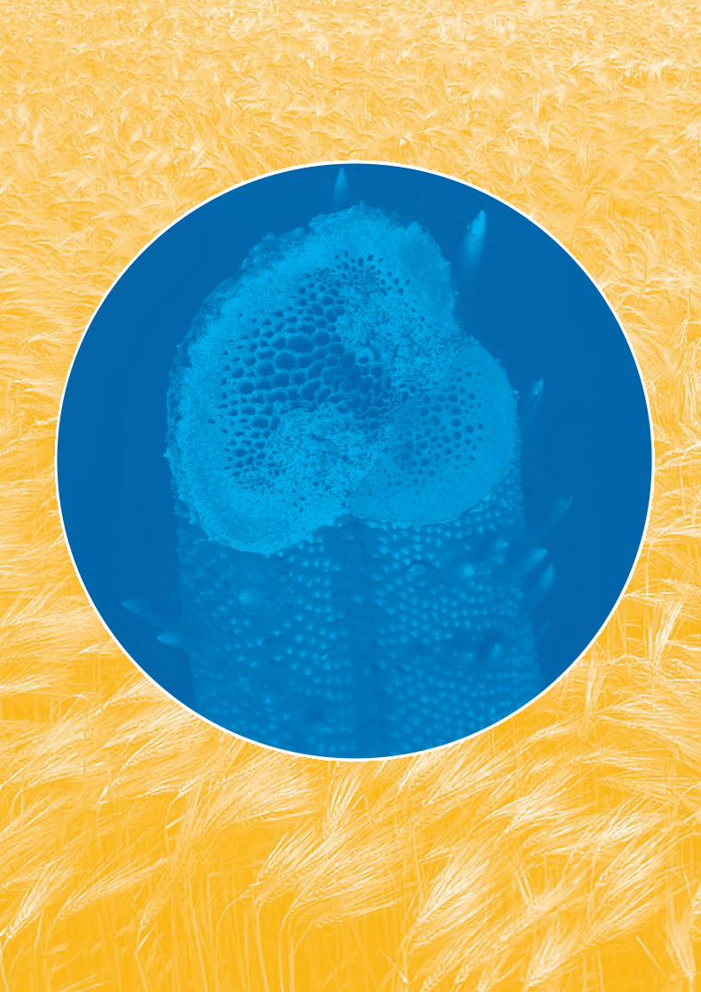
The Materials Design of Wheat Awns for Seed Dispersal (Rivka Elbaum)Awns evolved to direct seeds to a safe germination position.The dispersal unit of wild wheat (Triticum diccocoides) bearstwo pronounced awns that balance the seed as it falls to theground [9].

Fig. 4: A graphic illustration of the wild wheat plant and two dispersalunits (not in scale), are shown on the left. Each dispersal unit carriestwo pronounced awns that orient the dispersal unit as it falls. The redsquare indicates the location of the scanning electron micrograph on theright, and the active cellulose zone is indicated in pink.
Using X-ray diffraction we found that the cellulose fibrils,which construct the cell walls, are aligned mostly along thelong axis of the awn, except at a region close to the seeds(highlighted in Fig. 4). In this location the fibers are randomlyoriented. This design results in bending of the awns withchanges of humidity: water molecules that adsorb to the fiberscause mostly a lateral expansion. Thus, the whole structurewill expand laterally except for the region where the fibrils arerandomly oriented. This part of the awn will expand in alldirections, pushing the awns toward each other. With drying,this active region will contract, similarly to a muscle.
Fig. 5: Wild wheat awns at different levels of relative air humidity (r.h.)
Cycling the air humidity causes a periodic movement of theawns, resembling the swimming motion of frog legs. Fig. 5shows this principle of the dispersal unit movement. It isclearly visible that the average distance between the awnschanges as a function of air humidity. With the daily humiditycycle, the awns will move cyclically and thereby propel thedispersal unit forward. Silicified hairs that cover the awnsand point away from the seed are locking the awn in thisprocess and preventing a backward movement. This suggeststhe possibility that the daily humidity cycle may induce themotility required for seed dispersal. This also means that adead plant tissue can work as a motor fuelled just by theambient humidity cycle [10].
Structure-Function Relations of Human Teeth in 3D(Paul Zaslansky)Teeth are composed of mainly two carbonated hydroxyapatitebased composite materials (enamel and dentin), arranged in acomplex array of graded and varying micro structures. System-atic structural variations of tooth materials [11] lead to very dif-ferent responses to load within different parts of the crownand root. Consequently, the function of whole teeth and thenature of differences between different types of teeth are notwell understood. Our work focuses on trying to better under-stand design principles of human teeth by combining 2D imag-ing techniques (wet-mode environmental scanning electronmicroscopy, X-ray scattering techniques and Acoustic andRaman microscopy) of static and mechanically loaded toothsamples with 3D high resolution (sub µ) measurements (usingmicrotomography and speckle interferometry). Our workingpremise is that there is great importance to both arrangementand properties of features embedded in the microstructure[11,12] and both are needed to support the longevity of teeth.By matching structural and deformation patterns from 2Dslices with non-destructive measurements of the 3D samples,we hope to understand what allows teeth to function withoutremodeling or 'self-repair' the way that bones do.
Fig. 6: 3D virtual cube of dentin displaying tubules running upwardthrough the structure. The phase enhanced image, obtained at ID19 inthe ESRF clearly shows the distribution, density and orientations of the highly mineralized tubules. Scale bar: 100 µm
Much of our 3D characterization is based on imaging of inter-ference patterns and as a result we obtain spatial sub-micronresolution using currently available partially-coherent laserand X-ray sources [12]. As seen in Fig 6, our methods producedata with a resolution capable of resolving ~1 micrometerthick tubules in dentin (or similarly prisms in enamel). We arethus able to track displacements and image the microstruc-ture, and our efforts are aimed at correlating the deformationpatterns so as to understand the behavior of human teethunder physiological (daily) mechanical load.
B. Aichmayer [email protected]. Elbaum [email protected]. Zaslansky [email protected]. Fratzl [email protected]
45
References: [1] J. Aizenberg, J.C. Weaver, M.S.Thanawala, V.C. Sundar, D.E. Morse, P.Fratzl: Skeleton of Euplectella Sp.; struc-tural hierarchy from the nanoscale to themacoscale. Science 309, 275 - 278 (2005).[2] A. Woesz, J.C. Weaver, M. Kazanci,Y. Dauphin, J. Aizenberg, D. E. Morse, P.Fratzl: Micromechanical properties of bio-logical silica in skeltons of deep-sea spon-ges. J. Mater. Res. 21, 2068 -2078 (2006).[3] A. Sidorenko, T. Krupenkin, A. Taylor,P. Fratzl, J. Aizenberg: Reversible switch-ing of hydrogel-actuated nanostructuresinto complex micropatterns. Science 315, 487-490 (2007). [4] P. Fratzl, F.D. Fischer, J. Svoboda:Energy dissipation and stability of propagating surfaces. Phys. Rev. Lett. 95,195702 (2005).[5] G.A. Maier, G. Wallner, R.W. Lang, P.Fratzl: Structural changes during plasticdeformation at crack Tips in PVDF films.Macromol. 38, 6099 - 6105 (2005).[6] Aichmayer, B., Margolis, H.C., Sigel,R., Yamakoshi, Y., Simmer, J.P. and Fratzl,P.: The onset of amelogenin nanosphereaggregation studied by small-angle X-rayscattering and dynamic light scattering.J. Struct. Biol. 151, 239-249 (2005).[7] Wang, T., Antonietti, M. and Cölfen,H.: Calcite Mesocrystals: “Morphing”Crystals by a Polyelectrolyte. Chem. Eur. J. 12, 5722-5730 (2006).[8] B. Aichmayer, M. Mertig, A. Kirchner,O. Paris, P. Fratzl: Small-angle scatteringof S-layer mineralization. Adv. Mater. 18,915-919 (2006).[9] Chambers, J. C., MacMahon, J. A: A Day in the Life of a seed: Movementsand fates of seeds and their implicationsfor natural and managed system Annu.Rev. Ecol. Syst. 25, 263-292 (1994).[10] R. Elbaum, L. Zaltzman, I. Burgert, P. Fratzl: The role of wheat awns in the sead dispersal unit, Science 316,884-886 (2007).[11] P. Zaslansky, A. A. Friesem and S.Weiner: “Structure and mechanical properties of the soft zone separatingbulk dentin and enamel in crowns ofhuman teeth: Insight into toothfunction“, J. Struct. Biol. 153 (2), 188-199 (2006).[12] S. Zabler, H. Riesemeier, P. Fratzland P. Zaslansky: “Fresnel-propagatedimaging for the study of human toothdentin by partially coherent x-ray tomography”, Opt. Express 14 (19), 8584-8597 (2006).

Mesoscale materials exhibit particular struc-tural features at intermediate scales be-tween the atomic/molecular world andmacroscopic dimensions. Such systems mayshow novel properties and functions which
result directly from the size of the compart-ments and/or the interactions between the
individual structural units. Our research is direct-ed towards the structural characterization and the
understanding of structure-function relationships of hierar-chical mesoscale composites such as biological materials,and (biomimetic) carbons and ceramics. Moreover, we areinterested in the phase behavior of fluids in confined geome-try of mesopores, and in their elastic interaction with the sol-id pore walls. Our experimental approaches are essentiallybased on scattering techniques using synchrotron radiation.We develop sophisticated new in-situ methods to "watchmaterials at work", and we apply microbeam scanning tech-niques to map the local nanostructure in hierarchically organ-ized materials.
Biomimetic ProcessingThe aim here is to transform hierarchical plant tissues intoinorganic materials, and to characterize their structure andtransformation behavior. In a first approach, infiltration ofwood with ceria nanoparticles in a suitable acidic solutionwas successfully used to replace the lignin phase with thenanoparticles (nano-casting). Upon subsequent calcination,macroscopic ceramic replicas that reproduce four distincthierarchical levels of the original biological wood templatewere obtained [1]. In particular, it was shown for the firsttime with the aid of small-angle X-ray scattering (SAXS) thatthe spiraling cellulose microfibrils in wood could be cast withnanometer precision (Fig. 1). This opens new possibilities forthe simple and economical synthesis of novel ceramics withhierarchical and directional porosity.
Fig. 1: SAXS patterns illustrating that the microfibrillar cellulose orienta-tion is fully reproduced after nanoparticle casting of wood.
The second approach used direct conversion of tissues bypyrolytic decomposition of the plant biopolymers. We havestudied the structural and chemical development of pyrolysedwood as a function of temperature up to 2400°C. We couldshow that crack free carbon monoliths which fully resemblethe honeycomb-like cellular architecture of wood tissue,could be produced with a preferred carbon orientation alongthe cell axis [2]. The thermal decomposition of wood cellulosewas further investigated with in-situ X-ray diffraction. Byquantitatively analyzing the kinetics for different tempera-
tures it was found that decomposition of the crystalline cellu-lose in wood occurred mainly via a thermally activateddecrease of the microfibril diameter.
As a second model system for direct biomimetic conver-sion of plants, we have investigated the silica accumulatingstalks of Equisetum hyemale (horsetail or scouring rush).Besides a general interest in the function of silica in higherplants, we used horsetail for direct SiC synthesis by controlledpyrolysis taking the biopolymers as a carbon source and silicaas a Si- source. Ongoing work is focusing on the detailed char-acterization of the type and distribution of silica, and on theoptimization of the conversion process in terms of yield andtype of SiC.
Mesoscale CarbonsThe detailed origin of the extraordinary mechanical proper-ties of carbon fibers and the relation to their local mesoscalestructure are still largely unknown. In a pioneering experi-ment in cooperation with the University of Vienna we com-bined in-situ bending of single carbon fibers with high resolu-tion X-ray diffraction by scanning the bent fibers across a 0.1µm wide beam (Fig. 2). Strain redistribution across the fiberwith a shift of the neutral axis allowed a quantitative deter-mination of the elastic moduli in compression and in tension.A significant change of the preferred carbon orientation inthe compression regime proved that buckling of the carbonnanocrystallites is the physical origin of the difference in ten-sile and compressive properties. Differences between differ-ent carbon fiber types were attributed to different amounts ofcovalent cross-links connecting the crystallites [3].
Fig. 2: Sketch of the in-situ bending experiment to determine the localmechanical properties of carbon fibers.
Further work on mesoscale carbons included the localmechanical properties of pyrolysed wood at the level of singlecell walls using nanoindentation [4], and a critical examina-tion of the classical way to obtain carbon crystallite sizesfrom Raman band intensity ratios [5]. One of the future chal-lenges of our research in this field is related to the importantrole of covalent cross-links for the mechanical behavior indisordered carbons. In this respect, we have already startedin-situ high temperature creep studies of single carbon fiberswithin the synchrotron radiation X-ray microbeam at the µ-Spot beamline at BESSY.
46
Oskar Paris 26.01.19671993: Diploma, Physics (University of Vienna, Austria)Thesis: Internal Oxidation ofCu-Fe Alloys1996: PhD, Physics (University of Vienna, Austria)Thesis: Influence of Internal and External Stresses on Decomposition in Alloys 1996-1998: Postdoc (Federal Institute of Technology, Institute of Applied Physics, Zurich, Switzerland)1998-2003: University Assistant (University of Leoben, Austria)2003: Habilitation,(University of Leoben, Austria)Thesis: Structure and Properties of Complex Materials: SynchrotronRadiation and Neutrons as Local ProbesSince 2003: Group Leader (Max Planck Institute of Colloids and Interfaces, Potsdam)
Mesoscale Materials and Synchrotron Research
BIO-INSPIRED MATERIALS

Fluids in MesoporesMesoporous materials with narrow distributions of pores onhighly ordered lattices are ideal model systems to study thephase behavior of fluids in confinement. In cooperation withthe Technical University of Berlin, we have developed a sorp-tion device for in-situ small-angle X-ray diffraction. Sorptionof organic fluids in 2D hexagonal lattices of SBA-15 silicamaterials was investigated at Hamburger Synchrotron-strahlungslabor (HASYLAB), and more recently at the µ-Spotbeamline at BESSY. Analyzing the intensity of the diffractionpeaks as a function of vapor pressure along a sorptionisotherm provides detailed structural information on liquidfilm formation and on pore condensation [6]. Moreover, highcapillary pressures lead to a deformation of the pore walls atcapillary condensation. The adsorption strains related to thedeformation of the pore lattice can directly be obtained fromthe shift of the diffraction peaks as a function of vapor pres-sure (Fig. 3) [7].
In the future, we propose to investigate the dynamicinteraction between pore deformation as a consequence ofcapillary condensation, and the influence of these deforma-tions on the phase behavior of the fluid. Moreover, we plan todevelop a mechanical model that quantitatively explains theexperimentally observed behavior. This activity is embeddedin the framework of the Collaborative Research Center Sfb448 "Mesoscopically Organized Composites" of the GermanResearch Foundation (DFG).
Fig. 3: In-situ sorption of perfluoropentane (C5F12) in SBA-15 (red: adsorp-tion; blue: desorption). a) shows the integrated intensity of the 10 reflec-tion which is low for entirely filled pores due to partial contrast match-ing, and b) shows the corresponding lattice strains.
From Diffraction to ImagingA large part of our activities in the field of synchrotronresearch in the last two years were related to the commis-sioning of an experimental station for simultaneousmicrobeam small- and wide-angle scattering (SAXS/WAXS)at the microfocus (µ-Spot) beamline at BESSY in Berlin. Theinstrument is now fully operational and provides a routinemicrobeam of 10 µm diameter at a flux of more than 109 pho-tons per second [8]. A series of user experiments havealready been performed in cooperation with other researchgroups from the department and with external partners.Many of them were related to scanning SAXS/WAXS studieson biological or bio-inspired materials such as for instancebone, plant tissues, and biomimetic calcite.
A long-term goal of our research is to proceed frommicrobeam scanning SAXS/WAXS to a real imaging tech-nique. There has been considerable progress in our groupconcerning software development for interactive instrumentcontrol combined with online data analysis and onlineparameter imaging. This work was done in close collabora-tion with the European Synchrotron Radiation Facility (ESRF)in the framework of a long-term proposal. At present, severalsoftware tools are available that allow an automated extrac-tion and imaging of nanostructural parameters such as localfiber orientation from WAXS (Fig. 4) or particle size and orien-tation from SAXS. Future work is directed towards the imple-mentation of these tools into the beamline software atBESSY.
Furthermore, we plan to extend our scanning approach tothe third dimension in both, the reciprocal space (local tex-ture analysis) and in real space (SAXS tomography, confocal diffraction).
Fig. 4: SEM image (left) and an image based on scanning microbeam diffraction (right) of a flow sensing system in cricket appendices. Thecolor scale on the right is given by the intensity of the equatorial 040reflection from the crystalline chitin fibers in this chitin/proteinnanocomposite, and the bars indicate the local chitin fiber orientation as deduced from the individual diffraction patterns. The sample wasprepared by using a UV microlaser instrument and the diffraction experiments were performed at the ESRF [9].
O. Paris, A. Deshpande, A. Gourrier, C. Li, L. Sapei, R. Seidel,S. Siegel, G. Weseloh, I. Zenke, G. A. [email protected]
47
References: [1] Deshpande, A., Burgert, I. and ParisO.: Hierarchically structured ceramics byhigh-precision nanoparticle casting ofwood. Small 2, 994-998 (2006).[2] Paris, O., Zollfrank C. and ZicklerG.A.: Decomposition and carbonizationof wood biopolymers – A microstructur-al study of softwood pyrolysis. Carbon43, 53-66 (2005).[3] Loidl, D., Paris, O., Burghammer, M.,Riekel, C. and Peterlik, H.: Direct obser-vation of nanocrystallite buckling incarbon fibers. Phys. Rev. Lett. 95,225501 (2005).[4] Zickler, G., Schöberl, T and Paris, O.:Mechanical properties of pyrolysedwood: A nanoindentation study. Phil.Mag. A. 86, 1373-1386 (2006).[5] Zickler, G., Gierlinger, N., Smarsly,B., Peterlik, H. and Paris O.: A reconsid-eration of the relationship between thecrystallite size La of carbons determinedby X-ray diffraction and Raman spec-troscopy. Carbon 44, 3239-3246 (2006).[6] Zickler, G., Jähnert, S., Wagermeier,W., Funari, S., Findenegg, G. and Paris,O.: Physisorbed films in periodic meso-porous silica studied by in-situ synchro-tron radiation small-angle diffraction.Phys. Rev. B 73, 184109 (2006). [7] Zickler, G.A., Jähnert, S., Funari, S.,Findenegg, G. and Paris, O.: Pore latticedeformation in ordered mesoporoussilica studied by in-situ small-angle X-ray diffraction. J. Appl. Cryst. 40,522-526 (2007).[8] Paris, O., Li, C., Siegel, S., Weseloh,G., Emmerling, F., Riesemeier, H., Erko,A. and Fratzl, P.: A new experimentalstation for simultaneous microbeamscanning small-and wide-angle scatter-ing at BESSY. J. Appl. Cryst. 40,466-470 (2007).[9] Seidel R., Gourrier A., BurghammerM., Riekel C., Geronimidis G. and ParisO.: Mapping fibre orientation in com-plex-shaped biological systems withmicrometre resolution by scanning X-raymicrodiffraction, Micron (2007) in press(doi:10.1016/j.micron.2007.02.004).