biology of pseudomonas stutzeri - mmbr.asm.org · is always a danger of drawing taxonomic...
TRANSCRIPT

MICROBIOLOGY AND MOLECULAR BIOLOGY REVIEWS, June 2006, p. 510–547 Vol. 70, No. 21092-2172/06/$08.00�0 doi:10.1128/MMBR.00047-05Copyright © 2006, American Society for Microbiology. All Rights Reserved.
Biology of Pseudomonas stutzeriJorge Lalucat,1,2* Antoni Bennasar,1 Rafael Bosch,1 Elena Garcıa-Valdes,1,2 and Norberto J. Palleroni3
Departament de Biologia, Microbiologia, Universitat de les Illes Balears, Campus UIB, 07122 Palma de Mallorca, Spain1;Institut Mediterrani d’Estudis Avancats (CSIC-UIB), Campus UIB, 07122 Palma de Mallorca, Spain2; and
Department of Biochemistry and Microbiology, Rutgers University, Cook Campus,New Brunswick, New Jersey 08901-85203
INTRODUCTION .......................................................................................................................................................511DEFINITION OF THE SPECIES AND DIFFERENTIATION FROM OTHER PSEUDOMONAS SPECIES ....511
Definition .................................................................................................................................................................511Differentiation from Other Species ......................................................................................................................511
DISCOVERY AND NOMENCLATURAL PROBLEMS ........................................................................................512OCCURRENCE AND ISOLATION PROCEDURES ............................................................................................512PHENOTYPIC PROPERTIES..................................................................................................................................514
Colony Structures/Types ........................................................................................................................................514Morphological Characterization (Cells, Reserve Materials, Flagella, and Pili) and Chemotaxis ..............515Chemical Characterization and Chemotaxonomy..............................................................................................515
DNA base composition .......................................................................................................................................515Protein patterns ..................................................................................................................................................515LPS and immunological characteristics ..........................................................................................................515Fatty acid composition .......................................................................................................................................516Quinone and polyamine composition...............................................................................................................516PHA.......................................................................................................................................................................516
GENOMIC CHARACTERIZATION AND PHYLOGENY....................................................................................516DNA-DNA Hybridizations......................................................................................................................................516Genome Size and Organization ............................................................................................................................517Genotyping ...............................................................................................................................................................517Genetic Diversity: MLEE.......................................................................................................................................518Genetic Diversity: MLST .......................................................................................................................................518Phylogeny .................................................................................................................................................................519Clonality ...................................................................................................................................................................520
TAXONOMIC RANKS: GENOMOVARS ...............................................................................................................520IDENTIFICATION .....................................................................................................................................................521
Phenotypic Identification .......................................................................................................................................521Molecular DNA-Based Identification ...................................................................................................................521Polyphasic Identification........................................................................................................................................521
PHYSIOLOGICAL PROPERTIES...........................................................................................................................522Temperature, Pressure, pH, and O2 Relationships ...........................................................................................522Denitrification .........................................................................................................................................................522
Structural gene clusters and the nature of denitrification genes ................................................................522(i) nar genes.....................................................................................................................................................523(ii) nir genes ....................................................................................................................................................524(iii) nor genes ..................................................................................................................................................524(iv) nos genes ...................................................................................................................................................524
Metalloenzymes involved in the denitrification process................................................................................525(i) Nitrate respiration and NaRs..................................................................................................................525(ii) Properties of NarL and NarX proteins.................................................................................................526(iii) Nitrite respiration and NiRs .................................................................................................................526(iv) Nitric oxide respiration and NORs.......................................................................................................526(v) Nitrous oxide respiration and N2ORs ...................................................................................................527
Chlorate and Perchlorate as Terminal Electron Acceptors..............................................................................527Organic Compounds Used as the Sole Carbon and Energy Source................................................................528Inorganic Energy Sources (Thiosulfate)..............................................................................................................528Production of Siderophores...................................................................................................................................529Nitrogen Fixation ....................................................................................................................................................529Phosphite and Hypophosphite Oxidation............................................................................................................530
* Corresponding author. Mailing address: Institut Mediterranid’Estudis Avancats (CSIC-UIB), Campus UIB, 07122 Palma de Mallorca,Spain. Phone: 34 971 173140. Fax: 34 971 173184. E-mail: [email protected].
510
on January 9, 2020 by guesthttp://m
mbr.asm
.org/D
ownloaded from
Biodegradation and Useful Properties for Biotechnological Applications .....................................................530Metal cycling .......................................................................................................................................................530Crude oil, oil derivatives, and aliphatic hydrocarbons .................................................................................531Aromatic hydrocarbons ......................................................................................................................................531Biocides ................................................................................................................................................................533Proteolytic activity: applications for biorestoration...................................................................................................534
NATURAL TRANSFORMATION.............................................................................................................................534PATHOGENICITY AND ANTIBIOTIC RESISTANCE ........................................................................................535HABITATS AND ECOLOGICAL RELEVANCE....................................................................................................537
Soil, Rhizosphere, and Groundwater ...................................................................................................................537Marine Water and Sediment and Salt Marshes ................................................................................................538Wastewater Treatment Plants ...............................................................................................................................538
CONCLUSIONS .........................................................................................................................................................538ACKNOWLEDGMENTS ...........................................................................................................................................539REFERENCES ............................................................................................................................................................539
INTRODUCTION
Pseudomonas stutzeri was first described by Burri and Stutzerin 1895 (55). van Niel and Allen, in 1952 (371), preciselydefined its phenotypic features and discussed its definitive des-ignation as Pseudomonas stutzeri by Lehmann and Neumann(196). In spite of marked differences from the type strain of thegenus, the sequence similarities of the rRNAs, demonstratedinitially by DNA-rRNA hybridization, show the legitimacy ofthe inclusion of P. stutzeri in the genus Pseudomonas. Strains ofthe species have been identified among denitrifiers found innatural materials. Their inclusion in the phenotypic studiescarried out by Stanier et al. in 1966 (340) demonstrated that, inaddition to their typical colonies, the strains are nutritionallyversatile, using some carbon compounds seldom utilized byother pseudomonads (e.g., starch, maltose, and ethylene gly-col). Variations in DNA sequences, as shown by the results ofDNA-DNA hybridization experiments, were demonstrated inthe early studies of Palleroni et al., in 1970 (251). Work per-formed in recent years has clearly established firm bases forgrouping the strains into a number of genomic variants (geno-movars) that are phylogenetically closely related. Some strainshave received particular attention because of specific meta-bolic properties (such as denitrification, degradation of aro-matic compounds, and nitrogen fixation). Furthermore, somestrains have been shown to be naturally transformable andhave been studied extensively for their capacities for transfor-mation. P. stutzeri is distributed widely in the environment,occupying diverse ecological niches, and has also been isolatedas an opportunistic pathogen from humans. Based on resultsobtained in recent years, the biology of this species is dis-cussed.
DEFINITION OF THE SPECIES ANDDIFFERENTIATION FROM OTHER
PSEUDOMONAS SPECIES
Definition
Pseudomonas stutzeri is a member of the genus Pseudomonassensu stricto. It is in group I of Palleroni’s DNA-rRNA ho-mology group within the phylum Proteobacteria (252, 253). P.stutzeri is now recognized as belonging to the class Gamma-proteobacteria. Phylogenetic studies of P. stutzeri strains’ 16SrRNA sequences and other phylogenetic markers demonstrate
that they belong to the same branch, together with relatedspecies within the genus, such as P. mendocina, P. alcaligenes,P. pseudoalcaligenes, and P. balearica. Typically, cells are rodshaped, 1 to 3 �m in length and 0.5 �m in width, and have asingle polar flagellum. Under certain conditions, one or twolateral flagella with a short wavelength may be produced. Phe-notypic traits of the genus include a negative Gram stain,positive catalase and oxidase tests, and a strictly respiratorymetabolism. In addition, P. stutzeri strains are defined as deni-trifiers. They can grow on starch and maltose and have anegative reaction in arginine dihydrolase and glycogen hydro-lysis tests. The G�C content of their genomic DNA lies be-tween 60 and 66 mol%. DNA-DNA hybridizations enable atleast 17 genomic groups, called genomovars, to be distin-guished. Members of the same genomovar have more than70% similarity in DNA-DNA hybridizations. Members of dif-ferent genomovars usually have similarity values below 50%.
Differentiation from Other Species
No fluorescent pigments are produced, which differentiatesP. stutzeri from other members of the fluorescent group ofPseudomonas spp. Before the use of genomic approaches toidentifying bacteria became widespread, P. stutzeri strains weremisidentified with other species. This was due to the intrinsiclimitations of exclusively phenotypic identification procedureswithin the former genus Pseudomonas. P. stutzeri was mostcommonly confused with other Pseudomonas species (P. men-docina, P. pseudoalcaligenes, P. putida); with species actually inother genera (such as Delftia acidovorans and Ralstonia pick-ettii); or even with the flavobacteria, Alcaligenes or Achro-mobacter. Mandel proposed the species “Pseudomonas sta-nieri” for P. stutzeri strains with a low G�C content, around62% (212); however, G�C content alone is a weak parameterfor species differentiation. In some collections, P. stutzeri cul-tures were labeled P. saccharophila. The strain OX1 (ATCCBAA-172) was classified phenotypically as a P. stutzeri strain(13). It has been intensively studied due to its significant phe-notypic characteristics. However, when strain OX1 was char-acterized taxonomically in detail, it turned out to be a memberof the P. corrugata phylogenetic branch (73). Pseudomonas sp.strain OX1 may be confused phenotypically with P. stutzeribecause P. stutzeri is phenotypically diverse. However, OX1 isgenomically distinct.
VOL. 70, 2006 PSEUDOMONAS STUTZERI 511
on January 9, 2020 by guesthttp://m
mbr.asm
.org/D
ownloaded from
The species most closely related to P. stutzeri is P. balearica(formerly genomovar 6 of the species). It shares many basicphenotypic traits with P. stutzeri strains and belongs to thesame 16S rRNA phylogenetic branch. However, it can be dif-ferentiated chemotaxonomically from P. stutzeri by its ability togrow above 42°C and by a few other biochemical tests (23).
P. chloritidismutans is a member of genomovar 3. However,it has been proposed as the type strain of a new species (404)and is discussed below (see “Physiological properties”). Thereis always a danger of drawing taxonomic conclusions from theproperties of metabolic systems that are involved in the me-tabolism of unusual substrates or molecules.
The phylogenies of genes of the rrn operon, consideredindividually or with other housekeeping genes, demonstratethat all P. stutzeri strains are monophyletic. Such phylogeneticstudies are currently another good tool for discriminating P.stutzeri from the rest of the bacterial species. P. xanthomarinahas recently been described as a new species (289) with onlyone representative strain. It is located in the same 16S rRNAphylogenetic branch as P. stutzeri and P. balearica, with se-quence similarities above 98%. It can be differentiated pheno-typically from both species.
DISCOVERY AND NOMENCLATURAL PROBLEMS
In 1952, C. B. van Niel and M. B. Allen stated in their noteon the history of P. stutzeri: “During the two decades followingthe discovery of the denitrification process several notablepapers were published on the isolation and characterization ofdenitrifying bacteria. A study on this literature reveals thatBurri and Stutzer (1895) were the first to describe such organ-isms in sufficient detail to render them recognizable. This ap-plies particularly to their Bacillus denitrificans II, an organismof wide distribution and outstanding characteristics, which hasbeen isolated from straw, manure, soil, canal water, etc., andwhich students of the denitrification process have consideredas a very common and easily identifiable denitrifier” (371). Thedifferent names that this denitrifier has gained since its discov-ery are well documented in van Niel and Allen’s 1952 publi-cation (371). They include Bacterium stutzeri (196), Bacillusnitrogenes (229), Bacillus stutzeri (68), Achromobacter sewerinii(28), Pseudomonas stutzeri (322), and Achromobacter stutzeri(27). The species “Pseudomonas stanieri” was proposed in 1966by Mandel for those strains with a G�C content of around62% (212). However, no clear differences in phenotype can befound between P. stutzeri and “Pseudomonas stanieri.” It is notto be confused with Marinomonas stanieri, formerly considereda Pseudomonas species.
The type strain is Lautrop strain AB 201 (equivalent toStanier 221, ATCC 17588, CCUG11256, DSM 5190, ICMP12591, LMG11199, NCIB 11358, and WCPPB 1973). In addi-tion, a reference strain has been proposed for each genomovar(Table 1). Some relevant strains that were previously assignedto other species are Pseudomonas perfectomarina strain ZoBell(19), Alcaligenes faecalis A15 (380), and Flavobacterium lute-scens strain ATCC 27951 (24). Many, but not all, strains havebeen deposited in publicly recognized culture collections, areavailable for scientific research, and should be used as refer-ence strains.
OCCURRENCE AND ISOLATION PROCEDURES
Detection of P. stutzeri basically relies on two methods: (i)enrichment and isolation of pure cultures and (ii) direct anal-ysis without the need for culturing. Both methods are essentialto autoecological studies and to understanding the role of thespecies in the environment.
An elective culture method for the specific enrichment ofdenitrifiers and the isolation of P. stutzeri was developed byIterson in 1902 (described in 1952 by van Niel and Allen [371]).A mineral medium with 2% nitrate under anaerobic conditionsand tartrate (or malate, succinate, malonate, citrate, ethanol,or acetate) leads to a predominant population of P. stutzeri,even when some isolates are not able to grow on tartrate inpure culture. Tartrate may be converted anaerobically to anassimilable substrate by other bacteria in the sample. A selec-tion of cells producing colonies with the unusual morphologyof P. stutzeri permits an efficient isolation procedure from en-vironmental samples. Incubation temperatures of 37°C orabove allow a more selective enrichment, which can be com-bined with denitrifying conditions.
DNA methods based on 16S rRNA sequences have beenalso designed to detect P. stutzeri in DNA extracted directlyfrom environmental samples. Bennasar et al., in 1998, devel-oped PCR primers that were specific to all known genomovarsof P. stutzeri at that time (24). This served as a confirmationtest, as did amplicon cleavage using the restriction enzymeHindIII or a specific DNA probe targeted at the amplifiedproduct (24). Amann et al. considered the difficulty of obtain-ing a DNA probe to cover all of the P. stutzeri strains (5).However, they designed a DNA probe for specific 23S rRNAsequences. This is useful in fluorescence in situ hybridizationtechniques to detect and quantify P. stutzeri in environmentalsamples. Nevertheless, not all strains can be detected, due tothe high genetic diversity of the species, including the rrnoperon.
Besides the rrn genes, other genes are now used for func-tional analysis of ecosystems. These genes also detect P.stutzeri. They include nirS or nosZ for detecting denitrification(46) and nifH for analyzing diazotrophic bacteria in the rhizo-sphere (93). The usefulness of a conserved nosZ probe forscreening the distribution of denitrifying bacteria with similarN2O reductases in the environment has been described else-where (65, 386). In 2001, Gruntzig et al. developed a verysensitive method based on real-time PCR analysis of DNAisolated from soil and sediment samples (132). However, notall DNAs of the species’ strains could be amplified. Specificprimers for PCR and an internal probe of the denitrificationgene nirS enabled less than 100 cells per g of sample to bequantified.
In their analysis of P. stutzeri populations in marine wa-ters, Ward and Cockcroft used monoclonal antibodies raisedagainst outer membrane proteins of the strain ZoBell (388).ZoBell originally named this strain “Pseudomonas perfecto-marina.”
Sikorski et al. were able to isolate members of P. stutzerifrom aquatic habitats and terrestrial ecosystems in a two-stepprocedure. Firstly, the occurrence of P. stutzeri cells was as-sessed by a previously designed, slightly modified PCR proce-dure (24, 325). Secondly, the positive samples were screened
512 LALUCAT ET AL. MICROBIOL. MOL. BIOL. REV.
on January 9, 2020 by guesthttp://m
mbr.asm
.org/D
ownloaded from

TABLE 1. P. stutzeri strains cited in the text, with relevant characteristics, origins, and references
Strain Other designation(s) Taxonomy a Isolation Origin, geographic location, and/orphysiological characteristic(s) Reference(s)
CCUG11256 ATCC 17588, DSM5190,LMG11199, Stanier 221
Type strain, gv 1 Pre-1966 Clinical, spinal fluid; Copenhagen, Denmark;siderophore producer
340
ATCC 17591 Stanier 224 Ref, gv 2 1956 Clinical; Copenhagen, Denmark 340DSM50227 ATCC 11607, LMG1228 Ref, gv 3 Pre-1952 Garden soil; denitrifier 37119SMN4 DSM6084 Ref, gv 4 1988 Marine sediment; naphthalene degrader;
Barcelona, Spain291
DNSP21 DSM6082 Ref, gv 5 1988 Wastewater; denitrifier; Mallorca, Spain 291SP1402 DSM6083 Former Ref, gv 6;
P. balearica1988 Wastewater; 2-methylnaphthalene degrader;
Mallorca, Spain23
DSM50238 ATCC 17832 Ref, gv 7 Pre-1966 Soil; denitrifier; California 340JM300 DSM10701 Ref, gv 8 Pre-1980 Soil; California; natural transformation
model organism60
KC ATCC 55595, DSM7136 Ref, gv 9 1990 Aquifer; California 316CLN100 Ref, gv 10 1990 Chemical industry wastewater; Germany 11428a50 CCUG50544, DSM17089 Ref, gv 11 2002 Soil; Tel Aviv airport area, Israel 32528a39 CCUG50543, DSM17088 Ref, gv 12 2002 Soil; Tel Aviv airport area, Israel 32528a22 CCUG50542, DSM17087 Ref, gv 13 2002 Soil; Tel Aviv airport area, Israel 32528a3 CCUG50541, DSM17086 Ref, gv 14 2002 Soil; Tel Aviv airport area, Israel 3254C29 CCUG50538, DSM17082 Ref, gv 15 2002 Sea sediment; Dangast, Germany 32524a13 CCUG50539, DSM17083 Ref, gv 16 2002 Soil contaminated with mineral oil;
Espelkamp, Germany325
24a75 CCUG50540, DSM17084 Ref, gv 17 2002 Soil contaminated with mineral oil;Espelkamp, Germany
325
MT-1 CCUG50545, DSM17085 Ref, gv 18 1997 Marine sediment at 11,000-m depth, MarianaTrench
351
1317 1998 Accumulates PHA 1419A 2003 Alfalfa rhizosphere contaminated with coal
tar; Rubinsk, Russia; aromatics degrader;chemotactic
243
A15 LMG10652 gv 1 1981 Rice paddy; nitrogen fixer; Southeast Asia;formerly Alcaligenes faecalis
380
A29 2005 Proteolytic 273AG259 Soil; silver resistant 63AK61 Pre-1998 Cyanide degrader; metal-plating plant
wastewater; Japan390
AN10 gv 3 1983 Marine sediment; naphthalene degrader;Barcelona, Spain
42, 44
ATCC 14405 ZoBell, CCUG16156 gv 2 1944 Marine; Pacific Ocean, California; formerlyP. perfectomarina
412, 415
ATCC 17587 Stanier 220, LMG 5838 gv 2 Pre-1966 Clinical; Copenhagen, Denmark 340ATCC 17589 Stanier 222 gv 1 Pre-1966 Clinical; Copenhagen, Denmark 340ATCC 17594 Stanier 227 gv 1 Pre-1966 Clinical; Copenhagen, Denmark 340ATCC 27951 gv 1 1988 Formerly Flavobacterium lutescens 24AW1 DSM13592 gv 3; P. chloritidismutans 2002 Wastewater treatment plant; chlorate reducer 380
ATCC BAA-443BG 2 gv 4 1999 Sulfide-oxidizing bioreactor;
thiosulfate oxidizer337
CFPBD Pres gv 3 2001 Chlorate reducer 1ChG 5-2 gv 4 1999 Black Sea (southwest), 120-m depth;
thiosulfate oxidizer337
ChG 5-3 gv 3 1999 Black Sea (south), 120-m depth; thiosulfateoxidizer
337
CMT.9.A DSM4166 1987 Sorghum nutans rhizosphere; nitrogen fixer;Germany
189
JD4 gv 5 1995 Garden soil; denitrifier; Mallorca, Spain 24JJ 2003 1,2-Dichloroethane-contaminated soil; growth
on 2-chloroethanol as denitrifier95
NF13 1991 Deep-sea hydrothermal vent; sulfur oxidizer;Galapagos rift
300
P16 1994 Creosote-contaminated soil; phenanthrenedegrader
348
PDA Pres gv 1 or 5 2001 Primary digested wastewater on lactate;chlorate reducer
75
PDB Pres gv 1 or 5 2001 Primary digested wastewater on lactate;chlorate reducer
75
Continued on following page
VOL. 70, 2006 PSEUDOMONAS STUTZERI 513
on January 9, 2020 by guesthttp://m
mbr.asm
.org/D
ownloaded from

for P. stutzeri by means of plating on an artificial seawatermedium with ethylene glycol, starch, or maltose as the carbonsource under aerobic conditions (325). The characteristic col-ony morphology of P. stutzeri led to a highly efficient isolationprocedure: one P. stutzeri colony was detected among 9,100colonies of other bacteria.
However, many strains of P. stutzeri that have been studiedin detail were isolated by their metabolic peculiarities. Theywere not specifically isolated for denitrification ability or be-cause P. stutzeri was the target of the study.
PHENOTYPIC PROPERTIES
Apart from the 1952 study by van Niel and Allen, the onlypapers containing detailed descriptions of P. stutzeri’s pheno-typic properties are those by Stanier et al. in 1966 and Ros-sello-Mora et al. in 1994 (295, 340, 371).
Strains of P. stutzeri, like most recognized Pseudomonas spp.,can grow in minimal, chemically defined media, with ammo-nium ions or nitrate and a single organic molecule as the solecarbon and energy source. No additional growth factors arerequired. Some P. stutzeri strains can grow diazotrophically.This characteristic seems to be rare among the genus Pseudo-monas. None of the strains tolerate acidic conditions: they donot grow at pH 4.5. P. stutzeri has a respiratory metabolism,and oxygen is the terminal electron acceptor. However, allstrains can use nitrate as an alternative electron acceptor andcan carry out oxygen-repressible denitrification. Denitrificationmay be delayed or may appear only after serial transfers innitrate media under semiaerobic conditions (73, 340). Oxida-tive degradation of aromatic compounds involves the partici-pation of mono- and dioxygenases. Typically, catechol or pro-tocatechuate is the central intermediate in this reaction. Eachis cleaved through an ortho pathway when no accessory genesare involved in the degradation. Amylolytic activity is one ofthe phenotypic characteristics of the species. The enzymologyof the exo-amylase—which is responsible for the formation ofmaltotetraose as an end product—has been examined at themolecular level. This enzyme has also been cloned (231). Ob-radors and Aguilar demonstrated that polyethylene glycol wasdegraded to yield ethylene glycol, a substrate typically used byP. stutzeri strains (241).
The arginine deiminase system (“dihydrolase”) catalyzes theconversion of arginine to citrulline and of citrulline to orni-thine. It has been used by taxonomists to differentiate species.
All P. stutzeri strains give a negative test result for this reaction.They also fail to use glycogen and do not liquefy gelatin.
Colony Structures/Types
Colonies can be distinguished by their unusual shape andconsistency (Fig. 1). Freshly isolated colonies are adherent,have a characteristic wrinkled appearance, and are reddishbrown, not yellow, in color. They are typically hard, dry, andtenaciously coherent. It is easy to remove a colony in its en-tirety from a solid surface. Colonies generally resemble craterswith elevated ridges that often branch and merge, and theyhave been described as tenacious, with a coral structure. Theremay be more mucoid protuberances at the periphery than inother areas. The frequent occurrence of irregular polygon-likestructures or concentric zones has also been noted (371). The
FIG. 1. Colonial morphology. Several typical colonial morpholo-gies of P. stutzeri strains. (The image in panel A was taken fromreference 371.)
TABLE 1—Continued
Strain Other designation(s) Taxonomya Isolation Origin, geographic location, and/orphysiological characteristic(s) Reference(s)
PK Pres gv 3 1999 Soil or sediment; chlorate reducer 75RC7 1980 Catechol-like siderophore producer 224RS34 1984 Industrially polluted soil; zinc resistant 135ST27MN3 gv 4 1988 Marine sediment; naphthalene degrader;
Barcelona, Spain296
WM88 1998 Soil; Illinois; P oxidizer 223ZP6b 1997 Capparis spinosa Rhizosphere; nitrogen
fixer; Spain6
a Pres, presumptively; Ref, reference strain; gv, genomovar.
514 LALUCAT ET AL. MICROBIOL. MOL. BIOL. REV.
on January 9, 2020 by guesthttp://m
mbr.asm
.org/D
ownloaded from

shapes of colonies are neither uniform nor necessarily con-stant: they change appearance with time. After repeated trans-fers in laboratory media, colonies may become smooth, butyra-ceous, and pale in color. This has been described as colonialdissociation. Strain CMT.9.A hydrolyzes agar. This is a rareproperty and is mainly restricted to marine bacteria. However,the attack may be limited to what is known as “pitting” of theagar (3). Sorokin et al. give a very detailed description of thecolonial morphology, differentiating between R-type and S-type colonies (337). The R-type colonies are stable, but the Stype produces both colony types under appropriate conditions.Smooth colonies grown on plates at 30°C and stored at 4°C for24 h often develop a characteristic wrinkled appearance (A.Cladera, personal communication).
P. stutzeri is grouped with the nonpigmented species of thegenus, even though many strains’ colonies become dark brown.This is due to the high concentration of cytochrome c in thecells. No diffusible pigments are produced on agar plates.
Morphological Characterization (Cells, Reserve Materials,Flagella, and Pili) and Chemotaxis
Cells are typically motile and predominantly monotrichous.In some strains, lateral flagella with a short wavelength are alsoproduced. This particularly occurs in young cultures on com-plex solid media. These lateral flagella could easily be shedduring manipulations incidental to flagellar staining (251). Ithas been suggested that lateral flagella might be involved in thepopulation’s swarming or twitching motility on solid surfaces(319). However, type IV pili may also be responsible for thismovement. Statistically, the highest number of flagellated cellsis reached at the beginning of the exponential growth phase(192). Seventy percent of cells were flagellated in strain AN11:38% had only one flagellum, and 31% had one or more addi-tional flagella inserted laterally (80).
Caution should be exercised when only phenotypic traits areused for classification. This can clearly be seen in the case ofstrain ZoBell. This strain (ATCC 14405) was isolated as amarine bacterium and described by ZoBell and Upham as“Pseudomonas perfectomarinus” in 1944 (412). Subsequently,this organism became the only member of the species P. per-fectomarina. Its lack of flagella was emphasized by its assigna-tion to a new species, although the authors who first describedthis strain stated that it was motile (19, 412). After threepassages, enrichment for flagellated bacteria on semisolid tryp-tone agar enabled a population in which over 80% of cells wereflagellated to develop. This revertant strain is motile by meansof a single polar flagellum (294).
In a recently published chapter on chemotaxis in Pseudomo-nas, Parales et al. stated, “All Pseudomonas species are motileby one or more polar flagella and are highly chemotactic”(258). P. stutzeri is no exception. Chemotaxis machinery has notbeen studied in detail for any Pseudomonas species. Moreover,the ranges of attractants or repellents and environmental con-ditions to which Pseudomonas spp. respond remain largelyunexplored. They seem to be attracted to virtually all of theorganic compounds they can use as growth substrates. How-ever, they are also attracted to other compounds that they areunable to metabolize. Ortega-Calvo et al. studied the chemo-tactic response of several pseudomonads to polycyclic aromatic
hydrocarbon-degrading bacteria (243). Strain 9A of P. stutzeriwas included in the study. This strain degrades naphthalene,phenanthrene, and anthracene. It was concluded that chemo-taxis was positive to naphthalene and to the root exudates ofseveral plants. Chemotaxis may enhance the biodegradation ofpollutants in the rhizosphere, at least in laboratory-scale mi-crocosms. Strain KC mineralizes carbon tetrachloride, and mo-tility-enhanced bioremediation in aquifer sediments has beendemonstrated (401, 402).
Pseudomonas species have a range of different adhesins thatfunction during initial attachment to a substratum. This leadsto biofilm formation. Both flagella and pili seem to be impor-tant in the colonization of biotic and abiotic surfaces, particu-larly in the initial formation of microcolonies. P. aeruginosa’sinitial biofilm development appears to be conditionally depen-dent on type IV pili. P. stutzeri possesses both flagella and pilibut has not been described as a member of consortia that formnatural biofilms. Type IV pili confer twitching motility to P.stutzeri strains (a bacterial movement based on pilus extension/retraction). This is probably at least partly responsible formany colonies’ diffuse borders (J. Sikorski, personal commu-nication). These colonies also correspond to strains that havenatural transformation ability.
Chemical Characterization and Chemotaxonomy
DNA base composition. The G�C content of DNA is auseful characteristic in taxonomy for delineating species. It hasbeen proposed that if two strains differ by more than 5% inG�C content, then they should not be allocated to the samespecies (297). The limit for genus differentiation may be 10%.G�C content in P. stutzeri strains has been determined by thethermal denaturation temperature of the DNA and by enzy-matically hydrolyzing the DNA and subsequently analyzing itby high-performance liquid chromatography. Reported valuesvary widely: 60.7 to 66.3 mol% (251) and 60.9 to 65 mol%(291). However, variations are within the accepted limits formembers of the same species. The distribution of values wasinitially considered to be bimodal. This led to the suggestionthat P. stutzeri might be split into two species (212). Neverthe-less, the inclusion of novel strains resulted in a Gaussian dis-tribution.
Protein patterns. Whole-cell protein patterns obtained bydenaturing polyacrylamide gel electrophoresis (PAGE) arehighly characteristic at the strain level. They have been usedfor typing and classification purposes (265). P. stutzeristrains have been found to be particularly heterogeneous(271, 295). Computer-assisted analysis of the protein bandscreates a dendrogram that is in good agreement with thegenomovar subdivision of the species (366). This result isnot surprising, as whole-cell protein patterns reflect theprotein-encoding genes in the whole genome and the geno-movars were defined by the similarity values of total DNA-DNA hybridizations.
LPS and immunological characteristics. Lipopolysaccharide(LPS) is the main antigenic molecule on the cell surface. This isconsidered to be the heat-stable O-antigen of the genus. Thespecificity of antibodies is related to the composition of the poly-saccharide chains projecting outside the cells. Representative P.stutzeri strains of the seven known genomovars on which experi-
VOL. 70, 2006 PSEUDOMONAS STUTZERI 515
on January 9, 2020 by guesthttp://m
mbr.asm
.org/D
ownloaded from

ments were done showed marked serological diversity. This par-allels the LPS O side-chain heterogeneity between strains. In thestudy by Rossello et al., antigenic relatedness was observed onlybetween closely related strains of the same genomovar (292).
Outer membrane proteins analyzed by sodium dodecyl sul-fate-PAGE gave very similar results for all strains tested, re-gardless of genomovar ascription. Likewise, similar resultswere attained for immunoblotting using polyclonal antiseraagainst six representative strains’ whole cells. However, a sim-ilar procedure, based on Western blotting and immunologicalfingerprinting of whole-cell proteins using the polyclonal anti-body Ab160, raised against Pseudomonas fluorescens MT5—called Westprinting (360)—produced a typical protein profilefor each strain. Computer-assisted comparisons revealed a dis-tribution in groups that agreed with the strains’ genomovardistribution at different similarity levels (25).
Fatty acid composition. Fatty acid composition is a verygood taxonomic marker for distinguishing the genus fromother genera formerly included in Pseudomonas (e.g., Burk-holderia). These chemotaxonomic characteristics are very use-ful for identification purposes. Studies of the fatty acid com-position of Pseudomonas species (158, 246, 341, 367) revealedthat the straight-chain saturated fatty acid C16:0 and thestraight-chain unsaturated fatty acids C16:1 and C18:1 were themost abundant. These account for 82.3% of total fatty acids inP. stutzeri. Minor quantities of the hydroxylated fatty acids3-OH 10:0 and 3-OH 12:0 were also detected (295). Therewere no significant differences between genomovars in theother fatty acids. Members of genomovar 6 had a higher con-tent of cis-9,10-methylenehexadecanoate (17:0) and cis-9,10-methyleneoctadecanoate (19:0). This chemotaxonomic partic-ularity, together with other characteristics, helped to distinguishgenomovar 6 as a new species, Pseudomonas balearica (23).
Fatty acid composition must be determined under strictlycontrolled growth conditions, as it is highly dependent ongrowth substrates. Mrozik et al. describe the changes in fattyacid composition in strains of P. putida and P. stutzeri duringnaphthalene degradation (232, 233). The reaction of bothstrains to the addition of naphthalene was an increase in thesaturated/unsaturated ratio and alterations in the percentageof hydroxy, cyclopropane, and branched fatty acids. New fattyacids were detected when the strains were exposed to naph-thalene.
Quinone and polyamine composition. The determination ofpolyamine and quinone composition is a rapid chemotaxo-nomic identification tool. Putrescine is the main component ofall members of the genus Pseudomonas (57). Two major poly-amines were detected in P. stutzeri: putrescine (35.0 to 92.7�mol/g [dry weight]) and spermidine (8.9 to 29.2 �mol/g [dryweight]). Other polyamines were detected in very smallamounts only (1,3-diaminopropane, cadaverine, and spermine)(293). Ubiquinone Q-9 is the only quinone present in all of theP. stutzeri strains studied.
PHA. P. stutzeri cells do not accumulate polybetahydroxybu-tyrate. However, the production of novel polyhydroxyalkano-ates (PHA) by one strain of the species (strain 1317) has beendemonstrated (141). This strain was isolated from oil-contam-inated soil in an oil field in northern China. Another P. stutzeristrain, YM1006, has been isolated from seawater as a poly(3-hydroxybutyrate)-degrading bacterium, although it does not
seem to be able to accumulate this reserve material. The ex-tracellular polybetahydroxybutyrate depolymerase gene (phaZPst)has been well characterized (242).
Some combinations of unusual phenotypic properties can bevery helpful in the preliminary assignment of newly isolatedstrains to certain species. Alternatively, the absence of one ormore of the set’s properties suggests that the strain should beexcluded from the taxon. For example, in addition to the basiccharacteristics of a Pseudomonas species, the following char-acteristics strongly suggest that a culture is a strain of Pseudo-monas stutzeri: denitrification with copious gas emission; theformation of dark, folded, coherent colonies; and the capacityto grow at the expense of starch, maltose, or ethylene glycol.However, in our laboratories we have found that enrichmentconditions frequently yield cultures lacking one or more of thekey characteristics mentioned above. Such enrichment condi-tions included the use of aromatic compounds and some oftheir halogenated derivatives as the sole carbon and energysources. Although the general phenotypic properties of thesecultures could be used a priori as an argument for excludingthem from the species, it was surprising to find that some ofthem were phylogenetically very similar to P. stutzeri. This isprobably true in the case of a strain ascribed to Pseudomonasputida in a patent for the mineralization of halogenated aro-matic compounds (U.S. patent no. 4,803,166, 7 February 1989).Its DNA sequences most probably indicate its affiliation to P.stutzeri. Detailed analysis of atypical phenotypes (such as theabsence of either motility or denitrification) demonstrated insome cases that the characteristic was cryptic and could beexpressed when the cells were adapted.
An interesting example of variation to be taken into consid-eration may be the lack of folded colonies, which, in principle,is taken as an important primary criterion for the isolation. Infact, the discovery of P. mendocina at the University of Cuyo,Mendoza, Argentina, was linked to isolations of smooth colo-nies of Pseudomonas which at first were taken to be biovars ofP. stutzeri.
GENOMIC CHARACTERIZATION AND PHYLOGENY
DNA-DNA Hybridizations
The genomovar concept was originally defined for P. stutzerias a provisional taxonomic status for genotypically similarstrains within a bacterial species. Two strains classified pheno-typically as members of the Pseudomonas stutzeri species wereincluded in the same genomovar when their DNA-DNA sim-ilarity values were those generally accepted for members of thesame species (more than 70% similarity or less than 5°C dif-ference in thermal denaturation temperature [�Tm] values).Members of two different P. stutzeri genomovars have 15 to50% DNA-DNA similarity values or �Tm value differencesgreater than 5°C. Subsequently, this concept has been usedtaxonomically to group genotypically similar strains in otherspecies, such as Burkholderia cepacia and species in the generaXanthobacter, Azoarcus, and Shewanella, etc. It provides a use-ful provisional level of classification.
The methods used to calculate DNA-DNA similarity valueshave differed from one laboratory to another. Palleroni used125I labeling and/or membrane filters (251). Rossello et al. used
516 LALUCAT ET AL. MICROBIOL. MOL. BIOL. REV.
on January 9, 2020 by guesthttp://m
mbr.asm
.org/D
ownloaded from

the �Tm method, as described previously (291). Sikorski et al.used the method described by Ziemke et al. (411), with digoxi-genin and biotin labeling and quantification of the bindingratio in microtiter plates (327). Vermeiren et al. used DNA-DNA thermal reassociation, measured photometrically (380).The results were consistent with the genomovar subdivision ofthe species, regardless of the method used to estimate thesimilarity value.
To date, nine different genomovars have been well docu-mented. Eight new genomovars in the species P. stutzeri wereput forward recently (327). One reference strain has beenproposed for each genomovar and deposited in culture collec-tions. Most strains studied so far are included in genomovar 1(along with the species’ type strain). The genomovars 8 (strainJM300), 9 (strain KC), 10 (strain CLN100), and 18 (strainMT-1) each have only one representative strain. These mightbe considered genomospecies, sensu Brenner et al. (50). As anexample, we can consider strain CLN100, of genomovar 10. Itis a representative of a new species from a genomic perspec-tive, sharing many substantial phenotypic and phylogeneticcharacteristics with members of the P. stutzeri phylogeneticbranch. Some phenotypic traits can be used to discriminateCLN100 from the P. stutzeri and P. balearica strains describedto date (simultaneous degradation of chloro- and methyl-de-rivatives of naphthalene and absence of ortho cleavage of cat-echol, etc.). These characteristics could be the basis for de-scribing CLN100 as the type strain of a new species. However,some of these phenotypic traits could be strain specific; there-fore, it was preferred not to define a new species until morestrains that are genomically and phenotypically similar to strainCLN100 have been described (114).
Genome Size and Organization
Information on genome structure is a very important com-ponent of any comprehensive bacterial description. The com-parative analysis of bacterial chromosomes on intra- and in-terspecies levels can provide information about genomicdiversity, phylogenetic relationships, and chromosome dynam-ics. In the genus Pseudomonas, genome structure has beenstudied only for P. aeruginosa, P. fluorescens, P. putida, and P.stutzeri. Ginard et al. studied 20 strains of P. stutzeri in 1997,representing the seven genomovars known at that time (121).They also studied P. stutzeri’s closest relative, P. balearica. Thegenome of P. stutzeri strains is made up of one circular chro-mosome. It ranges from 3.75 to 4.64 Mb in size (20% differencein size). In comparison, P. aeruginosa genome sizes, calculatedby macrorestriction analysis, range from 6.345 to 6.606 kb, afluctuation of only about 4%. However, a more recent reporton P. aeruginosa genome sizes indicates a 20% fluctuation(from 5.2 to 7.1 Mb) (310). The I-CeuI, PacI, and SwaI low-resolution map of P. stutzeri’s type strain enabled 12 genes—including four rrn operons—and the origin of replication to belocated (121). The 20 strains’ enzyme digests were used tocompare rrn backbone organization within the genomovars.The four rrn operons seemed to be at similar locations withrespect to the origin of replication, as did the rest of the sixgenes analyzed. In most genomovar reference strains, rrn oper-ons are not arranged around the origin of replication but areequally distributed along the chromosome. Large chromo-
somal rearrangements and differences in genome size seem tobe responsible for the differences in genome structure. Thissuggests that they must have played an important role in P.stutzeri diversification and niche colonization. Strains belong-ing to the same genomovar have similar genome architecturesthat are well correlated with phylogenetic data (121).
From one to four plasmids were detected in 10 of the 20strains analyzed in this study (121). The Eckhardt method,using both conventional and pulsed-field gel electrophoresis,turned out to be the most reliable and useful technique forplasmid detection. Seventy-two percent of the plasmids ob-served were smaller than 50 kb, one plasmid was between 50and 95 kb, and four plasmids were larger than 95 kb. No twostrains shared the same plasmid profile, and no relation wasfound between genomovars and the distribution of plasmidsamong the strains. Seven of the 10 plasmid-containing strainswere isolated from polluted environments. This is not uncom-mon in plasmid analyses. A correlation between the degree ofcontamination and the incidence of plasmid occurrence wasfound in an environmental study by Baya et al. (20). Naphtha-lene degradation plasmids are common in Pseudomonas spp.However, in eight of the nine naphthalene-degrading strains ofP. stutzeri studied, the catabolic genes were inserted into anI-CeuI chromosomal fragment, as demonstrated by Southernblot hybridizations with nahA and nahH probes. The naphtha-lene genes seem to be plasmid encoded only in strain 19SMN4(120, 296).
Genotyping
Genotypic intraspecies relationships in P. stutzeri strainshave been determined by various genotyping methods. Theseare based on restriction fragment length polymorphism (RFLP)analysis of total DNA, PCR amplification of selected genes, orPCR amplification and restriction analysis. These analyticalmethods differ in discrimination level between strains. Theyhave been applied simultaneously to all P. stutzeri genomovars’reference strains; to P. balearica, the strains most closely re-lated to P. stutzeri; and to related type strains of the genusPseudomonas. In all methods, computer-assisted analysis gen-erates dendrograms that confirm the consistency of strain clus-tering with the genomovar subdivisions of the species. Addi-tional typing by multilocus enzyme electrophoresis (MLEE)and multilocus sequence typing (MLST) is discussed below.
Methods based on the electrophoretic patterns of macro-restriction fragments (low-frequency restriction fragment anal-ysis) have been used by two independent groups to examinerepresentative strains (121, 271). The restriction enzymes XbaIand SpeI cut the P. stutzeri genome of the strains studied into20 to 48 fragments. These fragments were resolved by pulsed-field gel electrophoresis. They are useful for generating finger-prints, which can be used to explore genome structures and todetermine the degree of relatedness of strains. No correlationwas found between the similarity of macrorestriction patternsand the subdivision of the species into genomovars. This wasdue to the high discriminatory power of the two enzymes andthe heterogeneity of the restriction patterns. However, somepatterns allowed clonal variants between strains to be distin-guished. In these cases the related strains belonged to the same
VOL. 70, 2006 PSEUDOMONAS STUTZERI 517
on January 9, 2020 by guesthttp://m
mbr.asm
.org/D
ownloaded from
genomovar. The marked heterogeneity was attributed, at leastin part, to large chromosomal rearrangements (121).
In the ribotyping procedure, total DNA is purified and thencleaved by restriction endonucleases. Brosch et al. (51) usedthe enzymes SmaI and HincII in their study of Pseudomonasstrains. Restriction fragments were separated by electrophore-sis, transferred to a nylon membrane, and hybridized with a16S-23S rRNA probe. Nine strains of P. stutzeri clustered to-gether in the dendrogram, which also showed 217 other strainsfrom different Pseudomonas species. Two identical bands weredetected by HincII in P. stutzeri. SmaI profiles were morediscriminative, distinguishing from four to eight bands. Mem-bers of a single genomovar were grouped in the same branch.
Bennasar et al. (25) revealed genetic diversity and the rela-tionships among P. stutzeri strains by rapid molecular typingmethods. Repetitive extragenic palindromic PCR and entero-bacterial repetitive intergenic consensus PCR analyses, basedon DNA consensus sequences, generated fingerprints thatwere then computer analyzed. Groupings were consistent withthe genomic groups that had previously been established byDNA-DNA hybridizations or 16S rRNA sequencing. Membersof other Pseudomonas species were clearly different. Sikorskiet al. (325) carried out random amplified polymorphic DNA(RAPD) PCR analysis in their study of P. stutzeri isolates frommarine sediments and soils in geographically restricted areas(local populations). The results demonstrated the complexcomposition and high strain diversity of the local populationsstudied.
Similar genomic relationships have been revealed by PCRamplification of several genes (16S rRNA, internal transcribedspacer region 1 [ITS1], ITS2, and rpoB) and by analyzing theRFLPs generated by several restriction enzymes (25, 133, 325).These methods have confirmed the high genetic diversity ofthe species, the consistency of genomic groups (genomo-vars), and the usefulness of the patterns generated for strainidentification.
Genetic Diversity: MLEE
Knowledge of the genomic structure of a population is es-sential to thoroughly understanding a species’ characteristics.Such knowledge is particularly important in studies of popu-lation dynamics or habitat colonization, as it is used to eluci-date genetic exchange in natural populations. The MLEE tech-nique involves determining allozyme variation in a variety ofhousekeeping enzymes. Codon changes within enzyme genes,leading to amino acid substitutions, are detected electro-phoretically by this technique (314). Thus, the variation inchromosomal genes is recorded, and the degree of gene trans-fer within a species is estimated. This enables relationshipsbetween bacterial isolates to be determined and a phylogeneticframework to be constructed.
Two independent research groups have used the MLEEapproach in studies of P. stutzeri (284, 324). In Sikorski’s study,16 P. stutzeri strains belonging to eight different genomovarswere analyzed for the allelic profiles of 21 enzymes. A distinc-tive multilocus genotype was detected in all strains, and up to11 alleles were detected per locus. In Rius’s analysis, 42 P.stutzeri strains from nine genomovars (including 9 strains pre-viously studied by Sikorski et al.) and 20 enzymes were studied.
The highest number of different alleles found per locus was 32,and all multilocus genotypes were represented by a singlestrain. Forty-two electrophoretic types were detected. In bothanalyses, P. stutzeri was shown to have a highly polymorphicstructure. If both groups’ results are combined, 49 different P.stutzeri strains have been studied with MLEE. A total of 33different enzymes were analyzed from these strains. An anal-ysis of this set of 49 strains again demonstrates that all of themultilocus genotypes were represented by a single strain.MLEE studies reveal that P. stutzeri is highly polymorphic. Thehighest genetic diversity described for a species is revealed(284) by an analysis of the members of genomovar 1 only. Ananalysis of source and place of isolation showed no clear as-sociation in clusters. When two subgroups of P. stutzeri popu-lations (clinical and environmental isolates) were compared,the mean levels of genetic diversity were not significantly dif-ferent. This indicates that clinical strains come from the samepopulations as environmental isolates. This may have impor-tant epidemiological implications for the microbiology of P.stutzeri infections. However, when two strains were grouped atmoderate genetic distances (below 0.55), each pair of strainsbelonged to the same genomovar.
Genetic Diversity: MLST
MLST has been proposed as a good method for populationgenetic analysis and for distinguishing clones within a species(98). This method employs the same principles as MLEE, as itdetects neutral genetic variation from multiple chromosomallocations. This variation is identified by nucleotide sequencedetermination of selected loci. Cladera et al. (72) attempted todifferentiate P. stutzeri populations and to establish the geneticdiversity and population structure of the species clearly. Theycarried out a comparative analysis of gene fragments, using theprinciples of multilocus sequence analysis. The genes wereselected from 26 strains belonging to nine genomovars of thespecies and from P. balearica strains, the species most closelyrelated to P. stutzeri. Seven representative chromosomal lociwere selected, corresponding to three kinds of genes: (i) house-keeping genes that are universally present in bacteria (16SrRNA and ITS1 region, representing the rrn operon, and thegyrB and rpoD genes, which interact with nucleic acid metab-olism, coding for gyrase B and DNA-directed RNA polymer-ase, respectively) and which have been included in previousPseudomonas taxonomic studies (408); (ii) genes that are char-acteristic of the species (catA, coding for catechol 1,2-dioxyge-nase, an enzyme responsible for the ortho cleavage of catecholin species of RNA group I of Pseudomonas, and nosZ, nitrousoxide reductase, a metabolically characteristic gene definingthis denitrifying species); and (iii) nahH, coding for catechol2,3-dioxygenase, responsible for the meta cleavage of catechol,a gene that is considered to be plasmid encoded in the genusPseudomonas but chromosomally encoded in most naphtha-lene-degrading P. stutzeri strains studied to date (296).
All loci were highly polymorphic in the 26 strains studied.The number of nucleotide substitutions per nucleotide sitevaried from 44.2% for catA to 21.8% for nahH. The number ofalleles varied in the different loci: 4 in nahH (16 strains), 18 incatA (24 strains), 20 in gyrB (26 strains), 17 in rpoD (26 strains),18 in nosZ (26 strains), 15 in 16S rRNA (26 strains), and 20 in
518 LALUCAT ET AL. MICROBIOL. MOL. BIOL. REV.
on January 9, 2020 by guesthttp://m
mbr.asm
.org/D
ownloaded from

ITS1 (26 strains). Apart from nahH (a gene that is probablyacquired through lateral transfer), the mean number of allelesper locus in the 26 strains was 18.7, an extremely high value.The average number of alleles per locus and strain was 0.72.
In this MLST study (72), the dN/dS ratio—the ratio of non-synonymous substitutions per nonsynonymous site which re-sulted in an amino acid replacement (dN) to synonymous sub-stitutions per synonymous site that did not change the aminoacid (dS)—was calculated for the genes encoding proteins as ameasure of the degree (amount and type) of selection in P.stutzeri populations. Changes are selectively neutral when theyare independent of the overlying phenotype and the selectionpressure dictated by the phenotype’s function. The ratio wasless than 0.1 in three genes (gyrB, rpoD, and nosZ). The highestdN/dS ratio corresponded to catA (0.18). All ratios were muchless than 1, indicating that these gene fragments are not underselection. In other words, most of the sequence variabilityidentified is selectively neutral. Synonymous substitutions wereat least 5.5 times (1/0.18) more frequent than amino acidchanges at any locus.
The number of nucleotide substitutions per nucleotide sitewas higher than in Campylobacter jejuni, Neisseria meningitidis,Streptococcus pneumoniae, Enterobacter faecium, and species ofthe Bacillus cereus complex. To our knowledge, the number ofnucleotide substitutions described for P. stutzeri is the highestrecorded to date (145). The average numbers of alleles perlocus and strain analyzed in the protein-coding genes were 0.72for P. stutzeri (an average of 18.7 alleles per locus in only 26strains), 0.18 for C. jejuni, and 0.43 for the B. cereus complex.These values are in good agreement with previous observationsmade in MLEE studies of most of the strains analyzed by theMLST technique. In such MLEE studies the genetic diversitywas the highest described for a species (284). Therefore, the
extremely high genetic diversity of the species manifested byMLEE was corroborated by the MLST study.
Figure 2 shows an analysis of the sequence types (STs)identified among 26 independent strains of P. stutzeri. Thisanalysis led to the assumption that one different ST per straincan be detected. This is the highest possible number of STs.Remarkably, when two strains had an allele in common theybelonged to the same genomovar. There was only one excep-tion: strain JM300 (genomovar 8) has an rpoD allele that isidentical to strain JD4, one of the two members of genomovar5. This can be explained by genomovars 5 and 8 having acommon ancestor or by a possible lateral gene transfer toJM300, a strain intensively studied due to its natural transfor-mation (206). Another strain, AN10 of genomovar 3, presentsa possible recombination event with members of the samegenomovar. Strains 19SMN4 and ST27MN3, of genomovar 4,were very closely related in the multilocus sequence analysis.They had identical 16S rRNA, rpoD, and gyrB genes. Bothstrains were isolated as naphthalene degraders from samplestaken in a wastewater treatment lagoon. However, they werefrom different habitats (water column and sediment). Molec-ular typing methods (25, 121, 133) and MLEE (284) had pre-viously demonstrated that both strains were genetically relatedbut different. Again, the enormous genetic diversity of thespecies was demonstrated in this study. Inclusion of nahH inthe analysis modifies the topography of the graph, indicatingmore possible events of lateral gene transfer (Fig. 2).
Phylogeny
Several genes have been used as phylogenetic markers in P.stutzeri studies. The most extensively used are the rRNAs, 16SrRNA in particular. However, other genes with different de-
FIG. 2. Split graphs showing the interrelationships of 26 strains of P. stutzeri distributed across nine genomovars. (A) The housekeeping genesanalyzed (16S rRNA, ITS1, catA, gyrB, rpoD, and nosZ) indicate an essentially clonal population structure, with limited recombinational events.(B) When nahH, a gene acquired most likely as a consequence of the adaptation of P. stutzeri strains to environmental pollutants, is included inthe analysis, new branches appear, indicating the transfer of this gene between 13 of the 17 naphthalene-degrading strains studied and the nonstrictclonality of P. stutzeri.
VOL. 70, 2006 PSEUDOMONAS STUTZERI 519
on January 9, 2020 by guesthttp://m
mbr.asm
.org/D
ownloaded from

grees of sequence variation have been studied, because theyprovide useful information for analyzing different phylogeneticlevels. Internal transcribed spacer regions ITS1 and ITS2, be-tween the 16S and 23S rRNAs and between the 23S and 5SrRNAs, respectively, in the rrn operon present more-variablepositions and are most useful in determining close relation-ships. Recently, Yamamoto et al. (408) studied the sequencesof other housekeeping genes (gyrB and rpoD). These genes areassumed to be less constant than the 16S rRNA moleculeamong species of the genus Pseudomonas. In most cases, thestudy confirmed the phylogenetic branches that were previ-ously defined by the 16S rRNA sequences in the genus.
Phylogenetic tree reconstructions of the same genes used inthe MLST method (16S rRNA, ITS1, gyrB, rpoD, nosZ, andcatA) were undertaken by Cladera et al. (72). Stability analysisusing bootstrap resampling showed that the trees were stableand well defined. Most strains of P. stutzeri clustered in thesame phylogenetic branch in the gene trees analyzed. Theywere usually separated from the other closely related speciesconsidered, P. balearica and P. mendocina. Strains belonging tothe same genomovar were usually located in the same branch.There were only a few exceptions, which varied depending onthe gene analyzed. A consensus phylogenetic tree was con-structed for the six genes to deduce a composite molecularphylogeny for P. stutzeri. All P. stutzeri strains are located in thesame phylogenetic branch, and members of each genomovarare clustered together, maintaining the genomovar subdivisionof the species. This tree is based on a sequence of no less than4,551 nucleotides, representing at least 9,546 nucleotides fromthe respective genomes, as there are four copies of the rrnoperon in P. stutzeri. Therefore, between 0.2 and 0.25% of thechromosome (depending on the strain’s genome size) has beencompared pairwise in 24 independent isolates.
Clonality
There is enormous genetic diversity in P. stutzeri. Despitethis, the topologies of the trees and the values of the house-keeping genes’ association indices, calculated from MLEE andMLST analyses, indicate that horizontal gene transfer andrecombination processes are not enough to disrupt allele as-sociations. This is because there is still a strong linkage dis-equilibrium among the P. stutzeri isolates. These results suggestthat the population structure of P. stutzeri is strongly clonal,indicating that there is no significant level of recombinationthrough independent assortment that might destroy linkagedisequilibrium. Some authors have suggested that recombina-tion events explain some of the diversity found in P. stutzeri(324). However, results of studies by Rius et al. (284) andCladera et al. (72) are clear on this point. They use evidencefrom linkage disequilibrium analysis to argue strongly againstthe presence of detectable recombination. In a study on thepotential for intraspecific horizontal gene exchange by naturalgenetic transformation, Lorenz and Sikorski (207) concludedthat, with regard to transformation, there is sexual isolationfrom other Pseudomonas species and other genomovars. Genetransfer between genomovars by transformation is limited bysequence divergence at least; heterogamic transformation wasreduced in competent cells. The potential to receive genes canalso vary greatly among strains. It appears that some strains
have a greater potential than others for gene acquisition. Itseems that genomovars are free to diverge in neutral sequencecharacters as a result of sexual isolation mechanisms. Thesemechanisms prevent randomization of alleles. Nevertheless,the authors consider this border to not be absolute, and foreignsequences may be acquired and fixed.
A careful analysis of some genes, based on incongruences inthe phylogenetic trees and/or what is known as relative codonusage, the codon bias index, or the G�C content of the genes,can help to define some metabolic pathways as genes acquiredthrough horizontal transfer. The following examples are con-sidered below: the aromatic degradative pathway, the nitroge-nase system, the ability to use chlorate as a terminal electronacceptor, and the energy-yielding reactions in the oxidation ofthiosulfate.
TAXONOMIC RANKS: GENOMOVARS
Strains ascribed to the species P. stutzeri share some pheno-typic traits that distinguish them from other species. In thisrespect, P. stutzeri is a well-defined species that is relativelyeasy to recognize. However, several intraspecific groups canbe delineated genomically and phylogenetically, even whenthey are monophyletic. In previous polyphasic taxonomic ap-proaches, groups that are phenotypically similar but genotyp-ically different have been referred to as “genospecies,” “geno-mospecies,” or “genomic species.” A genospecies has beendefined in bacteriology as a species that can be discerned onlyby comparison of nucleic acids. If a specific genospecies cannotbe differentiated from another genospecies on the basis of anyknown phenotypic trait, it should not be named until such adifferentiating trait is found (392). Brenner et al. (50) proposedthat the term “genospecies” be replaced by “genomospecies.”This would avoid confusion with the earlier definition ofgenospecies, which was a group of strains able to exchangegenetic materials. The term “genomic species” is also in use:it is a group of strains with high DNA-DNA hybridizationvalues (76, 297).
Subspecies designations can be used for organisms that aregenetically close but phenotypically divergent. In this way, theinfraspecific level seems to be phylogenetically valid. It can bedistinguished from the infrasubspecific concept of variety. Thisconcept is based solely on selected “utility” attributes thatcannot be demonstrated by DNA reassociation (392). Ranksbelow subspecies are often used to indicate groups of strainsthat can be distinguished by some special characteristic. Suchranks have no official standing in nomenclature but often havegreat practical usefulness. An infrasubspecific taxon is onestrain or a set of strains that have the same or similar proper-ties and are treated as a taxonomic group.
The “genomovar” concept was coined (291, 363) to clarifythe taxonomic status of P. stutzeri genomic subgroups. There-fore, the concept was first applied to P. stutzeri. It is a usefulpragmatic approach to classifying individual strains when theyare genomically different from phenotypically closely relatedstrains. It is also of use when phenotypic intragroup variabilitycannot be clearly established. This occurs when only a small setof strains (or just one) has been isolated. There is no clearphenotypic or biochemical relationship, or a common geo-graphical origin or source of isolation, between members of the
520 LALUCAT ET AL. MICROBIOL. MOL. BIOL. REV.
on January 9, 2020 by guesthttp://m
mbr.asm
.org/D
ownloaded from
same genomovar in P. stutzeri. The suffix “-var” refers to ataxonomic rank below the species level. Nine genomovars(114) have been intensively studied within the species. Mem-bers of two different genomovars are genomically distantenough to be considered different genomic species. However,due to the lack of discriminative phenotypic traits, the strainsare included in the same nomenspecies. Recent studies under-taken by Sikorski et al. (327) and Romanenko et al. (289) havedescribed some additional P. stutzeri isolates that belong topreviously described genomovars and others that represent atleast eight new genomovars. These results were obtained by16S rRNA phylogenetic analysis, RAPDs, and DNA-DNA hy-bridizations (327).
Since its definition, the genomovar concept has been appliedto other genomic groups in different bacterial species, suchBurkholderia cepacia (368) and Azoarcus spp. (336). It could beapplied to other well-defined genomic groups in species suchas Shewanella putrefaciens and Bacillus cereus, etc. Other au-thors (e.g., J. P. Euzeby [http://www.bacterio.cict.fr/]) consider“genomovar” to be an unfortunate term, as it assumes thatgenomic differentiation should be the basis for differentiatingbacterial species.
Due to the high genomic diversity of P. stutzeri strains, otherauthors prefer to use supraspecific terms to refer to all of them.Examples are the P. stutzeri “group” (337), the P. stutzeri “su-perspecies” (337), and the P. stutzeri “complex” (408).
IDENTIFICATION
Phenotypic Identification
Phenotypic identification based on the characteristics givenin the species definition and following dichotomous keys isusually satisfactory (26, 115). At present, studies of nutritionalproperties are frequently carried out with commercial kits de-signed to reduce the labor involved in traditional methods.Commercial procedures, such as the API 20NE, MicrobactNE, and Biolog GN tests, usually identify P. stutzeri strainscorrectly. The identification manuals consider important dis-tinguishing characteristics, such as denitrification or maltoseutilization, to not be universal (denitrification is 94% positive,maltose is 69% positive, arginine dihydrolase is 2% positive,and gelatin liquefaction is 1% positive in the API strips). It isassumed that some tests may not be in accordance with thespecies’ typical features. The strain sometimes has to be“adapted” to the test, by growing it under similar, but notstrictly selective, conditions prior to the test. Denitrification isa good example of this and is considered below. In a study ofthe presence and identification of P. stutzeri in clinical samples,Holmes (154) stated that routine clinical laboratories havedifficulty identifying this species.
A microbial cell expresses some 200 different proteins thatcan be separated by PAGE. This yields complex banding pat-terns, which are considered to be highly specific fingerprints(265). Strains with at least 70% DNA similarity tend to havesimilarities in protein electrophoretograms. Therefore, PAGEis thought to be a sensitive technique for gaining informationon the similarities between strains within the same species orsubspecies. Individual strains can often be recognized by pro-tein pattern. Under standard growth and PAGE conditions,
the patterns are reproducible. Computer-assisted analysis en-ables the information to be normalized and stored. Thismethod has been used to identify P. stutzeri strains when a widedatabase is available (366).
The Sherlock microbial identification system is based onanalyzing total fatty acid profiles. It gives satisfactory resultswithin the genus Pseudomonas, including P. stutzeri, if the cellsare cultured under strictly controlled conditions.
Molecular DNA-Based Identification
A PCR and an oligonucleotide probe method have beendeveloped specifically for detecting and identifying P.stutzeri. The amplification primers and the probe were de-signed from the analysis of available 16S rRNA sequences.Positions that were specific for P. stutzeri and differed fromthe rest of Pseudomonas species were selected from variableregions in the Pseudomonas 16S rRNA. Positions 743 (G)and 746 (A) fulfilled both criteria, and a 21-nucleotideprimer was designed (rps743). A second oligonucleotide,fps158 (17-mer), at positions 142 to 158, was selected as asecond specific primer. It produced a 625-bp amplicon inPCR. The specificity of the amplicon was further identifiedwith a DNA probe (17-mer) that included 12 bases of the 5�end of primer rps743 (25).
A second set of primers, fps158 and rps1271, was developedby Bennasar et al. (24). These primers produced a 1,159-bpamplicon containing a BamHI restriction site. The specificityof the amplicon for P. stutzeri was then corroborated by re-striction, giving two fragments, of 695 and 465 bp, respectively.A slightly modified set of primers in the same region was usedsuccessfully by Sikorski et al. (325).
The three methods permit good molecular differentiation ofP. stutzeri from other species. They have been used to identifyP. stutzeri and to detect it in environmental samples, as indi-cated below (see “Occurrence and Isolation Procedures”).
Polyphasic Identification
The species is well-defined phenotypically and chemo-taxonomically. However, some of its distinguishing traits arelacking in well-documented strains (starch hydrolysis, argi-nine dihydrolase activity, and motility, etc.). In addition,many biochemical properties are extremely variable withinthe species and are not correlated with the genomovargroupings. DNA-DNA similarity values of more than 70%(or less than 5°C difference in thermal denaturation tem-peratures) are required to definitively assign a strain to agiven species. In P. stutzeri, a polyphasic taxonomic ap-proach is needed for assigning a new strain to the species:the strain has to agree with the basic phenotypic traits of thespecies, has to be placed in the same branch as P. stutzerireference strains in the phylogenetic trees of one or morehousekeeping genes, and has to show DNA-DNA similarityvalues of more than 70% with a reference strain of a rec-ognized genomovar. If the last condition is not fulfilled, thestrain can be proposed as a new genomovar within thespecies. If it can be phenotypically distinguished from P.stutzeri strains, it can be proposed as a new species.
VOL. 70, 2006 PSEUDOMONAS STUTZERI 521
on January 9, 2020 by guesthttp://m
mbr.asm
.org/D
ownloaded from
PHYSIOLOGICAL PROPERTIES
Temperature, Pressure, pH, and O2 Relationships
The species has a wide range of growth temperatures. Tem-peratures from 4°C (strain NF13 grew at 4, 22, and 35 but not55°C [297]) to 45°C (CMT.9.A grows at 45°C) have been citedfor individual strains. However, growth at these extreme tem-peratures seems to be limited to selected strains. Strains thatgrow at low temperatures are mainly those isolated from coldhabitats. Most strains grow at 40°C and 41°C, some at 43°C.The optimum temperature for growth is approximately 35°C.Palleroni et al. (251) subdivided P. stutzeri into two biotypes:one clustered around 62% G�C that does not tolerate a tem-perature of 43°C, and a second of around 65 to 66% G�C thatgrows at 43°C or higher.
Some strains (NF13, MT-1) have been isolated from thedeep-sea bottom. Organisms adapted to the deep-sea environ-ment have to grow under conditions of 2°C and 100-MPapressure. On 28 February 1996, a sediment sample was ob-tained from the Mariana Trench by the unmanned submersibleKaiko. It seems likely that this was the first time sedimentsamples were collected from the world’s deepest point withoutany microbiological contamination from other depths (351).The analysis of amplified 16S rRNA sequences from DNAdirectly extracted from these sediment samples demonstratedthe presence of bacteria belonging to the P. aeruginosa branch(Mariana bacteria no. 2 [D87347] and no. 11 [D87346]). Pres-sure-regulated gene clusters were also amplified. Therefore, inaddition to being barotolerant, the bacteria from the Marianasediment may be barophilic microorganisms. Barophilic micro-organisms were isolated by maintaining the conditions of 100MPa and 4°C. Twenty-eight strains were selected. Strain MT-1,isolate HTA208, was grown on marine agar at 28°C and pH 7.6.Its 16S RNA sequence affiliates the strain with the P. stutzeriphylogenetic branch. It was able to grow at a hydrostatic pres-sure of 30 to 60 MPa, and slight growth occurred at 100 MPa.The growth rate of the P. stutzeri type strain was stronglyaffected by hydrostatic pressure. It must be clarified whetherthe isolated bacteria are active or inactive under high hydro-static pressure and low temperature or whether their presenceis simply a result of settling of flocculated organic matter.
As mentioned above, no strain tolerates acidic conditions:all fail to grow at pH 4.5. This is probably the reason why thereis a negative reaction to the oxidation/fermentation test forthe use of carbohydrates. Many P. stutzeri strains give a neutralresult, as the medium is not buffered and acidification inhibitsfurther growth, even when the strain might be able to use theadded sugar.
P. stutzeri strains grow well under atmospheric oxygen. How-ever, microaerophilic conditions have to be established whennitrogen-fixing strains are cultured as diazotrophs. All strainsdescribed to date are facultatively anaerobic with nitrate. Somestrains are also anaerobic, with chlorate or perchlorate as ter-minal electron acceptors. Both anaerobic properties are dis-cussed in the following section.
Denitrification
The denitrification process carried out by bacteria makes useof N oxides as terminal electron acceptors for cellular bioen-
ergetics under anaerobic, microaerophilic, and occasionallyeven aerobic conditions (for reviews, see references 45, 77, 184,263, and 420). During the denitrification process, which in-volves a pathway of four successive steps, several metallo-proteins catalyze the reduction of nitrate to nitrite, nitricoxide (NO), and finally nitrous oxide (N2O) to dinitrogen(N2). The metalloenzymes include nitrate reductase (EC1.7.99.4), nitrite reductase (EC 1.7.2.1 and EC 1.9.3.2), ni-tric oxide reductase (EC 1.7.99.7), and nitrous oxide (N2O)reductase (EC 1.7.99.6) (152).
In contrast to the assimilatory reduction of nitrate or nitriteto ammonia for biosynthetic purposes, denitrification in bac-teria is a dissimilatory transformation, associated with energyconservation (420). In other words, the enzymatic electrontransfer is coupled to ATP synthesis via proton translocationand the formation of a membrane potential (347). The bacte-rial process of denitrification is normally a facultative trait. Itprovides bacteria with a respiratory pathway for anaerobiclife (184, 420). The distribution of denitrification capabilitiesamong the prokaryotes does not follow a clear pattern (263).The former genus Pseudomonas is one of the largest taxonomicclusters of known denitrifying bacteria. This fact has largelyfavored the use of species of the genus Pseudomonas as modelorganisms for studying the denitrification process. Within thegenus Pseudomonas, and probably also within the pro-karyotes, much of the relevant work, advances in the bio-chemical characterization of denitrification, and essentialgenetics using highly interdisciplinary approaches have beenachieved with P. stutzeri. Denitrification is a stable trait forP. stutzeri; it is one of the most active denitrifying, hetero-trophic bacteria, and it has been considered a model systemfor the denitrification process (420).
Structural gene clusters and the nature of denitrificationgenes. A total of about 50 genes are needed in a single deni-trifying bacterium to encode the denitrification apparatus’ corestructures (384). The genes contain structural information forthe nitrogen oxide reductases and functions for metal process-ing, cofactor synthesis, electron donation, assembly processes,protein maturation, and regulation. These denitrification geneshave a chromosomal location in P. stutzeri.
The complete denitrification process that leads to N2 forma-tion starts with nitrate reduction. Therefore, many bacteriahave more than one of the three types of nitrate reductases:soluble assimilatory nitrate reductase and two dissimilatoryreductases that are further subdivided into respiratory andperiplasmic nitrate reductase (230). In P. stutzeri, the genescoding for the dissimilatory reductases, the nar genes, are notlinked to the denitrification genes sensu stricto (which includethe nir-nor loci and the nos genes; see below). Instead, theyform a separate locus in this bacterium (48, 420).
As in other denitrifying bacteria that depend on the cyto-chrome cd1 nitrite reductase, in P. stutzeri the genes encodingfunctions for nitrite respiration (nir) and nitric oxide (NO)respiration (nor) seem to be preferentially organized in amixed cluster made up of both types of genes (8, 48, 88, 169,170, 384, 414). Effectively, this is a single denitrification super-cluster of about 30 kb. It contains 33 genes and was located andmapped on a 56-kb BamHI fragment in P. stutzeri (strain ZoBell)(48). In addition to the nir-nor genes, this cluster harbors thenos genes, which are needed for the respiratory reduction of
522 LALUCAT ET AL. MICROBIOL. MOL. BIOL. REV.
on January 9, 2020 by guesthttp://m
mbr.asm
.org/D
ownloaded from

nitrous oxide (N2O) and are about 14 kb from the nir genes.Figure 3 shows that the gene organization in P. stutzeri sub-clusters is nos-nir-nor. Twenty-three of the 33 genes recognizedin the P. stutzeri denitrification cluster are transcribed in thesame direction. Two groups of seven (nirO, nirP, nirQ, nirJEN,and nirY) and three (fnrD and its two adjacent open readingframes [ORFs]) genes are transcribed in the opposite direc-tion. At least 10 transcriptional units have been clearly definedwith confidence. However, the definitive number remainsopen, since not all of the promoters have been mapped (420).Furthermore, polycistronic transcripts for norCB and nirSTB inP. stutzeri have been identified experimentally (418, 420).
(i) nar genes. The organization of the genes coding forrespiratory nitrate reduction (nar) in P. stutzeri has been almostcompletely determined (14, 137). Some of the nar genes havehomologs in the Escherichia coli nitrate respiration apparatus.These include narK (encoding a putative nitrite transporter/facilitator permease), narG, and narXL. The 5� end of narL
overlaps the 3� end of an ORF. Other characteristic E. coligenes that are either missing or unidentified in P. stutzeri arenarH, narJ, and narI. An additional gene (orf134) that, to-gether with narK, encodes a hypothetical transporter similarto fungal or plant nitrate transporters (narC) is missing in E.coli. An additional ORF immediately follows the narL gene.A sequence encoding an FNR family transcription factor,DnrE (137), follows directly from this ORF. Both the reg-ulatory and the structural genes have opposite orientationsin P. stutzeri (137).
Moreover, homologs of the structural genes encoding thesubunit NapA (a second dissimilatory nitrate reductase inthe form of a periplasmic, dissimilatory-type enzyme foundin many denitrifiers, such as Cupriavidus necator H16) havebeen detected by hybridizing a 109-kb SpeI fragment fromP. stutzeri genomic DNA. These loci are probably not linkedto the P. stutzeri 30-kb denitrification cluster (384). Addi-tionally, a partial napA gene for periplasmic nitrate reduc-
FIG. 3. Denitrification process. Shown is a hypothetical model of the denitrification pathway in P. stutzeri, which comprises respiratory nitratereductase (NAR), nitrite reductase (NIR), NO reductase (NOR), and N2O reductase (N2OR). Furthermore, evidence for the presence of aperiplasmic nitrate reductase (NAP) in P. stutzeri exists (420). The postulated nitrate/nitrite antiporter at the inner membrane has been alsoincluded in the model. The organization of the nar (137), nir, nor, and nos (420) genes of P. stutzeri is also shown. The numbers of amino acidsof the hypothetical gene products are used to label unassigned ORFs. Abbreviations: b, heme B; c, heme C; c551, cytochrome c551; CuA and CuZ,copper centers A and Z, respectively; Cyt c, unspecified c-type cytochromes accepting electrons from bc1; Cyt cb, cytochrome cb terminal oxidasecomplex; Cyt bc1, cytochrome bc1 complex; d1, heme D1; FeS, iron-sulfur centers; MGD, molybdenum cofactor; orf, open reading frames; Q, QH2quinone cycle. Gene abbreviations are described in the text. (Adapted from references 138 and 230; adapted from reference 420 with permission.)
VOL. 70, 2006 PSEUDOMONAS STUTZERI 523
on January 9, 2020 by guesthttp://m
mbr.asm
.org/D
ownloaded from
tase has been detected by PCR in the P. stutzeri strainZoBell (239). The amplified fragment (380 nucleotides) has85% identity on a 223-nucleotide stretch of the Cupriavidusnecator H16 megaplasmid, encoding key enzymes for H2-based lithoautotrophy and anaerobiosis. The features in thispart of the sequence belong to a large subunit of theperiplasmic nitrate reductase. Similarly, when a shorterfragment (92 nucleotides) from the complete genome ofErwinia carotovora subsp. atroseptica SCRI1043 is used, thesame fragment has 86% identity with the features that aredescribed for this part of the Erwinia carotovora genomesequence as a periplasmic nitrate reductase.
(ii) nir genes. The nitrite reductase gene sequence (cyto-chrome cd1), nirS, in two strains of P. stutzeri has been deter-mined. The cytochrome cd1 from P. stutzeri ZoBell was isolatedby a phage expression library, and JM300 was isolated withprotein-derived oligonucleotide probes (170, 332). In additionto the nirS gene, the nir gene subcluster contains nirTBMgenes. The nirT gene encodes tetraheme cytochrome, and nirBencodes diheme cytochrome (cytochrome c552). Tetraheme cy-tochrome encoded by nirT may have a putative electron donorfunction (170). However, it seems that in P. stutzeri, nirM en-codes the electron donor (cytochrome c551) for NirS. The nirDlocus has been established by Tn5 mutagenesis. The functionof its gene product(s) may be related to processing of cyto-chrome cd1 or to heme D1 biosynthesis (169, 413).
In addition, the nirS gene is part of clusters that harborgenes for the biosynthesis of heme D1, a cofactor of denitrifi-ers. The cytochrome cd1 nitrite reductase depends on thiscofactor. Such genes seem to be distributed over two loci inP. stutzeri: nirJEN and nirCFDLGH (123, 254). As in otherPseudomonas spp., several genes, e.g., nirDLGH, appear to beduplicated in P. stutzeri (254). The gene nirE, found in P.stutzeri upstream of nirS (transcribed in the opposite directionand part of the cluster nirQJENY [384]), encodes a putativemethyltransferase for the heme D1 pathway. Two P. stutzeriORFs, orf393 and orf507, are homologous to the nirJ and nirNgenes of P. aeruginosa, respectively (123). The gene product ofnirN (orf507) seems to affect anaerobic growth and in vivonitrite reduction (123). The nirF gene is preceded in the nirclusters by nirC (formerly orf5), which probably encodes aputatively periplasmic monoheme cytochrome c. The pre-sumed function of this cytochrome is related to the maturationof NirS (170). Both nirE and the putative nirMCFDLGH oper-ons have recognition motifs for the anaerobic regulator FNRin their promoter regions.
A putative LysR-type regulator has been located in an ORF(nirY) found between the nos and nir operons in P. stutzeri(123). The LysR-type regulator belongs to a family of factorsinvolved in the control of a wide variety of processes, includingoxygen stress regulation (309).
Finally, P. stutzeri has a nirR gene encoding a 25.6-kDaprotein that affects nitrite reduction, i.e., NirS synthesis (169).This nirR gene is not located in the described denitrificationgene cluster and has no significant similarity to known pro-teins.
(iii) nor genes. Generally, two groups of NO reductases(NORs) can be distinguished from a comparison of primarystructures: the so-called short-chain NORs (scNORs) (450amino acids) and the long-chain NORs (lcNORs) (about 760
amino acids). Furthermore, this division corresponds to elec-tron donor specificities: scNORs are part of a cytochromec complex, whereas lcNORs derive electrons from quinol.Thus, these two enzyme forms have been named “cNOR” and“qNOR,” respectively, in some studies (146). The genes en-coding the NO reductase (cytochrome bc-type complex),norCB, were the first scNOR genes to be identified. They wereestablished by reverse genetics on purified NOR from P.stutzeri (47, 48). A cosmid library of P. stutzeri was mapped byusing oligonucleotide probes derived from the N terminus ofthe purified NorC subunit (47).
The nor genes in P. stutzeri are arranged in three transcrip-tional units that consist of a norCB operon, a nirQOP operon,and a monocistronic norD transcript. The dnrN operon followsnorD downstream. It includes (encoded on the cDNA strand)a gene for the nor regulator, DnrD (385). The norC geneproduct functions as a cytochrome c subunit of scNORs. Im-mediately downstream from norC is an ORF (norB) that en-codes a strongly hydrophobic protein. In fact, the norB geneencodes the catalytic subunit of the scNOR and oxidizes cyto-chrome c. Evidence that this ORF represents the structuralgene of the cytochrome b subunit came from a deletion-re-placement mutation of this region. This rendered the Nor� P.stutzeri strain MK321 immunonegative for an antiserumagainst cytochrome b (47). The norD gene is downstream ofnorCB. This gene presumably encodes a cytoplasmic proteinthat affects the expression and function of both NirS andNorCB.
The P. stutzeri accessory genes nirQOP are situated upstreamfrom norCB and are separated by approximately 7 kb of the nirgenes, encoding the cytochrome cd1 nitrite reductase and thecomponents for heme D1 biosynthesis. The nirQ gene (norQ,formerly orf8) is positioned immediately upstream of nirS. ThenirQ gene has a certain sequence similarity to regulators in-cluded in the NtrC family (its product has been predicted to bea potential denitrification regulatory component), and it istranscribed in an orientation opposite that of nirS (48). Bothnitrite and NO respiratory reduction processes are affected bythe mutagenesis of nirQ (it affects the catalytic functions ofNirS and NorCB). This shows that there is a dependency be-tween these processes (171).
The nirO gene (formerly orf175) encodes a five-span mem-brane protein that affects the yield and rate of anaerobicgrowth. It is similar to the cytochrome c oxidase subunit III.Finally, nirP (norF, formerly orf82) encodes a two- or three-span membrane protein and is involved in NO and nitritereduction.
(iv) nos genes. The nos gene subcluster is located 9 kb up-stream of the nir cluster. P. stutzeri nos genes are needed forthe anaerobic respiration of nitrous oxide. This is the final partof the whole denitrification process. The nos-encoding regionis approximately 8 kb and contains nosZ (the structural genefor the copper-containing enzyme nitrous oxide reductase),genes for copper chromophore biosynthesis, and a supposedregulatory region (382, 414).
The nosR gene, located upstream from nosZ, encodes amembrane-bound transacting regulatory component that isnecessary for the transcription of nosZ (84–86, 416). Thecodon usage for NosR shows the characteristics of a typicalPseudomonas gene. There is a high overall G�C content (62.4
524 LALUCAT ET AL. MICROBIOL. MOL. BIOL. REV.
on January 9, 2020 by guesthttp://m
mbr.asm
.org/D
ownloaded from

mol%) and a preference for G or C at the third codon position(84). Furthermore, inverted repeats at the end of nosR are notprominent (84).
The synthesis of the functional multicopper enzyme nitrousoxide reductase (N2OR) needs an assembly apparatus and amaturation process to insert the prosthetic copper (155, 414).To achieve this, P. stutzeri has some genes immediately down-stream of the N2OR structural gene. They encode several ofthe accessory proteins required in the biosynthesis of the cat-alytically active enzyme. Three maturation genes, nosDFY, en-code the corresponding proteins. These products are involvedin acquiring or processing copper to form a catalytically activeN2O reductase (155, 414). The process includes the formationof a putative ABC transporter complex (consisting of NosD,NosF, and NosY) that extends to both sides of the cytoplasmicmembrane (155). Sequence similarity leads to the deductionthat nosF may encode an ATP/GTPase (155, 414). Moreover,the expressed and purified NosF protein has a structural sim-ilarity to the ATPase of maltose or histidine ABC transporters.If any of the nosDYF maturation genes are mutationally inac-tivated, the CuZ center is missing in the resulting enzyme (155,274, 420).
In addition, the nos operon encodes a Cu chaperone, NosL.The predicted presequence of the gene product derived fromnosL (formerly orf4) is similar to that of lipoproteins (48).Furthermore, like many other bacteria, P. stutzeri has a Tat(twin-arginine) (143, 305) translocation system for exportingproteins (in addition to the Sec system), which seem to betransported in a folded form (30, 285, 405). Downstream ofnosL, the nos region contains information that encodes a com-ponent of the Tat translocon, TatE (155). It is of note that theP. stutzeri tatE locus (formerly identified as orf57) is unlinked tothe rest of the tat genes (123, 143).
Finally, the nnrS operon is situated immediately downstreamof the nos operon. The nnrS operon includes a gene (orf396) thatencodes a putative heme-Cu protein (NnrS) and a member of theshort-chain dehydrogenase family (Orf247) (155). Additionally, inthis region the orf378 gene codes for a putative membrane-boundprotein (Orf378).
Metalloenzymes involved in the denitrification process. (i)Nitrate respiration and NaRs. Respiratory nitrate reductases(NaRs) are complexes that have either two or three subunits,depending on the isolation method. Pseudomonas stutzeristrain ZoBell (ATCC 14405) has a membrane-bound nitratereductase (EC 1.7.99.4) containing three subunits (���). It canbe prepared by detergent extraction (148). The heterotrimericenzyme has the electronic spectrum and absorbance intensityof a diheme protein. Nitrate reductase contains, per Mr
172,000, about 13 iron-sulfur groups and one atom of molyb-denum bound to a pterin cofactor (39). In P. stutzeri dissimi-latory nitrate reductases, the organic moiety of the molybde-num cofactor is a molybdopterin guanine dinucleotide (109).The name “bactopterin” was initially proposed for the modi-fied bacterial Mo cofactor (226). The cofactor in the molyb-doenzymes is thought to modulate the redox behavior of themetal. It also aids in electron transfer to or from other redoxcenters without the pterin cofactor and undergoes a redoxprocess itself (149). The pterin unit connects the electron flowbetween the molybdenum and the iron-sulfur center (148).Furthermore, the cofactor has a structural function: it fixes the
molybdenum core in the center of the protein matrix (94, 148).In addition, a two-subunit (��) form of dissimilatory nitrate
reductase can be separated from the membrane-residing �subunit by a heat solubilization step. The �� unit alone has thesame catalytic center as ���, the � subunit (NarG), whichconsists of molybdenum and two pterin cofactors. The ligandenvironment of molybdenum in the active center seems to beunaltered by heat treatment of �� and ��� preparations, asthe electron paramagnetic resonance spectrum properties forthe catalytically active molybdenum center signal, Mo(V), arealmost identical in both cases (39, 148).
The iron-sulfur complexes in the � subunit (NarH) seem toparticipate in electron transport from the membrane’s quinolpool. The small � subunit (NarI), which spans the membrane,is a cytochrome b protein that contains two b-type heme groups(148). The topology of the � subunit was predicted (by meansof an analogy with the NarI subunit from E. coli [89]) to be atransmembrane anchor which holds a two-subunit (��) formon the cytoplasmic side of the membrane (29). NarI is attrib-uted with quinol oxidation and electron transport to the �subunit.
The purified, soluble, �� form of the enzyme from P. stutzerihas been seen to have high specific activity (71 U/mg, when oneunit of nitrate reductase activity is defined as the production of1 �mol nitrite per min). The enzyme (��-NaR) has a pH rangefor optimum activity of 7.5 to 8.0, regardless of the NaClconcentration (1 mM to 1 M). NaR activity is strongly depen-dent on temperature. The maximum temperature is 76°C. Theenzyme is competitively inhibited by azide but not by chlorate(no inhibition was found in concentrations of up to 100 mMNaClO3) (148). The ��-NaR form of the enzyme has Km
values of 3.2 to 3.8 mM for nitrate (148). However, the Km
affinity of the P. stutzeri ��� nitrate reductase, determined withexogenous redox mediators, was 0.49 0.07 mM at saturatingmethyl viologen concentrations (41). These differences couldbe explained by the fact that electron donors first have toreduce the � subunit in the holoform. In contrast, electrondonors have easier access to the iron-sulfur centers in the ��form. As a result, the ��� form reaches substrate saturation ata lower nitrate concentration (148).
Many denitrifiers have a second dissimilatory nitrate reduc-tase, in the form of a periplasmic dissimilatory-type enzyme(e.g., napA in C. necator H16). There was no initial evidencefor such reductases in the denitrifying Pseudomonas species.However, sequence- and hybridization-based analyses havedemonstrated their presence in P. stutzeri (see above). In gen-eral, these periplasmic dissimilatory-type enzymes have aNapA subunit that binds a molybdenum cofactor. They mayalso have a four-cysteine motif near the N terminus, to attacha 4Fe-4S cluster. In addition, a small-subunit NapB with twopotential heme C-binding sites was detected in the sequence.This seems to be needed by these dissimilatory nitrate reduc-tases. Furthermore, a NapC protein belonging to a homolo-gous family of tetraheme c-type cytochromes was first reportedas P. stutzeri NirT (170, 420). The putative role of NapC in-volves electron transfer between a quinol and the periplasmicnitrate reductase. The physiological role of this periplasmicdissimilatory nitrate reductase could be to promote the tran-sition from aerobiosis to anaerobiosis (147, 240). Whereasmembrane-bound respiratory nitrate reductase is expressed
VOL. 70, 2006 PSEUDOMONAS STUTZERI 525
on January 9, 2020 by guesthttp://m
mbr.asm
.org/D
ownloaded from

only under anaerobic growth conditions, periplasmic nitratereductase is synthesized and active in the presence of oxygen(21, 321). In addition, both enzymes are under nitrate control,exerted via the sensor protein NarX or NarQ.
(ii) Properties of NarL and NarX proteins. NarL of P.stutzeri is a 218-amino-acid protein, Mr 24,378. It has 51 and47% positional identity with the E. coli proteins NarL andNarP, respectively (137). NarL regulates the narG operon. Itacts at the transcriptional level by activating the narG operon.However, it does not activate the other structural genes ofoxidoreductases involved in denitrification (137). Several of itsresidues (Asp13, Asp14, Asp59, and Lys109) correspond to aset of conserved amino acids found in response regulator pro-teins. In particular, the aspartic acid residues form an acidicpocket which is part of the phosphoryl acceptor chemistry (185,236, 259, 343).
The P. stutzeri-derived protein NarX consists of 648 aminoacids, Mr 71,791. It has 31% positional identity with NarX andNarQ of E. coli. Hydropathy and transmembrane predictionanalysis seem to suggest that it has two membrane-spanningregions. A carboxy-terminal cytoplasmic domain (C) and aninternal periplasmic domain (also known as the P box) havebeen delimited (137). The C domain has characteristic con-served regions (termed H, N, and D) for the histidine proteinkinase family (344) and is therefore a common feature ofsensor proteins. It is thought to be important in conferringspecificity on sensor response regulator interaction (259). Boththe periplasmic P and the cytoplasmic C regions (a stretch ofconserved residues intercalated between the aforementionedH and N regions) are conserved in either nitrate- or nitrite-responsive sensory kinases and are specific for NarX-type sen-sor proteins (137). NarX functions as a sensory componentthat has an approximately twofold preference for nitrate, i.e.,nitrite is about half as active an inducer as nitrate (399). The Pregion is responsible both for binding nitrate and nitrite andfor harboring the essential elements for distinguishing betweenthese ions (137).
(iii) Nitrite respiration and NiRs. Nitrite reductases (NiRs)are key periplasmic enzymes in denitrification. They are re-sponsible for catalyzing the first step of a process that leads tothe formation of a gaseous intermediate, which is no longeravailable to most living organisms. The nitrite reductase reac-tion in denitrifying bacteria is usually performed by the activityof two metalloenzymes. These enzymes are different in termsof their structure and prosthetic metal compounds. The me-talloenzymes are the cytochrome cd1 (which contains the hemes cand d1 as essential cofactors and is encoded by nirS) (83) anda copper-containing nitrite reductase at the active site (which isencoded by the nirK gene) (420). The two nitrite reductaseshave never been found within the same cell. Neither of themcould be exclusively associated with a particular member of theProteobacteria (416).
The nirS gene in P. stutzeri (186) encodes the 62-kDa subunitof the homodimeric cytochrome cd1 nitrite reductase (EC1.9.3.2) (393). P. stutzeri mutants that have lost cytochrome cd1
can no longer utilize nitrite (413). The enzyme has a quater-nary structure �2 and a molecular mass of 119 to 134 kDa. Theprosthetic groups are heme C and heme D1. Both of thesegroups are present in each subunit to render cytochrome cd1,a tetraheme protein. Heme D1 is noncovalently bound and is
extractable from the enzyme by acidified acetone. Reinsertionof heme D1 leads to restored activity and to spectroscopicproperties of the protein (393). Heme D1 of cytochrome cd1
consists of an unusual macrocycle with a set of oxo, methyl, andacrylate substituents. This has been proven by chemical syn-thesis (228, 406). Cytochrome cd1 catalyzes the reduction ofnitrite to NO through the oxidation of cytochrome c551 (elec-tron donor) (420). Its activity level with nitrite is 4.15 �mol ·min�1 · mg�1 (420).
The biogenesis of the periplasmic protein cytochrome cd1
involves translocation of the protein across the membrane.This is accompanied by the simultaneous or separate transportof prosthetic groups into the periplasm, covalent binding ofheme C and insertion of the noncovalent heme D1, and finallyfolding of the protein into its mature form. The translocationof cytochrome cd1 to the periplasm proceeds in the absence ofheme D1 (123, 413) and, probably as it occurs in other micro-organisms, heme C (248, 249). Heme attachment occurs on theperiplasmic side of the membrane (420).
Heme C biosynthesis in P. stutzeri proceeds via the gluta-mate (C-5) pathway (214). The central metabolite is glutamyl-tRNAGlu, formed by glutamyl-tRNA synthetase (EC 6.1.1.17)(162). A functional denitrification apparatus is dependent onthe expression of genes for heme D1 biosynthesis. It is assumedthat NirE catalyzes the methylation of uroporphyrinogen IIIduring heme D1 synthesis, yielding precorrin-2. Dehydrogena-tion of this intermediate gives sirohydrochlorin. Despite beinghomologous to CysG, NirE of P. stutzeri (123) needs an N-terminal domain to catalyze dehydrogenation. In addition, Fechelation in the siroheme pathway is missing. The conversionof precorrin-2 to heme D1 is probably catalyzed by nirD locusproducts (NirCFDLGH). Indeed, mutations in each particulargene of this locus result either in the absence of heme D1 fromthe enzyme or in a nonfunctional cytochrome cd1 (254, 413).
NirJ and the orf393 gene product have a certain similarity tothe PqqE/PqqIII/Pqq proteins, which have unknown functionsin the biosynthetic pathway of pyrroloquinoline quinone; NifBis involved in nitrogenase Mo cofactor biosynthesis. The N-terminal region of these proteins has a conserved cysteinemotif: CXXXCXYC. This motif is different in NirJ: CXXXCXXCY. Moreover, the C-terminal domain of NirJ is rich incysteines. Therefore, a metal-binding site has been postulated.An additional protein, NirN (formerly the orf507 product), hasan overall similarity to NirF and NirS, and its heme C-bindingdomain is comparable to that of NirC (123).
Finally, several of the nir gene products involved in biosyn-thetic or auxiliary functions have potential export signals,which indicates that periplasmic compartmentalization takespart in the final maturation steps of NirS.
(iv) Nitric oxide respiration and NORs. The respiratoryreduction of nitric oxide (NO) is part of a biogeochemicalprocess sustained by prokaryotes. NO is an essential substratefor nitrate and nitrite denitrifiers that release nitrous oxide ordinitrogen as products. Thus, NO is of bioenergetic impor-tance to denitrifying bacteria as both a respiratory substrateand an electron acceptor in anaerobic environments. TheNORs are integral membrane proteins. They are responsiblefor the reduction of NO to N2O and are members of theheme-copper oxidase superfamily.
The search for NORs in P. stutzeri led to the identification of
526 LALUCAT ET AL. MICROBIOL. MOL. BIOL. REV.
on January 9, 2020 by guesthttp://m
mbr.asm
.org/D
ownloaded from
the first purified and biochemically characterized enzyme, inthe form of a two-subunit cytochrome bc complex (144). Thehypothesis that NORs and heme-copper oxidases (369, 418,420) have a common ancestor was based on the followingevolutionary considerations: the reasonable association of ox-ygen (aerobic respiration) and nitrate respiration (bacterialdenitrification) and the relationship between nitrous oxide re-ductases and cytochrome c oxidases (421), proposed on thebasis of unexpected structural similarities between the P.stutzeri NOR (NorB sequence) and the cbb3 terminal oxidase.The high sequence similarity of the catalytic subunits of NORand cbb3 oxidases has enabled an accurate three-dimensionalstructural model of the transmembrane helix and cofactor ar-rangement of NorB to be constructed. This was achieved bycombining the amino acid sequence of the NorB protein fromP. stutzeri with crystallographic data from Paracoccus denitrifi-cans oxidase (161, 173). The proposed three-dimensionalmodel of the membrane topology is characterized by a corecatalytic subunit spanning the membrane 12 times (227, 417,418). The NOR catalytic subunit is a binuclear center (highand low spin). The high-spin center contains a heme iron anda second nonheme metal, which is Fe in NOR and Cu inoxidases. The NOR nonheme Fe is referred to as FeB, whichis clearly analogous to the oxidase CuB. The presence of thissecond metal (Cu or Fe) is a prerequisite for the catalyticactivity of both NOR and O2 reductases (122, 173). The secondmetal center is a six-coordinate low-spin heme. It acts as anelectron transfer center between the donor and the binuclearcenter. The catalytic subunit of NOR has six topologicallyconserved histidine residues, for coordinating the two hemegroups and a nonheme Fe (161). The resulting NorB model isa compact hydrophobic molecule that limits the exposure ofpolar surface areas on either side of the membrane.
The structural organization of the P. stutzeri NOR genesindicates that there is a single 2.2-kb transcript from the norCBoperon, rendering a two-subunit core complex (418). In fact,when NOR from P. stutzeri is purified, a two-subunit compo-sition and the results of activity assays showed that a minimalcomposition of two subunits is sufficient for catalysis (91, 176).
The absorption spectrum shows the characteristic spectralfeatures of both heme c and heme b (421). The isolated NorBsubunit, in its reduced form, has absorption maxima at 428,531, and 560 nm. The isolated, reduced NorC subunit hasabsorption maxima at 418, 523, and 551.5 nm. NorC is a mem-brane-bound, monoheme, c-type cytochrome. Its N terminus isoriented toward the cytoplasm, and it has a large heme c-binding domain of 120 amino acids residing in the periplasm. Asingle sequence motif, CXXCH, for covalent heme c attach-ment is located in the periplasmic domain. This motif followsthe single transmembrane helix for anchoring the protein inthe membrane (418, 421). The exposure of the heme c-bindingdomain toward the periplasm was shown by a topologicallysensitive reporter gene fusion into Leu67. This was carried outin the immediate vicinity of the heme attachment site, 61-CIGCH-65, of P. stutzeri NOR (173). The orientation of NorCtoward the outside allows this protein to interact with aperiplasmic cytochrome or (pseudo)azurin and supply elec-trons to the membrane-bound NorB.
(v) Nitrous oxide respiration and N2ORs. Nitrous oxidereduction is the final step in the denitrification pathway and is
catalyzed by the enzyme N2OR. The gene encoding N2OR(nosZ) is largely unique to denitrifying bacteria. The diversityof nosZ has been used to detect denitrifier-specific DNA inenvironmental samples (307, 308).
N2OR from P. stutzeri has probably been more intensivelystudied than N2ORs from other species (420). It is a periplas-mic dimer enzyme that exists in several forms. These forms aredistinguished by their redox and spectroscopic properties.Form I can be isolated anaerobically in high-activity “purple”species. What is known as form II (“pink” species) is obtainedwhen N2OR is purified under aerobic conditions. Form II haslow activity and a low Cu content, presumably due to oxygenaffecting the catalytic center (283, 420). Form I is converted tothe blue form III when dithionite is added. Form IV can beprepared from the apoenzyme by incubation with Cu(II).This form is catalytically inactive. Finally, form V of N2ORcarries only CuA. It was obtained from the P. stutzeri mutantMK402, which is unable to create the catalytic center (283,419).
Each subunit of the enzyme, which is encoded by nosZ,contains two binuclear copper centers. The metal ion contentis at least six Cu ions per subunit. The two binuclear centersare termed CuA, the entry site for electrons, and CuZ, thesubstrate-binding site (4). The model of the CuA center is welldescribed. Its properties are unique among proteins’ Cu cen-ters. It is thought to be restricted for cytochrome c oxidase(COX) by a mononuclear Cu(Cys)2(His)2 structure (for a de-tailed review, see reference 420). In the model, certain con-served histidine residues are involved in the mature protein’scopper-binding activity (153, 420). The histidines at positions583 and 626 coordinate with two cysteines (at positions 618 and622) and a methionine (at position 629) to bind the copperatoms in P. stutzeri (308).
Regarding the CuZ, or catalytic, center, eight conserved his-tidine residues that are likely to be involved in the coordinationof copper in the protein have been observed (416). The issue ofwhether Cu ligation is carried out by amino acids other thanhistidine is crucial to explaining the N2OR catalytic site (99,106). It seems that only one region in the amino acid sequencehas the spacing and sulfur-containing amino acid side chainsneeded to coordinate the additional binuclear copper center(308). For this reason, the region between amino acids 132 and178 has been proposed as the catalytic, or CuZ, site. This sitecontains four of the eight conserved histidines (308).
Chlorate and Perchlorate as Terminal Electron Acceptors
For over 50 years, it has been known that bacteria canreduce chlorate (ClO3
�) and perchlorate (ClO4�) under an-
aerobic conditions. Many nitrate-reducing bacteria in pure cul-tures reduce chlorate and perchlorate [which is usually re-ferred to as (per)chlorate] by means of membrane-boundrespiratory nitrate reductases and assimilatory nitrate reducta-ses. In all cases, chlorite (ClO2
�) is produced as a toxic endproduct. For many years, there was no evidence that thesebacteria could grow using this metabolism. It is now knownthat specialized bacteria that can grow by anaerobic reductivedissimilation of (per)chlorate into innocuous chloride haveevolved. For a recent review, see the work of Coates andAchenbach (75). All known dissimilatory (per)chlorate-reducing
VOL. 70, 2006 PSEUDOMONAS STUTZERI 527
on January 9, 2020 by guesthttp://m
mbr.asm
.org/D
ownloaded from
bacteria (DCRB) are facultatively anaerobic or microaerophilicbacteria. Some, but not all, are able to respire nitrate. TheDCRB are phylogenetically diverse. Some isolates belong tothe Gammaproteobacteria. The strains PK, CFPBD, PDA, andPDB have been affiliated with the P. stutzeri phylogeneticbranch by 16S rRNA sequence analysis. They also seem to beaffiliated with genomovars 3 and 1 or 5 in the phylogenetictrees. Strain AW1 is a member of P. stutzeri genomovar 3.Wolterink et al. have proposed this strain as the type and onlymember of a new species, P. chloritidismutans (404). Chlorateand perchlorate reduction specifically involves a c-type cyto-chrome in the transfer of electrons to (per)chlorate. Aperiplasmic oxygen-sensitive perchlorate reductase has beencharacterized recently (179). In addition to perchlorate, thisreductase also reduces chlorate, nitrate, iodate, and bromate.The next biochemical step is the quantitative dismutation ofchlorite into chloride and O2 by chlorite dismutase. Chloritedismutase is a highly specific enzyme that does not act on otheranalogous anions. Studies with a chlorite dismutase-specificimmunoprobe indicated that this enzyme is present on theouter membrane of all DPRB and that it is highly conservedamong these organisms, regardless of their phylogenetic affil-iation. Expression of the chlorite dismutase gene (cld) is con-stitutive in strains PDA and PK. Genomic organization hasbeen studied in strain PK: a gene encoding a c-type cyto-chrome lies between cld and a transposase gene. The trans-posase gene is followed by the chlorate reductase operon, withthe gene order clrABDC (� subunit, � subunit, chaperoneprotein, and � subunit). A recent comparison of a phylogenetictree based on 16S rRNA with a tree developed from the cldgene sequences of 11 diverse DPRB demonstrated significantdiscrepancies. The results of this comparison supported evo-lution through horizontal gene transfer (22). As mentionedabove, strain AW1 is a member of genomovar 3. The definitionof this strain as a new species (P. chloritidismutans) was basedonly on its ability to use chlorate, rather than nitrate, as theterminal electron acceptor. However, careful adaptation tonitrate use enabled a denitrifying variant to be isolated. Theonly difference between this new species and P. stutzeri is itsability to dechlorinate (74). This characteristic is most probablyacquired through horizontal gene transfer. This case demon-strates the danger of drawing taxonomic conclusions from ho-mologies that are found in metabolic systems involved in theuse of unusual substrates.
Organic Compounds Used as the SoleCarbon and Energy Source
Extensive nutritional studies of carbon substrates (morethan 150) used by P. stutzeri strains have been carried out byStanier et al (340). Conventional methodologies and commer-cial kits were later used by other investigators (131, 295). Theseshowed that intraspecies heterogeneity was high but P. stutzeristrains clustered separately from other phenons in the P.aeruginosa group, as pointed out by Palleroni and Doudoroff intheir review on the genus Pseudomonas (253a).
In an exhaustive phenotypic study, Rossello-Mora et al.(295) analyzed a total of 327 biochemical characteristics in 48strains. The expanded physiological analysis did not improvethe situation that was previously observed in the 102-charac-
teristic study (291). This high degree of intraspecies physiolog-ical heterogeneity had already been observed by Palleroni andcoworkers and by others (113, 251, 340, 371) and in other laternumerical studies (115, 291). Numerical analysis of the datadid not lead to genomovar-specific clusters but reflected con-siderable heterogeneity within genomovars. Characterizationof individual genomic groups on the basis of biochemical testswas not possible.
All strains tested were positive for the following activitieswith the indicated substrates: growth on gluconate, D-glucose,D-maltose, starch, glycerol, acetate, butyrate, isobutyrate,isovalerate, propionate, fumarate, glutarate, glycolate, glyoxylate,DL-3-hydroxybutyrate, itaconate, DL-lactate, DL-malate, malonate,oxaloacetate, 2-oxoglutarate, pyruvate, succinate, D-alanine, D-asparagine, L-glutamate, L-glutamine, L-isoleucine, and L-prolineand hydrolysis of L-alanine-para-nitroanilide. One hundredseven characteristics were variable in the 48 strains tested.The 48 phenotypic tests enabling the best discriminationbetween genomic groups of Pseudomonas stutzeri were se-lected. They are listed in the original publication by Ros-sello-Mora and coworkers (295).
The mineralization, or cometabolism, of xenobiotics or an-thropogenic substrates by specialized strains merits special at-tention. It is described in “Biodegradation and useful proper-ties for biotechnological applications,” below.
Inorganic Energy Sources (Thiosulfate)
The phylogenetic diversity of sulfur- and thiosulfate-oxidiz-ing bacteria has been of interest to several authors. The evo-lution of such bacteria can be clarified by molecular investiga-tion of the genes encoding these pathways’ enzymes, togetherwith an analysis of the 16S rRNA phylogenies of bacteria thathave these properties. Strain NF13 was isolated by Ruby andcoworkers from the Galapagos rift hydrothermal vents (190,300). It was assumed that H2S could be the predominant en-ergy source for chemosynthesis in this habitat. Enrichmentcultures under selective conditions for sulfur-oxidizing bacteriayielded several physiological groups of strains. One group,represented by strain NF13, was able to grow heterotrophicallyin media containing peptone or yeast extract either with orwithout thiosulfate. Acid was produced when thiosulfate waspresent. This group was unable to utilize CO2 as the solesource of carbon. Of all the sulfur-oxidizing bacteria isolated,only strain NF13 was found to fix nitrogen. Strain NF13 wasstudied phylogenetically by Lane and coworkers using the 16SrRNA sequence (190). It was classified as an unnamed “thio-bacillus” within the Gammaproteobacteria. Recent analysis (A.Cladera, personal communication) clearly placed isolate NF13in the P. stutzeri phylogenetic branch. The phenotypic proper-ties analyzed by Ruby and coworkers (300) were in accordancewith ascription to the species: gram-negative, denitrifier, mo-tile, nonfermentative.
More recently, Sorokin and coworkers analyzed the anaer-obic oxidation of thiosulfate to tetrathionate by obligately het-erotrophic bacteria (337). Strains were isolated from seawaterand freshwater and from a sulfide-oxidizing bioreactor. Allisolates were obligately heterotrophic. In phenotypic andgenomic identification analyses, they were classified as mem-bers of P. stutzeri (seven strains) and P. balearica (one strain).
528 LALUCAT ET AL. MICROBIOL. MOL. BIOL. REV.
on January 9, 2020 by guesthttp://m
mbr.asm
.org/D
ownloaded from

Of the seven P. stutzeri isolates, four were ascribed by DNA-DNA hybridizations to genomovars 3, 4, and 5. The remainingisolates seemed to cluster in another genomic group. StrainATCC 27951, of “Flavobacterium lutescens,” was included inthis study (it had previously been reclassified as P. stutzeri byBennasar et al. [25]). All seven strains oxidized thiosulfate totetrathionate using nitrite, nitrate, or N2O as an electron ac-ceptor. Thiosulfate oxidation under anaerobic conditions wasmuch slower than in the presence of oxygen. In addition, it wascontrolled by the availability of an organic electron donor. Theoxidation of thiosulfate to tetrathionate (yielding one electron)instead of sulfate (yielding eight electrons) does not generateenough energy to support autotrophic growth (a high-energy-requiring process). It is therefore not surprising that tetrathi-onate-forming isolates are obligate heterotrophs (337). Theygrow under these circumstances as chemolithoheterotrophs, asthe provision of thiosulfate increased their growth yield inacetate-limited continuous cultures.
Sijderius, in 1946, studied some P. stutzeri strains with thisability in his dissertation, “Heterotrophe Bacterien, die Thio-sulfaat oxydereeren” (322). Whether this chemolithotrophy isrestricted to specialized strains or is a general property of thespecies has not yet been studied. All strains studied were iso-lated from sulfur-rich environments, with the exception ofstrain ATCC 27951, which was isolated (surprisingly) from ayogurt in Algeria.
Production of Siderophores
Pyoverdines are important pigments from a taxonomic andphysiological perspective, as they function as efficient sid-erophores. Their production is enhanced under conditions ofiron starvation. Although they are not pigmented, some strainsof P. stutzeri synthesize siderophores. P. stutzeri ATCC 17588produces desferriferrioxamines E and D2 (224, 225). A differ-ent strain (RC7) produces a catechol-like siderophore (63). Nosiderophores have been detected in P. stutzeri YPL-1 (202).Siderotyping is also a powerful technique for discriminatingspecies within the genus Pseudomonas (225). It is interesting tonote that the internal heterogeneity of the nonfluorescent spe-cies P. stutzeri is also reflected in its siderophore productioncapacity.
The best studied siderophore is produced by strain KC, thereference strain and only member of genomovar 9 (316). It ishighly likely that this is a secondary siderophore in this strain(200). P. stutzeri KC can degrade carbon tetrachloride (CT)(trade names, Freon 10 and Halon 104) to carbon dioxide,chloride ions, and other nonvolatile compounds, such as for-mate. Chloroform is not formed in this process. CT is used inthe manufacture of fluorocarbon propellants, as a cleaner anddegreasing solvent, and as a fumigant. The EPA estimates that10% of U.S. groundwater may be contaminated with CT. CT isa toxic, carcinogenic, and ozone-depleting xenobiotic com-pound detected in groundwater. For CT transformation tooccur, P. stutzeri KC must be grown in an anaerobic, slightlyalkaline medium at around pH 8. The high pH lowers ironsolubility, limiting the iron concentration of the growth me-dium. The medium must also contain nitrate, an electron do-nor (such as acetate), and trace levels of copper. Strain KCsecretes the molecule PDTC (pyridine-2,6-bisthiocarboxylate),
which has a siderophore function. PDTC has been implicatedin the uptake of other transition metals in addition to iron(200). It is believed to be an iron chelator that is fortuitouslyinvolved in CT transformation. Because of its ability to effec-tively scavenge iron, strain KC has a competitive advantageover bacteria that lack this ability. The iron chelator alone maytransform CT. This is indicated by the fact that the supernatantalone (containing the chelator) from washed cells transformsCT. In its active form and in the presence of copper, PDTCtransforms CT to carbon dioxide and other nonvolatile prod-ucts, including formate and chloride ions.
A pdt locus corresponding to the PDTC biosynthetic path-way in the strain KC genome has been mapped and cloned asa 25,746-bp insert in the pT31 cosmid (199, 200, 315). The pdtoperon was sequenced, and a low-density PDTC microarrayconsisting of 17 PCR-amplified ORFs was printed on chemi-cally modified glass substrates. This was the first publishedattempt to apply microarray technology to the parallel detec-tion of multiple target genes in P. stutzeri for environmentalmonitoring (235).
Nitrogen Fixation
After years of controversy, it now seems that several strainsthat are unambiguously classified as true Pseudomonas speciescan be added to the list of nitrogen fixers, on the basis ofphysiological properties, nitrogenase assays, phylogenetic stud-ies, and the detection of nifH by hybridization or PCR ampli-fication and sequencing (66). All well-classified nitrogen-fixingPseudomonas strains described in the literature are membersof P. stutzeri (92). Former Pseudomonas strains from otherspecies able to fix N2 were transferred to other genera in the �-and �-Proteobacteria (“Pseudomonas paucimobilis,” “P. dia-zotrophicus,” and “P. saccharophila,” etc.). P. stutzeri’s simul-taneous capacity for nitrogen fixation and denitrification maybe of relevance to overall nitrogen cycling in several ecosys-tems. P. azotofigens has only one strain (6H33bT) and has beendescribed recently (140) as a novel nitrogen-fixing species iso-lated from a compost pile.
Pseudomonas stutzeri A15 is a member of genomovar 1. It isa particularly predominant diazotrophic strain. It was isolatedfrom the rice paddy rhizosphere and is widely used as a riceinoculant in China (136, 270, 380, 409). Previously identified asan Alcaligenes faecalis strain, it has been widely studied phys-iologically, biochemically, and genomically. Strain A15 is ableto colonize and infect rice roots and to grow endophytically(92). It may provide rice plants with fixed nitrogen and hencepromote plant growth. Another strain, P. stutzeri CMT.9.A,was isolated from the roots of a Sorghum nutans cultivar inGermany (189). It has not been assigned to any known geno-movar. P. stutzeri JM300, the only member of genomovar 8, isa denitrifying soil isolate (18, 60). Strain ZP6b, classified as P.stutzeri “var. mendocina,” was isolated from the rhizosphere ofcapers (Capparis spinosa) in Spain (6). Functional nitrogenaseactivity was tested by the acetylene reduction assay and by theincorporation of 15N. Rates of acetylene reduction in the ni-trogenase assay are in the range of those reported for otherstrains that are presently considered to be Pseudomonas spe-cies (66). Nitrogen fixation occurs at low oxygen tension undermicroaerobic conditions, as has been observed in many aerobic
VOL. 70, 2006 PSEUDOMONAS STUTZERI 529
on January 9, 2020 by guesthttp://m
mbr.asm
.org/D
ownloaded from

diazotrophs. The optimal pO2 for nitrogen fixation by strainCMT.9.A is 0.01 atm. In addition to nitrogen-fixing ability,some P. stutzeri strains, such as JM300 and CMT.9.A, showhydrogenase activity (18), supplying additional energy for me-tabolism.
The nifH gene, encoding the nitrogenase protein, is consid-ered to be the diagnostic gene for nitrogen fixation. It has beendetected in all diazotrophic P. stutzeri strains studied to date.A nifHDK probe from Klebsiella pneumoniae gave a positivesignal against the EcoRI-restricted total DNA of strain ZP6b(6). Both nifH and nifDK probes from Azospirillum brasilensealso gave a positive signal against strain A1501 (a derivative ofstrain A15). Partial nifH sequences from strains A15 andCMT.9.A are identical. The nifH phylogeny in the domainBacteria is largely congruent with phylogenetic trees based on16S rRNA. They are within the cluster of other strains of theGammaproteobacteria (380). Some strains have been analyzedfor the presence of alternative nitrogenase systems. GenomicDNA from strains CMT.9.A and JM300 did not hybridize witha nifDK probe encoding the large subunit of Mo-nitrogenase inAzotobacter chroococcum (105). In addition, no signal was de-tected with the genes of the alternative nitrogenases anfDGKand vnfDGK. The other known pseudomonad that fixes nitro-gen, P. azotofigens 6H33bT, has nifH and nifD genes that areclosely related to the corresponding genes of strain A15.
Deduced amino acid sequences for the P. stutzeri A15 nifHDKoperon had the highest identity (87 to 91%) with the respective A.vinelandii homologs (92). Moreover, the gene organization in thenifH region of P. stutzeri A15 was identical to the gene organiza-tion of A. vinelandii. The close relationship between the specieshas been revealed by a detailed phylogenetic analysis of A. vine-landii. This led to the suggestion that Azotobacter could be con-sidered a Pseudomonas in disguise (277).
The regulation of nif genes has been studied only for strainA1501. This strain is similar to A15 (if not identical). StrainA1501 was reisolated from rice roots inoculated with strain A15.Results suggest that P. stutzeri has a large number of NtrCfamily response regulators, as does P. aeruginosa. Differenceswere detected in a P. stutzeri NtrB mutant that had impairednitrogen fixation. No such differences were found in the case ofAzotobacter spp. Moreover, NifA controls NtrBC expression.This may reflect the fact that the regulatory circuit is differentfrom other organisms. The on/off switch of nitrogenase activityand the role of the nif genes also reflect interesting features ofthis particular strain. A study to identify which genes areswitched on during rice colonization and switched off duringfree-living growth on a synthetic medium has been conductedby Rediers et al. (276). Some of the corresponding genes areinvolved in stress response, chemotaxis metabolism, and globalregulation. Others encode putative proteins that have either un-known functions or no significant homology to known proteins.
A 50-kb plasmid that may carry some genetic informationfor nitrogen fixation in strain CMT.9.A has been identified.When this plasmid is cured, there is an associated loss of N2
fixation capability (189). No plasmid was detected in strainA1501. This is consistent with the assumption that nif geneshave a chromosomal localization in strain A1501. The originof the nitrogen-fixing genes in some strains of P. stutzeri canbe explained by a plausible lateral gene transfer acquisition.
This hypothesis seems to be supported by nifH phylogenies(380).
Phosphite and Hypophosphite Oxidation
A number of bacteria have been shown to be capable ofoxidizing reduced phosphorous compounds when these areprovided as the sole source of phosphorous. Inorganic phos-phite and hypophosphite can be used as such a source. Theyare oxidized to phosphate by several species, including mem-bers of the genus Pseudomonas. In a screening of bacteria thatoxidize reduced phosphorous compounds to phosphate, Met-calf and Wolfe, in 1998, were able to isolate 10 bacterial strainsafter enrichment under selective conditions (223). StrainWM88 was studied in detail. It is a P. stutzeri strain that isclosely related to the genomovar 3 reference strain DSM50227.Related collection strains, including four known P. stutzeristrains, were not able to oxidize hypophosphite. However,strain DSM50227 (a clinical isolate) was able to oxidize phos-phite. The genes required by strain WM88 have been clonedand studied, together with the respective enzymes. Two oper-ons in the Pho regulon (htx and ptx) are needed to use phos-phite and hypophosphite as alternative P sources (395). AnAlcaligenes faecalis strain was isolated recently. This strain hashtx genes that are virtually identical to their homologs in P.stutzeri, indicating that horizontal gene transfer may have oc-curred. However, pdtx in A. faecalis is different from the ho-molog in P. stutzeri (400). Whether the htx genes are widelydistributed in P. stutzeri or restricted to strain WM88 remainsto be studied.
Biodegradation and Useful Properties forBiotechnological Applications
Pseudomonas stutzeri is a ubiquitous bacterium with a highdegree of physiological and genetic adaptability. It is present ina large number of different natural environments (see “Habi-tats and ecological relevance,” below). Like other Pseudomo-nas species (e.g., P. putida), P. stutzeri is involved in environ-mentally important metabolic activities. Some of its majortasks are metal cycling and degradation of biogenic and xeno-biotic compounds (oil derivatives—aromatic and nonaromatichydrocarbons—and biocides).
Metal cycling. Although metals are essential nutrients, theycan be toxic in excess. Moreover, some metals are toxic andhave no beneficial purposes. As a result, bacteria have devel-oped systems to ensure the availability of essential metals and,simultaneously, to handle metal toxicity. P. stutzeri is no excep-tion. Three distinct types of siderophores (nocardamine, anarginine conjugate of 2,3-dihydroxybenzoic acid, and pyridine-2,6-dithiocarboxylic acid) have been described for this species.These siderophores ensure the availability of essential metals,such as cobalt, copper, iron, and nickel (63, 64, 224, 313, 345).Furthermore, several P. stutzeri strains have been describeddue to their high biosorption potential and resistance to metalssuch as aluminum (422), chromium (12, 172), cobalt (172),copper (69, 215), germanium (370), lead (215), manganese(172), nickel (272, 346), plutonium (255), selenium (157), silver(135), thallium (172), titanium (38), uranium (172), vanadium
530 LALUCAT ET AL. MICROBIOL. MOL. BIOL. REV.
on January 9, 2020 by guesthttp://m
mbr.asm
.org/D
ownloaded from

(172), and zinc (34, 172, 215).Nearly all metal resistance systems seem to be gene en-
coded, and they are found on plasmids in most cases (328).However, most reported P. stutzeri strains’ resistance mecha-nisms remain poorly understood. In fact, the only well-charac-terized systems are the mercury resistance mechanisms carriedon plasmid pPB of “P. stutzeri” OX1 (16, 280, 281). As men-tioned above, this strain does not belong to the P. stutzerispecies (see “Definition of the species and differentiation fromother Pseudomonas species,” above). Two distinct P. stutzeristrains have generated enormous biological and biotechnolog-ical interest: strain AG259 and strain RS34.
P. stutzeri AG259 is a silver-resistant strain isolated from thesoil of a silver mine in Utah (135). Its silver resistance mech-anism is still poorly understood, but it seems to be gene en-coded on the plasmid pKK1 (135, 362). Although plasmidpKK1 is still unsequenced, it has been demonstrated that theencoded silver resistance mechanism is energy dependent(329). This mechanism seems to produce intracellular silver-sulfide complexes (331). Interestingly, P. stutzeri AG259 is alsoable to accumulate large amounts of germanium, copper, lead,and zinc on the cells by energy-independent passive binding(215, 330). In contrast, a more recent report (183) shows thatP. stutzeri AG259 accumulates silver-based single crystals in theperiplasm. This suggests that silver resistance in P. stutzeriAG259 involves metal efflux and metal binding. This is also thecase in other well-characterized bacterial silver resistance sys-tems (134, 201). The size of the periplasmic silver-based crys-tals (up to 200 nm) and their well-defined compositions andshapes (equilateral triangles and hexagons) suggest that theyhave great potential as organic-metal composites in thin-filmand surface-coating technology (183).
P. stutzeri RS34 is a zinc-resistant strain isolated from anindustrially polluted soil in New Delhi, India (34). This strainefficiently accumulates large amounts of zinc on its outer mem-brane through morphological and ultrastructural changes (36).Its zinc resistance mechanism is still unknown and does notresemble any known mechanisms (70). However, its use inremoving zinc from solutions, low-grade ores, and ore tailingshas been demonstrated (35, 37).
An exhaustive study of nickel-resistant bacteria from anthro-pogenically nickel-polluted and naturally nickel-percolatedecosystems has been undertaken (346). This study analyzed aP. stutzeri strain isolated from a soil sample from New Cale-donia. The sample was taken from the rhizosphere of Sebertiaacuminata, a plant that hyperaccumulates nickel in its latex(25%) and leaves (1%). After anaerobic enrichment, the strainwas isolated as a denitrifier in the presence of 10 mM NiCl2. P.stutzeri was resistant to 3 mM Ni, as well as to Co, Zn, and Cu.Nickel resistance genes were detected by Southern blotting andDNA-DNA hybridization with DNA probes. These genes seemto be located in a plasmid detected by the Kado and Liumethod.
Crude oil, oil derivatives, and aliphatic hydrocarbons. Al-though P. stutzeri was one of the first alkane-degrading micro-organisms identified—it was identified as Bacterium stutzeri(333)—few reports of crude oil-, oil derivative-, and/or ali-phatic hydrocarbon-degrading P. stutzeri strains have appearedin the literature (81, 95, 156, 163, 166, 268). In contrast, muchinformation is available for other Pseudomonas species, such as
P. aeruginosa, P. fluorescens, P. oleovorans, and P. putida (re-viewed in reference 365). Nevertheless, one study directly iso-lated (with no prior enrichment) and identified 297 gasoline-degrading bacteria from a contaminated aquifer (282). Astrong predominance by Pseudomonas spp. was observed(86.9% of all strains). P. stutzeri was the third most frequentlyisolated Pseudomonas species in this study (7.4% of all strains,10.2% of Pseudomonas strains). In addition, a cultivation-in-dependent analysis (based on 16S rRNA amplification andsequence) of the dynamics of bacterial communities was car-ried out during a field-scale evaluation of bioremediation on amudflat beach contaminated with buried oil (288). This studydemonstrated that P. stutzeri and Alcanivorax borkumensis arekey microorganisms in dissipating hydrocarbon pollution onmaritime beaches. These results also show that enrichmentmethods may be biased toward the isolation of non-P. stutzeriPseudomonas spp.
In spite of the small number of aliphatic hydrocarbon-degrading P. stutzeri isolates, two distinct P. stutzeri strainsare of biological and biotechnological interest: strain KCand strain JJ.
As mentioned above, P. stutzeri strain KC is an aquifer iso-late that transforms CT to carbon dioxide, formate, and othernonvolatile products. This process occurs only under anoxicconditions, and no chloroform is formed (81, 101, 198, 355).CT has been found as a pollutant in soils and groundwater(159). It can also be mineralized biotechnologically. The fasttransformation of CT by strain KC has enabled it to be usedin bioaugmentation strategies. In such strategies it forms abiocurtain for the in situ remediation of a CT-contaminatedaquifer (102, 401, 402).
P. stutzeri strain JJ was isolated from 1,2-dichloroethane-contaminated soil (95). It is the first microorganism knownto grow anaerobically on 2-chloroethanol under denitrifyingconditions. Strain JJ requires anoxic conditions for 2-chloro-ethanol degradation. However, it has been suggested thatthe 2-chlorocatechol degradation pathway in this denitrifyingstrain is the same as that found in aerobic bacteria (96). Inindustry, 2-chloroethanol is used mainly in the synthesis ofinsecticides and as a solvent (95). It is metabolized by mam-malian alcohol dehydrogenase to 2-chloroacetaldehyde, whichis considered to be mutagenic (217). Very little is known aboutthe emission and fate of 2-chloroethanol in the environment,as this compound is not included in routine soil pollutionanalyses. However, the interest in strain JJ and its 2-chloro-ethanol anoxygenic degradation capability resides in the factthat (i) many soils contaminated with chlorinated aliphatics areanoxic and (ii) nitrate is often present in groundwater (79,116). Therefore, strain JJ may be of use in 2-chloroethanolbioremediation (95).
Aromatic hydrocarbons. The benzene ring is one of the mostwidely distributed chemical structures in nature, as it appearsin the recycling process of plant-derived material (139). Inaddition, its persistence in the environment is induced by itsthermodynamic stability (87). Aromatic compounds are con-sidered to be major environmental pollutants. Benzene andfour of its relatives [polychlorinated biphenyls (PCBs), pol-yaromatic hydrocarbons (PAHs), benzo(A)pyrene, and benzo-(B)fluoranthene] have been in the top 10 of the NationalPriority List of Hazardous Substances since 1997 (http://www
VOL. 70, 2006 PSEUDOMONAS STUTZERI 531
on January 9, 2020 by guesthttp://m
mbr.asm
.org/D
ownloaded from

.atsdr.cdc.gov/cercla/). This list was drawn up by the EPA andthe Agency for Toxic Substances and Disease Registry. Theability of Pseudomonas species to aerobically degrade benzeneand its relatives is well known (164, 175, 237, 264, 364, 398). Asshown in Fig. 4, the aerobic catabolism of these compoundsinvolves a wide variety of peripheral degradation pathways. Thesepathways channel substrates into a small number of commonintermediates (catechol, methylcatechol, chlorocatechol,protocatechuate, and gentisate). The intermediates are furtherprocessed by a few central pathways to tricarboxylic acidintermediates. The literature indicates that P. stutzeri strains areable to metabolize benzoate (2, 113, 311, 350); mono- and di-halogen Br, Cl, I, or F benzoates (188); 4-hydroxybenzoate (2,311); benzenesulfonate and 4-methyl-benzenesulfonate (15);carbazole (151, 245); cresol (13); dibenzothiophene (150);fluoranthene (178); fluorene (349); indan (349); naphthalene(113, 114, 218, 296, 311, 349) and its methyl (113, 114, 349) andchloro (114) derivatives; PCBs (90, 107); phenanthrene (218,348); phenol (2) and dimethylphenol (13); pyrene (177);
quinoline (320); salicylate (113, 114, 311) and its methyl (113,114) and chloro (114) derivatives; tetralin (311); toluate (113,296, 311); toluene (13); and xylene (97, 113, 296). However,several environments, such as subsurface organic-impactedsediments, are commonly anaerobic. Thus, biodegradation inthese anaerobic environments must occur in the absence ofoxygen. Information on aerobic degradation of aromaticcompounds is much more abundant than information onanaerobic degradation (reviews of anaerobic degradation ofaromatic hydrocarbons can be found in references 142, 339,396, and 219). However, several strains of P. stutzeri have beenisolated under nitrate-reducing conditions as degraders ofdibenzothiophene, pyrene, phenanthrene, naphthalene,benzoate, fluorobenzoate, and/or salicylate (150, 218, 286,378).
The most-studied “P. stutzeri” strain is the toluene/xylene-degradative strain OX1 (9–11, 13, 17, 32, 33, 40, 58, 97, 197,302, 303, 306, 312, 334, 353, 372–377). However, as mentionedabove, this strain does not belong to the P. stutzeri species (see
FIG. 4. Aerobic catabolism of aromatic compounds in P. stutzeri. The aerobic catabolism of these compounds involves a wide variety ofperipheral degradation pathways that channel substrates into a small number of common intermediates (catechol, methylcatechol, chlorocatechol,protocatechuate, and gentisate). The intermediates are further processed by a few central pathways to tricarboxylic acid intermediates. Degra-dation of PAHs proceeds through reductions in the number of aromatic rings (i.e., pyrene [four rings] is channeled to phenanthrene [three rings]-,naphthalene [two rings]-, and salicylate [one ring]-like structures. These compounds are indicated in gray).
532 LALUCAT ET AL. MICROBIOL. MOL. BIOL. REV.
on January 9, 2020 by guesthttp://m
mbr.asm
.org/D
ownloaded from
“Definition of the species and differentiation from otherPseudomonas species,” above). Two distinct P. stutzeri strainshave been well studied due to their biological and biotechno-logical interest: strain P16 and strain AN10.
P. stutzeri strain P16 is a PAH-degrading bacterium. It wasisolated from a phenanthrene enrichment culture of a creo-sote-contaminated soil (348). Strain P16 is able to grow, viasalicylate, using phenanthrene (three rings), fluorene (two rings),naphthalene, and methylnaphthalenes (two rings) as the onlycarbon and energy sources (348, 349). It is also able to transformpyrene (four rings) to nonmineral products (177). Interestingly,the phenanthrene bacterial growth rate increased in the presenceof Tergitol NP10, an anionic surfactant. The combination ofstrain P16, phenanthrene, and Tergitol has been proposed as amodel for understanding the physical-chemical effects of surfac-tants on nonaqueous hydrocarbon bioavailability (130).
P. stutzeri strain AN10 is a naphthalene-degrading bacteriumisolated from polluted marine sediments in the western Med-iterranean Sea (113). Strain AN10 is able to dissimilate naph-thalene, 2-methylnaphthalene, and salicylate as sole carbonand energy sources (295). In contrast to the usual plasmidlocation of the naphthalene-catabolic pathway (397, 398), itsdissimilatory genes are chromosomally encoded (296). Its en-tire naphthalene degradation pathway has been cloned andsequenced. It is organized into four operons: one coding forthe enzymes involved in the conversion of naphthalene tosalicylate (nahAaAbAcAdBFCED) (42), two coding for theconversion of salicylate to pyruvate and acetyl-coenzyme Athrough the meta cleavage pathway enzymes (nahW andnahGTHINLOMKJ) (43, 44), and the last containing the reg-ulatory gene nahR (44). Interestingly, two of these genes, nahGand nahW, encode two independent, inducible salicylate 1-hy-droxylases (43). The gene nahW is unique to P. stutzeri. Thetwo salicylate 1-hdyroxylases (NahG and NahW) from P.stutzeri AN10 were expressed upon incubation with salicylate.They are involved in naphthalene and salicylate metabolism(43). Both enzymes exhibited broad substrate specificities andmetabolized salicylate, methylsalicylates, and chlorosalicylates.However, the relative rates at which the substituted analogswere transformed differed considerably. NahW was better atconverting 3-chlorosalicylate, whereas NahG was more effi-cient at metabolizing methylsalicylates (43).
Biocides. A biocide is a chemical agent that, under carefullycontrolled conditions, can kill organisms on objects and mate-rials. Biocides are used extensively (in agricultural, clinical, andindustrial fields, etc.). The amount of biocides released fromhuman activities into the environment is extremely large (e.g.,the release of cyanide from industry has been estimated to beabove 14 million kg per year [103]). Biocides persist in natureand remain as a potential source of pollution. Bacterial pop-ulations could be useful in detoxifying these agents. Biocidesthat are degraded and/or resisted by P. stutzeri strains aretributyltin (167, 168, 299), an organostannic compound usedindustrially as a stabilizer in plastics and wood preservativesand as an antifouling agent in boat paints; nonoxidizing indus-trial water treatment bactericides (52–54, 194), used in indus-trial water cooling systems to avoid microbially induced corrosionof metal surfaces; �-cyfluthrin (304), a pesticide used in agricul-ture to control lepidopteran pests affecting solanaceous crops;
and cyanide and thiocyanates (129, 174, 389–391), used in petro-chemical refining, the synthesis of organic chemicals and plastics,electroplating, aluminum works, and metal mining.
Of all P. stutzeri strains involved in biocide resistance and/ordegradation, two are of biological and biotechnological inter-est: strain 5MP1 and strain AK61.
P. stutzeri 5MP1 is a tributyltin-resistant strain (MIC 1,000mg · liter�1) isolated from the sediment of Arcachon Harbor(France) (167). Tributyltin resistance was found to be associ-ated with the presence of the operon tbtABM. It is a memberof the resistance-nodulation-cell division efflux pump family(168). Interestingly, TbtABM conferred a multidrug resistancephenotype to strain 5MP1, including resistance to n-hexane,nalidixic acid, chloramphenicol, and sulfamethoxazole (168).
Many bacterial strains are capable of reducing the incidenceof plant diseases caused by soilborne organisms such as bacte-ria, fungi, and nematodes. Such bacteria therefore act as bio-control agents. Production of cyanide (HCN) by means ofhydrogen cyanide synthetase has been studied intensively asone of the antibiosis mechanisms in the rhizosphere. Cyanideis highly toxic to living organisms because it inactivates therespiration system by tightly binding to cytochrome c oxidase(335). Some HCN-producing Pseudomonas species are plantbeneficial, and others are plant deleterious. Such Pseudomonasspecies are common in soils. Thus, it is not surprising thatbacteria have evolved with the capacity to degrade or detoxifyHCN. P. stutzeri strain AK61 was isolated from wastewater ata metal-plating plant and was classified phenotypically andchemotaxonomically as a member of P. stutzeri (390). The aimof this study was to develop a biological treatment for cyanide.Such a treatment is needed, as cyanide is toxic and used inlarge amounts in the metal-plating, pharmaceutical, and agri-cultural-chemical industries. The biological treatment of cya-nide may be cheaper and more environmentally acceptablethan chemical methods such as alkaline chlorination, ozoniza-tion, and wet-air oxidation (100, 275). Whole cells of strainAK61 degraded cyanide rapidly in a 1 mM solution containingno organic substances. Induction of the cyanide-degrading ac-tivity was not dependent on the presence of cyanide. Thecyanide-degrading enzyme was purified and characterized, andits encoding gene and potential active site were identified(389–391). Results indicate that the only enzyme responsiblefor the hydrolysis of cyanide to ammonia and formate was thecyanide-degrading nitrilase (cyanidase). More recently, thequaternary structure of the cyanide-degrading nitrilase fromstrain AK61 was determined. It is considered to be the modelenzyme of the nitrilase superfamily (318). Enzymes from thenitrilase superfamily hydrolyze and condense a variety of non-peptide carbon-nitrogen bonds (49, 247). As a result, there isconsiderable interest in these enzymes as industrial catalysts.Therefore, uses include the production of nicotinic acid, R-(�)-mandelic acid, and S-(�)-ibuprofen and the detoxification ofcyanide waste (78).
P. stutzeri is also involved in cometabolic degradative pro-cesses. A P. stutzeri strain was isolated from chemostat enrich-ment on bacteria that degrade the organophosphate insecti-cide parathion. This strain was able to cleave the substrate inp-nitrophenol and diethylthiophosphate but could not useeither of the resulting molecules (234). Another P. aerugi-nosa strain in the consortium can mineralize p-nitrophenol
VOL. 70, 2006 PSEUDOMONAS STUTZERI 533
on January 9, 2020 by guesthttp://m
mbr.asm
.org/D
ownloaded from
but cannot attack intact parathion. The two-component en-richment degrades parathion synergistically with high effi-ciency. P. stutzeri apparently utilizes the products excretedby P. aeruginosa.
Proteolytic activity: applications for biorestoration. P. stutzeri isnot considered to be proteolytic, as discussed in “Phenotypicidentification,” above. Only 1% of the strains give a positivereaction in the gelatinase test. However, P. stutzeri strain A29was selected from a group of other Pseudomonas strains, as itexhibited good proteolytic activity in culture supernatant (273).
The aim of the study was to select the best proteolytic bac-terium for hydrolyzing the insoluble animal glue on a frescocalled Conversione di S. Efisio e Battaglia, painted by SpinelloAretino (1391 to 1392). The most abundant components inanimal glue are collagen and casein. The fresco was removedfrom its wall by a technique that involved the use of animalglue as a consolidating agent and treatment with formaldehydeas an antimicrobial agent. After the fresco was removed, thefront of it was treated with proteolytic enzymes to restore thepainting to view. The usual proteolytic treatments did notwork. However, spraying of the fresco with a high density ofviable P. stutzeri cells resulted in a satisfactory biorestorationprocess in 10 to 12 h. The most abundant proteolytic enzymewas 120 kDa in size and showed collagenase and caseinolyticactivities. This enzyme has been studied in detail (7). Thecurrent working hypothesis is that different proteases withunique activities may act cooperatively.
NATURAL TRANSFORMATION
Genome analysis and molecular microbial ecology studieshave shown that horizontal gene transfer is a relevant force inbacteria for continuous adaptation to environmental changes.Three broad mechanisms mediate the efficient movement ofDNA between cells: transduction, conjugation, and naturaltransformation. Natural transformation involves bacterial up-take of naked DNA from the surrounding environment and itsintegration into the genome. Natural transformation has beenobserved in the bacterial species of very different phylogeneticand trophic groups. Natural transformation is perhaps themost versatile mechanism of horizontal gene transfer (206).Pseudomonas stutzeri can be considered a naturally transform-able bacterium, as one-third of its members are naturally trans-formable (60, 207, 326). Its transformation capability has beenextensively studied during the last two decades. Competence isnot constitutive in most naturally transformable bacteria; itdepends on physiological state. P. stutzeri competence occurs inbroth-grown cultures during the transition from the log phaseto the stationary phase (60, 205). P. stutzeri competence is alsodeveloped in media prepared from aqueous extracts of varioussoils (204, 205). It is further stimulated under carbon-, nitro-gen-, and phosphorous-limited conditions (204, 205), such asthose frequently encountered by bacteria in soil. It has beendemonstrated that P. stutzeri can be transformed by mineral-associated DNA in laboratory-designed glass columns (203),DNA bound in autoclaved marine sediment (342), and DNAadsorbed in sterilized soil (250). P. stutzeri can also accessand take up DNA bound to soil particles in the presence ofindigenous DNases, in competition with native microorgan-isms (323).
P. stutzeri can be transformed by chromosomal and plasmidDNA. However, initial studies considered transformation onlyin the presence of homologous DNA, speculating that recog-nition sequences were necessary for DNA uptake (60, 61).Later studies reported natural transformation by P. stutzeriwith different broad-host-range plasmids formed only by het-erologous DNA (207, 326). Thus, it can be concluded thatcompetent cells of P. stutzeri take up foreign DNA as well asDNA from their own species. However, the frequency of for-eign DNA acquisition events was only 0.0003% of the valueobserved for fully homologous DNA transformation (221).The presence of a short (311-bp) homologous sequence on oneside of the foreign DNA increased this frequency by 200-fold.However, gene integration occurred mostly in the nonhomolo-gous region, with the help of an illegitimate recombinationevent involving 3- to 6-bp G�C-rich microhomologies (221).In addition, a recA mutation decreased transformation withone-sided homologous DNA by at least 100-fold (221). Theseresults suggest that genomic acquisition of foreign DNA byrecA-dependent illegitimate recombination occurs in P. stutzeri.
Transformability is widespread among environmental P.stutzeri strains. However, it has been shown that nontransform-ability and different levels of transformability are often associ-ated with distinct genomic groups (326). This suggests thattransformation capability may be associated with speciationin the highly diverse species P. stutzeri. In this respect, it hasbeen shown that the presence of DNA restriction-modifica-tion systems and mismatch repair mechanisms in P. stutzeriact as barriers to the uptake of foreign DNA. These mech-anisms may therefore contribute to sexual isolation andfurther speciation (31, 222).
Natural transformation capability requires the presence of aconsiderable number of gene products. Although much infor-mation has been obtained for Bacillus subtilis and Neisseriagonorrhoeae—see a review by Chen and Dubnau (67)—thetransformation machinery of P. stutzeri has been studied onlyrecently (125–128, 220). It has been demonstrated that P.stutzeri naturally transforms both duplex and single-strandedDNA using the same machinery. The levels of duplex DNAtransformation are 20- to 60-fold higher than the levels ofsingle-stranded DNA transformation (220).
It has been reported that type IV pili are essential to genetictransformation in P. stutzeri (125). In this study it was shownthat insertional inactivation of two genes, pilAI and pilC, abol-ished pilus formation. In addition, mutants of both genes werenot able to transform DNA. The pilAI gene showed high sim-ilarity to pilin genes of other species. Its product, PilAI, wasdefined as the structural protein of the P. stutzeri type IV pili.PilAI was involved in the first step of transformation: thecompetence-specific binding of duplex DNA, its transport intothe periplasm, and its transformation in a DNase-resistantstate (125). The pilC gene of P. stutzeri is transcribed with twoother pil genes, pilB and pilD. Its product, PilC, was shown tobe essential for DNA transformation. It seems to be a hydro-phobic protein involved in the transport of processed PilAIprotein (125). The pilB and pilD gene products, PilB and PilD,resemble accessory proteins in type IV pilus biogenesis. Theyare probably located in the cytoplasm and in the inner mem-brane, respectively (125). Interestingly, a new gene, pilAII, wasidentified downstream from the pilAI gene. Its product, PilAII,
534 LALUCAT ET AL. MICROBIOL. MOL. BIOL. REV.
on January 9, 2020 by guesthttp://m
mbr.asm
.org/D
ownloaded from
is 55% identical in amino acid sequence to that of PilAI (127).Although both genes were cotranscribed, the expression ofpilAII was only 10% of that observed for pilAI (127). Secondarypilin-coding genes have been found in other well-studied trans-formable bacteria, such as Neisseria gonorrhoeae, Acinetobactersp. strain BD4, Bacillus subtilis, Streptococcus pneumoniae, andS. gordonii. Their inactivation results in a loss of transforma-tion capability (56, 71, 210, 262, 403). Surprisingly, the geneticinactivation of P. stutzeri pilAII produced a hypertransforma-tion phenotype (127). It has been suggested that the role ofPilAII is to interfere with DNA transport within the cell fol-lowing DNA uptake. PilAII therefore acts as a factor that isantagonistic to genetic transformation. Its controlled expres-sion defines the level of transformability shown by naturallycompetent P. stutzeri cells (127).
The second step of transformation consists of the transloca-tion of DNA from the periplasm to the cytoplasm. In P. stutzeri,this step is totally dependent on the comA gene product (126).ComA is a polytopic integral membrane protein that is thoughtto form the pore through which single-stranded DNA reachesthe cytoplasm (126). The nuclease involved in the transforma-tion of duplex DNA into a single-stranded molecule remainsunknown (67). No ATP-binding site has been found in theComA amino acid sequence. This suggests that ComA is notthe driving force behind DNA translocation. Instead, ComAmay act in a protein complex with an energy-supplying enzyme(126). Inactivation in P. stutzeri of the exbB gene led to areduction in its natural transformation rate (126). The productof exbB has been described as a member of the TonB-ExbB-ExbD complex (126). In E. coli, this complex is thought tomediate energy transfer of the electrochemical potential fromthe cytoplasm to the periplasm (193). Thus, it has been sug-gested that ExbB interacts with ComA in P. stutzeri to supplythe energy needed for DNA translocation (126).
Finally, two other cotranscribed genes, pilT and pilU, havebeen identified and shown to be required for full transform-ability of P. stutzeri (128). In fact, pilT inactivation produces atransformation-deficient strain that is unable to take up DNA.A pilU mutant was only 10% naturally transformable com-pared with the wild-type strain (128). Both gene products, PilTand PilU, are homologous to components of a specializedprotein assembly system—competence traffic NTPases—that iswidely found in bacteria. This system is responsible for depo-lymerizing the pilus into pilin monomers. Consequently, it isalso responsible for pilus retraction (387). Thus, it has beensuggested that pilus retraction pulls DNA into the periplasmfrom the bacterial surface. Subsequently, DNA is somehowmoved to the ComA complex, where one strand is degraded.The resulting single-stranded DNA is finally translocated intothe cytoplasm (128).
PATHOGENICITY AND ANTIBIOTIC RESISTANCE
For a 15-year period after 1956, several reports describedthe isolation of P. stutzeri from clinical and pathological mate-rials. However, there was no clear association of this specieswith an infectious process (117, 118, 182, 191, 260, 340, 394). Infact, 15 of the 17 strains studied in 1966 by Stanier et al. (340)were of clinical origin. In 1973, the first well-documented caseof P. stutzeri infection appeared in the literature. It involved a
nonunion fracture of a tibia (119). Since then, a few cases of P.stutzeri infection have been reported in association with bacter-emia/septicemia (124, 180, 266, 267, 379); bone infection, i.e.,fracture infection, joint infection, osteomyelitis, and arthritis(119, 211, 279, 298, 361); endocarditis (290); eye infection, i.e.,endophthalmitis and panophthalmitis (165, 195); meningitis(287, 354); pneumonia and/or empyema (59, 62, 187, 244, 266,317, 407); skin infection, i.e., ecthyma gangrenosum (269);urinary tract infection (352); and ventriculitis (381). Only twoof the above cases resulted in death (62, 180). This reflects P.stutzeri’s relatively low degree of virulence. In fact, it is doubt-ful whether death was due to P. stutzeri infection in these twocases, as both patients had severe malfunctions caused by un-derlying conditions: chronic renal failure (180) and chronicliver disease (62). Interestingly, almost all patients with theaforementioned P. stutzeri infections had one or more of thefollowing predisposing risk factors: (i) underlying illness, (ii)previous surgery (implying probable nosocomial acquisition),(iii) previous trauma or skin infection, and (iv) immunocom-promise. Only two cases lacked any of these known risk factors:a man with vertebral osteomyelitis (279) and a 4-year-old boywith pneumonia and empyema (187).
Studies to determine the distribution rates of P. stutzeri inhospitals have also been carried out. Two different studieswere undertaken with all of the bacterial isolates obtained inuniversity hospitals during a defined period from samples ofwound pus, blood, urine, tracheal aspirates, and sputum. Bothstudies concluded that 1 to 2% of all the Pseudomonas spp.isolated were P. stutzeri (104, 238). Similar isolation rates(1.8%) were obtained in a study of Pseudomonas sp. infectionsin patients with human immunodeficiency virus disease (213).Interestingly, the highest rate of P. stutzeri isolation was re-ported by Tan et al. (352), who showed that 3% of all urine-isolated bacteria were P. stutzeri. Thus, it can be concluded thatP. stutzeri is also ubiquitous in hospital environments and thatthis species could be considered an opportunistic but rarepathogen.
Sensitivity tests for several antibiotics were performed innearly all of the epidemiological and case reports mentionedabove. There is a summary of these studies in Table 2. Nearlyall studies involving several antibiotics and bacterial speciesshowed that P. stutzeri was sensitive to many more antibioticsthan P. aeruginosa, its most closely related species and a well-known human pathogen (238, 352, 356). Its higher sensitivitywas explained by its reduced occurrence in clinical environ-ments and, consequently, its lower exposure to antibiotics. Inspite of these results, when bacterial isolates were obtainedfrom immunosuppressed patients (i.e., patients with humanimmunodeficiency virus disease) no significant differences inantibiotic susceptibility between P. aeruginosa and otherPseudomonas spp., including P. stutzeri, were detected (213).Immunosuppressed patients are normally hospitalized for longperiods. They are generally in contact with more types ofantibiotics at higher doses. This extensive use of antibioticscould be responsible for the higher rate of isolation of antibio-tic-resistant P. stutzeri strains. Interestingly, with the exceptionof fluoroquinolones, resistant P. stutzeri strains have been iso-lated for almost all antibiotic families (Table 2). This suggeststhat P. stutzeri has a wide range of antibiotic resistance mech-anisms. At least two such antibiotic resistance mechanisms in
VOL. 70, 2006 PSEUDOMONAS STUTZERI 535
on January 9, 2020 by guesthttp://m
mbr.asm
.org/D
ownloaded from
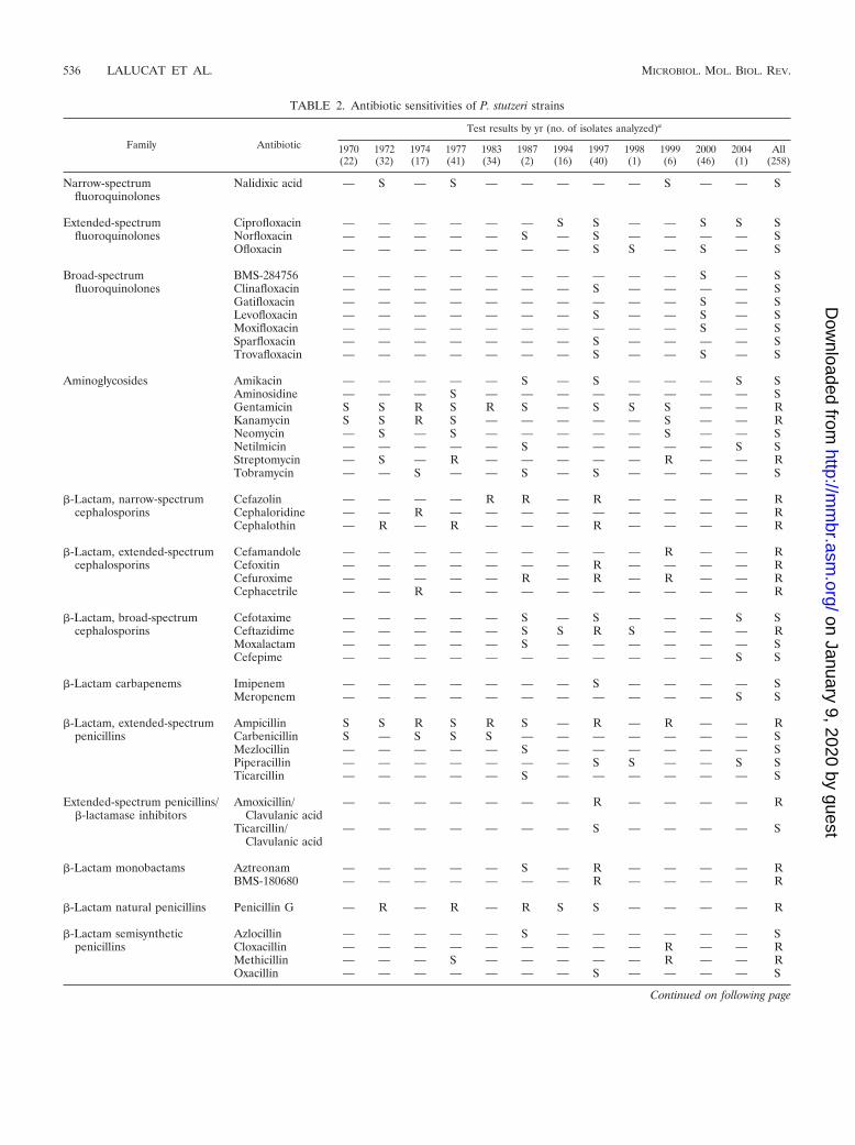
TABLE 2. Antibiotic sensitivities of P. stutzeri strains
Family Antibiotic
Test results by yr (no. of isolates analyzed)a
1970(22)
1972(32)
1974(17)
1977(41)
1983(34)
1987(2)
1994(16)
1997(40)
1998(1)
1999(6)
2000(46)
2004(1)
All(258)
Narrow-spectrumfluoroquinolones
Nalidixic acid — S — S — — — — — S — — S
Extended-spectrum Ciprofloxacin — — — — — — S S — — S S Sfluoroquinolones Norfloxacin — — — — — S — S — — — — S
Ofloxacin — — — — — — — S S — S — S
Broad-spectrum BMS-284756 — — — — — — — — — — S — Sfluoroquinolones Clinafloxacin — — — — — — — S — — — — S
Gatifloxacin — — — — — — — — — — S — SLevofloxacin — — — — — — — S — — S — SMoxifloxacin — — — — — — — — — — S — SSparfloxacin — — — — — — — S — — — — STrovafloxacin — — — — — — — S — — S — S
Aminoglycosides Amikacin — — — — — S — S — — — S SAminosidine — — — S — — — — — — — — SGentamicin S S R S R S — S S S — — RKanamycin S S R S — — — — — S — — RNeomycin — S — S — — — — — S — — SNetilmicin — — — — — S — — — — — S SStreptomycin — S — R — — — — — R — — RTobramycin — — S — — S — S — — — — S
�-Lactam, narrow-spectrum Cefazolin — — — — R R — R — — — — Rcephalosporins Cephaloridine — — R — — — — — — — — — R
Cephalothin — R — R — — — R — — — — R
�-Lactam, extended-spectrum Cefamandole — — — — — — — — — R — — Rcephalosporins Cefoxitin — — — — — — — R — — — — R
Cefuroxime — — — — — R — R — R — — RCephacetrile — — R — — — — — — — — — R
�-Lactam, broad-spectrum Cefotaxime — — — — — S — S — — — S Scephalosporins Ceftazidime — — — — — S S R S — — — R
Moxalactam — — — — — S — — — — — — SCefepime — — — — — — — — — — — S S
�-Lactam carbapenems Imipenem — — — — — — — S — — — — SMeropenem — — — — — — — — — — — S S
�-Lactam, extended-spectrum Ampicillin S S R S R S — R — R — — Rpenicillins Carbenicillin S — S S S — — — — — — — S
Mezlocillin — — — — — S — — — — — — SPiperacillin — — — — — — — S S — — S STicarcillin — — — — — S — — — — — — S
Extended-spectrum penicillins/�-lactamase inhibitors
Amoxicillin/Clavulanic acid
— — — — — — — R — — — — R
Ticarcillin/Clavulanic acid
— — — — — — — S — — — — S
�-Lactam monobactams Aztreonam — — — — — S — R — — — — RBMS-180680 — — — — — — — R — — — — R
�-Lactam natural penicillins Penicillin G — R — R — R S S — — — — R
�-Lactam semisynthetic Azlocillin — — — — — S — — — — — — Spenicillins Cloxacillin — — — — — — — — — R — — R
Methicillin — — — S — — — — — R — — ROxacillin — — — — — — — S — — — — S
Continued on following page
536 LALUCAT ET AL. MICROBIOL. MOL. BIOL. REV.
on January 9, 2020 by guesthttp://m
mbr.asm
.org/D
ownloaded from
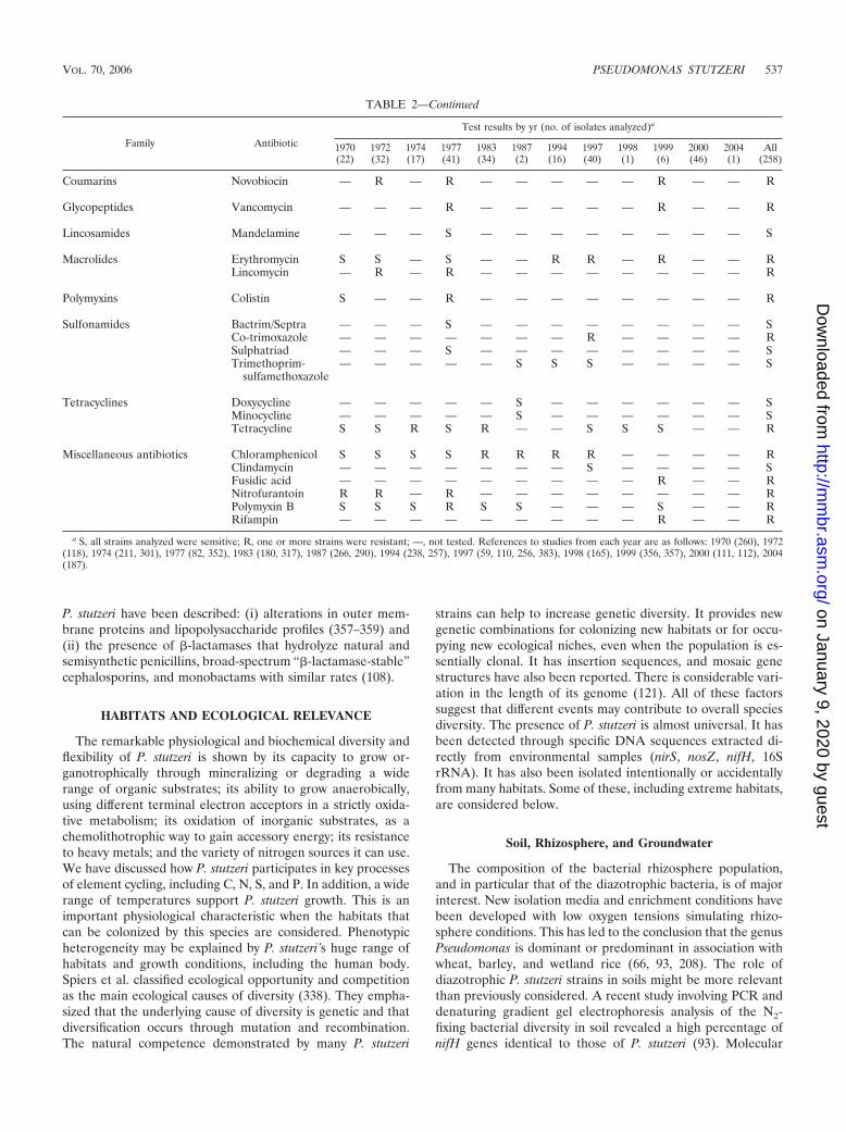
P. stutzeri have been described: (i) alterations in outer mem-brane proteins and lipopolysaccharide profiles (357–359) and(ii) the presence of �-lactamases that hydrolyze natural andsemisynthetic penicillins, broad-spectrum “�-lactamase-stable”cephalosporins, and monobactams with similar rates (108).
HABITATS AND ECOLOGICAL RELEVANCE
The remarkable physiological and biochemical diversity andflexibility of P. stutzeri is shown by its capacity to grow or-ganotrophically through mineralizing or degrading a widerange of organic substrates; its ability to grow anaerobically,using different terminal electron acceptors in a strictly oxida-tive metabolism; its oxidation of inorganic substrates, as achemolithotrophic way to gain accessory energy; its resistanceto heavy metals; and the variety of nitrogen sources it can use.We have discussed how P. stutzeri participates in key processesof element cycling, including C, N, S, and P. In addition, a widerange of temperatures support P. stutzeri growth. This is animportant physiological characteristic when the habitats thatcan be colonized by this species are considered. Phenotypicheterogeneity may be explained by P. stutzeri’s huge range ofhabitats and growth conditions, including the human body.Spiers et al. classified ecological opportunity and competitionas the main ecological causes of diversity (338). They empha-sized that the underlying cause of diversity is genetic and thatdiversification occurs through mutation and recombination.The natural competence demonstrated by many P. stutzeri
strains can help to increase genetic diversity. It provides newgenetic combinations for colonizing new habitats or for occu-pying new ecological niches, even when the population is es-sentially clonal. It has insertion sequences, and mosaic genestructures have also been reported. There is considerable vari-ation in the length of its genome (121). All of these factorssuggest that different events may contribute to overall speciesdiversity. The presence of P. stutzeri is almost universal. It hasbeen detected through specific DNA sequences extracted di-rectly from environmental samples (nirS, nosZ, nifH, 16SrRNA). It has also been isolated intentionally or accidentallyfrom many habitats. Some of these, including extreme habitats,are considered below.
Soil, Rhizosphere, and Groundwater
The composition of the bacterial rhizosphere population,and in particular that of the diazotrophic bacteria, is of majorinterest. New isolation media and enrichment conditions havebeen developed with low oxygen tensions simulating rhizo-sphere conditions. This has led to the conclusion that the genusPseudomonas is dominant or predominant in association withwheat, barley, and wetland rice (66, 93, 208). The role ofdiazotrophic P. stutzeri strains in soils might be more relevantthan previously considered. A recent study involving PCR anddenaturing gradient gel electrophoresis analysis of the N2-fixing bacterial diversity in soil revealed a high percentage ofnifH genes identical to those of P. stutzeri (93). Molecular
TABLE 2—Continued
Family Antibiotic
Test results by yr (no. of isolates analyzed)a
1970(22)
1972(32)
1974(17)
1977(41)
1983(34)
1987(2)
1994(16)
1997(40)
1998(1)
1999(6)
2000(46)
2004(1)
All(258)
Coumarins Novobiocin — R — R — — — — — R — — R
Glycopeptides Vancomycin — — — R — — — — — R — — R
Lincosamides Mandelamine — — — S — — — — — — — — S
Macrolides Erythromycin S S — S — — R R — R — — RLincomycin — R — R — — — — — — — — R
Polymyxins Colistin S — — R — — — — — — — — R
Sulfonamides Bactrim/Septra — — — S — — — — — — — — SCo-trimoxazole — — — — — — — R — — — — RSulphatriad — — — S — — — — — — — — STrimethoprim-
sulfamethoxazole— — — — — S S S — — — — S
Tetracyclines Doxycycline — — — — — S — — — — — — SMinocycline — — — — — S — — — — — — STetracycline S S R S R — — S S S — — R
Miscellaneous antibiotics Chloramphenicol S S S S R R R R — — — — RClindamycin — — — — — — — S — — — — SFusidic acid — — — — — — — — — R — — RNitrofurantoin R R — R — — — — — — — — RPolymyxin B S S S R S S — — — S — — RRifampin — — — — — — — — — R — — R
a S, all strains analyzed were sensitive; R, one or more strains were resistant; —, not tested. References to studies from each year are as follows: 1970 (260), 1972(118), 1974 (211, 301), 1977 (82, 352), 1983 (180, 317), 1987 (266, 290), 1994 (238, 257), 1997 (59, 110, 256, 383), 1998 (165), 1999 (356, 357), 2000 (111, 112), 2004(187).
VOL. 70, 2006 PSEUDOMONAS STUTZERI 537
on January 9, 2020 by guesthttp://m
mbr.asm
.org/D
ownloaded from
analysis of diazotroph diversity in the rhizosphere of smoothcordgrass (Spartina alterniflora) suggests that P. stutzeri-relatedstrains are present in the Spartina rhizosphere. Recently, anal-ysis of bacterial populations in the rhizosphere of cordgrass,based on PCR amplification of nifH sequences and separationof the amplicons by denaturing gel electrophoresis, revealednifH sequences highly similar to those of strains A1501 (aderivative of strain A15) and CMT.9.A (208, 209). The activityof an aromatic amino acid aminotransferase and the produc-tion of indole-3-acetic acid in P. stutzeri A15 have also beenreported. This may be involved in the production of growth-regulating substances in plants in addition to their nitrogen-fixing ability (261).
As mentioned above, many P. stutzeri strains have been isolatedfrom contaminated soil sites, where degradative and contami-nant-resistant strains have to develop relevant ecological activi-ties. Some strains, such as KC, and several methyl-naphthalene-degradative strains have been isolated in our laboratory fromgroundwaters contaminated with aircraft fuel (JetA1). The effi-cacy of strain KC in detoxifying groundwaters has been shownthrough bioaugmentation.
Marine Water and Sediment and Salt Marshes
Most strains isolated from marine environments and initiallyclassified in the genus Pseudomonas have been transferred toother genera after an analysis of their phylogenies. Thesetransfers include P. doudoroffii to Oceanimonas doudoroffii, P.nautica to Marinobacter hydrocarbonoclasticus, P. stanieri toMarinobacterium stanieri, P. elongata to Microbulbifer hydrolyti-cus, and P. marina to Cobetia marina. Not many species withinthe genus Pseudomonas sensu stricto have been detected inmarine waters. For a strain to be considered of marine origin,it must have the physiological characteristic of requiring, or atleast tolerating, NaCl. P. stutzeri (including strain ZoBell, for-merly P. perfectomarina), P. balearica, and P. xanthomarina(isolated from ascidian specimens in the Sea of Japan [289])seem to be true marine Pseudomonas species. In addition, P.alcaliphila and P. aeruginosa (181) have been isolated frommarine waters. Further research is required to define whetherthe latter pseudomonads might be considered marine bacteriaor allochthonous to the ecosystem.
Marine strains of P. stutzeri are located in the water columnand in sediment. The most relevant strains studied in detail areZoBell (isolated from the water column in the Pacific oceanand studied as a model denitrifier in marine environments),AN10 (isolated from polluted Mediterranean marine sedimentand studied as a naphthalene degrader), NF13 (isolated froma sample taken at 2,500- to 2,600-m depth in the Galapagos riftfrom near a hydrothermal vent and studied as a strain thatoxidizes sulfur chemolithotrophically), and strains MT-1 andHTA208 (isolated from deep-sea samples taken at the MarianaTrench at 10,897-m depth). The main ecological role of thesestrains seems to be denitrification, besides their specific phys-iological properties.
The study by Sikorski et al. (325) is the only one in which alarge number of P. stutzeri strains have been isolated from thesame sample, in this case marine sediment from the shore ofthe North Sea. This enabled a genetic study of the populationspresent in a single habitat to be undertaken.
The ability of P. stutzeri to oxidize thiosulfate to tetrathio-nate both aerobically and anaerobically was not known beforethe work of Sorokin et al. (337). Several strains were isolatedfrom the Black Sea at more than 100 m in depth. It wassuggested that this widespread bacterium could be importantin the turnover of thiosulfate in marine environments and thatit may compete with thiosulfate disproportionation and reduc-tion by thiosulfate-reducing bacteria.
Spartina marshes support high rates of macrophyte primaryproduction and microbially mediated nutrient cycling. Thepossible ecological role of P. stutzeri in such marshes seemsto be its contribution to global carbon and nitrogen budgets.Primary production and decomposition in Spartina marshesare nitrogen limited (208). In these systems, diazotrophy is akey source of new nitrogen, and denitrification completes thenitrogen cycle. P. stutzeri participates in both processes. Directmolecular analysis of diazotrophic diversity in the rhizosphereof Spartina alterniflora demonstrates that gene sequences ofnifH are highly similar to those of P. stutzeri. In addition, theyare located in the same phylogenetic branch as many othersequences of nif genes obtained from marine microorganisms.
Wastewater Treatment Plants
To screen bacteria with unusual metabolic properties, suchas the degradation of anthropogenic compounds for bioreme-diation purposes, it is common to examine samples taken fromwastewater treatment plants or to design bioreactors imitatingthe conditions of a treatment plant. Naphthalene degraders,thiosulfate oxidizers, chlorobenzoate degraders, and cyanideoxidizers have been isolated in this way. It has been demon-strated that P. stutzeri is also distributed in wastewater. How-ever, no attempt has been made to quantify P. stutzeri in suchhabitats or to determine its relevance.
CONCLUSIONS
P. stutzeri genomovars can be considered genomospecies, asdefined by J. P. Euzeby. According to his recommendations, ifa genomospecies has been identified it is possible to look forphenotypic traits that differentiate it from the other genomo-species. If the genomospecies can be identified phenotypically,it must receive a name and be converted into a new species. Ifno phenotypic characteristic can be used to identify the genomo-species easily, it is left without a name. We prefer to maintainthe genomovar concept for the genomic groups in P. stutzeri,because all of them share the basic phenotypic traits of thespecies. If DNA-DNA similarity results, or a multigenic se-quencing approach, are accepted as the only criteria for speciesdelineation, then P. stutzeri should be split into 17 differentspecies. However, in our opinion, this situation would not helpto clarify the taxonomic position of a phylogenetic and pheno-typically coherent group of strains, as is the case for membersof P. stutzeri.
As demonstrated, genomovars are monophyletic biologicaland evolutionary units in which different ecotypes may be dif-ferentiated by their adaptation to new environmental condi-tions. P. stutzeri is widely distributed in natural environmentsand shows great metabolic versatility, which is consistent witha large effective population size. This species shows very low
538 LALUCAT ET AL. MICROBIOL. MOL. BIOL. REV.
on January 9, 2020 by guesthttp://m
mbr.asm
.org/D
ownloaded from
recombination rates. When there is a large population size andno assortive recombination, bacterial clones diverge freely byaccumulating neutral mutations. The occurrence in a particu-lar population of adaptive mutations conferring selective ad-vantages in specific ecological situations leads to the elimina-tion of genetic diversity within the population. However, in thepresence of very low recombination rates, such mutations donot prevent genetic divergence between populations. Thus, theexceptionally high genetic diversity of P. stutzeri may be theresult of niche-specific selection that occurs during coloniza-tion and adaptation to a wide range of microenvironments.Horizontal gene transfer seems to be an efficient mechanismfor introducing new phenotypes into the genomes of P. stutzeri,without affecting the housekeeping genes. Integrons may playan important role in the acquisition of these new properties.
In conclusion, P. stutzeri exhibits exceptionally high diversitywithin a clonal population structure. In such cases, the exis-tence of a strong linkage disequilibrium can be explained byconsidering that P. stutzeri forms a metapopulation made up ofmultiple ecological populations. These populations occupy dif-ferent ecological niches. Although recombination is possiblewithin populations, it is rare or absent between different pop-ulations (216, 278, 410). More-extensive studies are required toassess the population structure of these ecological populationsof P. stutzeri. However, the results reported to date are consis-tent with the conclusion that this bacterial species represents agood example of a phenotypically cosmopolitan ecological spe-cies sensu Istock (160), i.e., a species characterized by limitedphenotypic variation, restricted local sets of genetic clones, andno or rare recombination. The clonal sets are genetically di-verse, but phenotypic resemblance is sufficient to make phen-etic classification and identification possible. P. stutzeri is thespecies with the highest genetic diversity described to date.MLEE and MLST data confirm the results obtained by othertechniques that have shown that some clones of P. stutzeri aredistinct enough to warrant taxonomic differentiation (23).
ACKNOWLEDGMENTS
We thank many colleagues for stimulating discussions and for pro-viding most of the strains described in the text.
The work in the Spanish laboratory was supported by researchgrants from the CICYT (Spain) and from the “Pla de Recerca iDesenvolupament Tecnologic de les Illes Balears” (PRDIB).
REFERENCES
1. Achenbach, L. A., U. Michaelidou, R. A. Bruce, J. Fryman, and J. D. Coates.2001. Dechloromonas agitata gen. nov., sp. nov. and Dechlorosoma suillumgen. nov., sp. nov., two novel environmentally dominant (per)chlorate-reducing bacteria and their phylogenetic position. Int. J. Syst. Evol. Micro-biol. 51:527–533.
2. Ahamad, P. Y. A., and A. A. M. Kunhi. 1996. Degradation of phenolthrough ortho-cleavage pathway by Pseudomonas stutzeri strain SPC2. Lett.Appl. Microbiol. 22:26–29.
3. Alexander, J. J., and J. F. Lewis. 1976. Pitting of agar surface by Pseudo-monas stutzeri. J. Clin. Microbiol. 3:381–385.
4. Alvarez, M. L., J. Ai, W. G. Zumft, J. Sanders-Loehr, and D. M. Dooley.2001. Characterization of the copper-sulfur chromophores in nitrous oxidereductase by resonance raman spectroscopy: evidence for sulfur coordina-tion in the catalytic cluster. J. Am. Chem. Soc. 123:576–587.
5. Amann, R., W. Ludwig, R. Shulze, S. Spring, E. Moore, and K.-H. Schleifer.1996. rRNA-targeted oligonucleotide probes for the identification of gen-uine and former Pseudomonas. Syst. Appl. Microbiol. 19:501–509.
6. Andrade, G., E. Esteban, L. Velasco, M. J. Lorite, and E. J. Bedmar. 1997.Isolation and identification of N2-fixing microorganisms from the rhizo-sphere of Capparis spinosa (L.). Plant Soil 197:19–23.
7. Antonioli, P., G. Zapparoli, P. Abbruscato, C. Sorlini, G. Ranalli, and P. G.
Righetti. 2005. Art-loving bugs: the resurrection of Spinello Aretino fromPisa’s cemetery. Proteomics 5:2453–2459.
8. Arai, H., Y. Igarashi, and T. Kodama. 1995. Expression of the nir and norgenes for denitrification of Pseudomonas aeruginosa requires a novel CRP/FNR-related transcriptional regulator, DNR, in addition to ANR. FEBSLett. 371:73–76.
9. Arenghi, F. L. G., M. Pinti, E. Galli, and P. Barbieri. 1999. Identification ofthe Pseudomonas stutzeri OX1 toluene-o-xylene monooxygenase regulatorygene (touR) and of its cognate promoter. Appl. Environ. Microbiol. 65:4057–4063.
10. Arenghi, F. L. G., P. Barbieri, G. Bertoni, and V. de Lorenzo. 2001. Newinsights into the activation of o-xylene biodegradation in Pseudomonasstutzeri OX1 by pathway substrates. EMBO Reps. 2:409–414.
11. Arenghi, F. L. G., D. Berlanda, E. Galli, G. Sello, and P. Barbieri. 2001.Organization and regulation of meta cleavage pathway genes for tolueneand o-xylene derivative degradation in Pseudomonas stutzeri OX1. Appl.Environ. Microbiol. 67:3304–3308.
12. Badar, U., N. Ahmed, A. J. Beswick, P. Pattanapipitpaisal, and L. E.Macaskie. 2000. Reduction of chromate by microorganisms isolated frommetal contaminated sites of Karachi, Pakistan. Biotechnol. Lett. 22:829–836.
13. Baggi, G., P. Barbieri, E. Galli, and S. Tollari. 1987. Isolation of a Pseudo-monas stutzeri strain that degrades ortho-xylene. Appl. Environ. Microbiol.53:2129–2132.
14. Baikalov, I., I. Schroder, M. Kaczor-Grzeskowiak, D. Cascio, R. P. Gunsalus,and R. E. Dickerson. 1998. NarL dimerization? Suggestive evidence from anew crystal form. Biochemistry 37:3665–3676.
15. Balashov, S. V., and A. M. Boronin. 1996. Sewage-sludge bacterial isolatesdecomposing sulfoaromatic compounds. Microbiology 65:549–552.
16. Barbieri, P., G. Galassi, and E. Galli. 1989. Plasmid-encoded mercuryresistance in a Pseudomonas stutzeri strain that degrades ortho-xylene.FEMS. Microbiol. Ecol. 62:375–383.
17. Barbieri, P., F. L. G. Arenghi, G. Bertoni, F. Bolognese, and E. Galli. 2001.Evolution of catabolic pathways and metabolic versatility in Pseudomonasstutzeri OX1. Antonie Leeuwenhoek 79:135–140.
18. Barraquio, W. L., A. Dumont, and R. Knowles. 1988. A method for count-ing free-living anaerobic N2-fixing H2-oxidizing bacteria. In H. Bothe, F. J.Bruijn, and W. E. Newton (ed.), Nitrogen fixation a hundred years after.Gustav-Fischer, Stuttgart, Germany.
19. Baumann, P., R. D. Bowditch, L. Baumann, and B. Beaman. 1983. Taxon-omy of marine Pseudomonas species: P. stanieri sp. nov.; P. perfectomarinasp. nov. nom. rev.; P. nautica; and P. doudoroffii. Int. J. Syst. Bacteriol.33:857–865.
20. Baya, A. M., P. R. Brayton, V. L. Brown, D. J. Grimes, E. Russek-Cohen,and R. R. Collwell. 1986. Coincident plasmids and antimicrobial resistancein marine bacteria isolated from polluted and unpolluted Atlantic Oceansamples. Appl. Environ. Microbiol. 51:1285–1292.
21. Bell, L. C., D. J. Richardson, and S. J. Ferguson. 1990. Periplasmic andmembrane-bound respiratory nitrate reductases in Thiosphaera pantotro-pha: the periplasmic enzyme catalyzes the first step in aerobic denitrifica-tion. FEBS Lett. 265:85–87.
22. Bender, K. S., M. R. Rice, W. H. Fugate, J. D. Coates, and L. A. Achenbach.2004. Metabolic primers for detection of (per)chlorate-reducing bacteria inthe environment and phylogenetic analysis of cld gene sequences. Appl.Environ. Microbiol. 70:5651–5658.
23. Bennasar, A., R. Rossello-Mora, J. Lalucat, and E. R. B. Moore. 1996. 16SrRNA gene sequence analysis relative to genomovars of Pseudomonasstutzeri and proposal of Pseudomonas balearica sp. nov. Int. J. Syst. Bacte-riol. 46:200–205.
24. Bennasar, A., C. Guasp, and J. Lalucat. 1998. Molecular methods for thedetection and identification of Pseudomonas stutzeri in pure culture andenvironmental samples. Microb. Ecol. 35:22–33.
25. Bennasar, A., C. Guasp, M. Tesar, and J. Lalucat. 1998. Genetic relation-ships among Pseudomonas stutzeri strains based on molecular typing meth-ods. J. Appl. Microbiol. 85:643–656.
26. Bergan, T. 1981. Human- and animal-pathogenic members of the genusPseudomonas, p. 666–700. In A. Balows, H. G. Truper, H. Dworkin, W.Harder, and K. H. Schleifer (ed.), The prokaryotes, vol. I. Springer-Verlag,Berlin, Germany.
27. Bergey, D. H. 1930. Bergey’s manual of determinative bacteriology, 3rd ed.The Williams and Wilkins Co., Baltimore, Md.
28. Bergey, D. H., F. C. Harrison, R. S. Breed, B. W. Hammer, and F. M.Huntoon. 1923. Bergey’s manual of determinative bacteriology, 1st ed. TheWilliams and Wilkins Co., Baltimore, Md.
29. Berks, B. C., M. D. Page, D. J. Richardson, A. Reilly, A. Cavill, F. Outen,and S. J. Ferguson. 1995. Sequence analysis of subunits of the membranebound nitrate reductase from a denitrifying bacterium: the integral mem-brane subunit provides a prototype for the dihaem electron-carrying arm ofa redox loop. Mol. Microbiol. 15:319–331.
30. Berks, B. C., F. Sargent, and T. Palmer. 2000. The Tat protein exportpathway. Mol. Microbiol. 35:260–274.
31. Berndt, C., P. Meier, and W. Wackernagel. 2003. DNA restriction is a
VOL. 70, 2006 PSEUDOMONAS STUTZERI 539
on January 9, 2020 by guesthttp://m
mbr.asm
.org/D
ownloaded from

barrier to natural transformation in Pseudomonas stutzeri JM300. Microbi-ology 149:895–901.
32. Bertoni, G., F. Bolognese, E. Galli, and P. Barbieri. 1996. Cloning of thegenes for and characterization of the early stages of toluene and o-xylenecatabolism in Pseudomonas stutzeri OX1. Appl. Environ. Microbiol. 62:3704–3711.
33. Bertoni, G., M. Martino, E. Galli, and P. Barbieri. 1998. Analysis of thegene cluster encoding toluene/o-xylene monooxygenase from Pseudomonasstutzeri OX1. Appl. Environ. Microbiol. 64:3626–3632.
34. Bhagat, R., and S. Srivastava. 1993. Growth response of Pseudomonasstutzeri RS34 to ethylenediaminetetraacetic acid (EDTA) and its interac-tion with zinc. Indian J. Exp. Biol. 31:590–594.
35. Bhagat, R., and S. Srivastava. 1993. Biohydrometallurgical technologies,vol. II. The Minerals, Metals and Materials Society, Warrendale, Pa.
36. Bhagat, R., and S. Srivastava. 1994. Effect of zinc on morphology andultrastructure of Pseudomonas stutzeri Rs34. J. Gen. Appl. Microbiol. 40:265–270.
37. Bhagat, R., and S. Srivastava. 1995. Environmental biotechnology: princi-ples and applications. Kluwer Academic Publishers, Amsterdam, The Neth-erlands.
38. Biguzzi, M., and G. Shama. 1994. Effect of titanium-dioxide concentrationon the survival of Pseudomonas stutzeri during irradiation with near-utra-violet light. Lett. Appl. Microbiol. 19:458–460.
39. Blumle, S., and W. G. Zumft. 1991. Respiratory nitrate reductase fromdenitrifying Pseudomonas stutzeri: purification, properties and target of prote-olysis. Biochim. Biophys. Acta 1057:102–108.
40. Bolognese, F., C. Di Lecce, E. Galli, and P. Barbieri. 1999. Activation andinactivation of Pseudomonas stutzeri methylbenzene catabolism pathwaysmediated by a transposable element. Appl. Environ. Microbiol. 65:1876–1882.
41. Borcherding, H., S. Leikefeld, C. Frey, S. Diekmann, and P. Steinrucke.2000. Enzymatic microtiter plate-based nitrate detection in environmentaland medical analysis. Anal. Biochem. 282:1–9.
42. Bosch, R., E. Garcia-Valdes, and E. R. B. Moore. 1999. Genetic character-ization and evolutionary implications of a chromosomally encoded naph-thalene-degradation upper pathway from Pseudomonas stutzeri AN10.Gene 236:149–157.
43. Bosch, R., E. R. B. Moore, E. Garcia-Valdes, and D. H. Pieper. 1999. NahW,a novel, inducible salicylate hydroxylase involved in mineralization of naph-thalene by Pseudomonas stutzeri AN10. J. Bacteriol. 181:2315–2322.
44. Bosch, R., E. Garcıa-Valdes, and E. R. B. Moore. 2000. Complete nucleo-tide sequence and evolutionary significance of a chromosomally encodednaphthalene-degradation lower pathway from Pseudomonas stutzeri AN10.Gene 245:65–74.
45. Bothe, H., G. Jost, M. Schloter, B. B. Ward, and K. Witzel. 2000. Molecularanalysis of ammonia oxidation and denitrification in natural environments.FEMS Microbiol. Rev. 24:673–690.
46. Braker, G., J. Z. Zhou, L. Wu, A. H. Devol, and J. M. Tiedje. 2000. Nitritereductase genes (nirK and nirS) as funtional markers to investigate diversityof denitrifying bacteria in Pacific Northwest marine sediment communities.Appl. Environ. Microbiol. 66:2096–2104.
47. Braun, C., and W. G. Zumft. 1991. Marker exchange of the structural genesfor nitric oxide reductase blocks the denitrification pathway of Pseudomo-nas stutzeri at nitric oxide. J. Biol. Chem. 266:22785–22788.
48. Braun, C., and W. G. Zumft. 1992. The structural genes of the nitric oxidereductase complex from Pseudomonas stutzeri are part of a 30-kilobase genecluster for denitrification. J. Bacteriol. 174:2394–2397.
49. Brenner, C. 2002. Catalysis in the nitrilase superfamily. Curr. Opin. Struct.Biol. 12:775–782.
50. Brenner, D. J., J. T. Staley, and N. R. Krieg. 2001. Classification of pro-karyotic species and the concept of bacterial speciation, p. 27–31. In G. M.Garrity (ed.), Bergey’s manual of systematic bacteriology, 2nd ed., vol. I.Springer, New York, N.Y.
51. Brosch, R., M. Lefevre, F. Grimont, and P. A. D. Grimont. 1996. Taxonomicdiversity of pseudomonads revealed by computer-interpretation of ribotyp-ing data. Syst. Appl. Microbiol. 19:541–555.
52. Brozel, V. S., and T. E. Cloete. 1991. Resistance of bacteria from coolingwaters to bactericides. J. Ind. Microbiol. 8:273–276.
53. Brozel, V. S., B. Pietersen, and T. E. Cloete. 1993. Adaptation of bacterialcultures to nonoxidizing water-treatment bactericides. Water SA 19:259–262.
54. Brozel, V. S., B. Pietersen, and T. E. Cloete. 1995. Resistance of bacterialcultures to nonoxidizing water-treatment bactericides by adaptation. WaterSci. Technol. 31:169–175.
55. Burri, R., and A. Stutzer. 1895. Ueber Nitrat zerstorende Bakterien undden durch dieselben bedingten Stickstoffverlust. Zentbl. Bakteriol. Para-sitenkd. Abt. II 1:257–265, 350–364, 392–398, 422–432.
56. Busch, S., C. Rosenplanter, and B. Averhoff. 1999. Identification and char-acterization of ComE and ComF, two novel pilin-like competence factorsinvolved in natural transformation of Acinetobacter sp. strain BD413. Appl.Environ. Microbiol. 65:4568–4574.
57. Busse, J., and G. Auling. 1988. Polyamine pattern as a chemotaxonomicmarker within the Proteobacteria. Syst. Appl. Microbiol. 11:1–8.
58. Cafaro, V., V. Izzo, R. Scognamiglio, E. Notomista, P. Capasso, A. Casbarra, P.Pucci, and A. Di Donato. 2004. Phenol hydroxylase and toluene/o-xylenemonooxygenase from Pseudomonas stutzeri OX1: interplay between twoenzymes. Appl. Environ. Microbiol. 70:2211–2219.
59. Campos-Herrero, M. I., A. Bordes, H. Rodrıguez, A. Perera, B. Gonzalez,and A. Conde. 1997. Pseudomonas stutzeri community-acquired pneumoniaassociated with empyema: case report and review. Clin. Infect. Dis. 25:325–326.
60. Carlson, C. A., L. S. Pierson, J. J. Rosen, and J. L. Ingraham. 1983.Pseudomonas stutzeri and related species undergo natural transformation. J.Bacteriol. 153:93–99.
61. Carlson, C. A., S. M. Steenbergen, and J. L. Ingraham. 1984. Naturaltransformation of Pseudomonas stutzeri by plasmids that contain clonedfragments of chromosomal deoxyribonucleic acid. Arch. Microbiol. 140:134–138.
62. Carratala, J., A. Salazar, J. Mascaro, and M. Santin. 1992. Community-acquired pneumonia due to Pseudomonas stutzeri. Clin. Infect. Dis. 14:792.
63. Chakraborty, R. N., H. N. Patel, and S. B. Desai. 1990. Isolation and partialcharacterization of catechol-type siderophore from Pseudomonas stutzeriRc-7. Curr. Microbiol. 20:283–286.
64. Chakraborty, R. N., H. N. Patel, and S. B. Desai. 1994. Studies on ironuptake and its effect on the outer-membrane proteins of Pseudomonasstutzeri Rc-7. Curr. Microbiol. 28:321–323.
65. Chan, Y., and R. Wheatcroft. 1993. Detection of nitrous oxide reductasestructural gene in Rhizobium meliloti strains and its location on the nodmegaplasmid of JJc10 and SU47. J. Bacteriol. 175:19–26.
66. Chan, Y.-K., W. L. Barraquio, and R. Knowles. 1994. N2-fixing pseudo-monads and related soil bacteria. FEMS Microbiol. Rev. 13:95–117.
67. Chen, I., and D. Dubnau. 2004. DNA uptake during bacterial transforma-tion. Nat. Rev. Microbiol. 2:241–249.
68. Chester, F. D. 1901. A manual of determinative bacteriology. Macmillan,New York, N.Y.
69. Cho, J. S., J. S. Hur, B. H. Kang, P. J. Kim, B. K. Sohn, H. J. Lee, Y. K.Jung, and J. S. Heo. 2001. Biosorption of copper by immobilized biomass ofPseudomonas stutzeri. J. Microbiol. Biotechnol. 11:964–972.
70. Choudhury, R., and S. Srivastava. 2001. Zinc resistance mechanisms inbacteria. Curr. Sci. 81:768–775.
71. Chung, Y. S., and D. Dubnau. 1998. All seven comG open reading framesare required for DNA binding during the transformation of competentBacillus subtilis. J. Bacteriol. 180:41–45.
72. Cladera, A. M., A. Bennasar, M. Barcelo, J. Lalucat, and E. Garcıa-Valdes.2004. Comparative genetic diversity of Pseudomonas stutzeri genomovars,clonal structure, and phylogeny of the species. J. Bacteriol. 186:5239–5248.
73. Cladera, A. M., L. C. Sepulveda, M. Valens-Vadell, J. M. Meyer, J. Lalucat,and E. Garcıa-Valdes. 6 December 2005, posting date. A detailed pheno-typic and genotypic description of Pseudomonas strain OX1. Syst. Appl.Microbiol. doi:10.1016/j.syapm.2005.11.007.
74. Cladera, A. M., E. Garcıa-Valdes, and J. Lalucat. 2006. Genotype versusphenotype in the circumscription of bacterial species: the case of Pseudo-monas stutzeri and P. chloritidismutans. Arch. Microbiol. 184:353–361.
75. Coates, J. D., and L. A. Achenbach. 2004. Microbial perchlorate reduction:rocket-fuelled metabolism. Nat. Rev. Microbiol. 2:569–580.
76. Collwell, R. R. 1970. Poliphasic taxonomy of bacteria, p. 431–436. In H.Iizuka and T. Hasegawa (ed.), Culture collections of microorganisms. Uni-versity of Tokyo Press, Tokyo, Japan.
77. Conrad, R. 1996. Soil microorganisms as controllers of atmospheric tracegases (H2, CO, CH4, OCS, N2O, and NO). Microbiol. Rev. 60:609–640.
78. Cowan, D., R. Cramp, R. Pereira, D. Graham, and Q. Almatawah. 1998.Biochemistry and biotechnology of mesophilic and thermophilic nitrile me-tabolizing enzymes. Extremophiles 2:207–216.
79. Cox, E. E., D. Major, and E. Edwards. 2000. Natural attenuation of 1,2-dichloroethane in groundwater at a chemical manufacturing facility. InG. B. Wickramanayake, A. R. Gavaskar, B. C. Alleman, and V. S. Magar(ed.), The second international conference on remediation of chlorinatedand recalcitrant compounds. Batelle, Monterey, Calif.
80. Cozar, E. 1987. Caracterizacion taxonomica, fisiologica y bioquımica denuevas Pseudomonas marinas degradadoras de hidrocarburos cıclicos.Ph.D. thesis. Universitat de les Illes Balears (UIB), Palma de Mallorca,Spain.
81. Criddle, C. S., J. T. DeWitt, D. Grbic-Galic, and P. L. McCarty. 1990.Transformation of carbon tetrachloride by Pseudomonas sp. strain KCunder denitrification conditions. Appl. Environ. Microbiol. 56:3240–3246.
82. Csukas, C., and P. Juhasz. 1977. Incidence of Pseudomonas stutzeri inpatients with pulmonary diseases. Acta Microbiol. (Hungary) 24:94–104.
83. Cutruzzola, F. 1999. Bacterial nitric oxide synthesis. Biochim. Biophys.Acta 1411:231–249.
84. Cuypers, H., A. Viebrock-Sambale, and W. G. Zumft. 1992. NosR, a mem-brane-bound regulatory component necessary for expression of nitrousoxide reductase in denitrifying Pseudomonas stutzeri. J. Bacteriol. 174:5332–5339.
540 LALUCAT ET AL. MICROBIOL. MOL. BIOL. REV.
on January 9, 2020 by guesthttp://m
mbr.asm
.org/D
ownloaded from
85. Cuypers, H., and W. G. Zumft. 1992. Regulatory components of the deni-trification gene cluster of Pseudomonas stutzeri, p. 188–197. In E. Galli, S.Silver, and B. Witholt (ed.), Pseudomonas: molecular biology and biotech-nology. ASM Press, Washington, D.C.
86. Cuypers, H., J. Berghofer, and W. G. Zumft. 1995. Multiple nosZ promotersand anaerobic expression of nos genes necessary for Pseudomonas stutzerinitrous oxide reductase and assembly of its copper centers. Biochim. Bio-phys. Acta 1264:183–190.
87. Dagley, S. 1986. Biochemistry of aromatic hydrocarbon degradation inpseudomonads, p. 527–555. In J. Sokatch and J. L. Ornston (ed.), Thebacteria, vol. 10. Academic Press Inc., Orlando, Fla.
88. de Boer, A. P., W. N. Reijnders, J. G. Kuenen, A. H. Stouthamer, and R. J.van Spanning. 1994. Isolation, sequencing and mutational analysis of a genecluster involved in nitrite reduction in Paracoccus denitrificans. AntonieLeeuwenhoek 66:111–127.
89. Demoss, J. A., T. Y. Fan, and R. H. Scott. 1981. Characterization of subunitstructural alterations which occur during purification of nitrate reductasefrom Escherichia coli. Arch. Biochem. Biophys. 206:54–64.
90. Dercova, K., B. Vrana, S. Balaz, and A. Sandorova. 1996. Biodegradationand evaporation of polychlorinated biphenyls (PCBs) in liquid media.J. Ind. Microbiol. 16:325–329.
91. Dermastia, M., T. Turk, and T. C. Hollocher. 1991. Nitric oxide reductase.Purification from Paracoccus denitrificans with use of a single column andsome characteristics. J. Biol. Chem. 266:10899–10905.
92. Desnoues, N., M. Lin, X. W. Guo, L. Y. Ma, R. Carreno-Lopez, and C.Elmerich. 2003. Nitrogen fixation genetics and regulation in a Pseudomonasstutzeri strain associated with rice. Microbiology 149:2251–2262.
93. Diallo, M. D., A. Willems, N. Vloemans, S. Cousin, T. T. Vandekerckhove,P. de Lajudie, W. Vyverman, M. Gillis, and K. Van der Gucht. 2004.Polymerase chain reaction denaturing gradient gel electrophoresis analysisof the N2-fixing bacterial diversity in soil under Acacia tortilis ssp. raddianaand Balanites aegyptiaca in the dryland part of Senegal. Environ. Microbiol.6:400–415.
94. Dias, J. M., M. E. Than, A. Humm, R. Huber, G. P. Bourenkov, H. D.Bartunik, S. Bursakov, J. Calvete, J. Caldeira, C. Carneiro, J. J. Moura, I.Moura, and M. J. Romao. 1999. Crystal structure of the first dissimilatorynitrate reductase at 1.9 A solved by MAD methods. Structure 7:65–79.
95. Dijk, J. A., A. J. M. Stams, G. Schraa, H. Ballerstedt, J. A. M. de Bont, andJ. Gerritse. 2003. Anaerobic oxidation of 2-chloroethanol under denitrify-ing conditions by Pseudomonas stutzeri strain JJ. Appl. Microbiol. Biotech-nol. 63:68–74.
96. Dijk, J. A., J. Gerritse, and G. Schraa. 2004. Degradation pathway of2-chloroethanol in Pseudomonas stutzeri strain JJ under denitrifying condi-tions. Arch. Microbiol. 182:514–519.
97. DiLecce, C., M. Accarino, F. Bolognese, E. Galli, and P. Barbieri. 1997.Isolation and metabolic characterization of a Pseudomonas stutzeri mutantable to grow on the three isomers of xylene. Appl. Environ. Microbiol.63:3279–3281.
98. Dingle, K. E., F. M. Colles, D. R. A. Wareing, R. Ure, A. J. Fox, F. E. Bolton,H. J. Bootsma, R. J. L. Willems, R. Urwin, and M. C. J. Maiden. 2001.Multilocus sequence typing system for Campylobacter jejuni. J. Clin. Micro-biol. 39:14–23.
99. Dreusch, A., J. Riester, P. M. Kroneck, and W. G. Zumft. 1996. Mutationof the conserved Cys165 outside of the CuA domain destabilizes nitrousoxide reductase but maintains its catalytic activity. Evidence for disulfidebridges and a putative protein disulfide isomerase gene. Eur. J. Biochem.237:447–453.
100. Dubey, S. K., and D. S. Holmes. 1995. Biological cyanide destruction me-diated by microorganisms. World J. Microbiol. Biotechnol. 11:257–265.
101. Dybas, M. J., G. M. Tatara, and C. S. Criddle. 1995. Localization andcharacterization of the carbon-tetrachloride transformation activity ofPseudomonas sp. strain KC. Appl. Environ. Microbiol. 61:758–762.
102. Dybas, M. J., M. Barcelona, S. Bezborodnikov, S. Davies, L. Forney,H. Heuer, O. Kawka, T. Mayotte, L. Sepulveda-Torres, K. Smalla, M.Sneathen, J. Tiedje, T. Voice, D. C. Wiggert, M. E. Witt, and C. S. Criddle.1998. Pilot-scale evaluation of bioaugmentation for in-situ remediation of acarbon tetrachloride contaminated aquifer. Environ. Sci. Technol. 32:3598–3611.
103. Ebbs, S. 2004. Biological degradation of cyanide compounds. Curr. Opin.Biotechnol. 15:231–236.
104. Ergin, C., and G. Mutlu. 1999. Clinical distribution and antibiotic resistanceof Pseudomonas species. East. J. Med. 4:65–69.
105. Fallik, E., Y. K. Chan, and R. L. Robson. 1991. Detection of alternativenitrogenases in aerobic gram-negative nitrogen-fixing bacteria. J. Bacteriol.173:365–371.
106. Farrar, J. A., W. G. Zumft, and A. J. Thomson. 1998. CuA and CuZ arevariants of the electron transfer center in nitrous oxide reductase. Proc.Natl. Acad. Sci. USA 95:9891–9896.
107. Fedi, S., M. Carnevali, F. Fava, A. Andracchio, S. Zappoli, and D. Zannoni.2001. Polychlorinated biphenyl degradation activities and hybridizationanalyses of fifteen aerobic strains isolated from a PCB-contaminated site.Res. Microbiol. 152:583–592.
108. Franceschini, N., M. Galleni, J. M. Frere, A. Oratore, and G. Amicosante.1993. A class-a beta-lactamase from Pseudomonas stutzeri that is highlyactive against monobactams and cefotaxime. Biochem. J. 292:697–700.
109. Frunzke, K., B. Heiss, O. Meyer, and W. G. Zumft. 1993. Molybdopteringuanine dinucleotide is the organic moiety of the molybdenum cofactor inrespiratory nitrate reductase from Pseudomonas stutzeri. FEMS Microbiol.Lett. 113:241–245.
110. Fung-Tomc, J., K. Bush, B. Minassian, B. Kolek, R. Flamm, E. Gradelski,and D. Bonner. 1997. Antibacterial activity of BMS-180680, a new catechol-containing monobactam. Antimicrob. Agents Chemother. 41:1010–1016.
111. Fung-Tomc, J., B. Minassian, B. Kolek, T. Washo, E. Huczko, and D.Bonner. 2000. In vitro antibacterial spectrum of a new broad-spectrum8-methoxy fluoroquinolone, gatifloxacin. J. Antimicrob. Chemother. 45:437–446.
112. Fung-Tomc, J. C., B. Minassian, B. Kolek, E. Huczko, L. Aleksunes, T.Stickle, T. Washo, E. Gradelski, L. Valera, and D. P. Bonner. 2000. Anti-bacterial spectrum of a novel des-fluoro(6) quinolone, BMS-284756. Anti-microb. Agents Chemother. 44:3351–3356.
113. Garcıa-Valdes, E., E. Cozar, R. Rotger, J. Lalucat, and J. Ursing. 1988.New naphthalene-degrading marine Pseudomonas strains. Appl. Environ.Microbiol. 54:2478–2485.
114. Garcıa-Valdes, E., M. M. Castillo, A. Bennasar, C. Guasp, A. M. Cladera,R. Bosch, K. H. Engesser, and J. Lalucat. 2003. Polyphasic characterizationof Pseudomonas stutzeri CLN100, which simultaneously degrades chloro-and methylaromatics: a new genomovar within the species. Syst. Appl.Microbiol. 26:390–403.
115. Gavini, F., B. Holmes, D. Izard, A. Beji, A. Bernigaud, and E. Jackubczac.1989. Numerical taxonomy of Pseudomonas alcaligenes, P. pseudoalcali-genes, P. mendocina, P. stutzeri and related bacteria. Int. J. Syst. Bacteriol.39:135–144.
116. Gerritse, J., A. Borger, E. van Heiningen, H. H. M. Rijnaarts, T. N. P.Bosma, J. Taat, B. van Winden, J. A. Dijk, and J. A. M. de Bont. 1999.Assesment and monitoring of 1,2-dichloroethane dechlorination, p. 73–80.In A. Leeson and B. C. Alleman (ed.), Engineered approaches for in situbioremediation of chlorinated solvent contamination. Batelle, Columbus,Ohio.
117. Gilardi, G. L. 1971. Characterization of Pseudomonas species isolated fromclinical specimens. Appl. Microbiol. 21:414.
118. Gilardi, G. L. 1972. Infrequently encountered Pseudomonas species causinginfections in humans. Ann. Intern. Med. 77:211–215.
119. Gilardi, G. L., and H. J. Mankin. 1973. Infection due to Pseudomonasstutzeri. N. Y. State J. Med. 73:2789–2791.
120. Ginard, M. 1997. Organizacion genomica de Pseudomonas stutzeri y susimplicaciones taxonomicas y evolutivas. Ph.D. thesis. Universitat de les IllesBalears (UIB), Palma de Mallorca, Spain.
121. Ginard, M., J. Lalucat, B. Tummler, and U. Romling. 1997. Genomeorganization of Pseudomonas stutzeri and resulting taxonomic and evolu-tionary considerations. Int. J. Syst. Bacteriol. 47:132–143.
122. Giuffre, A., G. Stubauer, P. Sarti, M. Brunori, W. G. Zumft, G. Buse, andT. Soulimane. 1999. The heme-copper oxidases of Thermus thermophiluscatalyze the reduction of nitric oxide: evolutionary implications. Proc. Natl.Acad. Sci. USA 96:14718–14723.
123. Glockner, A. B., and W. G. Zumft. 1996. Sequence analysis of an internal9.72-kb segment from the 30-kb denitrification gene cluster of Pseudomonasstutzeri. Biochim. Biophys. Acta 1277:6–12.
124. Goetz, A., V. L. Yu, J. E. Hanchett, and J. D. Rihs. 1983. Pseudomonasstutzeri bacteremia associated with hemodialysis. Arch. Intern. Med. 143:1909–1912.
125. Graupner, S., V. Frey, R. Hashemi, M. G. Lorenz, G. Brandes, and W.Wackernagel. 2000. Type IV pilus genes pilA and pilC of Pseudomonasstutzeri are required for natural genetic transformation, and pilA can bereplaced by corresponding genes from nontransformable species. J. Bacte-riol. 182:2184–2190.
126. Graupner, S., and W. Wackernagel. 2001. Identification and characteriza-tion of novel competence genes comA and exbB involved in natural genetictransformation of Pseudomonas stutzeri. Res. Microbiol. 152:451–460.
127. Graupner, S., and W. Wackernagel. 2001. Pseudomonas stutzeri has twoclosely related pilA genes (type IV pilus structural protein) with oppositeinfluences on natural genetic transformation. J. Bacteriol. 183:2359–2366.
128. Graupner, S., N. Weger, M. Sohni, and W. Wackernagel. 2001. Require-ment of novel competence genes pilT and pilU of Pseudomonas stutzeri fornatural transformation and suppression of pilT deficiency by a hexahistidinetag on the type IV pilus protein PilAI. J. Bacteriol. 183:4694–4701.
129. Grigor’eva, N. V., Z. A. Avakyan, T. P. Tourova, T. F. Kondrat’eva, and G. I.Karavaiko. 1999. The search for and study of microorganisms that degradecyanides and thiocyanates. Microbiology 68:393–399.
130. Grimberg, S. J., W. T. Stringfellow, and M. D. Aitken. 1996. Quantifying thebiodegradation of phenanthrene by Pseudomonas stutzeri P16 in the pres-ence of a nonionic surfactant. Appl. Environ. Microbiol. 62:2387–2392.
131. Grimont, P. A. D., M. Vancanneyt, M. Lefevre, K. Vandemeulebroecke, L.Vauterin, R. Brosch, K. Kersters, and F. Grimont. 1996. Ability of Biolog
VOL. 70, 2006 PSEUDOMONAS STUTZERI 541
on January 9, 2020 by guesthttp://m
mbr.asm
.org/D
ownloaded from

and Biotype-100 systems to reveal the taxonomic diversity of the pseudo-monads. Syst. Appl. Microbiol. 19:510–527.
132. Gruntzig, V., S. C. Nold, J. Zhou, and J. M. Tiedje. 2001. Pseudomonasstutzeri nitrite reductase gene abundance in environmental samples mea-sured by real-time PCR. Appl. Environ. Microbiol. 67:760–768.
133. Guasp, C., E. R. B. Moore, J. Lalucat, and A. Bennasar. 2000. Utility ofinternally transcribed 16S-23S rDNA spacer regions for the definition ofPseudomonas stutzeri genomovars and other Pseudomonas species. Int. J.Syst. Evol. Microbiol. 50:1629–1639.
134. Gupta, A., and S. Silver. 1998. Silver as a biocide: will resistance become aproblem? Nat. Biotechnol. 16:888.
135. Haefeli, C., C. Franklin, and K. Hardy. 1984. Plasmid-determined silverresistance in Pseudomonas stutzeri isolated from a silver mine. J. Bacteriol.158:389–392.
136. Hallmann, J., A. Quadt-Hallmann, W. F. Mahaffee, and J. W. Kloepper.1997. Bacterial endophytes in agricultural crops. Can. J. Microbiol. 43:895–914.
137. Hartig, E., U. Schiek, K. U. Vollack, and W. G. Zumft. 1999. Nitrate andnitrite control of respiratory nitrate reduction in denitrifying Pseudomonasstutzeri by a two-component regulatory system homologous to NarXL ofEscherichia coli. J. Bacteriol. 181:3658–3665.
138. Hartig, E., and W. G. Zumft. 1999. Kinetics of nirS expression (cytochromecd1 nitrite reductase) in Pseudomonas stutzeri during the transition fromaerobic respiration to denitrification: evidence for a denitrification-specificnitrate- and nitrite-responsive regulatory system. J. Bacteriol. 181:161–166.
139. Harwood, C. S., and R. E. Parales. 1996. The beta-ketoadipate pathway andthe biology of self-identity. Annu. Rev. Microbiol. 50:553–590.
140. Hatayama, K., S. Kawai, H. Shoun, and A. Nakamura. 2005. Pseudomonasazotofigens sp. nov., a novel nitrogen-fixing bacterium isolated from a com-post pile. Int. J. Syst. Evol. Microbiol. 55:1539–1544.
141. He, W. N., W. D. Tian, G. Zhang, G. Q. Chen, and Z. M. Zhang. 1998.Production of novel polyhydroxyalkanoates by Pseudomonas stutzeri 1317from glucose and soybean oil. FEMS Microbiol. Lett. 169:45–49.
142. Heider, J., A. M. Spormann, H. R. Beller, and F. Widdel. 1998. Anaerobicbacterial metabolism of hydrocarbons. FEMS Microbiol. Rev. 22:459–473.
143. Heikkila, M. P., U. Honisch, P. Wunsch, and W. G. Zumft. 2001. Role ofthe Tat transport system in nitrous oxide reductase translocation and cyto-chrome cd1 biosynthesis in Pseudomonas stutzeri. J. Bacteriol. 183:1663–1671.
144. Heiss, B., K. Frunzke, and W. G. Zumft. 1989. Formation of the N-N bondfrom nitric oxide by a membrane-bound cytochrome bc complex of nitrate-respiring (denitrifying) Pseudomonas stutzeri. J. Bacteriol. 171:3288–3297.
145. Helgason, E., N. J. Tourasse, R. Meisal, D. A. Caugant, and A. B. Kolsto.2004. Multilocus sequence typing scheme for bacteria of the Bacillus cereusgroup. Appl. Environ. Microbiol. 70:191–201.
146. Hendriks, J., A. Oubrie, J. Castresana, A. Urbani, S. Gemeinhardt, and M.Saraste. 2000. Nitric oxide reductases in bacteria. Biochim. Biophys. Acta1459:266–273.
147. Hernandez, D., and J. J. Rowe. 1988. Oxygen inhibition of nitrate uptake isa general regulatory mechanism in nitrate respiration. J. Biol. Chem. 263:7937–7939.
148. Hettmann, T., S. Anemuller, H. Borcherding, L. Mathe, P. Steinrucke, andS. Diekmann. 2003. Pseudomonas stutzeri soluble nitrate reductase alpha-beta-subunit is a soluble enzyme with a similar electronic structure at theactive site as the inner membrane-bound alpha-beta-gamma-holoenzyme.FEBS Lett. 534:143–150.
149. Hille, R. 1996. The mononuclear molybdenum enzymes. Chem. Rev. 96:2757–2816.
150. Hirano, S. I., F. Kitauchi, M. Haruki, T. Imanaka, M. Morikawa, and S.Kanaya. 2004. Isolation and characterization of Xanthobacter polyaromati-civorans sp. nov. 127W that degrades polycyclic and heterocyclic aromaticcompounds under extremely low oxygen conditions. Biosci. Biotechnol.Biochem. 68:557–564.
151. Hisatsuka, K., and M. Sato. 1994. Microbial transformation of carbazole toanthranilic acid by Pseudomonas stutzeri. Biosci. Biotechnol. Biochem. 58:213–214.
152. Hochstein, L. I., and G. A. Tomlinson. 1988. The enzymes associated withdenitrification. Annu. Rev. Microbiol. 42:231–261.
153. Hoeren, F. U., B. C. Berks, S. J. Ferguson, and J. E. McCarthy. 1993.Sequence and expression of the gene encoding the respiratory nitrous-oxidereductase from Paracoccus denitrificans. New and conserved structural andregulatory motifs. Eur. J. Biochem. 218:49–57.
154. Holmes, B. 1986. Identification and distribution of Pseudomonas stutzeri inclinical material. J. Appl. Bacteriol. 60:401–411.
155. Honisch, U., and W. G. Zumft. 2003. Operon structure and regulation of thenos gene region of Pseudomonas stutzeri, encoding an ABC-type ATPase formaturation of nitrous oxide reductase. J. Bacteriol. 185:1895–1902.
156. Hou, Y. F., Y. Kong, J. R. Yang, W. Xin, and H. W. Yu. 2004. Study onimmobilization of petroleum biodesulfurization catalyst Pseudomonasstutzeri UP-1. Acta Petrol. Sin. 20:75–80.
157. Ike, M., K. Takahashi, T. Fujita, M. Kashiwa, and M. Fujita. 2000. Selenate
reduction by bacteria isolated from aquatic environment free from sele-nium contamination. Water Res. 34:3019–3025.
158. Ikemoto, S., H. Kuraishi, K. Komagata, R. Ajuma, T. Suto, and H.Murooka. 1978. Cellular fatty acid composition in Pseudomonas species.J. Gen. Appl. Microbiol. 24:199–213.
159. Illman, D. L. 1993. Researchers take up environmental challenge at Han-ford. Chem. Eng. News 1993:9–21.
160. Istok, C. A., J. A. Bell, N. Ferguson, and N. L. Istok. 1996. Bacterial speciesand evolution: theoretical and practical prespectives. J. Ind. Microbiol.17:137–150.
161. Iwata, S., C. Ostermeier, B. Ludwig, and H. Michel. 1995. Structure at 2.8A resolution of cytochrome c oxidase from Paracoccus denitrificans. Nature376:660–669.
162. Jahn, D., E. Verkamp, and D. Soll. 1992. Glutamyl-transfer RNA: a pre-cursor of heme and chlorophyll biosynthesis. Trends Biochem. Sci. 17:215–218.
163. Janiyani, K. L., S. R. Wate, and S. R. Joshi. 1992. Surfactant production byPseudomonas stutzeri. J. Microb. Biotechnol. 7:18–21.
164. Jimenez, J. I., B. Minambres, J. L. Garcıa, and E. Dıaz. 2004. Genomicinsights in the metabolism of aromatic compounds in Pseudomonas, p.425–462. In J. L. Ramos (ed.), Pseudomonas, vol. 3. Kluwer Academic/Plenum Publishers, New York, N.Y.
165. Jiraskova, N., and P. Rozsival. 1998. Delayed-onset Pseudomonas stutzeriendophthalmitis after uncomplicated cataract surgery. J. Cataract Refr.Surg. 24:866–867.
166. Joo, C. S., Y. S. Oh, and W. J. Chung. 2001. Evaluation of bioremediationeffectiveness by resolving rate-limiting parameters in diesel-contaminatedsoil. J. Microbiol. Biotechnol. 11:607–613.
167. Jude, F., J. F. Lascourreges, M. Capdepuy, C. Quentin, and P. Caumette.1996. Evaluation of tributyltin resistance in marine sediment bacteria. Can.J. Microbiol. 42:525–532.
168. Jude, F., C. Arpin, C. Brachet-Castang, M. Capdepuy, P. Caumette, and C.Quentin. 2004. TbtABM, a multidrug efflux pump associated with tributyl-tin resistance in Pseudomonas stutzeri. FEMS Microbiol. Lett. 232:7–14.
169. Jungst, A., C. Braun, and W. G. Zumft. 1991. Close linkage in Pseudomonasstutzeri of the structural genes for respiratory nitrite reductase and nitrousoxide reductase, and other essential genes for denitrification. Mol. Gen.Genet. 225:241–248.
170. Jungst, A., S. Wakabayashi, H. Matsubara, and W. G. Zumft. 1991. ThenirSTBM region coding for cytochrome cd1-dependent nitrite respiration ofPseudomonas stutzeri consists of a cluster of mono-, di-, and tetrahemeproteins. FEBS Lett. 279:205–209.
171. Jungst, A., and W. G. Zumft. 1992. Interdependence of respiratory NOreduction and nitrite reduction revealed by mutagenesis of nirQ, a novelgene in the denitrification gene cluster of Pseudomonas stutzeri. FEBS Lett.314:308–314.
172. Kalinowski, B. E., A. Oskarsson, Y. Albinsson, J. Arlinger, A. Odegaard-Jensen, T. Andlid, and K. Pedersen. 2004. Microbial leaching of uraniumand other trace elements from shale mine tailings at Ranstad. Geoderma122:177–194.
173. Kannt, A., H. Michel, M. R. Cheesmn, A. J. Thomson, A. B. Dreusch, H.Korner, and W. G. Zumft. 1998. The electron transfer centers of nitric oxidereductase: homology with the heme-copper oxidase family, p. 279–291. InG. W. Canters and E. Vijgenboom (ed.), Biological electron transfer chains:genetics, composition and mode of operation. Kluwer Academic Publishers,Dordrecht, The Netherlands.
174. Karavaiko, G. I., T. F. Kondrat’eva, E. E. Savari, N. V. Grigor’eva, and Z. A.Avakyan. 2000. Microbial degradation of cyanide and thiocyanate. Micro-biology 69:167–173.
175. Kasai, Y., and S. Harayama. 2004. Catabolism of PAHs, p. 463–490. In J. L.Ramos (ed.), Pseudomonas, vol. 3. Kluwer Academic/Plenum Publishers,New York, N.Y.
176. Kastrau, D. H., B. Heiss, P. M. Kroneck, and W. G. Zumft. 1994. Nitricoxide reductase from Pseudomonas stutzeri, a novel cytochrome bc complex.Phospholipid requirement, electron paramagnetic resonance and redoxproperties. Eur. J. Biochem. 222:293–303.
177. Kazunga, C., and M. D. Aitken. 2000. Products from the incomplete me-tabolism of pyrene by polycyclic aromatic hydrocarbon-degrading bacteria.Appl. Environ. Microbiol. 66:1917–1922.
178. Kazunga, C., M. D. Aitken, A. Gold, and R. Sangaiah. 2001. Fluoranthene2,3- and 1,5-diones are novel products from the bacterial transformation offluoranthene. Environ. Sci. Technol. 35:917–922.
179. Kengen, S. W. M., G. B. Rikken, W. R. Hagen, C. G. van Ginkel, andA. J. M. Stams. 1999. Purification and characterization of (per)chloratereductase from chlorate-respiring strain GR-1. J. Bacteriol. 181:6706–6711.
180. Keys, T. F., L. J. Melton, M. D. Maker, and D. M. Ilstrup. 1983. Asuspected hospital outbreak of pseudobacteremia due to Pseudomonasstutzeri. J. Infect. Dis. 147:489–493.
181. Kimata, N., T. Nishino, S. Suzuki, and K. Kogure. 2004. Pseudomonasaeruginosa isolated from marine environments in Tokyo Bay. Microb. Ecol.47:41–47.
542 LALUCAT ET AL. MICROBIOL. MOL. BIOL. REV.
on January 9, 2020 by guesthttp://m
mbr.asm
.org/D
ownloaded from
182. King, E. O. 1964. The identification of unusual pathogenic gram-negativebacteria, 1st ed. Center for Disease Control, Atlanta, Ga.
183. Klaus, T., R. Joerger, E. Olsson, and C.-G. Granqvist. 1999. Silver-basedcrystalline nanoparticles, microbially fabricated. Proc. Natl. Acad. Sci. USA96:13611–13614.
184. Knowles, R. 1982. Denitrification. Microbiol. Rev. 46:43–70.185. Kofoid, E. C., and J. S. Parkinson. 1988. Transmitter and receiver modules
in bacterial signaling proteins. Proc. Natl. Acad. Sci. USA 85:4981–4985.186. Korner, H., K. Frunzke, K. Dohler, and W. G. Zumft. 1987. Immunochemi-
cal patterns of distribution of nitrous oxide reductase and nitrite reductase(cytochrome cd1) among denitrifying pseudomonads. Arch. Microbiol. 148:20–24.
187. Kose, M., M. Ozturk, T. Kuyucu, T. Gunes, M. Akcakus, and B. Sumerkan.2004. Community-acquired pneumonia and empyema caused by Pseudo-monas stutzeri: a case report. Turk. J. Pediatr. 46:177–178.
188. Kozlovsky, S. A., G. M. Zaitsev, and F. Kunc. 1993. Degradation of 2-chlo-robenzoic and 2,5-dichlorobenzoic acids in soil columns by Pseudomonasstutzeri. Folia Microbiol. 38:376–378.
189. Krotzky, A., and D. Werner. 1987. Nitrogen fixation in Pseudomonasstutzeri. Arch. Microbiol. 147:48–57.
190. Lane, D. J., A. P. Harrison, Jr., D. J. Stahl, B. Pace, S. J. Giovannoni, G. J.Olsen, and N. R. Pace. 1992. Evolutionary relationships among sulfur- andiron-oxidizing eubacteria. J. Bacteriol. 174:269–278.
191. Lapage, S. P., L. R. Hill, and J. D. Reeve. 1968. Pseudomonas stutzeri inpathological material. J. Med. Microbiol. 1:195–202.
192. Lautrop, H., and O. Jessen. 1964. On the distinction between polarmonotrichous and lophotrichous flagellation in green fluorescent pseudo-monads. Acta Pathol. Microbiol. Scand. 60:588–598.
193. Lazzaroni, J. C., P. Germon, M.-C. Ray, and A. Vianney. 1999. The Tolproteins of Escherichia coli and their involvement in the uptake of biomol-ecules and outer membrane stability. FEMS Microbiol. Lett. 177:191–197.
194. Lear, J. C., J. Y. Maillard, P. W. Dettmar, P. A. Goddard, and A. D. Russell.2002. Chloroxylenol- and triclosan-tolerant bacteria from industrial sources.J. Ind. Microbiol. Biotechnol. 29:238–242.
195. Lebowitz, D., R. Gurses-Ozden, R. F. Rothman, J. M. Liebmann, C. Tello,and R. Ritch. 2001. Late-onset bleb-related panophthalmitis with orbitalabscess caused by Pseudomonas stutzeri. Arch. Ophthalmol. 119:1723–1725.
196. Lehman, K. B., and Neumann. 1896–1927. Atlas und Grundriss der Bak-teriologie und Lehrbuch der speziellen bakteriologischen Diagnostik, 1st(1896), 2nd (1899), 3rd (1904), 5th (1912), 6th (1920), and 7th (1927) ed.J. F. Lehman, Munchen, Germany.
197. Leone, S., V. Izzo, A. Silipo, L. Sturiale, D. Garozzo, R. Lanzetta, M.Parrilli, A. Molinaro, and A. Di Donato. 2004. A novel type of highlynegatively charged lipooligosaccharide from Pseudomonas stutzeri OX1possessing two 4,6-O-(1-carboxy)-ethylidene residues in the outer core re-gion. Eur. J. Biochem. 271:2691–2704.
198. Lewis, T. A., and R. L. Crawford. 1993. Physiological factors affectingcarbon tetrachloride dehalogenation by the denitifying bacterium Pseudo-monas sp. strain KC. Appl. Environ. Microbiol. 59:1635–1641.
199. Lewis, T. A., M. S. Cortese, J. L. Sebat, T. L. Green, C. H. Lee, and R. L.Crawford. 2000. A Pseudomonas stutzeri gene cluster encoding the biosyn-thesis of C-CL4 dechlorination agent pyridine-2-6-bis(thiocarboxylic acid).Environ. Microbiol. 4:407–416.
200. Lewis, T. A., L. Leach, S. Morales, P. R. Austin, H. J. Hartwell, B. Kaplan,C. Forker, and J. M. Meyer. 2004. Physiological and molecular geneticevaluation of the dechlorination agent pyridine-2,6-bis(monothiocarboxylicacid) (PDTC) as a secondary siderophore of Pseudomonas. Environ. Mi-crobiol. 6:159–169.
201. Li, X. Z., H. Nikaido, and K. E. Williams. 1997. Silver-resistant mutants ofEscherichia coli display active efflux of Ag� and are deficient in porins. J.Bacteriol. 179:6127–6132.
202. Lim, H.-S., Y.-S. Kim, and S.-D. Kim. 1991. Pseudomonas stutzeri YPL-1genetic transformation and antifungal mechanism against Fusarium solani,an agent of plant root rot. Appl. Environ. Microbiol. 57:510–516.
203. Lorenz, M. G., and W. Wackernagel. 1990. Natural genetic transformationof Pseudomonas stutzeri by sand-adsorbed DNA. Arch. Microbiol. 154:380–385.
204. Lorenz, M. G., and W. Wackernagel. 1991. High frequency of naturalgenetic transformation of Pseudomonas stutzeri in soil extract supplementedwith a carbon energy and phosphorus source. Appl. Environ. Microbiol.57:1246–1251.
205. Lorenz, M. G., and W. Wackernagel. 1992. Stimulation of natural genetictransformation of Pseudomonas stutzeri in extracts of various soils by nitro-gen or phosphorus limitation and influence of temperature and pH. Microb.Releases 1:173–176.
206. Lorenz, M. G., and W. Wackernagel. 1994. Bacterial gene transfer bynatural genetic transformation in the environment. Microbiol. Rev. 58:563–602.
207. Lorenz, M. G., and J. Sikorski. 2000. The potential for intraspecific hori-zontal gene exchange by natural genetic transformation: sexual isolationamong genomovars of Pseudomonas stutzeri. Microbiology 146:3081–3090.
208. Lovell, C. R., Y. M. Piceno, J. M. Quattro, and C. E. Bagwell. 2000.
Molecular analysis of diazotroph diversity in the rhizosphere of the smoothcordgrass Spartina alterniflora. Appl. Environ. Microbiol. 66:3814–3822.
209. Lovell, C. R., M. J. Friez, J. W. Longshore, and C. E. Bagwell. 2001.Recovery and the phylogenetic analysis of the nifH sequences from dia-zotrophic bacteria associated with dead aboveground biomass of Spartinaalterniflora. Appl. Environ. Microbiol. 67:5308–5314.
210. Lunsford, R. D., and A. G. Roble. 1997. comYA, a gene similar to comGAof Bacillus subtilis, is essential for competence-factor-dependent DNAtransformation in Streptococcus gordonii. J. Bacteriol. 179:3122–3126.
211. Madhavan, T. 1974. Septic arthritis with Pseudomonas stutzeri. Ann. Intern.Med. 80:670–671.
212. Mandel, M. 1966. Desoxiribonucleic acid base composition in the genusPseudomonas. J. Gen. Microbiol. 43:273–292.
213. Manfredi, R., A. Nanetti, M. Ferri, and F. Chiodo. 2000. Pseudomonas spp.complications in patients with HIV disease: an eight-year clinical and mi-crobiological survey. Eur. J. Epidemiol. 16:111–118.
214. Matthews, J. C., and R. Timkovich. 1993. Biosynthetic origins of the carbonskeleton of heme d1. Bioorg. Chem. 21:71–82.
215. Mattuschka, B., G. Straube, and J. T. Trevors. 1994. Silver, copper, leadand zinc accumulation by Pseudomonas stutzeri AG259 and Streptomycesalbus—electron-microscopy and energy-dispersive X-ray studies. Biometals7:201–208.
216. Maynard-Smith, J., C. G. Dowson, and B. G. Spratt. 1991. Localized sex inbacteria. Nature 349:29–31.
217. McCann, J., V. Simmon, D. Streitwieser, and B. N. Ames. 1975. Mutage-nicity of chloroacetaldehyde, a possible metabolic product of 1,2-dichloro-ethane (ethylene chloride), chloroethanol (ethylene chlorohydrine), vinylchloride and cyclosphosphamide. Proc. Natl. Acad. Sci. USA 72:3190–3192.
218. McNally, D. L., J. R. Mihelcic, and D. R. Lueking. 1999. Biodegradation ofmixtures of polycyclic aromatic hydrocarbons under aerobic and nitrate-reducing conditions. Chemosphere 38:1313–1321.
219. Meckenstock, R. U. 2004. Anaerobic degradation of polycyclic aromatichydrocarbons. FEMS Microbiol. Ecol. 49:27–36.
220. Meier, P., C. Berndt, N. Weger, and W. Wackernagel. 2002. Natural trans-formation of Pseudomonas stutzeri by single-stranded DNA requires type IVpili, competence state and comA. FEMS Microbiol. Lett. 207:75–80.
221. Meier, P., and W. Wackernagel. 2003. Mechanisms of homology-facilitatedillegitimate recombination for foreign DNA acquisition in transformablePseudomonas stutzeri. Mol. Microbiol. 48:1107–1118.
222. Meier, P., and W. Wackernagel. 2005. Impact of mutS inactivation onforeign DNA acquisition by natural transformation in Pseudomonas stutzeri.J. Bacteriol. 187:143–154.
223. Metcalf, W. W., and R. S. Wolfe. 1998. Molecular genetic analysis of phos-phite and hypophosphite oxidation by Pseudomonas stutzeri WM88. J. Bac-teriol. 180:5547–5558.
224. Meyer, J.-M., and M. A. Abdallah. 1980. The siderochromes of non-fluo-rescent pseudomonads—production of nocardamine by Pseudomonasstutzeri. J. Gen. Microbiol. 118:125–129.
225. Meyer, J.-M., V. A. Geoffroy, N. Baida, L. Gardan, D. Izard, P. Lemanceau,W. Achouak, and N. J. Palleroni. 2002. Siderophore typing, a powerful toolfor the identification of fluorescent and nonfluorescent pseudomonads.Appl. Environ. Microbiol. 68:2747–2753.
226. Meyer, O., S. Jacobitz, and B. Kruger. 1986. Biochemistry and physiologyof aerobic carbon monoxide-utilizing bacteria. FEMS Microbiol. Rev. 39:161–179.
227. Michel, H., J. Behr, A. Harrenga, and A. Kannt. 1998. Cytochrome coxidase: structure and spectroscopy. Annu. Rev. Biophys. Biomol. Struct.27:329–356.
228. Micklefield, J., M. Beckmann, R. L. Mackman, M. H. Block, F. J. Leeper,and A. R. Battersby. 1997. Haem d1: stereoselective synthesis of the mac-rocycle to establish its absolute configuration as 2R,7R. J. Chem. Soc. A1:2123–2138.
229. Migula, W. 1900. System der Bakterien, vol. 2. Gustav Fisher, Jena, Ger-many.
230. Moreno-Vivian, C., P. Cabello, M. Martınez-Luque, R. Blasco, and F.Castillo. 1999. Prokaryotic nitrate reduction: molecular properties andfunctional distinction among bacterial nitrate reductases. J. Bacteriol. 181:6573–6584.
231. Morishita, Y., K. Hasegawa, Y. Matsuura, Y. Katsube, M. Kubota, and S.Saka. 1997. Crystal structure of a maltotetraose-forming exo-amylase fromPseudomonas stutzeri. J. Mol. Biol. 257:661–672.
232. Mrozik, A., S. Labuzek, and Z. Piotrowska-Seget. 2005. Whole cell-derivedfatty acid profiles of Pseudomonas sp. JS150 during naphthalene degrada-tion. Pol. J. Microbiol. 54:137–144.
233. Mrozik, A., S. Labuzek, and Z. Piotrowska-Seget. 2005. Changes in fattyacid composition in Pseudomonas putida and Pseudomonas stutzeri duringnaphthalene degradation. Microbiol. Res. 160:149–157.
234. Munnecke, D. M., and D. P. H. Hsieh. 1976. Pathways of microbial metab-olism of parathion. Appl. Environ. Microbiol. 31:63–69.
235. Musarrat, J., and S. A. Hashsham. 2003. Customized cDNA microarray forexpression profiling of environmentally important genes of Pseudomonasstutzeri strain KC. Teratog. Carcinog. Mutagen. 23(Suppl. 1):283–294.
VOL. 70, 2006 PSEUDOMONAS STUTZERI 543
on January 9, 2020 by guesthttp://m
mbr.asm
.org/D
ownloaded from

236. Ninfa, A. J. 1991. Protein phosphorylation and the regulation of cellularprocesses by the homologous two-component regulatory systems of bacte-ria. Genet. Eng. 13:39–72.
237. Nishino, S. F., and J. C. Spain. 2004. Catabolism of nitroaromatic com-pounds. In J. L. Ramos (ed.), Pseudomonas, vol. 3. Kluwer Academic/Plenum Publishers, New York, N.Y.
238. Noble, R. C., and S. B. Overman. 1994. Pseudomonas stutzeri infection—areview of hospital isolates and a review of the literature. Diagn. Microbiol.Infect. Dis. 19:51–56.
239. Nogales, B., K. N. Timmis, D. B. Nedwell, and A. M. Osborn. 2002. Detec-tion and diversity of expressed denitrification genes in estuarine sedimentsafter reverse transcription-PCR amplification from mRNA. Appl. Environ.Microbiol. 68:5017–5025.
240. Noji, S., and S. Taniguchi. 1987. Molecular oxygen controls nitrate trans-port of Escherichia coli nitrate-respiring cells. J. Biol. Chem. 262:9441–9443.
241. Obradors, N., and J. Aguilar. 1991. Efficient biodegradation of high-molecular-weight polyethylene glycols by pure cultures of Pseudomonas stutzeri. Appl.Environ. Microbiol. 57:2383–2388.
242. Ohura, T., K. I. Kasuya, and Y. Doi. 1999. Cloning and characterization ofthe polyhydroxybutyrate depolymerase gene of Pseudomonas stutzeri andanalysis of the function of substrate-binding domains. Appl. Environ. Mi-crobiol. 65:189–197.
243. Ortega-Calvo, J. J., A. I. Marchenko, A. V. Vorobyov, and R. V. Borovick.2003. Chemotaxis in polycyclic aromatic hydrocarbon-degrading bacteriaisolated from coal-tar- and oil-polluted rhizospheres. FEMS Microbiol.Ecol. 44:373–381.
244. Ostergaard, K., and P. L. Andersen. 1993. Etiology of community-acquiredpneumonia: evaluation by transtracheal aspiration, blood, culture and se-rology. Chest 104:1400–1407.
245. Ouchiyama, N., S. Miyachi, and T. Omori. 1998. Cloning and nucleotidesequence of carbazole catabolic genes from Pseudomonas stutzeri strainOM1, isolated from activated sludge. J. Gen. Appl. Microbiol. 44:57–63.
246. Oyaizu, H., and T. Komagata. 1983. Grouping of Pseudomonas species onthe basis of cellular fatty acid composition and the quinone system withspecial reference to the existence of 3-hydroxy fatty acids. J. Gen. Appl.Microbiol. 29:17–40.
247. Pace, H. C., and C. Brenner. 2001. The nitrilase superfamily: classification,structure and function. Genome Biol. 2:reviews0001.1–reviews0001.9.
248. Page, M. D., and S. J. Ferguson. 1989. A bacterial c-type cytochrome can betranslocated to the periplasm as an apo form: the biosynthesis of cyto-chrome cd1 (nitrite reductase) from Paracoccus denitrificans. Mol. Micro-biol. 3:653–661.
249. Page, M. D., and S. J. Ferguson. 1990. Apo forms of cytochrome c550 andcytochrome cd1 translocated to the periplasm of Paracoccus denitrificans inthe absence of haeme incorporation caused either mutation or inhibition ofhaeme synthesis. Mol. Microbiol. 4:1181–1192.
250. Paget, E., and P. Simonet. 1997. Development of engineered genomic DNAto monitor the natural transformation of Pseudomonas stutzeri in soil-likemicrocosms. Can. J. Microbiol. 43:78–84.
251. Palleroni, N. J., M. Doudoroff, R. Y. Stanier, R. E. Solanes, and M. Mandel.1970. Taxonomy of the aerobic pseudomonads: the properties of thePseudomonas stutzeri group. J. Gen. Microbiol. 60:215–231.
252. Palleroni, N. J., R. Kunisawa, R. Contopoulou, and M. Doudoroff. 1973.Nucleic acid homologies in the genus Pseudomonas. Int. J. Syst. Bacteriol.23:333–339.
253. Palleroni, N. J. 1984. Pseudomonas, p. 141–199. In N. R. Krieg (ed.),Bergey’s manual of systematic bacteriology, vol. I. Williams and Wilkins,Baltimore, Md.
253a.Palleroni, N. J., and M. Doudoroff. 1972. Some properties and taxonomicsub-divisions of the genus Pseudomonas. Anmu. Rev. Phytopathol. 10:73–100.
254. Palmedo, G., P. Seither, H. Korner, J. C. Matthews, R. S. Burkhalter, R.Timkovich, and W. G. Zumft. 1995. Resolution of the nirD locus for hemed1 synthesis of cytochrome cd1 (respiratory nitrite reductase) from Pseudo-monas stutzeri. Eur. J. Biochem. 232:737–746.
255. Panak, P. J., and H. Nitsche. 2001. Interaction of aerobic soil bacteria withplutonium (VI). Radiochim. Acta 89:499–504.
256. Papadopoulou, C., D. Dimitriou, S. Levidiotou, H. Gessouli, A. Panagiou,S. Golegou, and G. Antoniades. 1997. Bacterial strains isolated from eggsand their resistance to currently used antibiotics: is there a health hazardfor consumers? Comp. Immunol. Microbiol. Infect. Dis. 20:35–40.
257. Papapetropoulou, M., J. Iliopoulou, G. Rodopoulou, J. Detorakis, and O.Paniara. 1994. Occurrence and antibiotic-resistance of Pseudomonas spe-cies isolated from drinking-water in southern Greece. J. Chemother. 6:111–116.
258. Parales, R. E., A. Ferrandez, and C. S. Harwood. 2004. Chemotaxis inPseudomonas, p. 793–816. In J. L. Ramos (ed.), Pseudomonas, vol. 1. Klu-wer Academic/Plenum Publishers, New York, N.Y.
259. Parkinson, J. S., and E. C. Kofoid. 1992. Communication modules inbacterial signaling proteins. Annu. Rev. Genet. 26:71–112.
260. Pedersen, M. M., E. Marso, and M. J. Pickett. 1970. Non-fermentative
bacilli associated with man. 3. Pathogenicity and antibiotic susceptibility.Am. J. Clin. Pathol. 54:178–192.
261. Pedraza, R. O., A. Ramirez-Mata, M. L. Xiqui, and B. E. Baca. 2004.Aromatic amino acid aminotransferase activity and indole-3-acetic acidproduction by associative nitrogen-fixing bacteria. FEMS Microbiol. Lett.233:15–21.
262. Pestova, E. V., and D. A. Morrison. 1998. Isolation and characterization ofthree Streptococcus pneumoniae transformation-specific loci by use of a lacZreporter insertion vector. J. Bacteriol. 180:2701–2710.
263. Philippot, L. 2002. Denitrifying genes in bacterial and archaeal genomes.Biochim. Biophys. Acta 1577:355–376.
264. Pieper, D. H., and W. Reineke. 2004. Degradation of chloroaromatics bypseudomonads, p. 509–574. In J. L. Ramos (ed.), Pseudomonas, vol. 3.Kluwer Academic/Plenum Publishers, New York, N.Y.
265. Pot, B., P. Vandamme, and K. Kersters. 1994. Analysis of electrophoreticwhole-organism protein fingerprints. In M. Goodfellow and A. G. O’Donell(ed.), Chemical methods in prokaryotic systematics. John Wiley and Sons,Ltd., New York, N.Y.
266. Potvliege, C., J. Jonckheer, C. Lenclud, and W. Hansen. 1987. Pseudomonasstutzeri pneumonia and septicemia in a patient with multiple-myeloma.J. Clin. Microbiol. 25:458–459.
267. Priestley, G. S., J. Holland, B. Marsh, and R. Wilson. 1996. Pseudomonasstutzeri septicaemia in association with a bullous skin eruption. Anaesth.Intensive Care 24:710–713.
268. Pucci, O. H., M. A. Bak, S. R. Peressutti, I. Klein, C. Hartig, H. M. Alvarez,and L. Wunsche. 2000. Influence of crude oil contamination on the bacte-rial community of semiarid soils of Patagonia (Argentina). Acta Biotechnol.20:129–146.
269. Puzenat, E., C. Chirouze, N. Khayat, F. Aubin, J. M. Estavoyer, P. Hum-bert, and B. Hoen. 2004. Ecthyma gangrenosum caused by Pseudomonasstutzeri with bacteraemia and systemic vascularitis. Rev. Med. Interne. 25:315–318. (In French.)
270. Qiu, Y. S., S. P. Zhou, X. Z. Mo, D. Wang, and J. H. Hong. 1981. Study ofnitrogen bacteria associated with rice root. Acta Microbiol. Sin. 21:468–472.
271. Rainey, P. B., I. P. Thompson, and N. Palleroni. 1994. Genome and fattyacid analyis of Pseudomonas stutzeri. Int. J. Syst. Bacteriol. 44:54–61.
272. Ramteke, P. W. 2000. Biosorption of nickel (II) by Pseudomonas stutzeri. J.Environ. Biol. 21:219–221.
273. Ranalli, G., G. Alfano, C. Belli, G. Lustrato, M. P. Colombini, I. Bonaduce,E. Zanardini, P. Abbruscato, F. Cappitelli, and C. Sorlini. 2005. Biotech-nology applied to cultural heritage: biorestoration of frescoes using viablebacterial cells and enzymes. J. Appl. Microbiol. 98:73–83.
274. Rasmussen, T., B. C. Berks, J. Sanders-Loehr, D. M. Dooley, W. G. Zumft,and A. J. Thomson. 2000. The catalytic center in nitrous oxide reductase,CuZ, is a copper-sulfide cluster. Biochemistry 39:12753–12756.
275. Raybuck, S. A. 1992. Microbes and microbial enzymes for cyanide degra-dation. Biodegradation 3:3–18.
276. Rediers, H., V. Bonnecarrere, P. B. Rainey, K. Hamonts, J. Vanderleyden,and R. de Mot. 2003. Development and application of a dapB-based in vivoexpression technology system to study colonization of rice by the endo-phytic nitrogen-fixing bacterium Pseudomonas stutzeri A15. Appl. Environ.Microbiol. 69:6864–6874.
277. Rediers, H., J. Vanderleyden, and R. de Mot. 2004. Azotobacter vinelandii:a Pseudomonas in disguise? Microbiology 150:1117–1119.
278. Reeves, P. R. 1992. Variations in O-antigens, niche specific selection andbacterial populations. FEMS Microbiol. Lett. 100:509–516.
279. Reisler, R. B., and H. Blumberg. 1999. Community-acquired Pseudomonasstutzeri vertebral osteomyelitis in a previously healthy patient: case reportand review. Clin. Infect. Dis. 29:667–669.
280. Reniero, D., E. Galli, and P. Barbieri. 1995. Cloning and comparison ofmercury-resistance and organomercurial-resistance determinants from aPseudomonas stutzeri plasmid. Gene 166:77–82.
281. Reniero, D., E. Mozzon, E. Galli, and P. Barbieri. 1998. Two aberrantmercury resistance transposons in the Pseudomonas stutzeri plasmid pPB.Gene 208:37–42.
282. Ridgway, H. F., J. Safarik, D. Phipps, P. Carl, and D. Clark. 1990. Identi-fication and catabolic activity of well-derived gasoline-degrading bacteriafrom a contaminated aquifer. Appl. Environ. Microbiol. 56:3565–3575.
283. Riester, J., W. G. Zumft, and P. M. Kroneck. 1989. Nitrous oxide reductasefrom Pseudomonas stutzeri. Redox properties and spectroscopic character-ization of different forms of the multicopper enzyme. Eur. J. Biochem.178:751–762.
284. Rius, N., M. C. Fuste, C. Guasp, J. Lalucat, and J. G. Loren. 2001. Clonalpopulation structure of Pseudomonas stutzeri: a species with exceptionalgenetic diversity. J. Bacteriol. 183:736–744.
285. Robinson, C. 2000. The twin-arginine translocation system: a novel meansof transporting folded proteins in chloroplasts and bacteria. J. Biol. Chem.381:89–93.
286. Rockne, K. J., J. C. Chee-Sanford, R. A. Sanford, B. P. Hedlund, J. T.Staley, and S. E. Strand. 2000. Anaerobic naphthalene degradation bymicrobial pure cultures under nitrate-reducing conditions. Appl. Environ.Microbiol. 66:1595–1601.
544 LALUCAT ET AL. MICROBIOL. MOL. BIOL. REV.
on January 9, 2020 by guesthttp://m
mbr.asm
.org/D
ownloaded from

287. Roig, P., A. Orti, and V. Navarro. 1996. Meningitis due to Pseudomonasstutzeri in a patient infected with human immunodeficiency virus. Clin.Infect. Dis. 22:587–588.
288. Roling, W. F. M., M. G. Milner, D. M. Jones, F. Fratepietro, R. P. J.Swannell, F. Daniel, and I. M. Head. 2004. Bacterial community dynamicsand hydrocarbon degradation during a field-scale evaluation of bioreme-diation on a mudflat beach contaminated with buried oil. Appl. Environ.Microbiol. 70:2603–2613.
289. Romanenko, L. A., M. Uchino, E. Falsen, A. M. Lysenko, N. V. Zhukova,and V. V. Mikhailov. 2005. Pseudomonas xanthomarina sp. nov., a novelbacterium isolated from marine ascidian. J. Gen. Appl. Microbiol. 51:65–71.
290. Rosenberg, I., L. Leibovici, F. Mor, C. Block, and A. J. Wysenbeek. 1987.Pseudomonas stutzeri causing late prosthetic valve endocarditis. J. R. Soc.Med. 80:457–459.
291. Rossello, R., E. Garcıa-Valdes, J. Lalucat, and J. Ursing. 1991. Genotypicand phenotypic diversity of Pseudomonas stutzeri. Syst. Appl. Microbiol.14:150–157.
292. Rossello, R., E. Garcıa-Valdes, A. J. L. Macario, J. Lalucat, and E. Conwayde Macario. 1992. Antigenic diversity of Pseudomonas stutzeri. Syst. Appl.Microbiol. 15:617–623.
293. Rossello-Mora, R. A. 1992. Caracterizacion taxonomica de cepas dePseudomonas stutzeri degradadoras y no degradadoras de naftaleno. Ph.D.thesis. Universitat de les Illes Balears (UIB), Palma de Mallorca, Spain.
294. Rossello-Mora, R. A., E. Garcıa-Valdes, and J. Lalucat. 1993. Taxonomicrelationship between Pseudomonas perfectomarina Zobell and Pseudomo-nas stutzeri. Int. J. Syst. Bacteriol. 43:852–854.
295. Rossello-Mora, R. A., J. Lalucat, W. Dott, and P. Kampfer. 1994. Biochem-ical and chemotaxonomic characterization of Pseudomonas stutzeri geno-movars. J. Appl. Bacteriol. 76:226–233.
296. Rossello-Mora, R. A., J. Lalucat, and E. Garcıa-Valdes. 1994. Comparativebiochemical and genetic analysis of naphthalene degradation amongPseudomonas stutzeri strains. Appl. Environ. Microbiol. 60:966–972.
297. Rossello-Mora, R. A., and R. Amann. 2001. The species concept for pro-karyotes. FEMS Microbiol. Rev. 25:39–67.
298. Rowley, A. H., L. D. Dias, E. G. Chadwick, and S. T. Shulman. 1987.Pseudomonas stutzeri—an unusual cause of calcaneal Pseudomonas osteo-myelitis. Pediatr. Infect. Dis. J. 6:296–297.
299. Roy, U., S. K. Dubey, and S. Bhosle. 2004. Tributyltin chloride-utilizingbacteria from marine ecosystem of west coast of India. Curr. Sci. 86:702–705.
300. Ruby, E. G., C. O. Wirsen, and W. Jannasch. 1981. Chemolithotrophicsulfur-oxidizing bacteria from the Galapagos rift hydrothermal vents. Appl.Environ. Microbiol. 42:317–324.
301. Russell, A. D., and A. P. Mills. 1974. Comparative sensitivity and resistanceof some strains of Pseudomonas aeruginosa and Pseudomonas stutzeri toantibacterial agents. J. Clin. Pathol. 27:463–466.
302. Ryoo, D., H. Shim, K. Canada, P. Barbieri, and T. K. Wood. 2000. Aerobicdegradation of tetrachloroethylene by toluene-o-xylene monooxygenase ofPseudomonas stutzeri OX1. Nat. Biotechnol. 18:775–778.
303. Ryoo, D., H. Shim, F. L. G. Arenghi, P. Barbieri, and T. K. Wood. 2001.Tetrachloroethylene, trichloroethylene, and chlorinated phenols inducetoluene-o-xylene monooxygenase activity in Pseudomonas stutzeri OX1.Appl. Microbiol. Biotechnol. 56:545–549.
304. Saikia, N., S. K. Das, B. K. C. Patel, R. Niwas, A. Singh, and M. Gopal.2005. Biodegradation of beta-cyfluthrin by Pseudomonas stutzeri strain S1.Biodegradation 16:581–589.
305. Sargent, F., E. G. Bogsch, N. R. Stanley, M. Wexler, C. Robinson, B. C.Berks, and T. Palmer. 1998. Overlapping functions of components of abacterial Sec-independent protein export pathway. EMBO J. 17:3640–3650.
306. Sazinsky, M. H., J. Bard, A. Di Donato, and S. J. Lippard. 2004. Crystalstructure of the toluene/o-xylene monooxygenase hydroxylase from Pseudo-monas stutzeri OX1—insight into the substrate specificity, substrate chan-neling, and active site tuning of multicomponent monooxygenases. J. Biol.Chem. 279:30600–30610.
307. Scala, D. J., and L. J. Kerkhof. 1998. Nitrous oxide reductase (nosZ)gene-specific PCR primers for detection of denitrifiers and three nosZgenes from marine sediments. FEMS Microbiol. Lett. 162:61–68.
308. Scala, D. J., and L. J. Kerkhof. 1999. Diversity of nitrous oxide reductase(nosZ) genes in continental shelf sediments. Appl. Environ. Microbiol.65:1681–1687.
309. Schell, M. A. 1993. Molecular biology of the LysR family of transcriptionalregulators. Annu. Rev. Microbiol. 47:597–626.
310. Schmidt, K. D., B. Tummler, and U. Romling. 1996. Comparative genomemapping of Pseudomonas aeruginosa PAO with P. aeruginosa C, whichbelongs to a major clone, in cystic fibrosis patients and aquatic habitats. J.Bacteriol. 178:85–93.
311. Schreiber, A. F., and U. K. Winkler. 1983. Transformation of tetralin bywhole cells of Pseudomonas stutzeri AS39. Eur. J. Appl. Microbiol. Biotech-nol. 18:6–10.
312. Scognamiglio, R., E. Notomista, P. Barbieri, P. Pucci, F. Dal Piaz, A.Tramontano, and A. Di Donato. 2001. Conformational analysis of putative
regulatory subunit D of the toluene/o-xylene-monooxygenase complex fromPseudomonas stutzeri OX1. Protein Sci. 10:482–490.
313. Sebat, J. L., A. J. Paszczynski, M. S. Cortese, and R. L. Crawford. 2001.Antimicrobial properties of pyridine-2,6-dithiocarboxylic acid, a metalchelator produced by Pseudomonas spp. Appl. Environ. Microbiol. 67:3934–3942.
314. Selander, R. K., D. A. Caugant, H. Ochman, J. M. Musser, M. N. Gilmour,and T. S. Whittam. 1986. Methods of multilocus enzyme electrophoresis forbacterial population genetics and systematics. Appl. Environ. Microbiol.51:873–884.
315. Sepulveda-Torres, L., A. Huang, H. Kim, and C. S. Criddle. 2002. Analysisof regulatory elements and genes required for carbon tetrachloride degra-dation in Pseudomonas stutzeri strain KC. J. Mol. Microbiol. Biotechnol.4:151–161.
316. Sepulveda-Torres, L. D., J. Z. Zhou, C. Guasp, J. Lalucat, D. Knaebel, J. L.Plank, and C. S. Criddle. 2001. Pseudomonas sp. strain KC represents a newgenomovar within Pseudomonas stutzeri. Int. J. Syst. Evol. Microbiol. 51:2013–2019.
317. Serkova, G. P., and B. A. Shenderov. 1984. Pleuropneumonia caused byPseudomonas stutzeri. Zh. Mikrobiol. Epidemiol. Immunobiol. 1984:59–62.(In Russian.)
318. Sewell, B. T., M. N. Berman, P. R. Meyers, D. Jandhyala, and M. J.Benedik. 2003. The cyanide degrading nitrilase from Pseudomonas stutzeriAK61 is a two-fold symmetric, 14-subunit spiral. Structure 11:1413–1422.
319. Shinoda, S., and K. Okamoto. 1977. Formation and function of Vibrioparahaemolyticus lateral flagella. J. Bacteriol. 129:1266–1271.
320. Shukla, O. P. 1989. Microbiological degradation of quinoline by Pseudo-monas stutzeri. The coumarin pathway of quinoline catabolism. Microbios59:47–63.
321. Siddiqui, R. A., U. Warnecke-Eberz, A. Hengsberger, B. Schneider, S.Kostka, and B. Friedrich. 1993. Structure and function of a periplasmicnitrate reductase in Alcaligenes eutrophus H16. J. Bacteriol. 175:5867–5876.
322. Sijderius, R. 1946. Heterotrophe bacterien, die thiosulfaat oxydeeren.Ph.D. thesis. University of Amsterdam, Amsterdam, The Netherlands.
323. Sikorski, J., S. Graupner, M. G. Lorenz, and W. Wackernagel. 1998. Nat-ural genetic transformation of Pseudomonas stutzeri in a non-sterile soil.Microbiology 144:569–576.
324. Sikorski, J., R. A. Rossello-Mora, and M. G. Lorenz. 1999. Analysis ofgenotypic diversity and relationships among Pseudomonas stutzeri strains byPCR-based genomic fingerprinting and multilocus enzyme electrophoresis.Syst. Appl. Microbiol. 22:393–402.
325. Sikorski, J., M. Mohle, and W. Wackernagel. 2002. Identification of com-plex composition, strong strain diversity and directional selection in localPseudomonas stutzeri populations from marine sediment and soils. Environ.Microbiol. 4:465–476.
326. Sikorski, J., N. Teschner, and W. Wackernagel. 2002. Highly different levelsof natural transformation are associated with genomic subgroups within alocal population of Pseudomonas stutzeri from soil. Appl. Environ. Micro-biol. 68:865–873.
327. Sikorski, J., J. Lalucat, and W. Wackernagel. 2005. Genomovars 11 to 18 ofPseudomonas stutzeri, identified among isolates from soil and marine sed-iment. Int. J. Syst. Evol. Microbiol. 55:1767–1770.
328. Silver, S. 1996. Bacterial resistance to toxic metal ions—a review. Gene179:9–19.
329. Slawson, R. M., J. T. Trevors, and H. Lee. 1992. Silver accumulation andresistance in Pseudomonas stutzeri. Arch. Microbiol. 158:398–404.
330. Slawson, R. M., M. I. Vandyke, H. Lee, and J. T. Trevors. 1992. Germaniumand silver resistance, accumulation, and toxicity in microorganisms. Plasmid27:72–79.
331. Slawson, R. M., E. M. Lohmeiervogel, H. Lee, and J. T. Trevors. 1994. Silverresistance in Pseudomonas stutzeri. Biometals 7:30–40.
332. Smith, G. B., and J. M. Tiedje. 1992. Isolation and characterization of anitrite reductase gene and its use as a probe for denitrifying bacteria. Appl.Environ. Microbiol. 58:376–384.
333. Sohngen, N. L. 1913. Benzin, Petroleum, Paraffinol und Paraffins als Kohlen-stoff und Energiequelle fur Mikroben. Zentbl. Bakteriol. Parasitenkd. Abt. II37:595–609.
334. Solera, D., F. L. G. Arenghi, T. Woelk, E. Galli, and P. Barbieri. 2004.TouR-mediated effector-independent growth phase-dependent activationof the �54 Ptou promoter of Pseudomonas stutzeri OX1. J. Bacteriol. 186:7353–7363.
335. Solomonson, L. P., and A. M. Spehar. 1981. Cyanide as a metabolic inhib-itor, p. 11–28. In B. Vennesland, E. E. Conn, C. J. Knowles, J. Westley, andF. Wissing (ed.), Cyanide in biology. Academic Press, London, England.
336. Song, B., M. M. Haggblom, J. Zhou, J. M. Tiedje, and N. J. Palleroni. 1999.Taxonomic chraracterization of denitrifying bacteria that degrade aromaticcompounds and description of Azoarcus toluvorans sp. nov. and Azoarcustoluclasticus sp. nov. Int. J. Syst. Bacteriol. 49:1129–1140.
337. Sorokin, D. Y., A. Teske, L. A. Robertson, and J. G. Kuenen. 1999. Anaer-obic oxidation of thiosulphate to tetrathionate by obligately heterotrophicbacteria, belonging to the Pseudomonas stutzeri complex. FEMS Microbiol.Ecol. 30:113–123.
VOL. 70, 2006 PSEUDOMONAS STUTZERI 545
on January 9, 2020 by guesthttp://m
mbr.asm
.org/D
ownloaded from
338. Spiers, A. J., A. Buckling, and P. B. Rainey. 2000. The causes of Pseudo-monas diversity. Microbiology 146:2345–2350.
339. Spormann, A. M., and F. Widdel. 2000. Metabolism of alkylbenzenes,alkanes, and other hydrocarbons in anaerobic bacteria. Biodegradation11:85–105.
340. Stanier, R. Y., N. J. Palleroni, and M. Doudoroff. 1966. The aerobic Pseudo-monas: a taxonomic study. J. Gen. Microbiol. 43:159.
341. Stead, D. E. 1992. Grouping of plant-pathogenic and some other Pseudo-monas spp. by using cellular fatty acid profiles. Int. J. Syst. Bacteriol.42:281–295.
342. Stewart, G. J., C. D. Sinigalliano, and K. A. Garko. 1991. Binding ofexogenous DNA to marine-sediments and the effect of DNA/sedimentbinding on natural transformation of Pseudomonas stutzeri strain Zobell insediment columns. FEMS Microbiol. Ecol. 85:1–8.
343. Stock, J. B., A. J. Ninfa, and A. M. Stock. 1989. Protein phosphorylation andregulation of adaptive responses in bacteria. Microbiol. Rev. 53:450–490.
344. Stock, J. B., M. G. Surette, M. Levit, and P. Park. 1995. Two-componentsignal transduction systems: structure-function relationships and mecha-nisms of catalysis, p. 25–51. In J. A. Hoch and T. J. Silhavy (ed.), Two-component signal transduction. ASM Press, Washington, D.C.
345. Stolworthy, J. C., A. Paszczynski, R. Korus, and R. L. Crawford. 2001.Metal binding by pyridine-2,6-bis(monothiocarboxylic acid), a biochelatorproduced by Pseudomonas stutzeri and Pseudomonas putida. Biodegrada-tion 12:411–418.
346. Stoppel, R. D., and H. G. Schlegel. 1995. Nickel-resistant bacteria fromanthropogenically nickel-polluted and naturally nickel-percolated ecosys-tems. Appl. Environ. Microbiol. 61:2276–2285.
347. Stouthamer, A. H. 1991. Metabolic regulation including anaerobic metab-olism in Paracoccus denitrificans. J. Bioenerg. Biomembr. 23:163–185.
348. Stringfellow, W. T., and M. D. Aitken. 1994. Comparative physiology ofphenanthrene degradation by 2 dissimilar pseudomonads isolated from acreosote-contaminated soil. Can. J. Microbiol. 40:432–438.
349. Stringfellow, W. T., and M. D. Aitken. 1995. Competitive metabolism ofnaphthalene, methylnaphthalenes, and fluorene by phenanthrene-degrad-ing pseudomonads. Appl. Environ. Microbiol. 61:357–362.
350. Takami, H., T. Kudo, and K. Horikoshi. 1997. Isolation of extradiol dioxy-genase genes that are phylogenetically distant from other meta-cleavagedioxygenase genes. Biosci. Biotechnol. Biochem. 61:530–532.
351. Tamegai, H., L. Li, N. Masui, and C. Kato. 1997. A denitrifing bacteriumfrom the deep sea at 11,000-m depth. Extremophiles 1:207–211.
352. Tan, R. J. S., E. W. Lim, and R. Sakazaki. 1977. Unusual cause of urinary-tract infection by Pseudomonas stutzeri in Singapore. Jpn. J. Exp. Med.47:311–313.
353. Tao, Y., A. Fishman, W. E. Bentley, and T. K. Wood. 2004. Altering toluene4-monooxygenase by active-site engineering for the synthesis of 3-methoxy-catechol, methoxyhydroquinone, and methylhydroquinone. J. Bacteriol.186:4705–4713.
354. Tasdelen Fisgin, N., I. C. Acuner, A. Y. Coban, T. Fisgin, A. Birinci, and B.Durupinar. 2004. Meningitis due to Pseudomonas stutzeri: a case report.Mikrobiyol. Bul. 38:261–264.
355. Tatara, G. M., M. J. Dybas, and C. S. Criddle. 1993. Effects of medium andtrace metals on kinetics of carbon tetrachloride transformation by Pseudo-monas sp. strain KC. Appl. Environ. Microbiol. 59:2126–2131.
356. Tattawasart, U., J. Y. Maillard, J. R. Furr, and A. D. Russell. 1999. Com-parative responses of Pseudomonas stutzeri and Pseudomonas aeruginosa toantibacterial agents. J. Appl. Microbiol. 87:323–331.
357. Tattawasart, U., J. Y. Maillard, J. R. Furr, and A. D. Russell. 1999. De-velopment of resistance to chlorhexidine diacetate and cetylpyridiniumchloride in Pseudomonas stutzeri and changes in antibiotic susceptibility. J.Hosp. Infect. 42:219–229.
358. Tattawasart, U., A. C. Hann, J. Y. Maillard, J. R. Furr, and A. D. Russell.2000. Cytological changes in chlorhexidine-resistant isolates of Pseudomo-nas stutzeri. J. Antimicrob. Chemother. 45:145–152.
359. Tattawasart, U., J. Y. Maillard, J. R. Furr, and A. D. Russell. 2000. Outermembrane changes in Pseudomonas stutzeri resistant to chlorhexidine diac-etate and cetylpyridinium chloride. Int. J. Antimicrob. Agents 16:233–238.
360. Tesar, M., C. Hoch, E. R. B. Moore, and K. N. Timmis. 1996. Westprinting:development of a rapid identification for species within the genus Pseudo-monas (rRNA-DNA homology group I). Syst. Appl. Microbiol. 19:577–588.
361. Thangkhiew, I. 1986. Pseudomonas stutzeri infection of the hip-joint. J. In-fect. 12:183–184.
362. Trevors, J. T., and M. E. Starodub. 1990. Electroporation of Pkk1 silver-resistance plasmid from Pseudomonas stutzeri AG259 into Pseudomonasputida CYM318. Curr. Microbiol. 21:103–107.
363. Ursing, J. B., R. A. Rossello-Mora, E. Garcıa-Valdes, and J. Lalucat. 1995.A pragmatic approach to the nomenclature of phenotypically similargenomic groups. Int. J. Syst. Bacteriol. 45:604.
364. Vaillancourt, F. H., J. T. Bolin, and L. D. Eltis. 2004. Ring-cleavage dioxy-genases, p. 359–395. In J. L. Ramos (ed.), Pseudomonas, vol. 3. KluwerAcademic/Plenum Publishers, New York, N.Y.
365. van Beilen, J. B., and B. Witholt. 2004. Alkane degradation by pseudo-
monads, p. 397–423. In J. L. Ramos (ed.), Pseudomonas, vol. 3. KluwerAcademic/Plenum Publishers, New York, N.Y.
366. Vancanneyt, M., U. Torck, D. Dewettinck, M. Vaerewijck, and K. Kersters.1996. Grouping of pseudomonads by SDS-PAGE of whole-cell proteins.Syst. Appl. Microbiol. 19:556–568.
367. Vancanneyt, M., S. Witt, W. R. Abraham, K. Kersters, and H. L. Fredrickson.1996. Fatty acid content in whole-cell hydrolysates and phospholipid frac-tions of pseudomonads: a taxonomic evaluation. Syst. Appl. Microbiol.19:528–540.
368. Vandamme, P., B. Holmes, M. Vancanneyt, T. Coenye, B. Hoste, R. Coopman,H. Revets, M. Gillis, K. Kesters, and J. R. W. Govan. 1997. Occurrence ofmultiple genomovars of Burkholderia cepacia in cystic fibrosis patients andproposal of Burkholderia multivorans sp. nov. Int. J. Syst. Bacteriol. 47:1188–1200.
369. van der Oost, J., A. P. de Boer, J. W. de Gier, W. G. Zumft, A. H.Stouthamer, and R. J. van Spanning. 1994. The heme-copper oxidasefamily consists of three distinct types of terminal oxidases and is related tonitric oxide reductase. FEMS Microbiol. Lett. 121:1–9.
370. Vandyke, M. I., W. Parker, H. Lee, and J. T. Trevors. 1990. Germaniumaccumulation by Pseudomonas stutzeri AG259. Appl. Microbiol. Biotechnol.33:716–720.
371. van Niel, C. B., and M. B. Allen. 1952. A note on Pseudomonas stutzeri. J.Bacteriol. 64:413–422.
372. Vardar, G., and T. K. Wood. 2002. Directed evolution of toluene-O-mono-oxygenase of Pseudomonas stutzeri OX1 for the degradation of chlorinatedaliphatics. Abstr. Pap. Am. Chem. Soc. 224:225.
373. Vardar, G., and T. K. Wood. 2004. Protein engineering of toluene-o-xylenemonooxygenase from Pseudomonas stutzeri OX1 for synthesizing 4-methyl-resorcinol, methylhydroquinone, and pyrogallol. Appl. Environ. Microbiol.70:3253–3262.
374. Vardar, G., P. Barbieri, and T. K. Wood. 2005. Chemotaxis of Pseudomonasstutzeri OX1 and Burkholderia cepacia G4 toward chlorinated ethenes.Appl. Microbiol. Biotechnol. 66:696–701.
375. Vardar, G., K. Ryu, and T. K. Wood. 2005. Protein engineering of toluene-o-xylene monooxygenase from Pseudomonas stutzeri OX1 for oxidizing ni-trobenzene to 3-nitrocatechol, 4-nitrocatechol, and nitrohydroquinone.J. Biotechnol. 115:145–156.
376. Vardar, G., and T. K. Wood. 2005. Alpha-subunit positions methionine 180and glutamate 214 of Pseudomonas stutzeri OX1 toluene-o-xylene monoox-ygenase influence catalysis. J. Bacteriol. 187:1511–1514.
377. Vardar, G., and T. K. Wood. 2005. Protein engineering of toluene-o-xylenemonooxygenase from Pseudomonas stutzeri OX1 for enhanced chlorinatedethene degradation and o-xylene oxidation. Appl. Microbiol. Biotechnol.68:510–517.
378. Vargas, C., B. Song, M. Camps, and M. M. Haggblom. 2000. Anaerobicdegradation of fluorinated aromatic compounds. Appl. Microbiol. Biotech-nol. 53:342–347.
379. Vela, D., J. M. Ribera, M. P. Lopez, and M. Gimenez. 1995. Pseudomonasstutzeri septicemia in a patient with acute leukemia. Med. Clin. 104:438–439. (In Spanish.)
380. Vermeiren, H., A. Willems, G. Schoofs, R. de Mot, V. Keijers, W. L. Hai,and J. Vanderleyden. 1999. The rice inoculant strain Alcaligenes faecalisA15 is a nitrogen-fixing Pseudomonas stutzeri. Syst. Appl. Microbiol. 22:215–224.
381. Veyssier, P., P. Y. Lallement, A. Devillers, E. Kaloustian, J. P. Darchis, andA. Bryskier. 1985. Cefmenoxime concentrations in cerebral-ventricles dur-ing systemic treatment of a Pseudomonas stutzeri ventriculitis. Presse Med.14:747. (In French.)
382. Viebrock, A., and W. G. Zumft. 1988. Molecular cloning, heterologousexpression, and primary structure of the structural gene for the copperenzyme nitrous oxide reductase from denitrifying Pseudomonas stutzeri. J.Bacteriol. 170:4658–4668.
383. Visalli, M. A., S. Bajaksouzian, M. R. Jacobs, and P. C. Appelbaum. 1997.Comparative activity of trovafloxacin, alone and in combination with otheragents, against gram-negative nonfermentative rods. Antimicrob. AgentsChemother. 41:1475–1481.
384. Vollack, K. U., J. Xie, E. Hartig, U. Romling, and W. G. Zumft. 1998.Localization of denitrification genes on the chromosomal map of Pseudo-monas aeruginosa. Microbiology 144:441–448.
385. Vollack, K. U., and W. G. Zumft. 2001. Nitric oxide signaling and transcrip-tional control of denitrification genes in Pseudomonas stutzeri. J. Bacteriol.183:2516–2526.
386. von Berg, K.-H. L., and H. Bothe. 1992. The distribution of denitrifyingbacteria in soils monitored by DNA-probing. FEMS Microbiol. Ecol. 86:31–34.
387. Wall, D., and D. Kaiser. 1999. Type IV pili and cell motility. Mol. Micro-biol. 32:1–10.
388. Ward, B. B., and A. R. Cockcroft. 1993. Immunofluorescence detection ofthe denitrifying strain Pseudomonas stutzeri (ATCC 14405) in sea water andintertidal sediment environments. Microb. Ecol. 25:233–246.
389. Watanabe, A., K. Yano, K. Ikebukuro, and I. Karube. 1998. Cloning and
546 LALUCAT ET AL. MICROBIOL. MOL. BIOL. REV.
on January 9, 2020 by guesthttp://m
mbr.asm
.org/D
ownloaded from
expression of a gene encoding cyanidase from Pseudomonas stutzeri AK61.Appl. Microbiol. Biotechnol. 50:93–97.
390. Watanabe, A., K. Yano, K. Ikebukuro, and I. Karube. 1998. Cyanide hy-drolysis in a cyanide-degrading bacterium, Pseudomonas stutzeri AK61, bycyanidase. Microbiology 144:1677–1682.
391. Watanabe, A., K. Yano, K. Ikebukuro, and I. Karube. 1998. Investigation ofthe potential active site of a cyanide dihydratase using site-directed mu-tagenesis. Biochim. Biophys. Acta 1382:1–4.
392. Wayne, L. G., D. J. Brenner, R. R. Collwell, P. A. D. Grimont, O. Kandler,M. I. Krichevsky, L. H. Moore, W. E. C. Moore, R. G. E. Murray, E.Stakebrandt, M. P. Starr, and H. G. Truper. 1987. Report of the ad hoccommittee on reconciliation of approaches to bacterial systematics. Int. J.Syst. Bacteriol. 37:463–464.
393. Weeg-Aerssens, E., W. S. Wu, R. W. Ye, J. M. Tiedje, and C. K. Chang.1991. Purification of cytochrome cd1 nitrite reductase from Pseudomonasstutzeri JM300 and reconstitution with native and synthetic heme d1. J. Biol.Chem. 266:7496–7502.
394. Wetmore, P. W., and W. S. J. Gochenour. 1956. Comparative studies of thegenus Malleomyces, and selected Pseudomonas species. I. Morphologicaland cultural characteristics. J. Bacteriol. 72:79–89.
395. White, A. K., and W. W. Metcalf. 2004. The htx and ptx operons of Pseudo-monas stutzeri WM88 are new members of the pho regulon. J. Bacteriol.186:5876–5882.
396. Widdel, F., and R. Rabus. 2001. Anaerobic biodegradation of saturated andaromatic hydrocarbons. Curr. Opin. Biotechnol. 12:259–276.
397. Williams, P. A., and J. R. Sayers. 1994. The evolution of pathways foraromatic hydrocarbon oxidation in Pseudomonas. Biodegradation 5:195–217.
398. Williams, P. A., R. M. Jones, and G. J. Zylstra. 2004. Genomics of catabolicplasmids, p. 165–196. In J. L. Ramos (ed.), Pseudomonas, vol. 1. KluwerAcademic/Plenum Publishers, New York, N.Y.
399. Williams, S. B., and V. Stewart. 1997. Discrimination between structurallyrelated ligands, nitrate and nitrite, controls autokinase activity of the NarXtransmembrane signal transducer of Escherichia coli K-12. Mol. Microbiol.26:911–925.
400. Wilson, M. M., and W. W. Metcalf. 2005. Genetic diversity and horizontaltransfer of genes involved in oxidation of reduced phosphorus compoundsby Alcaligenes faecalis WM2072. Appl. Environ. Microbiol. 71:290–296.
401. Witt, M. E., M. J. Dybas, D. C. Wiggert, and C. S. Criddle. 1999. Use ofbioaugmentation for continuous removal of carbon tetrachloride in modelaquifer columns. Environ. Eng. Sci. 16:475–485.
402. Witt, M. E., M. J. Dybas, R. M. Worden, and C. S. Criddle. 1999. Motility-enhanced bioremediation of carbon tetrachloride-contaminated aquifersediments. Environ. Sci. Technol. 33:2958–2964.
403. Wolfgang, M., J. P. M. van Putten, S. F. Hayes, and M. Koomey. 1999. ThecomP locus of Neisseria gonorrhoeae encodes a type IV prepilin that isdispensable for pilus biogenesis but essential for natural transformation.Mol. Microbiol. 31:1345–1357.
404. Wolterink, A. F. W. M., A. B. Jonker, S. W. M. Kengen, and A. J. M. Stams.2002. Pseudomonas chloritidismutans sp. nov., a nondenitrifying, chlorate-reducing bacterium. Int. J. Syst. Evol. Microbiol. 52:2183–2190.
405. Wu, L. F., B. Ize, A. Chanal, Y. Quentin, and G. Fichant. 2000. Bacterialtwin-arginine signal peptide-dependent protein translocation pathway: evo-lution and mechanism. J. Mol. Microbiol. Biotechnol. 2:179–189.
406. Wu, W., and C. K. Chang. 1987. Structure of “dioneheme.” Total synthesisof the green heme prosthetic group in cytochrome cd1 dissimilatory nitritereductase. J. Am. Chem. Soc. 109:3149–3150.
407. Xiol, X., J. M. Castellvi, J. Guardiola, E. Sese, J. Castellote, A. Perello, X.Cervantes, and M. J. Iborra. 1996. Spontaneous bacterial empyema incirrhotic patients: a prospective study. Hepatology 23:719–723.
408. Yamamoto, S., H. Ksai, D. L. Arnold, R. W. Jackson, A. Vivian, and S.Harayama. 2000. Phylogeny of the genus Pseudomonas: intrageneric struc-ture reconstructed from the nucleotide sequences of gyrB and rpoD genes.Microbiology 146:2385–2394.
409. You, C. B., X. Li, Y. W. Wang, Y. S. Qiu, X. Z. Mo, and Y. L. Zhang. 1983.Associative nitrogen fixation of Alcaligenes faecalis with rice plant. Biol. N2Fixation Newslett. 11:92–103.
410. Young, J. P. W. 1989. The population genetics of bacteria, p. 418–438. InD. A. Harpwood and K. E. Chater (ed.), Genetics of bacterial diversity.Academic Press, London, England.
411. Ziemke, F., M. G. Hofle, J. Lalucat, and R. A. Rossello-Mora. 1998.Reclassification of Shewanella putrefaciens Owens genomic group II asShewanella baltica sp. nov. Int. J. Syst. Bacteriol. 48:179–186.
412. Zobell, C. E., and H. C. Upham. 1944. A list of marine bacteria includingdescription of sixty species. Bull. Scripps Inst. Oceanogr. 5:239–292.
413. Zumft, W. G., K. Dohler, H. Korner, S. Lochelt, A. Viebrock, and K.Frunzke. 1988. Defects in cytochrome cd1-dependent nitrite respiration oftransposon Tn5-induced mutants from Pseudomonas stutzeri. Arch. Micro-biol. 149:492–498.
414. Zumft, W. G., A. Viebrock-Sambale, and C. Braun. 1990. Nitrous oxidereductase from denitrifying Pseudomonas stutzeri. Genes for copper-pro-cessing and properties of the deduced products, including a new member ofthe family of ATP/GTP-binding proteins. Eur. J. Biochem. 192:591–599.
415. Zumft, W. G. 1992. The denitrifying prokaryotes, p. 554–582. In A. Balows,H. G. Truper, M. Dworkin, W. Harder, and K. H. Schleifer (ed.), Theprokaryotes, 2nd ed. Springer-Verlag, Berlin, Germany.
416. Zumft, W. G., A. Dreusch, S. Lochelt, H. Cuypers, B. Friedrich, and B.Schneider. 1992. Derived amino acid sequences of the nosZ gene (respira-tory N2O reductase) from Alcaligenes eutrophus, Pseudomonas aeruginosaand Pseudomonas stutzeri reveal potential copper-binding residues. Impli-cations for the CuA site of N2O reductase and cytochrome-c oxidase. Eur.J. Biochem. 208:31–40.
417. Zumft, W. G. 1993. The biological role of nitric oxide in bacteria. Arch.Microbiol. 160:253–264.
418. Zumft, W. G., C. Braun, and H. Cuypers. 1994. Nitric oxide reductase fromPseudomonas stutzeri. Primary structure and gene organization of a novelbacterial cytochrome bc complex. Eur. J. Biochem. 219:481–490.
419. Zumft, W. G., and P. M. H. Kroneck. 1996. Metal-center assembly of thebacterial multicopper enzyme nitrous oxide reductase. Adv. Inorg. Bio-chem. 11:193–221.
420. Zumft, W. G. 1997. Cell biology and molecular basis of denitrification.Microbiol. Mol. Biol. Rev. 61:533–616.
421. Zumft, W. G. 2005. Nitric oxide reductases of prokaryotes with emphasis onthe respiratory, heme-copper oxidase type. J. Inorg. Biochem. 99:194–215.
422. Zwarun, A. A., and G. W. Thomas. 1973. Effect of soluble and exchangeablealuminum on Pseudomonas stutzeri. Soil Sci. Soc. Am. J. 37:386–387.
VOL. 70, 2006 PSEUDOMONAS STUTZERI 547
on January 9, 2020 by guesthttp://m
mbr.asm
.org/D
ownloaded from