biology highlights – keys - zanichelli
TRANSCRIPT
Curtis et al. Il nuovo Invito alla biologia.blu
© Zanichelli 2017
1
Curtis et al. Il nuovo Invito alla biologia.blu
BIOLOGY HIGHLIGHTS – KEYS
Watch the videos and download the transcripts of this section at: online.scuola.zanichelli.it/curtisnuovoinvitoblu/clil
> GENETIC CODE
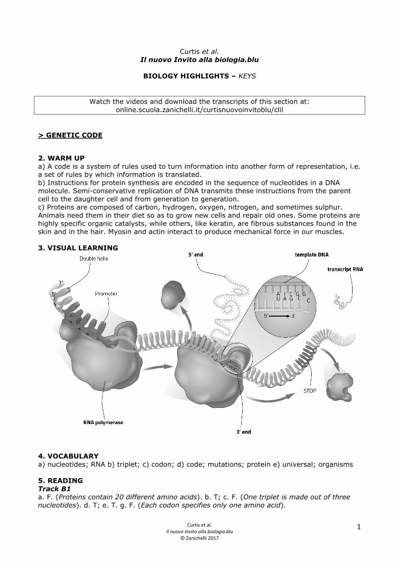
2. WARM UP a) A code is a system of rules used to turn information into another form of representation, i.e. a set of rules by which information is translated. b) Instructions for protein synthesis are encoded in the sequence of nucleotides in a DNA molecule. Semi-conservative replication of DNA transmits these instructions from the parent cell to the daughter cell and from generation to generation. c) Proteins are composed of carbon, hydrogen, oxygen, nitrogen, and sometimes sulphur. Animals need them in their diet so as to grow new cells and repair old ones. Some proteins are highly specific organic catalysts, while others, like keratin, are fibrous substances found in the skin and in the hair. Myosin and actin interact to produce mechanical force in our muscles. 3. VISUAL LEARNING
4. VOCABULARY a) nucleotides; RNA b) triplet; c) codon; d) code; mutations; protein e) universal; organisms 5. READING Track B1 a. F. (Proteins contain 20 different amino acids). b. T; c. F. (One triplet is made out of three nucleotides). d. T; e. T. g. F. (Each codon specifies only one amino acid).
Curtis et al. Il nuovo Invito alla biologia.blu
© Zanichelli 2017
2
6. VOCABULARY a) purines; pyrimidines; b) guanine; cytosine; c) adenine; d) amino acids; e) information; f) bases; amino acid 7. DISCUSSION a) Every living organism stores its genetic information in DNA, whose structure and components are the same for all organisms. For any sequence of three nucleotides, there is just one amino acid. b) Codons (or triplets) are sequences of three nucleotides in messenger RNA that code for a single amino acid. c) The genetic code consists of triplet combinations, or codons, and their corresponding amino acid. Each triplet specifies only one amino acid. A scheme using three bases, a triplet code, is the minimum necessary to encode for the 20 amino acids. A triplet code is the sequence of three nucleotides (bases) for the synthesis of a specific amino acid. 8. VIDEO Video B1 Transcript The instructions for cell functioning are contained in DNA and are organized in genes, i.e. specific sequences of nucleotides. The information needed to synthesise a polypeptide is contained in a gene. One or more polypeptides form proteins. The vehicle between a gene and a polypeptide is RNA: a single strand molecule obtained from a molecule of DNA by a process called transcription. This RNA is modified and becomes the messenger for genetic information. Messenger RNA leaves the nucleus and is involved in the process of translation, which leads to the production of a polypeptide. The processes of transcription and translation are similar in prokaryotes and eukaryotes, with just a few differences. In this video we will look at transcription in eukaryotes. The process of transcription can be divided into 3 phases: initiation, elongation and termination. Transcription can only initiate in certain regions of DNA called promoters. The proteins which are indispensable for initiation are called transcription factors. Each promoter is made up of different sequences; one of these is the TATA BOX, which is rich in adenine and thymine. Unlike cytosine and guanine, which have three hydrogen bonds holding them together, adenine and thymine only have two. This makes it is easier to separate the bases and open the double helix. Initiation of transcription occurs at the TATA BOX, due to the action of the transcription factors. When these regulatory proteins bind to the TATA BOX they change shape and induce conformational changes in the DNA. At this point other transcription factors and RNA polymerase, the enzyme that catalyses the synthesis of RNA, also attach, forming the preinitiation complex. One of the factors opens the DNA double helix at the initiation site, forming an open complex. RNA polymerase begins to join the nucleotides together to form a strand of RNA. One nucleotide is added at a time, using the DNA as a template. The criteria for choosing which base to add next is the pairing of complementary nitrogen bases: a purine base is always paired with a pyrimidine base. Let’s have a look in more detail. At the bottom we can see the DNA, with the chain of sugars and the nitrogen bases represented by letters. At the top RNA is being formed: uracil is paired with adenine and cytosine with guanine. Uracil and cytosine are bonded together at their sugars, as are the other nucleotides being added. The carbon at the 3’-end binds to the carbon at the 5’-end of the next nucleotide, releasing water and pyrophosphate. During elongation RNA polymerase moves along the strand of DNA, unwinding the double helix and winding it up again behind itself. When the chain of RNA is about 20 nucleotides long its 5’-end is modified chemically by a process known as capping, which we will look at more closely later. The final phase of transcription is called termination. RNA polymerase runs along the strand of DNA until it comes across a certain sequence called a terminator or stop. The strand of RNA is released and the polymerase detaches from the DNA.
Curtis et al. Il nuovo Invito alla biologia.blu
© Zanichelli 2017
3
The strand of RNA matures by undergoing some modification which transforms it into messenger RNA. As we have already mentioned, one change has already taken place during transcription: capping. A cap has been added at the 5’-end. The cap is a modified nucleotide: a methyl group has bonded to the nitrogen base guanine. At the end of transcription the 3’-end of the strand of RNA is also modified, by polyadenylation. This consists of the synthesis of a chain of adenine bases about 200 nucleotides long. These are added by poly-A polymerase which adds bases without a template. The poly-A tail stabilises the RNA molecule, particularly in the cytoplasm, where the RNA will be transferred to for translation. The RNA undergoes another transformation called splicing. With this process it becomes to all effects messenger RNA, and leaves the nucleus. Keys a. T; b. F (One or more polypeptides form proteins); c. F (The vehicle between a gene and a polypeptide is RNA); d. T; e. F (The processes are similar, but there are a few differences); f. T; g. F (Proteins are transcription factors); h. F (Uracil pairs with adenine); i. F (It is called termination); j. T; k. F (Splicing is a transformation affecting RNA). 9. REVISION Free answer 10. EXPLORE FURTHER Free answer FINAL TEST 1-B; 2-C; 3-C; 4-C; 5-C; 6-F; 7-T; 8-T; 9-F; 10-T
> PROTEIN SYNTHESIS
12. WARM UP a) Messenger RNA carries the instructions for protein manufacture from the DNA in the nucleus to the ribosomes in the cytoplasm. b) Transcription is the making of mRNA from a single strand of DNA and occurs in the nucleus. c) Messenger RNA are copies (transcripts) of DNA sequences. Each new mRNA molecule is copied from one of the two strands of DNA. This process is known as transcription. 13. VIDEO Video B2 Transcript We know that in DNA transcription a molecule of RNA is obtained after a process of maturation. This molecule of RNA is called messenger RNA and contains the information for synthesising a polypeptide chain. One or more chains of polypeptides will then form a protein. Synthesis, called translation, takes place in the cytoplasm. Let’s look now at the main stages of this process, taking the prokaryotes as a model. Translation occurs on structures called ribosomes. Each ribosome is made of two subunits, the large and the small subunit. The subunits combine with each other and the mRNA. Each one is made of a particular type of RNA, coloured blue, and proteins, coloured orange. The ribosomes move along the messenger RNA “reading” the nucleotide sequence and constructing a sequence of amino acids. This is how the polypeptide chain is made. After decades of research and experiments it was found that genetic information is organised into groups of three nucleotides, or triplets. Each triplet, called a codon, codes for a specific
Curtis et al. Il nuovo Invito alla biologia.blu
© Zanichelli 2017
4
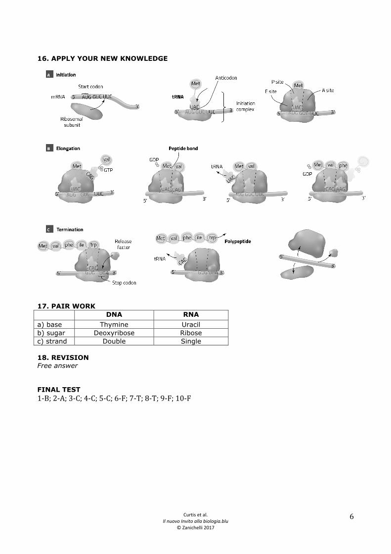
amino acid. The correlation between codons-amino acids is called the genetic code and can be represented as a table. In the table we can see how the four types of nucleotides which make up RNA can be combined into groups of three: the possible combinations are four raised to the power of 3, i.e. 64. Three of these codons do not code for an amino acid, but are stop codons which indicate the end of translation. So 61 codons are left to code for 20 amino acids: it seems obvious that there is not a one-to-one relationship between codons and amino acids. A codon codes for a single amino acid, but an amino acid can be coded for by several codons. For example, CUA codes only for the amino acid leucine. Leucine however is coded for by six different codons. Nearly all the amino acids are associated to more than one codon, therefore we say that the genetic code is redundant or degenerate. Let’s return to the process of translation. Messenger RNA not only interacts with ribosomes but also with another type of molecule: transfer RNA (or tRNA). As the name implies, the function of tRNA is to transport amino acids. Here we can see different structural representations of tRNA. The first shows the internal regions of the molecule and the way the bases are paired. The second is a compact model showing the three-dimensional structure of tRNA. This molecule is fundamental in the process of translation because it ensures that each amino acid is added in the right place. We can see why if we take a look at a more schematic model of its structure. The binding site for the amino acid is found at the 3’-end of tRNA. At the other end there is the sequence of three nucleotides which bond to three complementary nucleotides on the messenger RNA. This is the anticodon, a complementary triplet to the codon on the mRNA. There are many types of tRNA: at least one for each of the 20 amino acids. Their binding sites and anticodons differ. Each tRNA can only transfer one type of amino acid, but may have different anticodons. This is in line with the degeneracy of the genetic code: several triplets code for one amino acid. The bond between tRNA and the amino acid is made by a family of enzymes called aminoacyl-tRNA-synthetases. Each enzyme is specific for a single amino acid and for the corresponding tRNA. The bond is made between the amino acid and the 3’-end of the tRNA and uses the energy of one molecule of ATP. Now that we have seen the main components involved in translation let’s look at the main stages of this process. At initiation the small subunit of the ribosome bonds to an initiation factor and together they bond with the 5’-end of the mRNA.The subunit moves in the 5’ à 3’ direction until it reaches a specific codon, called the start codon. At this point a tRNA, called initiator RNA, binds to the start codon. Another protein completes the initiation complex. The start codon is a single triplet: AUG. Therefore, initiator tRNA always has the complementary sequence UAC. This tRNA always binds to the amino acid methionine. Once recognition has taken place the initiation factors detach and the large subunit of the ribosome attaches itself. At this point elongation begins. Let’s look inside the large subunit to see what’s going on. The initiator tRNA is in the region of the ribosome called the peptide site (or P site). Next to this, on the right in this representation, is the aminoacyl site (or A site). This is where the next tRNA will arrive and bind to the messenger RNA with its anticodon. Due to the structure of the tRNA molecules the two amino acids are situated very close to each other and can bond together. The second codon, in this case AUA, codes for tyrosine. The bond is a peptide bond and occurs between the amine group of tyrosine and the carboxyl group of methionine, releasing one molecule of water. The large subunit catalyses the formation of the bond between the two amino acids. Through a series of coordinated movements, called traslocation, the tRNA molecules move between the different sites of the ribosome. The tRNA that binds the amino acid chain is now in the P site, while the tRNA which carried methionine, which it has unloaded, moves from the P site to the exit or E site. Here it leaves the ribosome and returns to the cytoplasm where aminoacyl-tRNA-synthetase bonds a new molecule of methionine to it. A new tRNA molecule attaches to the A site, and the amino acid is bonded to the nascent polypeptide chain. The cycle is repeated: translocation occurs, the unloaded tRNA is transferred to the E site and a new loaded tRNA binds to the A site. In this way the polypeptide chain grows progressively longer.
Curtis et al. Il nuovo Invito alla biologia.blu
© Zanichelli 2017
5
Translation concludes with the phase called termination. A stop codon exists on the messenger RNA that tells the ribosome to stop. Unlike the start codon the stop codon is not unique. In this case it is UAA, but it can also be coded for by the triplets UAG or UGA. As we saw in the table of the genetic code no tRNA has a complementary anticodon to these three triplets. When a stop codon is in the A site some proteins, called release factors, bind to the ribosome. This bond modifies the activity of the ribosome, which separates the polypeptide chain from the tRNA at the P site. The two ribosome subunits and the tRNA detach and take part in another cycle of protein synthesis. Now let’s look at the polypeptide chain made by translation. The linear sequence of amino acids shows the so-called primary structure of the protein. Even during synthesis the polypeptide begins to fold and take on its three-dimensional structure. The adjacent amino acids in the sequence react with each other. Regular repetitive structures are formed, which make up the protein’s secondary structure. The main types of secondary structures are the alpha-helix and the beta-strand. These structures are held together by hydrogen bonds. Other bonds, such as hydrogen or disulphuric bonds may also form between amino acids which are not next to each other in the sequence. Together these interactions determine the tertiary structure of the protein. In practice this corresponds to the global structure of a single chain of amino acids, i.e. the way the atoms are spatially arranged. Some proteins are formed by the combination of several polypeptides. In this case the way the chains combine with each other is the quaternary structure. Keys a) DNA molecule is a code that contains instructions for biological structures and functions. DNA specifies mRNA. RNA specifies proteins. mRNA molecule is the “working” copy of DNA to determine the amino acid sequence; it is long 500 to 10000 nucleotides and is single stranded. tRNA molecules are comparatively small, ranging from 75 to 85 nucleotides. There are more than 20 different kinds in every cell, at least one for each of the kinds of amino acids. The 3’ end of the tRNA molecule attaches to a particular amino acid. The anticodon binds to the codon on the mRNA molecule. For this reason the tRNA molecules provide the crucial link between nucleic acids and protein. Ribosomes are composed of specific rRNA. b) Translation occurs in the rough endoplasmatic reticulum and ribosomes in the cytoplasm. Ribosomes consist of two subunits, one large and one small. Each subunit is composed of specific rRNA and protein molecules. c) A tRNA molecule consists of about 80 nucleotides linked together in a single chain. The chain always terminates in a (5’)-CCA-(3’) sequence. The molecule folds over itself producing a tri-dimensional structure. The chain has three loops. Three of the unpaired nucleotides in the loop at the bottom form the anticodon. 14. VOCABULARY 1-A; 2-D; 3-E; 4-F; 5-C; 6-B 15. READING Track B2 a) The synthesis of proteins is known as translation, since it is the transfer of information from one language (nucleotides) to another (amino acids). It takes place in thee stages: initiation, elongation and termination. b) The three molecules involved are: mRNA, tRNA and rRNA. c) A codon (triplet) is a sequence of three nucleotides in mRNA that codes for a single amino acid. d) mRNA carries the code to the ribosome, a cell organelle. Ribosome consists of two subunits, one large and one small. Each subunit is composed by rRNA and proteins. e) Three bases. f) The anticodon, one of the two important attachment site of the tRNA molecule, binds to the codon of the mRNA on the ribosome.
Curtis et al. Il nuovo Invito alla biologia.blu
© Zanichelli 2017
6
16. APPLY YOUR NEW KNOWLEDGE
17. PAIR WORK DNA RNA a) base Thymine Uracil b) sugar Deoxyribose Ribose c) strand Double Single 18. REVISION Free answer FINAL TEST 1-‐B; 2-‐A; 3-‐C; 4-‐C; 5-‐C; 6-‐F; 7-‐T; 8-‐T; 9-‐F; 10-‐F