biological oxidation (electron transport chain)
TRANSCRIPT

BIOCHEMISTRY
Biological Oxidation(Electron Transport Chain)
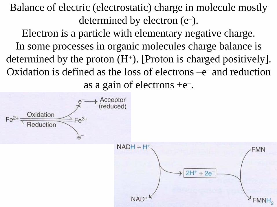
Balance of electric (electrostatic) charge in molecule mostly
determined by electron (e–).
Electron is a particle with elementary negative charge.
In some processes in organic molecules charge balance is
determined by the proton (H+). [Proton is charged positively].
Oxidation is defined as the loss of electrons –e– and reduction
as a gain of electrons +e–.

Biological oxidation – also named as respiration – it is an
ATP-generating process in which an inorganic compound
serves as the ultimate electron (e–) acceptor (i.g. O2 [i.e. during
biological oxidation O2 reduced to H2O]). The electron donor
can be either an organic compound or inorganic one.

Standard redox
potential (E0) of
some oxidation-
reduction systems

Sponsored
Medical Lecture Notes – All Subjects
USMLE Exam (America) – Practice

Mitochondria
Mitochondria are
oval-shaped cell
organelles, contain
the respiratory
assembly – that is the
enzymes of the citric
acid cycle, and the
enzymes of fatty acid
oxidation.
Mitochondria are
typically about 2μm
in length and 0.5μm
in diameter

Mitochondria electron microscope photo

Distribution of protons H+ – between matrix and
intermembrane space [inner crista space] forms electrostatic
potential on inner mitochondria membrane.

Four complexes of mitochondria electron transporting
chain and (fifth) ATP-synthesis complex.
[NADH and Succinate-Fumarate entrances into ETC]

Cellular respiration and biological oxidation based on
NADH and FADH2 (which are gained from: glycolysis, pyruvate
processing, citric acid cycle, amino acid oxidation, and β-oxidation

Sugars (carbohydrates) are the main oxidation substrate

Two main possible ways of sugar oxidation[energy gradient during biological oxidation and direct burn of sugar]

Amino
Acids
enter
TCA

Glycolysis
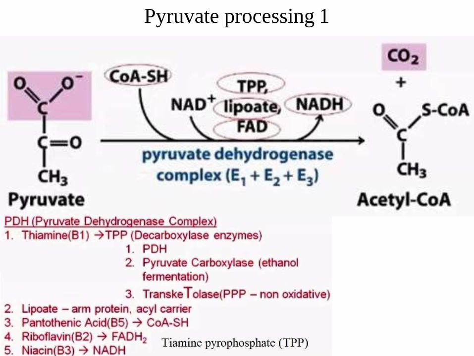
Pyruvate processing 1

Pyruvate processing (phases)

Coenzyme A (3 parts of CoA)
nucleotide, panthotenic acid (Vitamin B5), β-mercapto
ethylamine)

CAC
[TCA]
Krebs
cycle

CAC (Krebs Cycle)

Electron Transport Chain sequence

Levels of Redox
potendial and Free
energy in Electron
Transport Chain
Complexes

Chemical potential energy decreasing in ETC

Electron Transport Chain [I, III, IV] proton pomp
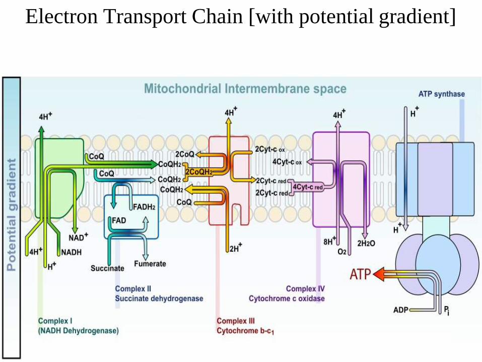
Electron Transport Chain [with potential gradient]

Complexes I-II-III-IV, V in Electron Transport Chain

Complex I in Electron Transport Chain 1
NADH ubiquinone
oxidoreductase (Complex I)
[usually have ‘L’ shape
consists of 46 polypeptides]
NADH – is an oxidized
molecule
Ubiquinone – is reduced
Complex I acts as proton pomp

Complex I in Electron Transport Chain
[this complex is pumping H+ into intermembrane
space: each NADH+H+ – give two protons 2H+]
Source of NADH+H+ for complex I :
-Glycolysis
-TCAcycle
-Amino Acid oxidation
-β-oxidation
All of these reactions increas concentration of
NADH+H+, which is transporting into mitochondrial
matrix, where it is oxidated by enzyme oxidoreductase
NADH+H+ → NAD+
in the same time FMN is reducing:
FMN(oxidated form) → FMNH2 (reduced form)

Nicotinamide dinucleotid (NAD)
Nicotinamide
(niacin – Vitamin B3)
ribose
Adenine
ribose

Nicotinamide dinucleotid (NAD) oxidated form

Nicotinamide dinucleotid (NAD+)
oxidation of niacin (Vitamin B3 part)
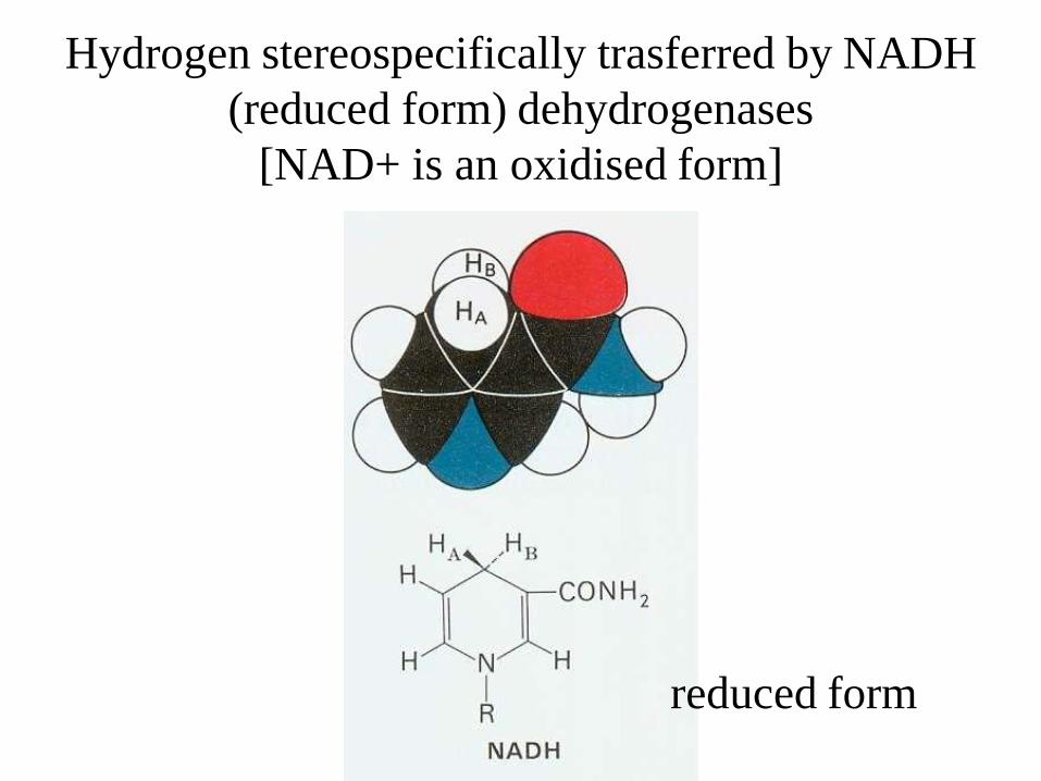
Hydrogen stereospecifically trasferred by NADH
(reduced form) dehydrogenases
[NAD+ is an oxidised form]
reduced form

Flavin Mono Nucleotide
(FMN)[reduced form is shown here ]
dimethylisoalloxazine
(flavin)
ribose

Reduction of dimethylisoalloxazine (flavin)
in Flavin mono nucleotide (FMN)
reduced form
oxidized form

Complex I with the help of FMN and Iron-Sulfur
centers [clusters] (Fe–S) do transfer electron
to Ubiquinone (reducting it into Ubiquinol)
FMNred → FMNox
Fe+3–S(ox) → Fe+2–S(red)Ferric (+3) oxidized form → Ferros (+2) reduced form
Fe+2–S(red) → Fe+3–S(ox)
…
Fe+3–S(ox) → Fe+2–S(red)
Fe+2–S(red) → Fe+3–S(ox)
…
Coenzyme Qn (Ubiquinone(ox))→ Coenzyme QnH2 (Ubiquinol(red))
this all increasing ubiquinol pool

Molecular models of iron-sulfur complex: (A) cluster
containing one Fe; (B) containing [2Fe-2S] cluster;
(C) [4Fe-4S] cluster. Iron atoms are shown in red, cistein sulfur
atoms shown in yellow; inorganic sulfur atoms in green.

Iron-sulfur complex: cluster containing
one Fe; containing [2Fe-2S] cluster;
[4Fe-4S] cluster
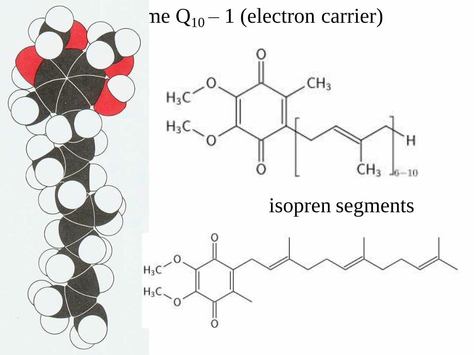
Coezyme Q10 – 1 (electron carrier)
isopren segments

Coezyme Q10 – oxidized and reduced forms
(with intermediate semiquinone form)
[to be reduced receives 2 electrons (from Complex I
or Complex II) and 2 protons (from matrix)]

Complex II (Succinate dehydrogenese) of ETC
Complex II do not transfer
protons (H+) into intermembrane
space
but reduces FAD to FADH2 and
through Fe-S clusters reduce
coenzyme Q10 (to ubiquinol)

Succinate dehydrogenese
(it is the same enzyme as in TCA cycle)
Succinate (alkane) oxidized to (alkine) Fumarate

Adenin mono
phosphat
(AMP)
Flavin Adenin
Dinucleotide (FAD)
[dimethylisoalloxazine]
Flavin
([ribo]flavin – Vitamin B2)

Structure of FAD(ox) – FADH2(red)

Flavin part of
Flavin adenin
dinucleotide
(FAD) is active
part of molecule
– oxidized
(consists of
flavin
mononucleotid
(FMN) unit
[green] and
adenin mono
phosphate
(AMP) [red]

Flavin adenin
dinucleotide
(FAD) oxidized
(consists no
hydrogen)

dimethylisoalloxazine in FAD [oxidized]
and in FADH2 [redused]

Iron Sulfur center (Fe–S) in Complex II in ETC
Succinate → FumarateFAD(ox) → FADH2(red)
Ferric (+3) oxidized form → Ferros (+2) reduced form
Fe+3–S(ox) → Fe+2–S(red)
Fe+2–S(red) → Fe+3–S(ox)
…
Fe+3–S(ox) → Fe+2–S(red)
Fe+2–S(red) → Fe+3–S(ox)
…
Coenzyme Qn (Ubiquinone(ox))→ Coenzyme QnH2(Ubiquinol(red))
this all increasing ubiquinol pool

Complex III in Electron Transport Chain
Cytochrome c Ubiquinol
oxidoreductase [Q cycle]
12 polypeptide chains
Complex III do transfer
protons (2H+) into
intermembrane space

Cytochrome c Ubiquinol oxidoreductase [Q cycle]
Complex III
QH2 →–→ e → 2Fe–2S
2Fe–2S→Heme c→Cytochrome c(red)→complex IV
e– → Heme blowpotencial → Heme bhighpotencial
Semiquinol intermediate radical
[Q` ubiquinol with one electron]

Heme (AmE) or haem (BrE) is a cofactor has an Fe2+ (ferrous) ion in the
middle of a large heterocyclic organic ring called a porphyrin, made up
of 4 pyrrolic groups joined together by methine bridges(5,10,15,20).
Not all porphyrins contain iron, but a
substantial fraction of porphyrin-
containing metalloproteins have heme
as their prosthetic group; these are
known as hemoproteins. Hemes are
most commonly recognized as
components of hemoglobin, the red
pigment in blood, but are also found in
a number of other biologically
important hemoproteins such as
myoglobin, cytochrome, catalase, and
endothelial nitric oxide synthase.

The heme in cytochrome c and c1 is covalently
attached to two cystein side chains

The heme b structure

Complex IV in Electron Transport Chain
Cytochrome c oxidaze
Complex IV do transfer
protons (2H+) into
intermembrane space

Cytochrome reductaze
(main phases of electron transferring)

Three-dimensional structure of reduced cytochrome c.The heme group (red), methionine 80 (green), histidine 18 (blue), and the
α-carbon atoms are shown. [from tuna]

Cytochrome. The heme group histidine and oxygen atoms are shown.

Chemical redox
potential and free
energy of
compounds
decreasing through
ETC.

Gradient of protons (pH) on inner membrane of
mitochondria

ATP [adenosine triphosphate]
[GTP]guanine
triphosphate
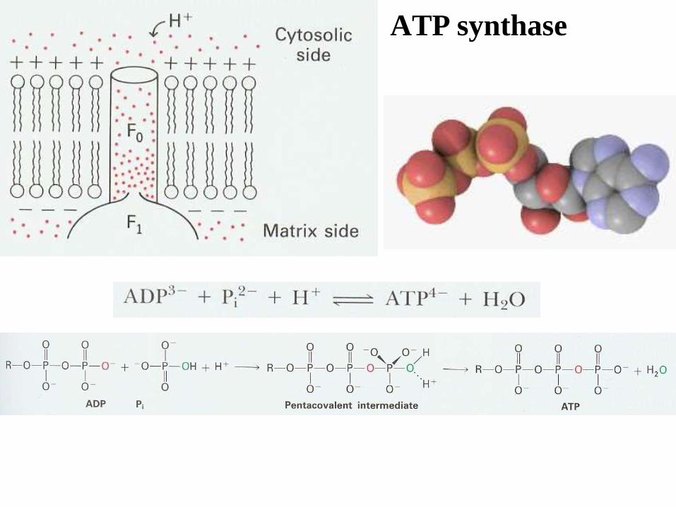
ATP synthase

ATP synthase and arrangment
of gens encoding the subunits
of ATP synthase in E.coli. This
cluster of genes is called
uncoupled operon (unc)

ATP synthase
Complex V

ATP synthase – the three catalytic sites cyclr through
three conformational states: O [open], L[loose
binding], T [tight binding]. Proton flux through the
syntase drives this interconversion of states. The
essence of this proposed mechanism is that proton flux
lead to the release of tightly bound ATP.

ATP
synthase 1

ATP
synthase
interacting with
actin
filament

Thank YOU for ATTENTION