bioinformatics for a better tomorrowscfbio-iitd.res.in/tutorial/bioinformatics_2010_new.pdf ·...
TRANSCRIPT
1
BIOINFORMATICS FOR A BETTER TOMORROWB. Jayaram and Priyanka Dhingra
Department of Chemistry &Supercomputing Facility for Bioinformatics & Computational Biology,
Indian Institute of Technology,Hauz Khas, New Delhi - 110016, India.Email: [email protected] site: www.scfbio-iitd.res.in
I. What is Bioinformatics?
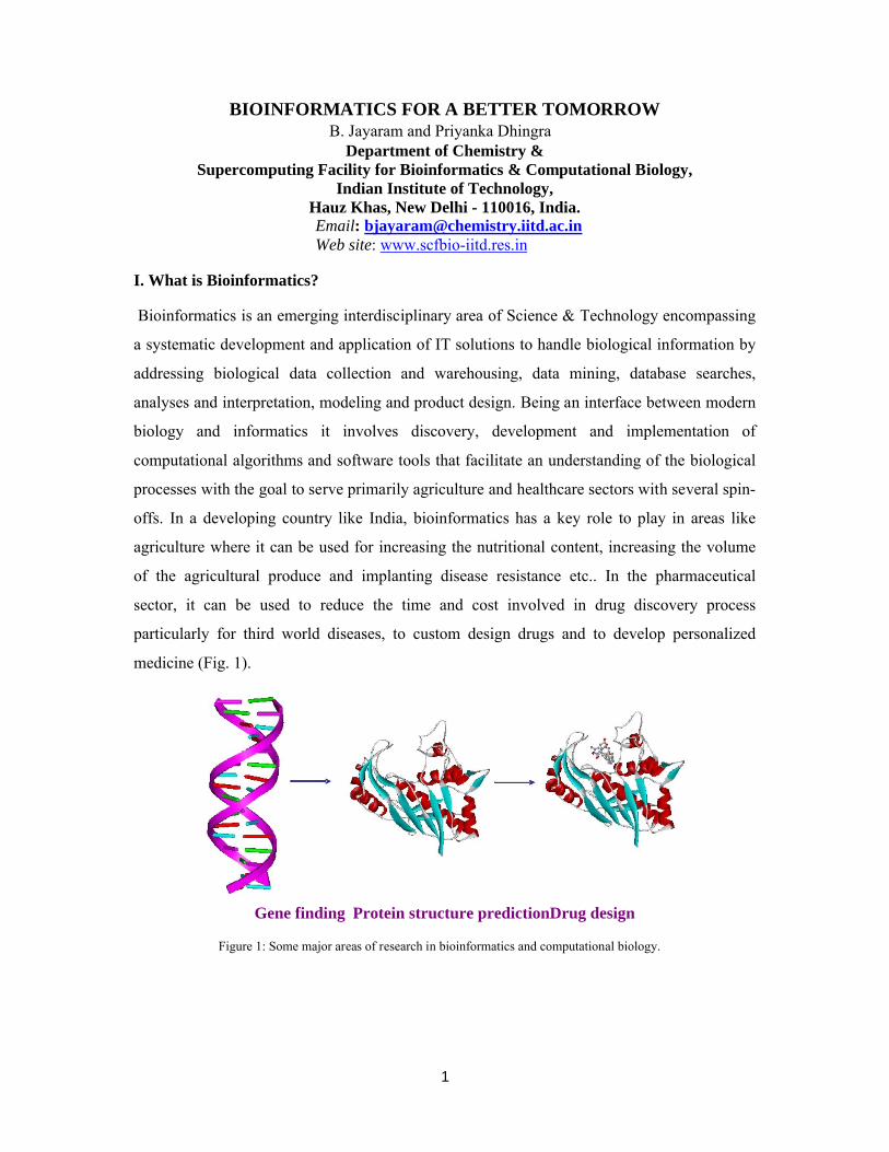
Bioinformatics is an emerging interdisciplinary area of Science & Technology encompassing
a systematic development and application of IT solutions to handle biological information by
addressing biological data collection and warehousing, data mining, database searches,
analyses and interpretation, modeling and product design. Being an interface between modern
biology and informatics it involves discovery, development and implementation of
computational algorithms and software tools that facilitate an understanding of the biological
processes with the goal to serve primarily agriculture and healthcare sectors with several spin-
offs. In a developing country like India, bioinformatics has a key role to play in areas like
agriculture where it can be used for increasing the nutritional content, increasing the volume
of the agricultural produce and implanting disease resistance etc.. In the pharmaceutical
sector, it can be used to reduce the time and cost involved in drug discovery process
particularly for third world diseases, to custom design drugs and to develop personalized
medicine (Fig. 1).
Gene finding Protein structure predictionDrug design
Figure 1: Some major areas of research in bioinformatics and computational biology.

2
A n g io sp erm s G y m n o sp e rm s
A sc o b u lo u s
N eu ro s p o ra
F ern s
G re e n A lg a eY ea st
D iato m s
B ro w n A lg a e
R e d A lg ae
C h lo ro p la st
M ito ch o n d riaC ya n o b ac te ria
M yc o b ac te riuP u rp le b a c teria
V e rte b rates
U ro ch o rd a te s
E c h in o d e rm s
A rth ro p o d s
M o llu sk s
N e m ato d e
C o e le n te rate sS p o n g es
S lim e M o ld s
A m o e b a
H elio zo a n s
C iliate s
D in o flag e lle te s
A rc h a eb a cteria
G ra m p o sitiv eb a cteria
A n cestral P ro k aryo te
Figure 2: The tree of life depicting evolutionary relationships among organisms from the major biological kingdoms. A possible evolutionary path from a common ancestral cell to the diverse species present in the modern world can be deduced from DNA sequence analysis. The branches of the evolutionary tree show paths of descent. The length of paths does not indicate the passage of time and the vertical axis shows only major categories of organisms, not evolutionary age. Dotted lines indicate the supposed incorporation of some cell types into others, transferring all of their genes and giving the tree some web-like features (adopted from: A Alberts, D Bray, J Lewis, M Raff, K Roberts & J D Watson, Molecular Biology of the Cell, p38, Garland, New York (1994)).
3
It is assumed that life originated from a common ancestor and all the higher organisms
evolved from a common unicellular prokaryotic organism. Subsequent division of different
forms of life from this makes the diversity in the morphological and genetic characters (Fig.
2).
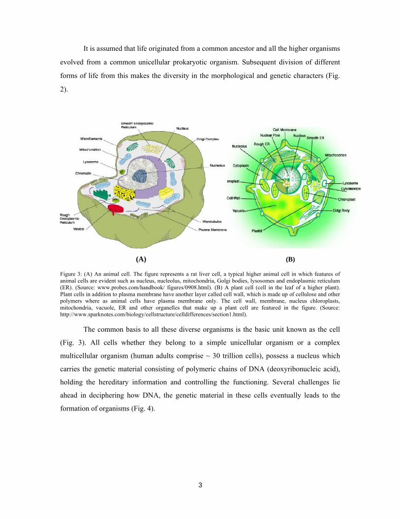
(A) (B)
Figure 3: (A) An animal cell. The figure represents a rat liver cell, a typical higher animal cell in which features of animal cells are evident such as nucleus, nucleolus, mitochondria, Golgi bodies, lysosomes and endoplasmic reticulum (ER). (Source: www.probes.com/handbook/ figures/0908.html). (B) A plant cell (cell in the leaf of a higher plant). Plant cells in addition to plasma membrane have another layer called cell wall, which is made up of cellulose and other polymers where as animal cells have plasma membrane only. The cell wall, membrane, nucleus chloroplasts, mitochondria, vacuole, ER and other organelles that make up a plant cell are featured in the figure. (Source: http://www.sparknotes.com/biology/cellstructure/celldifferences/section1.html).
The common basis to all these diverse organisms is the basic unit known as the cell
(Fig. 3). All cells whether they belong to a simple unicellular organism or a complex
multicellular organism (human adults comprise ~ 30 trillion cells), possess a nucleus which
carries the genetic material consisting of polymeric chains of DNA (deoxyribonucleic acid),
holding the hereditary information and controlling the functioning. Several challenges lie
ahead in deciphering how DNA, the genetic material in these cells eventually leads to the
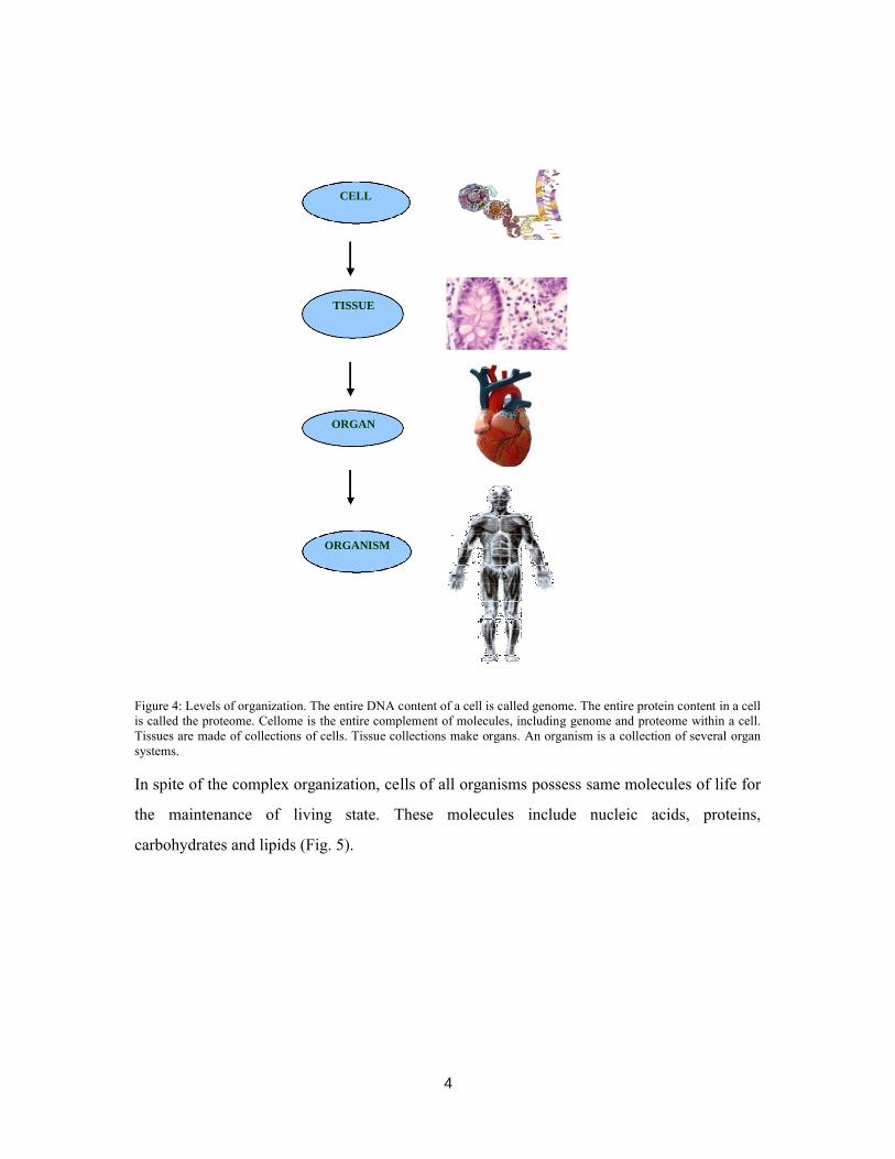
formation of organisms (Fig. 4).
4
CELL
TISSUE
ORGAN
ORGANISM
Figure 4: Levels of organization. The entire DNA content of a cell is called genome. The entire protein content in a cell is called the proteome. Cellome is the entire complement of molecules, including genome and proteome within a cell. Tissues are made of collections of cells. Tissue collections make organs. An organism is a collection of several organ systems.
In spite of the complex organization, cells of all organisms possess same molecules of life for
the maintenance of living state. These molecules include nucleic acids, proteins,
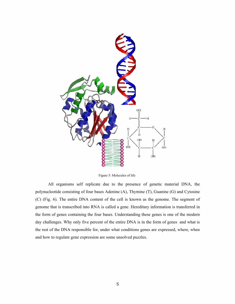
carbohydrates and lipids (Fig. 5).
5
Figure 5: Molecules of life
All organisms self replicate due to the presence of genetic material DNA, the
polynucleotide consisting of four bases Adenine (A), Thymine (T), Guanine (G) and Cytosine
(C) (Fig. 6). The entire DNA content of the cell is known as the genome. The segment of
genome that is transcribed into RNA is called a gene. Hereditary information is transferred in
the form of genes containing the four bases. Understanding these genes is one of the modern
day challenges. Why only five percent of the entire DNA is in the form of genes and what is
the rest of the DNA responsible for, under what conditions genes are expressed, where, when
and how to regulate gene expression are some unsolved puzzles.

6
Figure 6: DNA and its alphabets – the nucleic acid bases: A, T, G and C
Some major areas of research in bioinformatics
Genome Analysis: Segments of genome coding for messenger ribonucleic acids (mRNAs),
transfer ribonucleic acids (tRNAs), ribosomal ribonucleic acids (rRNAs) are called genes.
Among these mRNAs determine the sequence of amino acids in proteins. The mechanism is
simple for the prokaryotic cell where all the genes are converted into the corresponding
mRNA (messenger ribonucleic acid) and then into proteins. The process is more complex for
eukaryotic cells where rather than full DNA sequence, some parts of genes called exons are
expressed in the form of mRNA interrupted at places by random DNA sequences called
introns. Of the several questions posed here, one is that how some parts of the genome are
expressed as proteins and yet other parts (introns as well as intergenic regions) are not
expressed and which exons are combined under what conditions to make proteins necessary
for the organism.
Several genome projects are being carried out worldwide in order to identify all the
genes in a specified organism. Human genome project [1, 2] is one such global effort to
identify all the alphabets on the human genome, initiated in 1990 by the US government. A
comparison of the various genome sizes of different organisms (Table 1) raises questions like
what types of genetic modifications are responsible for the four times larger genome size of
wheat plant and seven times smaller size of the rice plant [3] as compared to that of humans.

7
Mice and humans contain roughly the same number of genes – about 28K protein coding
regions, about 90% of the human genome is in large blocks of homology with mouse [4]. The
chimpanzee and human genomes vary by an average of just 5% [5].
Table 1: Genome sizes of some organisms.
(http://users.rcn.com/jkimball.ma.ultranet/BiologyPages/G/GenomeSizes.html)
Several genetic disorders like Huntington’s disease, Parkinson’s disease, sickle cell
anemia etc. are caused due to mutations in the genes or a set of genes inherited from one
generation to another (Table 2). There is a need to understand the genetic origins for such
disorders. Why nature carries such disorders and how to prevent these are some of the areas
where extensive research is in progress.
OrganismGenome size
((Mb)=Mega base)
H.Influenza 1.83M tuberculosis 4.4
Eschericia coli 4.6
Sacchromyces cerevisiae (Yeast) 12.5
Plasmodium falciparum 23
C. elegans (Nematode) 100
Drosophila melanogaster (Fruit fly) 122
Gallus gallus (Chicken) 120
Oryza sativa (Rice) 390
Canis lupis familiaris (Dog) 2400
Pan troglodytes (Chimpanzee) 2700
Mus musculus (Mouse) 3000
Homo sapiens (Humans) 3300
Triticum aestivum(Wheat) 13500

8
Table 2: Specific genetic disorders
Genetic Disorder Reason
Sickle Cell Anemia Mutation in hemoglobin-b gene on chromosome 11
Brukitt lymphoma Translocations on chromosome 8
Hemophilia A Mutation of the HEMA gene on the X chromosome
Breast Cancer Mutation on genes found on chromosomes 13 & 17
LeukemiaExchange of genetic material between the long arms
of chromosome 6 & 22
Colon cancerProteins MSH2, MSH6 on chromosome 2 & MLH1
on chromosome 3 are mutated.
Cystic Fibrosis Mutations in a single (CFTR) gene
Best disease Mutation in one copy of a gene located on chromosome 11
Rett Syndrome Disfunctioning of a gene on the X chromosome.
Pendred Syndrome Defective gene on chromosome 7.
Asthma Disfunctioning of genes on chromosome 5, 6, 11, 14&12.
Diastrophic dysplasia Mutation in a gene on chromosome 5
Angelman Syndrome Deletion of a segment on maternally derived chromosome 15
Werner Syndrome Mutations on genes located on chromosome 8.
Alzheimer disease Mutations on four genes located on chromosome 1, 14, 19 & 21
Parkinson’s Disease Variations in genes on chromosomes 4, 6.
Huntington’s DiseaseExcessive repeats of a three-base sequence, "CAG"
on chromosome 4.
Tay-Sachs Disease Controlled by a pair of genes on chromosome 15
William’s syndromeDeletion of the gene for elastin and LIM kinase from
chromosome 7.
Zellweger syndrome Mutations in the PXR1 gene on chromosome 12.
Gaucher disease Mutation in a gene on chromosome 1
Achondroplasia Mutations in the gene encoding FGFR3 situated onchromosome 4
Familial Mediterranean fever Mutation in a gene on chromosome 16(Source: http://www.ncbi.nlm.nih.gov)
9
An understanding of the genome organization can lead to progresses in drug-target
identification. Genome level comparisons of healthy individuals with those carrying some
disorder can help identify drug targets. If the genome for humans and a pathogen, a virus
causing harm is identified, comparative genomics can predict possible drug-targets for the
invader without causing side effects to humans. SNPs (single nucleotide polymorphisms) are
common DNA sequence variations that occur when a single nucleotide in the genome
sequence changes. SNPs occur every 100 to 300 bases along the human genome. The SNP
variants promise to significantly advance our ability to understand and treat human diseases.
National Center for Biotechnology Information in collaboration with the National Human
Genome Research Institute, has established the dbSNP database
(http://www.ncbi.nlm.nih.gov/snp) to serve as a central repository for both single base
nucleotide subsitutions and short deletion and insertion polymorphisms. The database includes
single-base nucleotide substitutions (SNPs), small-scale multi-base deletions or insertions
(Deletion insertion polymorphisms or DIPs), and retroposable element insertions and
microsatellite repeat variations (short tandem repeats or STRs). Each dbSNP entry includes
the sequence context of the polymorphism (i.e., the surrounding sequence), the occurrence
frequency of the polymorphism (by population or individual), and the experimental method(s),
protocols, and conditions used to assay the variation [6]. Once discovered, these
polymorphisms will assist in the study of the structure and history of human genome.
Comparative genomics is the establishment of the relation between two genes from different
organisms. Comparison of series of sequences between two genomes generates intergenomic
maps which help in identifying the evolutionary process responsible for divergence of two
genomes / species. Functional genomics involves identification of gene function. DNA micro
array [7] data analysis is another research area for quantifying the levels of gene expression in
various tissues or at different stages in the development of diseases.
Over the past two decades, genetic modifications have enabled plant breeders to
develop new varieties of crops like cereals, soya, and maize at a faster rate. Genes are
transferred from one species to another species called as transgenic varieties, engineered to
possess special characteristics that make them better. Research is in progress world-wide,
utilizing GM (genetically modified) crops to produce therapeutic plants [8]. Modern plant
biotechnology faces a challenge of feeding an increasing world population. The emerging
field of genomics has provided huge information to improve crop characteristics like size and
height of the plant, seed and flower color (phenotypes) [9].

10
In order to contribute to the sustainability of rural agriculture, studies are being
conducted to identify medicinal substances based on indigenous knowledge and publicly
available databases, to critically evaluate these products using controlled functional genomics
experiments and bioinformatics and to increase awareness and assess perceptions about the
technology used and to disseminate outcomes. Studies are also in progress to evaluate the
effectiveness of traditional therapeutics on inflammatory and parasitic processes in livestock
(cows and goats) and to establish models for comparative genomic analyses of functional
consequences of exposure using cell and molecular biology, bioinformatics and micro array
techniques. Neem, wormwood and garlic are some examples of plants used in traditional
medicine that are known to possess anti-helmintic and anti-inflammatory properties The main
biologically active constituents of these selected agents are presented in Table 3.
Table 3: Selected medicinals and their biologically active constituents
(Source: 9th ICABR International Conference on Agricultural Biotechnology: Ten years later, indigenous knowledge, bioinformatics and rural agriculture technology)
Comparative genomics of plant genomes has suggested that the organization of genes
has been conserved during evolution. The complete genomes of many crop plants (e.g. Oryza
sativa, wheat) help in providing information about the agronomically important genes which
could be used for further improvement in food crops. Genes from Bacillus thuringiensis which
control a number of pests are successfully transferred to crops like cotton, maize and potatoes.
This helps plants to become insect resistant and the amount of insecticides being used is
reduced thus improving overall economics.
Given the whole genome of an organism, finding the genes is a challenging task.
Various sophisticated mathematical methods have been proposed. Most of these approaches
are database driven which rely on the existing experimental information. Some of the
Medicinal Main Biologically Active ConstituentGarlic Allicin
Tobacco Nicotine Anabasine
NeemLimonoids (e.g. azadirachtin, salannin, meliantriol, nimbin,
nimbidin)
Wormwood ThujoneShitake 1-3 beta glucans and lentinan
Diatomaceous earth DiatomitesWhey Proteins

11
successful strategies are based on short range correlations between bases along the genome
(e.g. Markov models) and some others are based on long range / global correlations (e.g.
Fourier transform techniques). Table 4 lists some of the common gene prediction software’s
available freely over the internet.
Table 4: A list of gene prediction software’s available freely over the internet
Sl. No. Name of the Software URL Remarks
1. FGENESH http://www.softberry.com/all.htm HMM
2. GeneID http://www1.imim.es/geneid.html Ab initio
3. GeneParser http://beagle.colorado.edu/~eesnyder/GeneParser.html Ab initio
4. GeneWise www.sanger.ac.uk/Software/Wise2/genewiseform.shtml Homology
5. GeneMark http://exon.gatech.edu/GeneMark Ab initio
6. GENSCAN http://genes.mit.edu/GENSCAN.html Ab initio
7. Glimmer http://www.tigr.org/software/glimmer/ Ab initio
8. GlimmerHMM http://www.tigr.org/software/GlimmerHMM/ Ab initio
9. GRAILEXP http://compbio.ornl.gov/grailexp Ab initio
10. GENVIEW www.itba.mi.cnr.it/webgene Ab initio
11. GenSeqer http://bioinformatics.iastate.edu/cgi-bin/gs.cgi Homology
12. ORFgene http://125itba.mi.cnr.it/~webgene/wwworfgene2.html Homology
13. MORGAN http://www.cs.jhu.edu/labs/compbio/morgan.html Ab initio
14. PredictGenes http://mendel.ethz.ch:8080/Server/subsection3_1_8.html Homology
15. MZEF http://rulai.cshl.edu/software/index1.htm Ab initio
16. Rosetta http://crossspecies.lcs.mit.edu Homology
17. VEIL http://www.tigr.org/~salzberg/veil.html Ab initio
18. PROCRUSTES http://hto-13.usc.edu/software/procrustes/index.html Homology
19. Xpound ftp://igs-server.cnrs-mrs.fr/pub/Banbury/xpound Ab initio
20. Chemgenome http://www.scfbio-iitd.res.in/chemgenome/chemgenomenew.jsp Ab initio
We have recently developed a hypothesis driven physico-chemical model for gene
prediction. In this model a physico-chemical vector which attempts to capture forces
responsible for DNA structure and protein-nucleic acid interactions walks along the genome
identifying the potential protein coding regions. As of now, the physico-chemical model
ChemGenome 2.0 is seen to predict genes reasonably well with fairly high specificities and
sensitivities. The methodology has been web enabled at http://www.scfbio-

12
iitd.res.in/chemgenome/chemgenomenew.jsp. Also software for distinguishing genes from
non-genes is available for free access at http://www.scfbio-iitd.res.in/chemgenome/index.jsp
[10, 11]. It is hoped that bioinformatics research in genomics will eventually generate a
molecular view of genome organization and genetic networks.
Protein Folding
Proteins are polymers of amino acids with unlimited potential variety of structures and
metabolic activities. Proteins may be classified into structural proteins, enzymes, hormones,
transport proteins, protective proteins, contractile proteins, storage proteins, toxins. Each
protein possesses a characteristic three-dimensional shape and function which is defined by
the sequence of amino acids constituting it (Fig. 7). This in turn is genetically controlled by
the sequences of bases in DNA of the cell through the genetic code. Substitution of a single
amino acid can cause a major alteration in function as in the well known case of sickle cell
anemia. Study of proteins with related folds and related sequences (homologous proteins)
from different species is of interest in constructing taxonomic relationships.
Figure 7: Folding of amino acid chain into a 3-D structure.
Protein folding can be considered to involve changes in the polypeptide chain
conformation to attain a stable conformation corresponding to the global minimum in free
energy which is about 10 to 15 kcal/mol lower relative to the unfolded state (Fig. 8) [12].
13
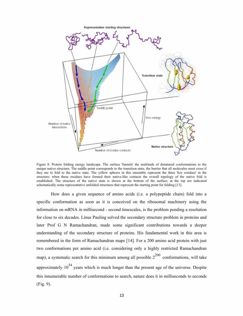
Figure 8: Protein folding energy landscape. The surface 'funnels' the multitude of denatured conformations to the unique native structure. The saddle point corresponds to the transition state, the barrier that all molecules must cross if they are to fold to the native state. The yellow spheres in this ensemble represent the three 'key residues' in the structure; when these residues have formed their native-like contacts the overall topology of the native fold is established. The structure of the native state is shown at the bottom of the surface; at the top are indicated schematically some representative unfolded structures that represent the starting point for folding [13].
How does a given sequence of amino acids (i.e. a polypeptide chain) fold into a
specific conformation as soon as it is conceived on the ribosomal machinery using the
information on mRNA in millisecond - second timescales, is the problem pending a resolution
for close to six decades. Linus Pauling solved the secondary structure problem in proteins and
later Prof G N Ramachandran, made some significant contributions towards a deeper
understanding of the secondary structure of proteins. His fundamental work in this area is
remembered in the form of Ramachandran maps [14]. For a 200 amino acid protein with just
two conformations per amino acid (i.e. considering only a highly restricted Ramachandran
map), a systematic search for this minimum among all possible 2200
conformations, will take
approximately 1054
years which is much longer than the present age of the universe. Despite
this innumerable number of conformations to search, nature does it in milliseconds to seconds
(Fig. 9).
14
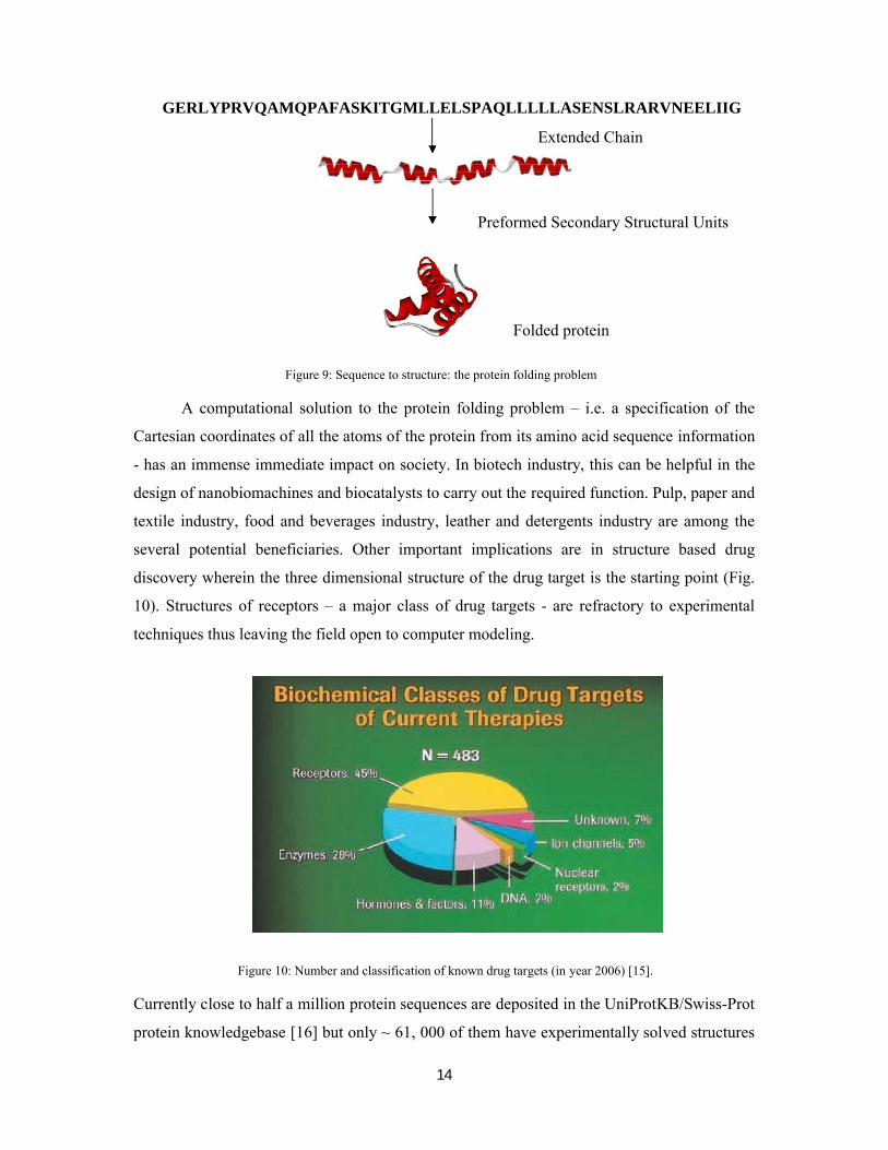
GERLYPRVQAMQPAFASKITGMLLELSPAQLLLLLASENSLRARVNEELIIG
Extended Chain
Preformed Secondary Structural Units
Folded protein
Figure 9: Sequence to structure: the protein folding problem
A computational solution to the protein folding problem – i.e. a specification of the
Cartesian coordinates of all the atoms of the protein from its amino acid sequence information
- has an immense immediate impact on society. In biotech industry, this can be helpful in the
design of nanobiomachines and biocatalysts to carry out the required function. Pulp, paper and
textile industry, food and beverages industry, leather and detergents industry are among the
several potential beneficiaries. Other important implications are in structure based drug
discovery wherein the three dimensional structure of the drug target is the starting point (Fig.
10). Structures of receptors – a major class of drug targets - are refractory to experimental
techniques thus leaving the field open to computer modeling.
Figure 10: Number and classification of known drug targets (in year 2006) [15].
Currently close to half a million protein sequences are deposited in the UniProtKB/Swiss-Prot
protein knowledgebase [16] but only ~ 61, 000 of them have experimentally solved structures

15
[17]. The numbers present the immediate demand for faster and better algorithms for protein
structure prediction. There are two major ways in which protein structure prediction attempts
are currently progressing viz. comparative modeling, and de novo approaches. The former is
database dependent methodology relying on known structures and the latter is independent of
the databases and starts from the physical principles. Comparative modeling relies on the
principle that sequences, which are related evolutionarily, exhibit similar three dimensional
folded structures that is sequence similarity suggests structural similarity [18]. Comparative
modeling techniques are extremely popular, reliable and fast where sequence homologues
exist in the database. With increasing structural information, these techniques should prove
more useful.
The de novo methods utilize first principles as well as the database information
(directly or indirectly) to predict the three dimensional structure of proteins. Although the de
novo techniques till date are able to predict structures of only small proteins, because of their
first principles approach and the concurrent computational requirements, they have the
potential to predict new / novel folds and structures. The time required to fold a 200 amino
acid protein which evolves ~10-11
sec per day per processor according to Newton’s laws of
motion will require approximately a million years to fold. If one can envision a million
processors working together, a single mid-sized protein can be folded in one year computer
time. Against this backdrop, IBM has launched a five year Blue Gene project
(www.research.ibm.com/bluegene/) in the year 1999 to address complex biomolecular
phenomena such as protein folding. The full Blue Gene/L machine was designed and built in
collaboration with the Department of Energy's NNSA/Lawrence Livermore National
Laboratory in California, and has a peak speed of 360 Teraflops. Blue Gene is one of the
fastest supercomputing systems in the world, giving scientists access to unprecedented
computing power. Table 5 lists some of the freely available comparative modeling as well as
de novo protein structure prediction software’s available over the internet.

16
Table 5: A list of protein structure prediction software’s available freely over the internet
Sl.No
Name of thesoftware
Description URL
1. PSI-BLAST
The Basic Local Alignment Search Tool(BLAST) finds regions of local similaritybetween sequences. The program comparesnucleotide or protein sequences to sequencedatabases and calculates the statisticalsignificance of matches
http://www.ncbi.nlm.nih.gov/BLAST/
2. CPHModels2.0 An automated protein structure homologymodeling server - http://www.cbs.dtu.dk/services/CPHmodels/
3. Swiss-Model A fully automated protein structure homology -modeling server
http://swissmodel.expasy.org/SWISS-MODEL.html
4. ModWebA web server implementation of MODELLER (comparative protein structure modeling by satisfaction of spatial restraints)
http://alto.compbio.ucsf.edu/modweb-cgi/main.cgi
5. 3DJigSawAn automated server to build three-dimensional models for proteins based on homologues of known structure
http://www.bmm.icnet.uk/servers/3djigsaw/
6. GenTHREADER
A combination of methods such as sequence alignment with structure based scoring functions and neural network based jury system to calculate final score for the alignment
http://bioinf.cs.ucl.ac.uk/psipred/
7. 3D PSSMThreading approach using 1D and 3D profiles coupled with secondary structure and solvation potential
http://www.sbg.bio.ic.ac.uk/~3dps sm
8. ROBETTADe novo Automated structure predictionanalysis tool used to infer protein structural information from protein sequence data
http://robetta.bakerlab.org
9. PROTINFO
De novo protein structure prediction web server utilizing simulated annealing for generation and different scoring functions for selection of final five conformers
http://protinfo.compbio.washington.edu/
10. SCRATCH
Protein structure and structural features prediction server which utilizes recursive neural networks, evolutionary information,fragment libraries and energy
http://scratch.proteomics.ics.uci.edu/
11. ROKKYDe novo structure prediction by the simfold energy function with the multi-canonical ensemble fragment assembly
http://www.proteinsilico.org/rokky/rokky-p/
12. BHAGEERATHEnergy based methodology for narrowing down the search space of small globular proteins
http://www.scfbio-iitd.res.in/bhageerath/index.jsp

17
We have recently developed a computational protocol for modeling and predicting
protein structures of small globular proteins. Here a combination of bioinformatics tools,
physicochemical properties of proteins and de novo approaches are used. This suite of
programs is named Bhageerath (http://www.scfbio-iitd.res.in/bhageerath) [19, 20]. Starting
with the sequence of amino acids, for 50 small globular proteins, 5 candidate structures for
each protein within 3-6 Å of the native are predicted within less than 3 hours on a 280
processor cluster. Attempts are in progress to further improve the prediction accuracies of the
structures to within a root mean square deviation of <3Å from the native structures via explicit
solvent molecular dynamics and Metropolis Monte Carlo simulations.
Function follows form [21] and hence the need for structures. Stated alternatively,
sequence to consequence [22] is the major challenge in proteomics investigations.
Drug Design
The information present in DNA is expressed via RNA molecules into proteins which
are responsible for carrying out various activities. This information flow is called the central
dogma of molecular biology (Fig. 11). Potential drugs can bind to DNA, RNA or proteins to
suppress or enhance the action at any stage in the pathway.
Genome
Protein
Gene = DNARNA Primary Sequence
AGTMGLKPVLYDSMLTMPGLKKPGYDSMGTMLYTMGPVLLYVL
DNA binding drugs RNA binding drugsDrugsGene therapy
Inhibitors/activators
Figure 11: Central dogma of modern drug discovery

18
As structures of more and more protein targets become available through
crystallography, NMR and bioinformatics methods, there is an increasing demand for
computational tools that can identify and analyze active sites and suggest potential drug
molecules that can bind to these sites specifically (Fig. 12).
Active Site
Figure 12: Active-site directed drug-design
Also to combat life-threatening diseases such as AIDS, tuberculosis, malaria etc., a
global push is essential (Table 6). “Millions for Viagra and pennies for the diseases of the
poor” [23] is the current situation of investment in Pharma Research & Development.
Table 6: Leading causes of death
Leading causes of death in millions per year due toinfectious diseases ( for the year 2002)
Percentage (%)
Lower respiratory infections 3.9 19
HIV-AIDS 2.8 3
Diarrheal diseases 1.8 17
Tuberculosis 1.6 n/a
Malaria 1.2 8
Measles 0.6 4
Neonatal Causes N/A 37
Others( includingnoncommunicable diseases)
N/A 10
Source: World health report, 2004, WHO, www.globalhealth.org

19
Time and cost required for designing a new drug are immense and at an unacceptable level.
According to some estimates it costs, on an average, about $1.2-1.4 billion and 14 years of
research to develop a new drug before it is introduced in the market (Fig 13).
Figure 13: Cost and time involved in drug discovery. (Source: PAREXEL [24])
Intervention of computers at some plausible steps is imperative to bring down the cost
and time involved in the drug discovery process (Table 8). Making a drug is more like
designing a key for a lock to jam or open the lock, except that both the lock and the key are
dynamic and made of atoms and are susceptible to environmental effects such as solvent, salt
and other small or biomolecules.

20
Table 8: High End Computing Needs for In Silico Drug Design: Estimates of current computational requirements to complete a binding affinity calculation for a given drug
Modelingcomplexity Method Size of library
Required computing time
Molecular Mechanics SPECITOPE 140,000 ~1 hour
Rigid ligand/target LUDI 30,000 1-4 hours
CLIX 30,000 33 hours
Molecular Mechanics Hammerhead 80,000 3-4 days
Partially flexible ligand DOCK 17,000 3-4 days
Rigid target DOCK 53,000 14 days
Molecular Mechanics ICM 50,000 21 days
Fully flexble ligand
Rigid target
AMBER 1 ~several daysMolecular Mechanics
Free energy CHARMM
perturbation
QM Active site and Gaussian, 1 >several weeks
MM protein Q-Chem
(Source: http://cbcg.lbl.gov)
The involvement of genomics, proteomics, bioinformatics and efficient technologies
like, combinatorial chemistry, high throughput screening (HTS), virtual screening, in vitro, in
silico ADMET screening, de novo and structure-based drug design serves to expedite as well as
economize the modern day drug discovery process [25]. Structure based computational drug
design methods mainly focus on the design of molecules for a target site/active sites with
known three dimensional structure, generate candidate molecules, check the molecules for
their drug-likeness, dock these molecules with the target, rank them according to their
binding affinities, further optimize the molecules to improve binding characteristics,
studies on newer drug delivery methods and design principles to cut down on toxicity
(Fig. 14).

21
Figure 14: Protein ligand docking
The advent of high performance computing environments, data management software
and internet offer the advantage of delivering new drug candidates more quickly and at lower
costs. Over 40 plus compounds have been discovered with the aid of structure based drug
design method [26]. Captopril was the first drug whose discovery was based on the concept of
structure based drug design. It is an angiotensin converting enzyme inhibitor used for the
treatment of hypertension and some types of congestive heart failure. Table 9 shows list of
drugs whose discovery has been aided by structure based drug design methods [26].
Table 9: List of drugs whose discovery has been aided by structure based drug designing methods
Drugs Structure
Captopril
Zanamivir
Nelfinavir
Ligand
Protein Ligand complex
Protein
Docking

22
Several drug design softwares are now available in public domain. Table 10 lists some
popular softwares for drug designing.
Table 10: Comprehensive software’s for drug design
S. No. Name of the Software
Description URL
1 Insight II, Discovery Studio
Cerius, ADME/Tox Package Syby
A suite for molecular modeling and de novodrug design.
Provides computational models for prediction of ADME properties derived from chemical structures
http://accelrys.com/products/insight/
http://accelrys.com/products/cerius2/
2 Sybyl Computational informatics software for drug discovery
http://www.tripos.com/
Amprenavir
Lopinavir
Dorzolamide
Oseltamivir
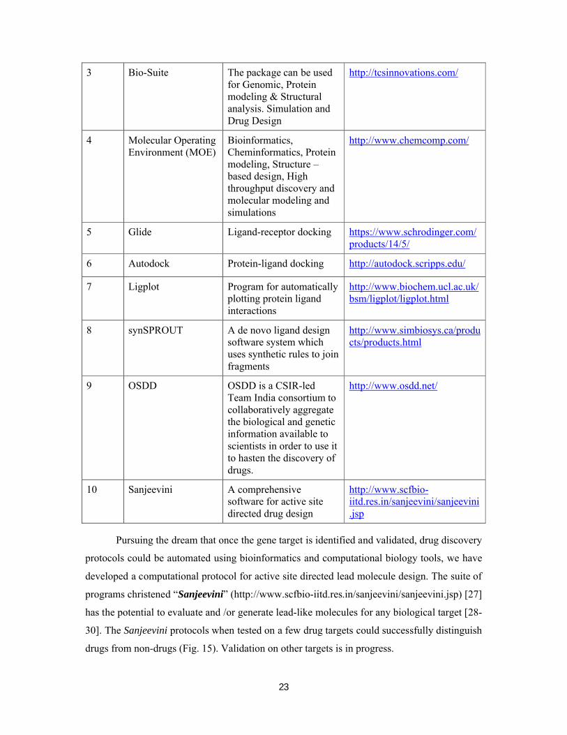
23
3 Bio-Suite The package can be used for Genomic, Protein modeling & Structural analysis. Simulation and Drug Design
http://tcsinnovations.com/
4 Molecular Operating Environment (MOE)
Bioinformatics, Cheminformatics, Protein modeling, Structure –based design, High throughput discovery and molecular modeling and simulations
http://www.chemcomp.com/
5 Glide Ligand-receptor docking https://www.schrodinger.com/products/14/5/
6 Autodock Protein-ligand docking http://autodock.scripps.edu/
7 Ligplot Program for automatically plotting protein ligand interactions
http://www.biochem.ucl.ac.uk/bsm/ligplot/ligplot.html
8 synSPROUT A de novo ligand design software system which uses synthetic rules to join fragments
http://www.simbiosys.ca/products/products.html
9 OSDD OSDD is a CSIR-led Team India consortium to collaboratively aggregate the biological and genetic information available to scientists in order to use it to hasten the discovery of drugs.
http://www.osdd.net/
10 Sanjeevini A comprehensive software for active site directed drug design
http://www.scfbio-iitd.res.in/sanjeevini/sanjeevini.jsp
Pursuing the dream that once the gene target is identified and validated, drug discovery
protocols could be automated using bioinformatics and computational biology tools, we have
developed a computational protocol for active site directed lead molecule design. The suite of
programs christened “Sanjeevini” (http://www.scfbio-iitd.res.in/sanjeevini/sanjeevini.jsp) [27]
has the potential to evaluate and /or generate lead-like molecules for any biological target [28-
30]. The Sanjeevini protocols when tested on a few drug targets could successfully distinguish
drugs from non-drugs (Fig. 15). Validation on other targets is in progress.
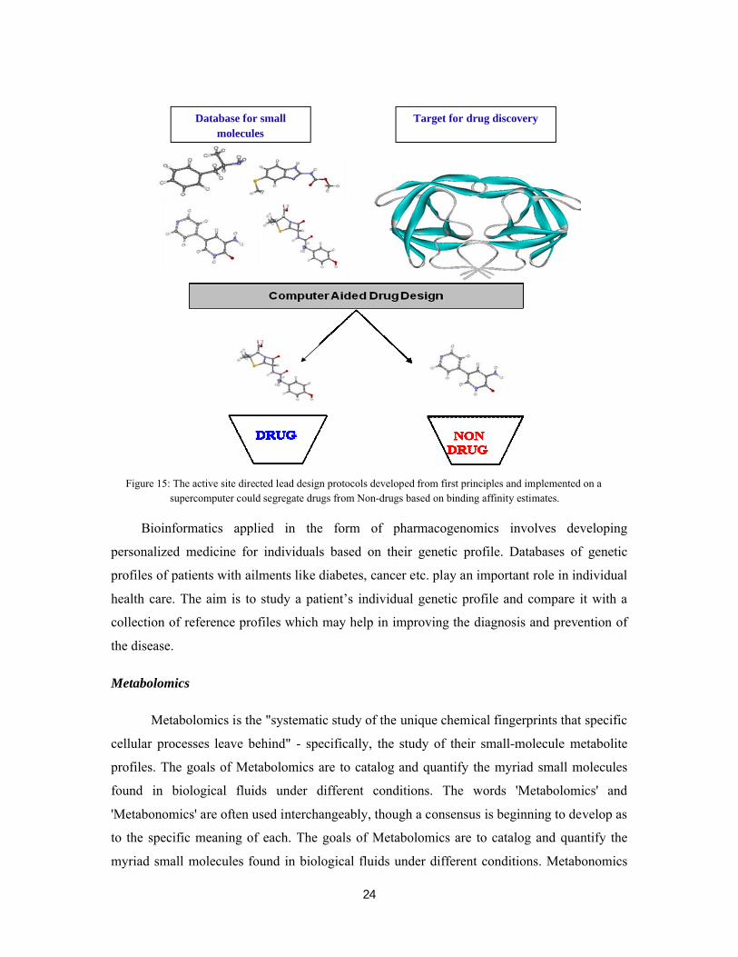
24
Figure 15: The active site directed lead design protocols developed from first principles and implemented on a supercomputer could segregate drugs from Non-drugs based on binding affinity estimates.
Bioinformatics applied in the form of pharmacogenomics involves developing
personalized medicine for individuals based on their genetic profile. Databases of genetic
profiles of patients with ailments like diabetes, cancer etc. play an important role in individual
health care. The aim is to study a patient’s individual genetic profile and compare it with a
collection of reference profiles which may help in improving the diagnosis and prevention of
the disease.
Metabolomics
Metabolomics is the "systematic study of the unique chemical fingerprints that specific
cellular processes leave behind" - specifically, the study of their small-molecule metabolite
profiles. The goals of Metabolomics are to catalog and quantify the myriad small molecules
found in biological fluids under different conditions. The words 'Metabolomics' and
'Metabonomics' are often used interchangeably, though a consensus is beginning to develop as
to the specific meaning of each. The goals of Metabolomics are to catalog and quantify the
myriad small molecules found in biological fluids under different conditions. Metabonomics
Target for drug discoveryDatabase for small molecules

25
is the study of how the metabolic profile of a complex biological system changes in response
to stresses like disease, toxic exposure, or dietary change.
The metabolome represents the collection of all metabolites (such as metabolic
intermediates, hormones and other signaling molecules, and secondary metabolites) in a
biological organism, which are the end products of its gene expression. While the genome,
transcriptome and proteome are estimated to be quite large, the metabolome is relatively small
by comparison, and tightly conserved across organisms. There are estimated to be 25,000
genes, 100,000 transcripts and more than one million proteins in humans but, according to
recent reports, there are only approximately 2,500 metabolites in the human metabolome [31,
32] (Fig 16). Thus, while mRNA gene expression data and proteomic analyses do not tell the
whole story of what might be happening in a cell, metabolic profiling can give an
instantaneous snapshot of the physiology of that cell. Metabolites are the intermediates and
products of metabolism. The term metabolite is usually restricted to small molecules. A
primary metabolite is directly involved in the normal growth, development, and reproduction.
A secondary metabolite is not directly involved in those processes, but usually has important
ecological function. Examples include antibiotics and pigments. Various small molecule
databases have been created and a few have been listed in Table 11.
Figure 16: General schematic of the omic organisation. The flow of information is from genes to transcripts to proteins to metabolites to function [33].
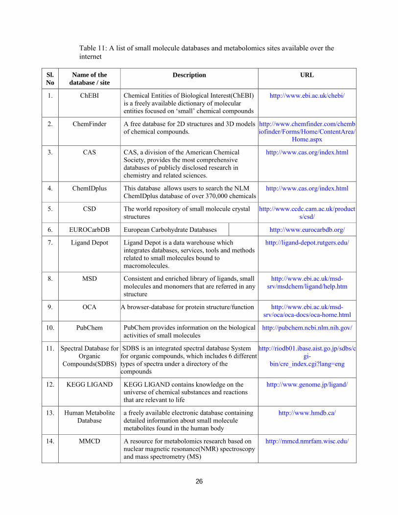
26
Table 11: A list of small molecule databases and metabolomics sites available over the internet
Sl. No
Name of the database / site
Description URL
1. ChEBI Chemical Entities of Biological Interest(ChEBI) is a freely available dictionary of molecular entities focused on ‘small’ chemical compounds
http://www.ebi.ac.uk/chebi/
2. ChemFinder A free database for 2D structures and 3D models of chemical compounds.
http://www.chemfinder.com/chembiofinder/Forms/Home/ContentArea/
Home.aspx
3. CAS CAS, a division of the American ChemicalSociety, provides the most comprehensive databases of publicly disclosed research in chemistry and related sciences.
http://www.cas.org/index.html
4. ChemIDplus This database allows users to search the NLM ChemIDplus database of over 370,000 chemicals
http://www.cas.org/index.html
5. CSD The world repository of small molecule crystal structures
http://www.ccdc.cam.ac.uk/products/csd/
6. EUROCarbDB European Carbohydrate Databases http://www.eurocarbdb.org/
7. Ligand Depot Ligand Depot is a data warehouse whichintegrates databases, services, tools and methods related to small molecules bound to macromolecules.
http://ligand-depot.rutgers.edu/
8. MSD Consistent and enriched library of ligands, small molecules and monomers that are referred in any structure
http://www.ebi.ac.uk/msd-srv/msdchem/ligand/help.htm
9. OCA A browser-database for protein structure/function http://www.ebi.ac.uk/msd-srv/oca/oca-docs/oca-home.html
10. PubChem PubChem provides information on the biological activities of small molecules
http://pubchem.ncbi.nlm.nih.gov/
11. Spectral Database for Organic
Compounds(SDBS)
SDBS is an integrated spectral database Systemfor organic compounds, which includes 6 different types of spectra under a directory of the compounds
http://riodb01.ibase.aist.go.jp/sdbs/cgi-
bin/cre_index.cgi?lang=eng
12. KEGG LIGAND KEGG LIGAND contains knowledge on theuniverse of chemical substances and reactions that are relevant to life
http://www.genome.jp/ligand/
13. Human MetaboliteDatabase
a freely available electronic database containingdetailed information about small molecule metabolites found in the human body
http://www.hmdb.ca/
14. MMCD A resource for metabolomics research based on nuclear magnetic resonance(NMR) spectroscopy and mass spectrometry (MS)
http://mmcd.nmrfam.wisc.edu/

27
The comprehensive qualitative and quantitative analyses of the primary and secondary
metabolites provides a holistic view of the biochemical status or biochemical phenotype of an
organism. The correlations of biochemical information with genetic and molecular data are
very useful in providing better insight into the functions of unknown gene or systems response
to external stimuli. Metabolomic studies also offer unique opportunities to study regulation
and signaling under the control of small molecules (i.e., metabolites). Quite often, signaling
and regulation are transparent at the transcriptome and/or proteome level. Finally,
metabolomics offers the unbiased ability to differentiate organisms or cell states based on
metabolite levels that may or may not produce visible phenotypes/genotypes. Although
metabolomics is quite promising, several challenges still exist that influence the
implementation of a metabolomic approach, including chemical complexity, analytical and
biological variance, and dynamic range. The success of metablomic analysis lies in the
interpretation of the biological importance of the measurements of identified chemicals in the
samples. The ability to understand the data in a biochemical context can provide important
insights into the mechanism and biological functions involved in the experimental condition.
The key application of metabolomics is in the toxicity assessment / toxicology of
potential drug candidates. Metabolic profiling (especially of urine or blood plasma samples)
can be used to detect the physiological changes caused by toxic insult of a chemical (or
mixture of chemicals). In many cases, the observed changes can be related to specific
syndromes, e.g. a specific lesion in liver or kidney. This is of particular relevance to
pharmaceutical companies wanting to test the toxicity of potential drug candidates: if a
compound can be eliminated before it reaches clinical trials on the grounds of adverse
toxicity, it saves the enormous expense of the trials.
Metabolomics can be an excellent tool in functional genomics for determining the
phenotype caused by a genetic manipulation, such as gene deletion or insertion. Sometimes
this can be a sufficient goal in itself -- for instance, to detect any phenotypic changes in a
genetically-modified plant intended for human or animal consumption. More exciting is the
prospect of predicting the function of unknown genes by comparison with the metabolic
perturbations caused by deletion/insertion of known genes. Such advances are most likely to
come from model organisms such as Saccharomyces cerevisiae and Arabidopsis thaliana.
Nutrigenomics is a generalized term which links genomics, transcriptomics,
proteomics and metabolomics to human nutrition. In general a metabolome in a given body

28
fluid is influenced by endogenous factors such as age, sex, body composition and genetics as
well as underlying pathologies. The large bowel microflora are also a very significant
potential confounder of metabolic profiles and could be classified as either an endogenous or
exogenous factor. The main exogenous factors are diet and drugs. Diet can then be broken
down to nutrients and non- nutrients. Metabolomics is one means to determine a biological
endpoint, or metabolic fingerprint, which reflects the balance of all these forces on an
individual's metabolism.
Bioinformatics endeavors in India
Owing to the well acknowledged IT skills and a spate of upcoming software, biotech and
pharma industries and active support from Government organizations, the field of
Bioinformatics in India appears promising. The Indian bioinformatics market has grown from
$18 million in 2003-04 to $35 million in 2006-07, at a CAGR (Compound annual growth rate)
of 25%. About 90% of bioinformatics revenues in India are derived from outsourcing
activities and the Indian bioinformatics outsourcing services opportunity is estimated to grow
at 25% per annum during 2007-2010 raising its share of the global market from 1.4% in 2007
to 1.7% in 2010 [34](Fig. 17). These projections clearly indicate the growth potential of
Indian bioinformatics market in a global scenario.
Figure 17: Growth potential of Bioinformatics off-shore market in India. (Source: Pharma Asia [34])
29
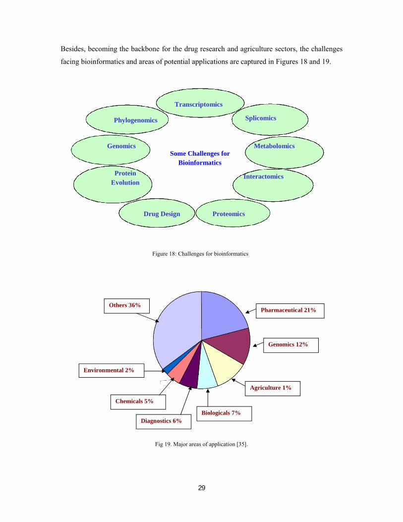
Besides, becoming the backbone for the drug research and agriculture sectors, the challenges
facing bioinformatics and areas of potential applications are captured in Figures 18 and 19.
Transcriptomics
Phylogenomics Splicomics
Genomics MetabolomicsSome Challenges for
Bioinformatics
Protein InteractomicsEvolution
Drug Design Proteomics
Figure 18: Challenges for bioinformatics
Fig 19. Major areas of application [35].
Pharmaceutical 21%
Environmental 2%
Genomics 12%
Agriculture 1%
Biologicals 7%
Others 36%
Diagnostics 6%
Chemicals 5%

30
Bioinformatics and Biodiversity
Biodiversity informatics harnesses the power of computational and information
technologies to organize and analyze data on plants and animals at the macro and at genome
levels. India ranks among the top twelve nations of the world in terms of biological diversity
(Fig. 20 and Fig. 21)
Figure 20: Biodiversity in India (Source: http://edugreen.teri.res.in/explore/maps/biodivin.htm)
Figure 21: Biodiversity bioinformatics is essential to preserve the natural balance of flora and fauna on the planet and to prevent the extinction of species (Source: www.hku.hk/ecology/envsci.htm)
31
Bioinformatics and environment
Deinococcus radiodurans is known for radiation resistance and being used for cleaning
up the waste sites that contain toxic chemicals. Bioinformatics is also helping in climate
change studies. There are many organisms which use carbon dioxide as their sole carbon
source and increasing levels of carbon dioxide emission is one of the major causes of the
global climate change. The study of genomes of these microbial organisms, which is possible
through bioinformatics, helps in proposing ways to decrease the carbon dioxide content. The
program launched by Department of Energy, USA (DOE) started Microbial Genome Project
aimed at sequencing the genomes of bacteria useful in environmental cleanup. This project
started in the year 1994 has brought a revolution in the field of microbiology. According to
NCBI, about 100 genomes have been sequenced so far. According to some estimates,
microbes constitute about 60% of the earth’s biomass and play an important role in natural
biogeochemical cycles. Scientists have now started realizing their potential and role in global
climate processes. Several applications of microbes have been conceived, such as in cleaning
up toxic waste-sites worldwide, energy generation and development of renewable energy
sources, management of environmental carbon dioxide related to climate change, detection of
disease-causing organisms and monitoring of the safety of food and water supplies, use of
genetically altered bacteria as living sensors (biosensors) to detect harmful chemicals in soil,
air or water and understanding of specialized systems used by microbial cells to live in natural
environments with other cells.
Bioinformatics and Diagnostics
Microarray experiments generate the sort of data where the number of measurements
of each sample is much greater than the number of samples. Bioinformatics helps in building
new statistical techniques specifically for microarray data to cope up with the multivariate
nature of microarray data and to extract meaningful information from it. These tools enable
identification of diagnostic markers that are based on a very small number of genes. The fewer
genes that are required to diagnose a disease, the simpler and cheaper the diagnostic tests can
be.
Bioinformatics and biotechnology
The microbes Archaeoglobus fulgidus, Thermotoga maritima and Corynebacterium
glutamicum have the potential for practical applications in industry and environmental

32
projects. These microorganisms thrive in water temperatures above the boiling point and
therefore may provide heat-stable enzymes suitable for use in industrial processes.
Corynebacterium glutamicum is used by the chemical industry for the biotechnological
production of the amino acid lysine. The substance is employed as a source of protein in
animal nutrition. Lysine is one of the essential amino acids in animal nutrition.
Biotechnologically produced lysine is an alternative to soybeans and bonemeal.
Bioinformatics and veterinary sciences
Sequencing projects of farm animals like pig, cow and others are aimed at
understanding the biology of these animals which thus helps in improving their health and
therefore benefits in human nutrition. Conservation of extinct species is another area where
bioinformatics finds applications.
Bioinformatics and systems biology
It is anticipated that many more computational innovations will ensue in going from
genome to the organism and systems biology is that all encompassing field. It is a
multidisciplinary approach to integrate different levels of information to understand how
biological systems function. By studying the relationships and interactions between various
parts of a biological system (e.g., gene and protein networks involved in cell signaling,
metabolic pathways, organelles, cells, physiological systems, organisms, etc.) [36], this
nascent field is expected to provide a prior knowledge about the whole system including
response of the system to external perturbations at both individual and collective levels.
Conclusion
With the increasingly large amounts of biological data, integration with information
technology has become essential. Originally started as a speciality for storage of data and as a
tool kit for analyzing data, bioinformatics now encompasses many emerging areas like,
evolutionary studies, protein structure-function prediction, gene expression studies etc. [37-
58]. It may not be long before bioinformatics becomes a hypothesis driven molecular science
bridging the gap between the genome and the organism, with data providing a platform for
validation and new product development.
33
References
1. Venter, J.C. et al. The sequence of the human genome. Science, 2001, 291, 1304–1351.
2. Schmutz, J. et al. Human genome: Quality assessment of the human genome sequence. Nature, 2004, 429, 365-368.
3. Vij, S., Gupta, V., Kumar, D., Vydianathan, R., Raghuvanshi, S., Khurana, P., Khurana, J.P. and Tyagi, A.K. Decoding the rice genome. Bioessays, 2006, 28, 421-432.
4. Ross, C. Hardison. Compartivs genomics. PLoS Biology, 2003, 1, 156-160.
5. Roy, J. Britten. Divergence between samples of chimpanzee and human DNA sequences is 5%, counting indels. Proceedings of the National Academy of Sciences, 2002, 99, 13633-13635.
6. McEntyre J, Ostell J, (Eds). The NCBI handbook. Bethesda (MD): National Library of Medicine (US), NCBI, 2005.
7. Hanash, S. Disease Proteomics. Nature, 2003, 422, 226-232.
8. Chevre, A., Eber, F., Baranger, A. and Renard, M. Gene flow from transgenic crops. Nature, 1997, 389, 924.
9. Cutanda, M.C. Hernandez-Acosta P. Culianez-Macia F.A. Bioinformatics for crop improvement. Proceedings of the World Congress of Computers in Agriculture and Natural Resources, 2002, 773-778.
10. Dutta, S., Singhal, P., Agrawal, P., Tomer, R., Kritee, Khurana, E. and Jayaram, B. A Physico-Chemical Model for Analyzing DNA sequences. J. Chem. Inf. Model., 2006, 46(1), 78-85.
11. Singhal P., Jayaram B., Dixit S.B. and Beveridge D.L., Prokaryotic Gene Finding based on Physicochemical Characteristics of Codons Calculated from Molecular Dynamics Simulations, Biophys J., 2008, 94, 11, 4173-4183.
12. Anfinsen, C.B. Principles that govern the folding of protein chains. Science, 1973, 181,223.
13. Dobson, C.M. Protein folding and misfolding. Nature, 2003, 426, 884-890.
14. Nelson, D.L., Cox, M. M. Lehninger- Principles of Biochemistry 4th Edition, W.H. Freeman, 2005.
15. Peter, Lmming., Christian, Sinning. and Achim, Meyer. Drugs, their targets and the nature and number of drug targets. Nature, 2006, 5,821-834.
16. UniProtKB/Swiss-Prot Available at http://ca.expasy.org/sprot/relnotes/relstat.html
17. Berman, H. M., Westbrook, J., Feng, Z., Gilliland, G., Bhat, T.N., Weissig, H., Shindyalov, I. N. and Bourne, P. E. The Protein Data Bank. Nucleic Acids Res., 2000, 28, 235–242.
18. Floudas, C. A. Computational Methods in Protein Structure Prediction, Biotechnol Bioeng., 2007, 97, 207-213.
19. Jayaram, B., Bhushan, K., Shenoy, S. R., Narang, P., Bose, S., Agrawal, P., Sahu, D., Pandey, V.S. Bhageerath : An Energy Based Web Enabled Computer Software Suite for Limiting the Search Space of Tertiary Structures of Small Globular Proteins. Nucleic Acids Res., 2006, 34, 6195-6204.
34
20. Thukral, L., shenoy, S.R., Bhushan, K. and Jayaram, B. ProRegIn: A regularity index for the selection of native-like tertiary structures of proteins. J. Biosci., 2007, 32, 71-81.
21. Dickerson, R.E. and Geis,I. The Structure and Action of Proteins, Menlo Park, 1969.
22. Petsko, G.A. From sequence to consequence. Genome Biol., 2000, 1, 406.
23. Silvenstein, K. Millions for Viagra, Pennies for Diseases of the Poor. The Nation, 1999, 269, 3, 13.
24. PAREXEL’s Pharmaceutical R&D Statistical Sourcebook, 2001, p96.
25. Shaikh, S.A., Jain, T., Sandhu, G., Latha, N., Jayaram, B. From Drug Target to Leads-Sketching, A Physicochemical Pathway for Lead Molecule Design In Silico. Current Pharmaceutical Design. 2007, 13, 3454-3470.
26. Hardy, L.W. and Malikayil, A. The impact of structure-guided drug design on clinical agents. Curr. Drug Dicov. , 2003, 15, 20.
27. Jayaram, B., Latha, N., Jain, T., Sharma, P., Gandhimathi, A. and Pandey, V.S., Sanjeevini: A Comprehensive Active-Site Directed Lead Design Software. Ind. J. Chem.-A, 2006, 45A, 1834-1837.
28. Shaikh, S.A. and Jayaram, B., A Swift All-atom Energy based Computational Protocol to Predict DNA-Drug Binding Affinity and ∆Tm. J. Med. Chem., 2007, 50, 2240-2244.
29. Jain, T. and Jayaram, B. An all atom energy based computational protocol for predicting binding affinities of protein-ligand complexes. FEBS Letters, 2005, 579, 6659-6666.
30. Jain, T. and Jayaram,B. A computational protocol for predicting the binding affinities of zinc containing metalloprotein-ligand complexes. Proteins, 2007, 67, 1167-1178.
31. Beecher, C. The Human Metabolome in Metabolic Profiling: Its Role in Biomarker Discovery and Gene Function Analysis. Academic Publishers (Boston), 2003, 311-319.
32. Wishart, D.S. et al. HMDB: the Human Metabolome Database. Nucleic Acids Research. 2007, 35, Database issue, D521 - D526.
33. Goodacre, R. Metabolomics-the forward way. Metabolomics.2005, 1, 1573-3882.
34. Bhana, P. The Indian Bioinformatics Landscape. Pharma Asia, 2008, May.
35. Paolo, Saviotti, P., Marie-Angele deLooze, Michelland, S., and Catherine,D., Nature Biotechnology, 2000, 18, 1247 - 1249.
36. Chong L et al., Whole-istic Biology. Science, 2002, 295, 1661.
37. Shenoy, S.R. and Jayaram, B. "Proteins: Sequence to Structure and Function". Current Status.Current Protein and Peptide Science, 2009, Accepted.
38. Philip E. Bourne, Structural Bioinformatics, John Wiley & Sons; 2002.
39. Westhead, D.R., Parish, J. H., Twyman, R.M. Instant Notes in Bioinformatics, Bios Scientific Pub Ltd, 2002.
40. Des Higgins, Willie Taylor (eds.), Bioinformatics: Sequence, structure and databanks, Oxford University Press, 2000.
41. Hooman H. Rashidi, Lukas K. Buehler, Bioinformatics Basics: Applications in Biological Science and Medicine, CRC Press, 2000.
42. Jin Xiong, Essential Bioinformatics, Cambridge University Press, 2006.
35
43. Krane, D.E., Raymer, M.L., Marieb, E.N. Fundamental Concepts of Bioinformatics, Benjamin/Cummings, 2002.
44. Andreas D. Baxevanis and B. F. F. Ouellette (eds.), Bioinformatics: a Practical Guide to the Analysis of Genes and Proteins, Wiley Interscience, 1998.
45. Arthur, M. Lesk, Introduction to Bioinformatics, Oxford University Press, 2002.
46. Stephen, A.K., and David, D.W. Introduction to Bioinformatics: A Theoretical and Practical Approach, Humana Press, 2002.
47. Pierre Baldi and Soren Brunak, Bioinformatics The Machine Learning Approach, The MIT Press, 2001.
48. Hans-Werner Mewes, H., Seidel, B., Weiss, U., Karrenberg, A., Bioinformatics and Genome Analysis, Springer Verlag, 2002.
49. Sensen, C. W. Essentials of Genomics and Bioinformatics, John Wiley & Sons, 2002.
50. Thomas, Lengauer., Bioinformatics: From Genomes to Drugs, John Wiley & Sons, 2001.
51. Stephen Misener and Stephen A. Krawetz (Eds.), Bioinformatics Methods and Protocols, Humana Press, 2001.
52. Mount, D.W. Bioinformatics: Sequence and Genome Analysis, Cold Spring Harbor Laboratory, 2001.
53. Cynthia Gibas, Per Jambeck, Developing Bioinformatics Computer Skills, O'Reilly & Associates, 2001.
54. Zoe Lacroix and T. Critchlow, Bioinformatics: Managing Scientific Data, Morgan Kauffman Publishers, 2003.
55. Gary Benson, Roderic Page (Eds.), Algorithms in Bioinformatics, Springer International Edition, 2004.
56. Rastogi S.C, Mendiratta N, Rastogi P, Bioinformatics Methods and Applications, Prentice Hall of India Pvt Ltd, 2004.
57. Gautham, N. Databases and Algorithms, Narosa Publishing House, 2006.
58. M.M. Gromiha, Protein Bioinformatics: From Sequence to Function, Elsevier,Academic Press, 2010.