biocorrosion of dental alloys due to … of dental alloys due to desulfotomaculum nigrificans...
TRANSCRIPT
Acta of Bioengineering and Biomechanics Original paperVol. 18, No. 4, 2016 DOI: 10.5277/ABB-00499-2015-03
Biocorrosion of dental alloysdue to Desulfotomaculum nigrificans bacteria
JOANNA MYSTKOWSKA*
Department of Materials Science and Biomedical Engineering, Faculty of Mechanical Engineering,Bialystok University of Technology, Bialystok, Poland.
Purpose: Degradation processes of metallic biomaterials in the oral cavity limit the stability and reliability of dental materials. Theinfluence of environment bacteria Desulfotomaculum nigrificans sulfate reducing bacteria on the corrosion processes of Co-Cr-Mo andTi-6Al-4V alloys was assessed. Methods: After 28 and 56 days of contact of the materials with the bacterial environment, the surfaces ofthe biomaterials tested were observed by means of confocal scanning laser microscopy (CSLM), and their chemical composition wasstudied using X-Ray Photoelectron Spectrometry (XPS). Results: Corrosive changes and the presence of sulfur (with medium atomicconcentration of 0.5% for Co-Cr-Mo and 0.3% for Ti-6AL-4V) were observed on the surface of the biomaterials. Image analysis con-ducted using Aphelion software indicated that corrosion pits took up approx. 2.3% and 1.8% (after 28 days) and 4.2% and 3.1% (after56 days) of the total test surfaces of cobalt and titanium alloys respectively. The greatest number of corrosion pits had a surface areawithin the range of 1–50 m2. They constituted from 37% up to 83% of all changes, depending on the type of material. Conclusions: Anevident influence of the SRB on the surfaces of cobalt and titanium alloys was observed. Significant corrosive losses caused by the ac-tivity of microorganisms were observed on the metallic surfaces under study. The results of this study have much cognitive and utilitar-ian significance.
Key words: biocorrosion, sulfate-reducing bacteria, Desulfotomaculum nigrificans, XPS, Aphelion
1. Introduction
Oral cavity is an environment where high humid-ity, constant temperature and source of food ingredi-ents promote the development of complex and differ-entiated microorganisms, which can colonize, i.e.,surfaces of dental materials. The composition of theoral microbiota can be stable over time or can changedepending of the local environment, lifestyle, age, con-tact with prosthesis or dental implants, etc. [18]. Humansaliva, which plays a significant role in oral mouth, i.e.,provides optimal pH and buffer properties [6], is alsoresponsible for forming special film, called biofilm [1].
In this environment, due to the contact of differentmicroorganisms (bacteria and fungi) and products oftheir metabolic activity [27], the process of microbialcorrosion of metallic biomaterials (prosthodontic ele-
ments, medical implants, etc.) can occur, which causesdestruction of biomaterials, as shown in the work ofKameda et al. [11]. This process is a serious problemand still is little known. Information on this subject isscarce in the literature and insufficient for a meritori-ous description of these processes, especially as regardscontact with anaerobic bacteria – sulfate-reducingbacteria (SRB), which have the ability to oxidizemetal, and this finally leads to microbiologically in-fluenced corrosion (MIC).
Numerous research works described several types ofsulfate reducing bacteria [8], [14], which can occur in theoral cavity as transient flora, and can be involved in bio-corrosion: Desulfovibrio (desulfuricans, fairfieldensis)and Desulfotomaculum nigrificans genera [17], [20].
One of the mechanisms of MIC shows that sulfatereducing bacteria exhibit the capability of inducingcorrosion. Under anaerobic conditions, a metal surface
______________________________
* Corresponding author: Joanna Mystkowska, Department of Materials and Biomedical Engineering, Faculty of Mechanical Engineering,Bialystok University of Technology, Wiejska 45C, 15-351 Bialystok, Poland. Tel: +48 571-443-083, e-mail: [email protected]
Received: October 30th, 2015Accepted for publication: January 5th, 2016

J. MYSTKOWSKA88
acts as the anode in an electrochemical reaction and isoxidized, yielding Me2+ ions. Bacteria produce S2– ions,which enter into a reaction with Me2+, resulting in theformation of metal sulfide. In the cathode area, H+ ionsare produced and they react with hydroxyl groups pres-ent in aqueous environment. As a result, oxygen fromsulfates is consumed for oxidizing metal, which leadsto the formation of metal oxides. A study by Lata et al.[15] shows that the aqueous environment of the oralcavity additionally fosters adhesion of bacteria to metalsurfaces. It was also stated [13] that the activity of SRBcauses a difference in potential between surfaces in/notin contact with bacteria, which leads to the formation oflocal corrosion pits (corrosion characterized as deeplocalized penetration) on the surfaces of metals [28].
Corrosion of biomedical metals in the human bodyis critical, because it can adversely affect their bio-compatibility and mechanical integrity, so studies inthis area are very important [12].
The subject of this study was the evaluation of the in-fluence of Desulfotomaculum nigrificans sulfate reduc-ing bacteria on corrosion processes of commonly usedmetallic biomaterials for dental applications: Co-Cr-Moand Ti-6Al-4V alloys.
The studies of bacterial proliferation and an analy-sis of the chemical composition of sample surfaceswere conducted.
2. Materials and methods
Strains and growth conditions
The standard ATCC 7946 D. nigrificans strain,originating from the collection of the Department ofMicrobiology of the Medical University of Białystok(Poland), was used in this study.
The D. nigrificans strain was cultivated on a TSImedium (Triple Sugar Iron Agar, BBL BD BiosciencesSparks) with the following composition (quantities givenper 1 liter of purified water): 10 g enzymatic caseinhydrolysate, 10 g peptone, 5 g sodium chloride, 10 glactose, 10 g sucrose, 1 g glucose, 0.2 g ferrous ammo-nium sulfate, 0.2 g sodium thiosulfate, 0.025 g phenolred, 13 g agar at a temperature of 37 °C, for a period of72 hours, under anaerobic conditions [7] with the use ofa Genbox anaer generator (bioMerieux). In order to ob-tain semi-liquid medium, the primary medium was di-luted three times. This medium provides an appropriategrowth conditions for D. nigrificans. Next, 10 ml ofculture was transferred to a thrice diluted TSI mediumand incubated for another 72 hours at a temperatureof 37 °C under anaerobic conditions, like in work [19].
Biomaterials
Samples of Co-Cr-Mo and Ti-6Al-4V alloys(SANDVIK, Sweden) were tested in the present study.For each test, twelve samples of each material, witha diameter of 8 mm and a height of 3 mm, were pol-ished progressively with coarse to fine (up to 2000 grit)polishing papers. The polished samples were ultra-sonically degreased in acetone/alcohol and sterilizedin an autoclave at a temperature of 121 C for 15 min-utes. Six samples were tested in a bacterial environ-ment, and six in environment without bacteria.
Biofilm formation
The D. nigrificans biofilm was formed as describedin works [14], [17]. After sterilization process, alloysamples were put into containers, one disk per container,incubated in thrice diluted TSI medium, and inoculumwas cultivated, adjusted to 0.5 on the McFarland scale(approximate cell density 1.5×10⁸ cfu/ml). The negativecontrol was also prepared. In this case steel sampleswere placed into containers with 3-fold diluted TSImedium without bacteria. Afterwards prepared sam-ples were incubated for a period of 28 and 56 days inan incubator at a temperature of 37 °C under anaerobicconditions. After the set period of time passed, sampleswere removed from the solution and rinsed three timesin sterile PBS (pH 7.2) in order to remove the bacteriamaking up the plankton suspension and rinse the sur-face which was held in an environment without bacte-ria. A dark color of the solution in contact with sulfatereducing bacteria, in which the test materials were held,was observed, as for stainless steel [19].
Confocal scanning laser microscopy
Confocal scanning laser microscopy (CSLM, LEXTOLS 4000, Olympus, KeyMed House Stock Road SS25QH Southend-on-Sea, U.K.), a non-destructive real-time imaging technique, was used for evaluation ofsample surfaces, which was based on observation ofadsorbed bacteria and the formation of corrosion cen-ters after process of incubation in inoculum.
Samples were observed in two stages:(a) the first stage – just after being rinsed three times
in sterile PBS in order to remove free bacteriafrom the surface of steel,
(b) the second stage, before CSLM microscope obser-vations, samples were additionally rinsed in an ul-trasonic cleaner in an acetone/alcohol solution inorder to remove adsorbed bacteria from the surfaceof the test materials.The images obtained were analyzed using image
analysis software (Aphelion 3.1, ADCIS, France). The

Biocorrosion of dental alloys due to Desulfotomaculum nigrificans bacteria 89
entire surface of the sample was examined, and im-ages were taken from the representative area (threeplaces) of the sample. The final results of area andamount of biocorrosion pits are the average of allmeasurements.
XPS
X-ray Photoelectron Spectroscopy (XPS) usinga PHI 5000 VersaProbe – Scanning ESCA Micro-probe™ (ULVAC-PHI, Japan/USA) was utilized toanalyze chemical composition [9] at corrosive loca-tions. The aim of this study was to evaluate the pres-ence of sulfur on the surfaces of test materials aftercontact with the D. nigrificans strain.
Statistical analysis
In this study, all experiments were carried out us-ing six samples (six in the environment of bacteriaand six in the environment without bacteria) for tests.Collected data was statistically analyzed and differ-ences were determined using the one-tailed Student’st-test. Statistical analyses were performed using Sta-tistica 12. A p-value < 0.05 was considered statisti-cally significant.
3. Results
Microscope observations of the surfaces of cobaltand titanium alloys after being held in an environ-ment with/without sulfate reducing bacteria (positiveand negative control) for 28 and 56 days were carried
out using confocal scanning laser microscopy (CSLM).Figure 1 presents the surfaces of biomaterials testedas negative control after 28 days of contact with TSImedium only. The surface of Ti-6Al-4V alloy (Fig. 1b;CSLM, bars 20 m) is more uniform without visiblechanges of structure in comparison to the surface ofCo-Cr-Mo alloy (Fig. 1a; CSLM, bars 20 m).
As mentioned earlier, microscope observations ofsurfaces treated with sulfate-reducing bacteria wereperformed in two steps. In the first step, samples wereobserved using CSLM without cleaning in an ultrasoniccleaner in order to present the quantity of D. nigrificansbacteria adsorbed onto surfaces and of corrosion prod-ucts on the test surfaces. Figure 2 shows examplephotographs of the surfaces of Co-Cr-Mo (Fig. 2a)and Ti-6Al-4V (Fig. 2b) alloys. Adsorbed products ofthe biocorrosion reaction and non-uniformly distrib-uted bacteria were observed. The amounts of bacteriaattached to the surface were similar between the mate-rials studied. As in other studies [19], an increasedamount of biocorrosion products was observed on thesurfaces of materials at locations where SRB colonieswere present. Each of these products took up an areaof about a few m2 on the surface, as shown in Fig. 2(CSLM, bars, 10 m).
In the second step of studies, the bacteria and ad-sorbed products were removed in an ultrasonic bath.This process reveals the extent of biologically inducedcorrosion of alloys (Figs. 3–6). After 28 days of contactwith sulfate-reducing bacteria, small dark points on thesurfaces of Co-Cr-Mo (Fig. 3a; CSLM, bars, 400 m)and Ti-6Al-4V (Fig. 4a; CSLM, bars, 400 m) wererevealed, and they appear to be similar. But when thesesurfaces are examined under higher magnification
Fig. 1. Confocal scanning laser micrographs of: (a) Co-Cr-Mo, (b) Ti-6Al-4Vafter 28 days of contact with solution without bacteria (negative control);
images represent typical field of view; bars, 20 m

J. MYSTKOWSKA90
Fig. 2. Confocal scanning laser micrographs of D. nigrificans and biocorrosion products in the presence of bacterial inoculum.Biofilm was grown on the surface of Co-Cr-Mo (a) and Ti-6Al-4V (b) discs for 28 days, surfaces without cleaning in the ultrasonic cleaner.
Image represents typical field of view. Bars, 10 m
Fig. 3. Biocorrosion changes on the surface of Co-Cr-Mo (CSLM) after immersion in D. nigrificans for 28 days;(a) bars, 200 m, (b) bars, 10 m
Fig. 4. Biocorrosion changes on the surface of Ti-6Al-4V (CSLM) after immersion inD. nigrificans for 28 days; (a) bars, 200 m, (b) bars, 10 m
Biocorrosion of dental alloys due to Desulfotomaculum nigrificans bacteria 91
(Fig. 3b, 4b), some differences between both materi-als are revealed.
Corrosion pits (marked with white arrows in thefigures) were more evident in the case of cobalt alloy(Fig. 3b; CSLM, bars, 10 m) in comparison to thetitanium alloy (Fig. 4b; CSLM, bars, 10 m). Also,some differences in their shapes were observed whenboth tested alloys were compared.
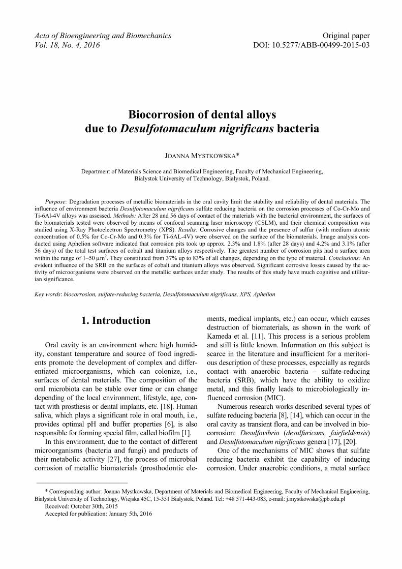
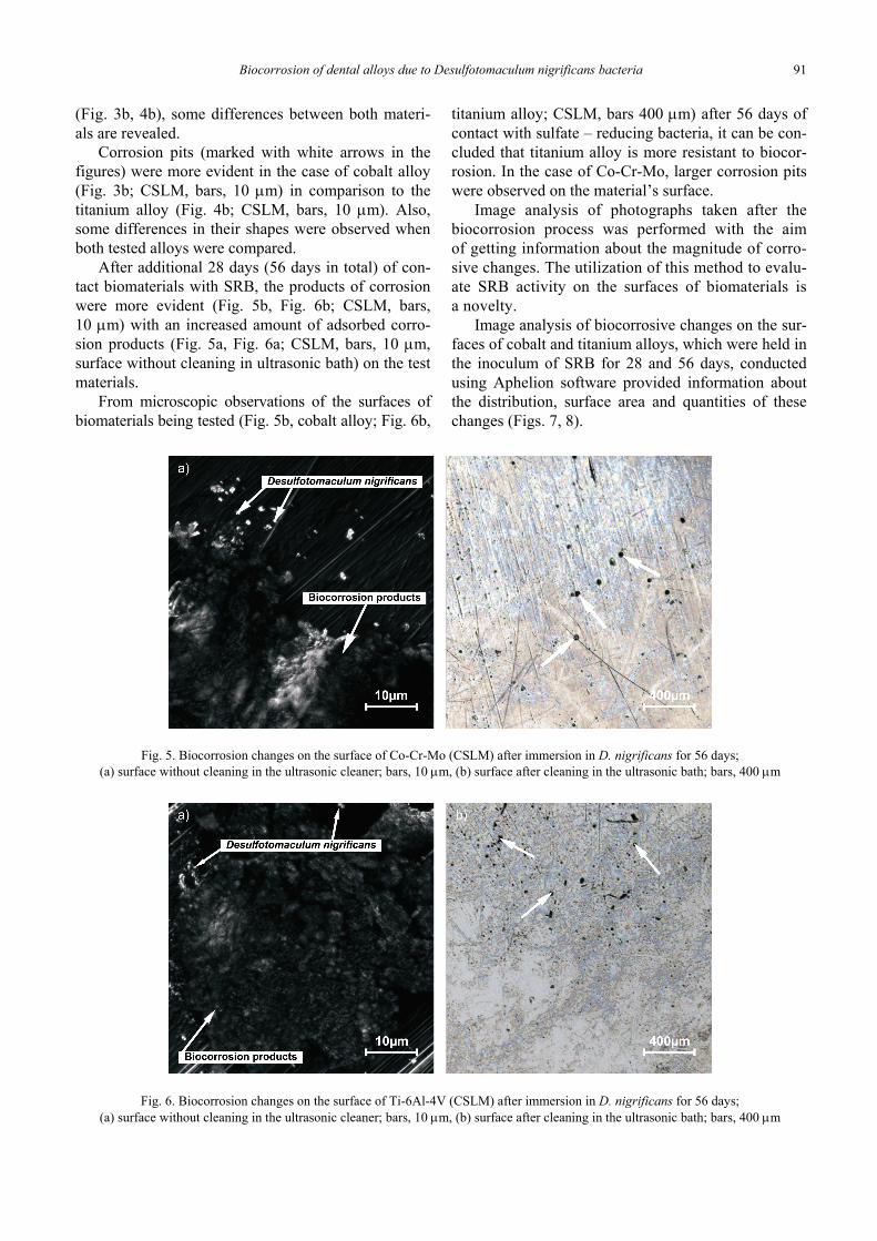
After additional 28 days (56 days in total) of con-tact biomaterials with SRB, the products of corrosionwere more evident (Fig. 5b, Fig. 6b; CSLM, bars,10 m) with an increased amount of adsorbed corro-sion products (Fig. 5a, Fig. 6a; CSLM, bars, 10 m,surface without cleaning in ultrasonic bath) on the testmaterials.
From microscopic observations of the surfaces ofbiomaterials being tested (Fig. 5b, cobalt alloy; Fig. 6b,
titanium alloy; CSLM, bars 400 m) after 56 days ofcontact with sulfate – reducing bacteria, it can be con-cluded that titanium alloy is more resistant to biocor-rosion. In the case of Co-Cr-Mo, larger corrosion pitswere observed on the material’s surface.
Image analysis of photographs taken after thebiocorrosion process was performed with the aimof getting information about the magnitude of corro-sive changes. The utilization of this method to evalu-ate SRB activity on the surfaces of biomaterials isa novelty.
Image analysis of biocorrosive changes on the sur-faces of cobalt and titanium alloys, which were held inthe inoculum of SRB for 28 and 56 days, conductedusing Aphelion software provided information aboutthe distribution, surface area and quantities of thesechanges (Figs. 7, 8).
Fig. 5. Biocorrosion changes on the surface of Co-Cr-Mo (CSLM) after immersion in D. nigrificans for 56 days;(a) surface without cleaning in the ultrasonic cleaner; bars, 10 m, (b) surface after cleaning in the ultrasonic bath; bars, 400 m
Fig. 6. Biocorrosion changes on the surface of Ti-6Al-4V (CSLM) after immersion in D. nigrificans for 56 days;(a) surface without cleaning in the ultrasonic cleaner; bars, 10 m, (b) surface after cleaning in the ultrasonic bath; bars, 400 m

J. MYSTKOWSKA92
Fig. 7. Image analysis of example surfaces of (a) Co-Cr-Mo and (b) Ti-6Al-4Vafter 28 days of the biocorrosion process, Aphelion 3.1; bars, 200 m
Fig. 8. Image analysis of example surfaces of (a) Co-Cr-Mo and (b) Ti-6Al-4Vafter 56 days of the biocorrosion process, Aphelion 3.1; bars, 200 m
Fig. 9. The amount and area of biocorrosion pits on surfaces of test materials;(a) 28 days; (b) 56 days of contact with inoculum

Biocorrosion of dental alloys due to Desulfotomaculum nigrificans bacteria 93
From Figs. 7 and 8 it can be concluded that thearea of corrosion pits on materials after 28 days ofcontact with the bacterial inoculum was smaller com-pared to materials after 56 days of contact. It is alsoshown that the cobalt alloy was characterized by a moreintensive process of surface corrosion, with a higheramount of small biocorrosion changes in comparisonto the titanium alloy.
On the surface areas of the test alloys (each totalarea tested was approximately 1.3 mm2), most corro-sive changes had a surface area within the range of1–50 m2, which constituted from 37% up to 83%of all changes, depending on the type of material. Theleast changes had a surface area within the range of2001–3000 m2, which constituted 0.3% of all changesand was observed only after 56 days of contact withSRB. A large percentage was also noted for corrosionpits with a surface area within the range of 51–100 m2
(8–16% of all changes). Thus, after comparing thetotal surface of these changes to the total surface of theimage subjected to analysis, it can be concluded thatcorrosion pits took up approx.: 2.3% for Co-Cr-Mo(after 28 days), 1.8% for Ti-6Al-4V (after 28 days),4.2% for Co-Cr-Mo (after 56 days), 3.1% for Ti-6Al-4V(after 56 days) of the total surface of the test materials.
Fig. 10. XPS analysis results:on the surface of Co-Cr-Mo, green line-sulfur,
after 28 days of contact with inoculum
In addition, to confirm SRB activity on the sur-faces of biomaterials, chemical composition analysiswas performed at points of corrosive changes. XPSanalysis (an example figure of Co-Cr-Mo surfaceanalysis is given in Fig. 10) indicated the presence ofsulfur on both materials tested, probably in the formof chemical compounds, which explains the dark colorof the contact solution in which biomaterial sampleswere held.
4. Discussion
Numerous metal alloys have been employed asmaterials for treatment of dental disorders. The majormaterials used as dental implants, prosthetic or ortho-dontic materials are: 316 stainless steel, cobalt/titaniumalloys. Among these metals, titanium alloys are char-acterized by the highest corrosion resistance and pas-sivity in biological media due to the presence of titaniumoxide (TiO2) film (naturally, chemically or electro-chemically formed) on their surfaces [21], [22]. Therutile-type tetragonal structure of this oxide is lessreactive in biological media due to its stronger struc-ture and has better biocompatibility with surroundingtissues. As mentioned in the work of Hsu et al. [10],the cobalt alloy (Co-Cr-Mo) also exhibits attractiveproperties such as biocompatibility and corrosionresistance due to the presence of a passive oxide filmon its surface. Reduction of the loss of dental materi-als arising from corrosion phenomena can increase thelong-term success of, i.e., dental implant systems.Most corrosion tests on these alloys are performedelectrochemically. However, oral cavity also containsmicroflora composed of different bacteria strains,which can exacerbate corrosion of metallic biomateri-als. Kameda et al. [11] studied the influence of repre-sentative indigenous oral bacteria, Streptococcus mu-tans and Streptococcus sanguinis, on orthodonticmetallic appliances. In the work of Wilson et al. [25],it was stated that bacteria induce corrosion by severalmechanisms, such as: their presence on the metal sur-face can establish cathodic or anodic regions, whichcan result in the generation of corrosion currents, anda wide range of metabolic products, such as organicacids, can react directly with the metal.
The results of Kameda et al. [11] show that micro-biologically induced corrosion of dental materials byStreptococcus mutans and Streptococcus sanguiniswas found in stainless steel materials but not in tita-nium materials. However, the work of Souza et al.[23], involving electrochemical tests (polarizationresistance of the passive titanium film), shows that thepresence of S.mutans colonies on the titanium surfacenegatively affected corrosion resistance.
The process of biocorrosion in the environmentof sulfate-reducing bacteria [17] was investigated forstainless steel materials. However, no literature datawas found regarding the impact of sulfate-reducingbacteria (SRB) on dental alloys (cobalt or titaniumalloys). Thus, the aim of this work was to study theinfluence of the environment of Desulfotomaculumnigrificans bacteria, which, as mentioned earlier, can

J. MYSTKOWSKA94
also be found in the oral cavity as transient flora,on the corrosion resistance of two different dentalalloys.
Besides adsorbed D. nigrificans bacteria and bio-corrosion products (Fig. 2), corrosion pits (Figs. 3, 5and Figs. 4, 6 for cobalt and titanium alloys after 28and 56 days respectively) were also observed on thetest surfaces of biomaterials used to make elements ofdental prostheses or dental implants [10], [12], as themain causes of destruction of materials of this type.As shown in Figs. 3–6, changes in the structure ofthe test alloys were observed on their surfaces aftercontact with a solution containing sulfate-reducingD. nigrificans bacteria compared to a surface nottreated with SRB (Fig. 1). CLSM analysis indicatedthe presence of biocorrosion centers just after 28 daysof contact of the materials with the environment ofsulfate-reducing bacteria. After additional 28 days(56 days in total), these changes were more evident.What is more, biocorrosion products and corrosionpits were greater on the cobalt alloy surface (Fig. 3and Fig. 5 after 28 and 56 days, respectively) in com-parison to the titanium alloy surface (Fig. 4 and Fig. 6after 28 and 56 days, respectively). This phenomenonconfirms the higher corrosion resistance of titaniumalloy when compared with other metallic biomaterials,including cobalt alloy [22].
Also, changes in the structure of the material wereobserved, being manifested, above all, as discolora-tions of the surface of the biomaterial and small corro-sion pits, which are the effect of biocorrosion occur-ring (Fig. 3b, 4b). A longer time of contact (56 days)of the test biometals with sulfate-reducing bacteria ledto the exacerbation of these processes, and as a result,to the formation of larger corrosion pits on surfacesbeing tested (Fig. 5b, 6b). This may indicate an inten-sively progressing process of microbiologically in-duced corrosion, which occurs in the structure of bio-film, as indicated also in work [26]. Observedcorrosion pits were non-uniformly distributed over thesurfaces of the materials and had surface areas rangingfrom 1 m2 to 3000 m2.
The phenomenon of biocorrosion of metals is ex-plained in many ways, e.g., in the work of Lata et al.[14] it was stated that the activity of corrosive bacteriacreates a difference of electrical potential between thearea attacked by micro-organisms and the area freefrom bacterial activity, which leads to the formationof local pits on the surface of metals. Additionally,when combined with organic substances, their meta-bolic activity may lead to the formation of aggressivecorrosion products, such as incomplete metabolicorganic acid [2].
One of the mechanisms of MIC is cathodic depo-larization, as suggested by Kuhr and Vlugt in 1934[16]. Also, according to work of Bryant [4] the roleof hydrogenase was considered as a unique mecha-nism of MIC by SRB metabolic activities. The utili-zation of hydrogen in their cells can be expressed asfollows [16]
NAD+ + H2 (hydrogenase activity) = NADH + H+ (1)
The XPS analysis shown in Fig. 9 indicates thatthe corrosion film on cobalt alloy’s surface was com-posed of Cr, O, C, S. The dark color of the contactsolution and the presence of sulfur (probably in theform of compounds) on the surface of the materials isa sign of bacterial corrosion [19]. This corrosionproduct may be deposited and become a cathode witha large surface area relative to the unreacted cobaltalloy, accelerating the dissolution of the surface. Asa possible energy source, hydrogen was an importantelectron donor for sulfate reduction. It was shown inthe work of Chen and Clayton [5] that the passive filmon steel surface may be deteriorated by bacteriallyinduced sulfides and by the removal of alloying ele-ments. A similar effect can occur in the case of metalalloys, where the increased amount of oxygen near thesurface of the material may be an effect of the forma-tion of a layer of oxides, including titanium or cobaltoxides. These oxide films are protective layers thatmay contribute to reduction of corrosion. However,the dissolution of TiO2 may occur in certain media,such as those containing high fluoride concentrations,hydrogen peroxide (H2O2) and lactic acid, e.g., it canoccur in the oral cavity [23].
Corrosive changes are one of the main causes ofdestruction of materials used in dentistry in the oralcavity. This process causes the release of metal ionsinto the surrounding medium and deterioration of themetal, which in fact reduces the functional propertiesof the material and its mechanical strength [3]. Corro-sion of metallic appliances in the oral cavity by bacte-ria releases metal ions, which may migrate within theentire organism, be deposited in organs of the humanbody, stimulate an initial inflammatory response, andact as allergens or carcinogens [11], [24].
The image analysis applied for evaluation of thesize and quantity of formed corrosion pits is a nov-elty in this work, compared to other works dealingwith biocorrosion. The results of this analysis pro-vide valuable information about the proportion of thesurface area of these changes to the total surface areaof the dental alloys. Biocorrosion covered between1.8% and 4.2% of the total surface of the test materi-als, which gives a global perspective on this process

Biocorrosion of dental alloys due to Desulfotomaculum nigrificans bacteria 95
in terms of material destruction and allows its inten-sity to be estimated depending on the type of alloy.
An evident influence of the alloy type on bacterialproliferation was observed. Significant corrosionlosses caused by the activity of microorganisms wereobserved on the metallic surfaces under study. Thematerials studied exhibited varying resistance to bio-corrosive destruction. Based on data found in the lit-erature, it was concluded that Ti6Al4V was the mostsuitable material for implant applications in the hu-man body [22] when considering its corrosion resis-tance. Similar results were obtained in this work,however resistance to biocorrosion was assessed. Al-though the cobalt alloys do not passivate as titaniumalloys do, they also have good resistance against cor-rosion in a biological environment, compared, i.e., tostainless steel [19]. The results of this study havemuch cognitive and utilitarian significance.
Acknowledgement
This scientific work was partially financed with researchfunds of Statutory Work of the Department of Materials andBiomedical Engineering (Faculty of Mechanical Engineering,Białystok University of Technology), number S/WM/1/14 andpartially by the National Centre for Research and Development(Poland) under research project No. N R15 0117 10/NCBR.Special thanks are due to Prof. Katarzyna Leszczynska from theDepartment of Microbiology of the Medical University ofBiałystok (Poland) for biological research.
References
[1] ALMAGUER-FLORES A., Biofilms in the oral environment,Bio-Tribocorrosion in Biomaterials and Medical Implants,Woodhead Publishing Series in Biomaterials, 2013, 169–186.
[2] BASKARAN V., NEMATI M., Anaerobic reduction of sulfate inimmobilized cell bioreactors, using a microbial culture origi-nated from an oil reservoir, Biochem. Eng. J., 2006, 31(2), 148–159.
[3] BEECH I.B., SUNNER J.A., HIRAOKE K., Biocorrosion: to-wards understanding interactions between biofilms and met-als, Curr. Opin. Biotech., 2004, 15, 181–186.
[4] BRYANT R.D., LAISHLEY E.J., The role of hydrogenase in an-aerobic biocorrosion, Can. J. Microbiol., 1990, 36, 259–264.
[5] CHEN G., CLAYTON C.R., Influence of sulfate-reducing bac-teria on the passivity of type 304 austenitic stainless steel,J. Electrochem. Soc., 1997, 144(9), 3140–3146.
[6] DODDS M.W.J., JOHNSON D.A., YEH C.-K., Health benefitsof saliva: a review, J. Dent., 2005, 33, 223–233.
[7] DONNELLY L.S., BUSTA F.F., Heat resistance of Desulfoto-maculum nigrificans spores in soy protein infant formula prepa-rations, Appl. Environ. Microbiol., 1980, 40(4), 721–725.
[8] DZIERŻEWICZ Z., CWALINA B., GAWLIK B., WILCZOK T., Iso-lation and evaluation of susceptibility of sulphasalazine ofDesufovibrio desulfuricans strains from the human digestivetract, Pol. J. Microbiol., 1997, 46, 175–187.
[9] GUO W.Y., SUN J., WU J.S., Electrochemical and XPS stud-ies of corrosion behavior of Ti–23Nb–0.7Ta–2Zr–O alloyin Ringer’s solution, Mater. Chem. Phys., 2009, 113(2–3),816–820.
[10] HSU R.W.-W., YANG CH.-H., HUANG CH.-A., CHEN Y.-S.,Electrochemical corrosion studies on CoCrMo implant alloy inbiological solutions, Mater. Chem. Phys., 2005, 93, 531–538.
[11] KAMEDA T., ODA H., OKHUMA K., SANO N., BATBAYAR N.,TARASHIMA Y., SATO S., TERADA K., Microbiologically in-fluenced corrosion of orthodontic metallic appliances, Dent.Mater. J., 2014, 33(2), 187–195.
[12] KUPHASUK C.H., OSHIDA Y., ANDRES C.J., HOVIJITRA S.T.,BARCO M.T., BROWN D.T., Electrochemical corrosion of ti-tanium and titanium-based alloys, J. Prosthet. Dent., 2001,85, 195–202.
[13] KURTZ W., RACKI J., Korozja mikrobiologiczna oraz rolabakterii w przemyśle kopalin chemicznych, Wiadomości bo-taniczne, 1964, t. VIII, z. 2., 163–169 (in Polish).
[14] LATA S., SHARMA C.H., SINGH A.K., Comparison of Biocor-rosion due to Desulfovibrio desulfuricans and Desulfoto-maculum nigrificans Bacteria, J. Mater. Eng. Perform., 2013,22, 463–469.
[15] LATA S., SHARMA C.H., SINGH A.K., Effect of host media onmicrobial influenced corrosion due to Desulfotomaculumnigrificans, J. Mater. Eng. Perform., 2013, 22, 1120–1128.
[16] LIU Y., WANG Q., SONG Y., ZHANG D., YU S., ZHU X.,A study on the corrosion behavior of Ce-modified cast AZ91magnesium alloy in the presence of sulfate-reducing bacte-ria, J. Alloy Compd., 2009, 473, 550–556.
[17] LOPES F.A., MORIN P., OLIVEIRA R., MELO L.F., Interactionof Desulfovibrio desulfuricans biofilms with stainlees steelsurface and its impact on bacterial metabolism, J. Appl. Mi-crobiol., 2006, 101, 1087–1095.
[18] MARSH P.D., HEAD D.A., DEVINE D.A., Dental plaque asa biofilm and a microbial community-Implications fortreatment, J. Oral Biosci., 2015, http://dx.doi.org/10.1016/j.job.2015.08.002.
[19] MYSTKOWSKA J., FERREIRA J.A., LESZCZYŃSKA K.,CHMIELEWSKA S., DĄBROWSKI J.R., WIECIŃSKI P.,KURZYDŁOWSKI K.J., Biocorrosion of 316LV steel used in oralcavity due to Desulfotomaculum nigrificans bacteria, J. Bio-med. Mater. Res. B, 2015. DOI: 10.1002/jbm.b.33518.
[20] NAZINA T.N., ROZANOVA E.P., BELYAKOVA E.V., LYSENKO A.M.,POLTARAUS A.B., TOUROVA T.P., OSIPOV G.A., BELYAEV S.S.,Description of “Desulfotomaculum nigrificans subsp. sali-nus” as a New Species, Desulfotomaculum salinum sp. nov,Microbiol., 2005, 74(5), 567–574.
[21] OSHIDA Y., SACHDEVA R.C., MIYAZAKI S., Microanalyticalcharacterization and surface modification of TiNi orthodon-tic archwires, Biomed. Mater Eng., 1992, 2, 51–69.
[22] SONGUR M., CELIKKAN H., GOKMESE F., SIMSEK S.A., ALTUNN.S., AKSU M.L., Electrochemical corrosion properties ofmetal alloys used in orthopaedic implants, J. Appl. Electro-chem., 2009, 39, 1259–1265.
[23] SOUZA J.C.M., PONTHIAUX P., HENRIQUES M., OLIVEIRA R.,TEUGHELS W., CELIS J.-P., ROCHA L.A., Corrosion behaviorof titanium in the presence of Streptococcus mutans, J. Dent.,2013, 41, 528–534.
[24] WANG J.J., SANDERSON B.J.S., WANG H., Cyto- and geno-toxicity of ultrafine TiO2 particles in cultures human lympho-blastoid cells, Mutation Research, 2007, 628, 99–106.
[25] WILSON M., KPENDEMA H., NOAR J.H., HUNT N., MORDAN N.J.,Corrosion of intra-oral magnets in the presence and absence

J. MYSTKOWSKA96
of biofilms of Streptococcus sanguis, Biomaterials, 1995, 16,721–725.
[26] XU LI.-CH., CHAN K.-Y., FANG H.H.P., Applicationof atomic force microscopy in the study of microbiologi-cally influenced corrosion, Mater. Charact., 2002, 48,195–203.
[27] YAN Y., NEVILLE A., DOWSON D., Biotribocorrosion of Co-CrMo orthopaedic implant materials-Assessing the formationand effect of the biofilm, Tribol. Int., 2007, 40, 1492–1499.
[28] ZUO R., Biofilms: strategies for metal corrosion inhibitionemploying microorganisms, Appl. Microbiol. Biotechnol.,2007, 76, 1245–1253.