biocompatibility and osteoconductivity of the … bility...biocompatibility and osteoconductivity of...
TRANSCRIPT
Histol Histopathol (1997) 12: 19-24
001: 10.14670/HH-12.19
http://www.hh.um.es
Histology and Histopathology
From Cell Biology to Tissue Engineering
Biocompatibility and osteoconductivity of the pyrost bone substitutes V-H. Tsuang1,2, F-H. Lin3, H-C. TaP, J-S. Sunl, H-C. Liu1 and V-So Hang1
1 Department of Orthopedic Surgery, National Taiwan University Hospital, Taipei, Taiwan, R.O.C.,
20epartment of Orthopedic Surgery, Taiwan Provincial Tao-Yuan General Hospital Tao-Yuan, Taiwan, R.O.C. and
3Center for Biomedical Engineering, College of Medicine, National Taiwan University, Taipei , Taiwan, R.O.C.
Summary. The purpose of this study was to re-evaluate the bone regeneration power and the in vitro biocompatibility of the Pyrost bone substitute. Twentyfour adult New Zealand White rabbits were used. Bony defect over both iliac crest and mid-diaphyseal portion of the ulna bone were created. Appropriate sized-block of Pyrost bone substitute were implanted. Four of the animals were killed at each postoperative month to evaluate its bone regeneration power by histologic study. The Pyrost bones were co-cultured with osteoblasts to evaluate its biocompatibility. The results showed that Pyrost bone substitute was quite stable and incorporated well with active bone regeneration. The Pyrost heal better at the iliac crest than at the ulnar defect. The Pyrost was compatible to the osteoblasts . Osteoblasts had successfully seeded and mitotically expanded on the porous surface of the Pyrost bone graft. The result showed that Pyrost bone obviously exerts an intense stimulus on osteo-regeneration in the presence of osteoblasts. We consider Pyrost to be an alternate to the conventional preserved allografts that is occasionally necessary.
Key words: Pyrost bone substitute , Biocompatibility, Osteoconductivity
Introduction
Congenital or acquired bone defect is a major problem in the orthopedic surgery. In order to accelerate the healing of bone defect, bone grafting is required in about 15 % of the reconstructive operations on the locomotor system (Urist et aI., 1994). Autografts are preferably used because of their superior efficacy and capability of avoiding transmission of infection. However, it does have drawbacks including donor site morbidity and limited availability , particularly in
Offprint requests to: Dr. Hwa·Chang Liu , Department of Orthopedic
Surgery, National Taiwan University Hospital, No.7, Chung-Shan South
Road, Taipei , Taiwan, 100, ROC
children (Glowacki et aI. , 1981; Dutting et aI., 1988; Grob , 1988). The effectiveness of allogenous bone graft is limited by the problems of high cost of bone banking, potential of graft-related disease transfer, the high rates of nonunion and infection , and allograft fracture s (Kotani et aI., 1992).
A variety of bone grafts substitute materials are commercially available : both mineralized and demineralized forms (Doherty et aI. , 1994). Pure mineral bone , Pyrost , had been reported as a promising bonereplacement material (Katthgen and Mittelmeier, 1983). Osteoblasts have recently been grown in vitro on the bone-replacement materials (Guillemin et aI. , 1987 ; Matsuda and Daves, 1987; Gregoire et aI., 1990; Sautier et aI. , 1990). In the study of Katthgen and Mittelmeier (1983), the experimental model used was mainly the condylar portion of distal femur. It is not a common location for harvesting bone graft. Filling with plenty of osteogenic cells, the iliac crest is one of the most commonly used donor site for harvesting autogenous bone graft. The significant difference between autologous bone and these bone-replacement materials is that autologous bone contained a variety of living cells, including osteoblasts. When being used to replace bony defects , the biocompatibility of bone substitutes is mandatory for achieving bone regeneration .
The purpose of this study were to re-examine the bone regeneration power of Pyrost bone substitute at the site of cancellous and cortical bone defects; also to determine the in vitro biocompatibility of the Pyrost bone substitute . Our hypothesis is that the Pyrost is a suitable carrier system for osteoblasts proliferation and also a potential bone substitute for the iliac bone defect created after harvesting bone graft.
Materials and methods
Animal experiment
Twenty-four adult New Zealand White rabbits , weighing 2.0 to 2.5 kg ., were used in this experiment. The animals were fed Purina Laboratory Chow ad
20
Biocompafibilify and osfeoconductivity of Pyrost
libitum and housed in a temperature-, humidity- , and light-controlled environment. Surgical procedures and experimental protocols were approved and under supervision by the Medical College 's Animal Research Committee of the National Taiwan University. The rabbits were premedicated with 0.5 mg atropine and then anesthetized using ketamine (50 mg/kg) and rompun [2-(2 ,6-xy I id ino- )-5 ,6-d i hyd ro-4H -1 ,3-th i azi ne hydrochloride] (12 mg/kg). After shaving , the operating sites were disinfected and draped . Bilateral iliac crests were exposed. A 5xLO mm sized bony defect was created at mid-portion of the iliac crest. An appropriatesized Pyrost bone substitute was inserted to the osteotomy site, and then the wound was closed in layer. The opposite side of iliac crest was sham-operated but without insertion Pyrost block . The bilateral forelimbs were then surgically treated to create a l.5-cm bone defect at the middle diaphyseal portion of ulna as described by Grundel (1991) . One limb serves as the control without any further management at the bony defect. The experimental limb received a block of Pyrost bone substitute with an artificially-created central canal for fixation with an 1.0 mm intramedullary K-wire. Intravital staining of tetracycline was performed since the 7th postoperative day with intramuscular injection of Vibramycin (Doxycycline hyclate: 15 mg/kg b .w.) and then repeated every 14 days till the date of sacrifice.
Four animals, selected by random , were sacrificed at each postoperative month using an overdose of pentobarbital. Blocks of the pelvic wings and forelimb two bones were excised . All of the harvested specimens received roentogenographic examination . The specimens were fixed with 3% phosphate-buffered formaline for seven days, progressive alcohol dehydration (in 70, 80, and 90% alcohol, and twice in 100% alcohol for three days) , and then embedded in methyl-methacrylate polymer. Sections of 500 jlm thickness, parallel to the plane of iliac wing, were prepared using a diamond band saw with water cooling (Buehler Isomet™ , low speed saw, Lake Bluff, Illinois, USA), and were grinded to a thickness of 50 ]I m . The section was glued onto sl ide with Entellan and examined by fluorescence photomicroscopy (Nikon-Microphot-Fx, Japan).
For light microscopy examination, sections were further grounded to 5-10 jl m thickness, deplasti fication , and sequentially rehydrated down to distilled water. They were then transferred to 5% silver nitrate , reduced with Na2C03 - formaldehyde solution, followed by staining in methyL green pyronin (Von Kossa stain) (Recker, 1983); and observed with light microscopy.
Osteoblasts culture
Sequential digestion of newborn Wi star-rat calvaria was performed by using a modification of the methods described by Wong and Cohn (Boonekamp et aI., 1984). Briefly, after pretreatment of the dissected calvaria with 4mM Na2 EDTA in a pre-warmed (37 DC) solution containing 137mM NaCl, 2 .7mM KCI, 3mM NaH2P04 ,
pH 7.2 (solution A), for 10 minutes three times, the fragments were sequentially digested with collagenase (180 U/ml, Sigma Co.) in solution A with EDTA . The sequential digestion consisted of four times treatments for 5 minutes followed by two times treatments for 10 minutes. The cells released after each treatment were immediately harvested by centrifugation and resuspended in culture medium. The osteoblastic phenotype of the cells was assessed by the tests of alkaline phosphatase assay (Doty and Schofield, 1976; Begley et aI., 1993).
Three Pyrost bones (lOx IOxO.5 mm) were placed in sterile dish. Confluent rat osteoblast cultures were passaged by trypsin-EDTA and were seeded into each well on top of the implant materials at a seeding density of 3x I 04 cells/cm3. A large area of each well was left uncovered to allow assessment of cell plating efficiency by microscopy. The culture media used was Dulbecco 's modified Eagle's medium supplemented with 10% fetal calf serum (Gibco , UK), penicillin (100 units/ml) and streptomycin (100 mg/ml). The dishes were incubated at 37 DC in an atmosphere supplemented with 5% CO2 and fed with complete changes of medium twice a week. The days of plating was considered as the first day of culture. The test samples were removed from wells at days 3, 5 , 9, fixed in 3% formaldehyde in O.IM PBS buffer (pH 7.4). Each test composed of triplicate samples and separate control at each of the time points. The samples were stained with Hematoxyline-Eosin and then observed and photographed with inverted microscopy.
For electron microscopic examination , the Pyrost bone substitute blocks were fixed with 3% phosphatebuffered formaline solution, dehydrated through a graded ethanol series and critical dried from CO2 (Ladd Research Industries Inc . , Burlington, VT, USA). Specimens were sputter-coated with gold (Model 18-2 Ion Coater, Eiko Engineering, K.K. Japan) and examined in a ISA ABT (model SX-30 E , International Scientific Instruments, Inc, CA , USA) scanning electron microscope at an accelerating voltage of 15 kYo
Results
In vivo gross morphological and histological evaluation
Pyrost bone grafts were stably fixed into rabbits' pelvis. After implantation of Pyrost for six months, the location of Pyrost still can be identified, but had been closely incorporated into the host bone. It was further approved by the roentogenographic assessment that did not show any radiolucent space surrounding the Pyrost bone substitute . The sham-operated site of pelvic wing showed a bony defect filled with fibrous tissue. The defect was quite obvious in the roentogenogram.
At the ulnar defects grafting with the Pyrost bone substitute, visible callus formation was observed at the end of the first month. The amount of callus format ion was much increased at the end of the 6th month . At the control limb without Pyrost bone substitute, callus
21
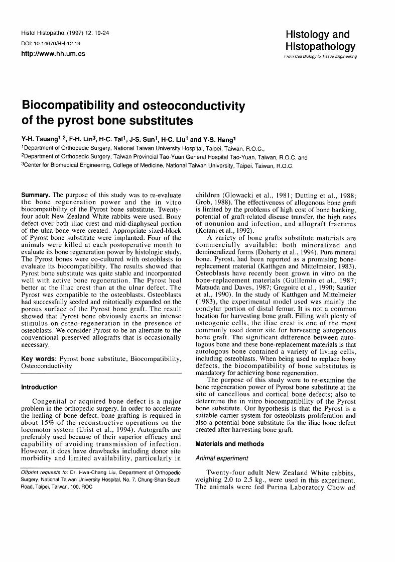
Fig. 1. Light micrograph of bone regeneration on Pyrost at the iliac defect. The darkly colored Pyrost implant (P) is surrounded everywhere, by regenerated bone (R) and osteoid tissue (aT). Von Kossa stain . Bar: 200 !-1m.
Fig. 2. Fluorescence micrograph of bone regeneration on Pyros!. Vigorous bone regeneration on the surface of the Pyrost implant (P). There is no connective tissue layer between implant and bone; adjacent to it is inconspicuous regenerated marrow tissue . Von Kossa stain. Bar: 200 !-1m .
Fig. 3. Vigorous bone regeneration on the surface of the Pyrost implant (P). There is minimal connective tissue layer between implant and bone. Note that even after 3 months' implantation , the darkly colored Pyrost is still almost totally preserved. The entire bone-defect area is filled with regenerated bone. Pyrost and regenerated bone are closely linked together. Von Kossa stain . Bar: 80 !-1m.
Fig. 4. At high power magnification , Pyrost (P) surrounded by regenerated bone (R) . Intensive bone regenerat ion with massive osteoblasts (OB) can be seen . Von Kossa stain. Bar: 20 !-1m.
Fig. 5. Histological examination of the Pyrost bone grafted at the ulnar defect. At six months after implantation, there is little callus formation (C) of the adjacent cortical bone into the Pyrost (P) observed with intervening fibrous tissue (F) . Von Kossa stain. Bar: 200 !-1m .
22
Biocompatibility and osteoconductivity of Pyrost
formation at both ends of the defect toward the center was observed. However, complete bridging of the defect did not occur. Neither the ulnar defects with Pyrost block nor that without Pyrost block was completely united with autogenous bone formation.
Pyrost bone substitute, incorporated well with the pelvis, showed active bone regeneration. Histological examination revealed that the Pyrost was infiltrated with regenerated bone trabeculae (Fig . L). The entire bonedefect area is filled with regenerated bone . There is vigorous bone regeneration on the surface of the Pyrost with little connective ti ss ue layer (Fig. 2). After deplastication, vigorous bone regeneration on the sUlface of the Pyrost implant with mass ive osteoid tissue can be seen. Pyrost and regenerated bone are closely linked together. Also there is inconspicuous regenerated marrow tissue adjacent to this regenerated bone (Fig. 3). At higher magnification, early phases of bone regeneration with many osteoblasts at the surface of the Pyrost implant were visible (Fig. 4).
In the ulnar defects grafted with Pyrost block, solid incorporation of the Pyrost block into the adjacent cOI1icai bone was not observed. Even after six months' implantation , there is little callus formation of the adjacent cortical bone into the Pyrost observed with intervening fibrous tissue (Fig. 5). This suggested that the bone regeneration and ingrowth was incomplete at the end of the six month.
In vitro biocompatibility of the Pyrost bone graft
At the 3rd day of culture, the Pyrost block exhibited a sparse population of osteoblasts (Fig. 6). The number of cells had increased by the 5th day, the cells began to contact with each other. On the more porous surfaces, the osteoblasts were observed to suspended across the pores by the Pyrost block (Fig. 7). By the 9th day , a nearly complete layer of osteoblasts coated over porous surface (Fig . 8).
The cells, growing on the Pyrost block, typically exhibited numerous pseudopodial and filopodial extensions anchoring the cells to the surface (Fig. 9). Later, layer of flattened polygonal cells developed and covered almost the whole surface of the Pyrost bone substitute (Fig . 10) . The osteoblasts successfully seeded and mitotically expanded on the porous surface of Pyrost bone substitute block. The Pyrost bone graft exhibited good biocompatibility in the osteoblast culture.
Discussion
The basic functions of a bone graft or bone substitute are (L) the osteogenic performance , the induction or conduction of bone regeneration, (2) the bony fixation and stabilization and (3) the filling of the bone defect itself (Morscher, 1982). In many cases there are problems with the filling of fairly large bone cavities, so soughing of the alternatives to conventional bone grafts are indicated. In the past decade , many synthetic
biomaterials have been reported for bone substitutes, including bio-inert ceramic and bio-active ceramics (Kotani et aI., 1992), synthetic calcium phosphates (Kitsugi et aI., 1988), plastic and metal implants (Katthagen, 1986). Because in case of requiring bone graft or bone substitute, the ideal approach is not longterm or permanent bone replacement , but the stimulation of bone regeneration followed by filling the defect with the body 's own bone. This principle still cannot be adeq uately realized with above-mentioned synthetic biomaterials . In this study, we focused on the os teoconduction induced by Pyrost bone at the region of iliac crest for its possible role in bone substitute after harvesting autogenous iliac bone graft.
In our study, Pyrost bone graft incorporated well with the bone actively regenerated at the defect of pelvic osteotomy site. The implanted material was resolved slowly, and was still present almost in its entirety after 6 months. Histological examination revealed vigorous bone regeneration on the surface of the Pyrost implant (Fig. I). The regenerated bone contains little connective tissue layer between implant and bone (Fig. 2) with massive osteoid tissue (Fig . 3) and many osteoblasts at the surface of the Pyrost implant (Fig . 4). The resulting network of regenerated bone surrounding the implant and fi lling the interstices was as dense as, and sometimes even denser than the surrounding intact bone structures. Since the bone defect chosen in the rabbit cannot be filled spontaneously with regenerated bone , the bony regeneration observed in the experimental groups were mainly under the influence of the Pyrost substitutes (Karaca et at. , 1994; Matsuda et aI., 1995). In those of ulnar defects , there is little callus formation of the adjacent cortical bone into the Pyrost even after six months' implantation. This suggested that ingrowth was incomplete even after six months. The Pyrost heal better at the iliac crest than at the ulnar defect. In the clinical medicine, the usual site for harvesting autogenous bone graft was the iliac crest. The loss of bone in the donor site and the related cosmetic problem may bothering in so me degree . The previous harvested site was also impossible for further harvesting graft. The present results provide the evidence that Pyrost bone substitute is good osteoconductive at the iliac crest. We suggest that in appropriate indications, implantation of Pyrost substit ute can be a very useful and effective bone substitute in the donor site after harvesting bone graft. This makes this site possible to reuse in .later situation.
An important prerequisite for a bone graft substitute materia] is that the material must be able to support osteob last attachment and even able to promote proliferation of the attached cells. Doherty suggested that cell attachment and proliferation is faster and dimineralized bone as opposed to mineralized form (Doherty et aI., 1994). In the present study, we elucidated that the biocompatibility of the porous surface of mineralized Pyrost bone substitute to osteoblasts was rather good (Figs. 5-8). The osteoblasts had successfully seeded onto and mitotically expanded on the Pyrost bone
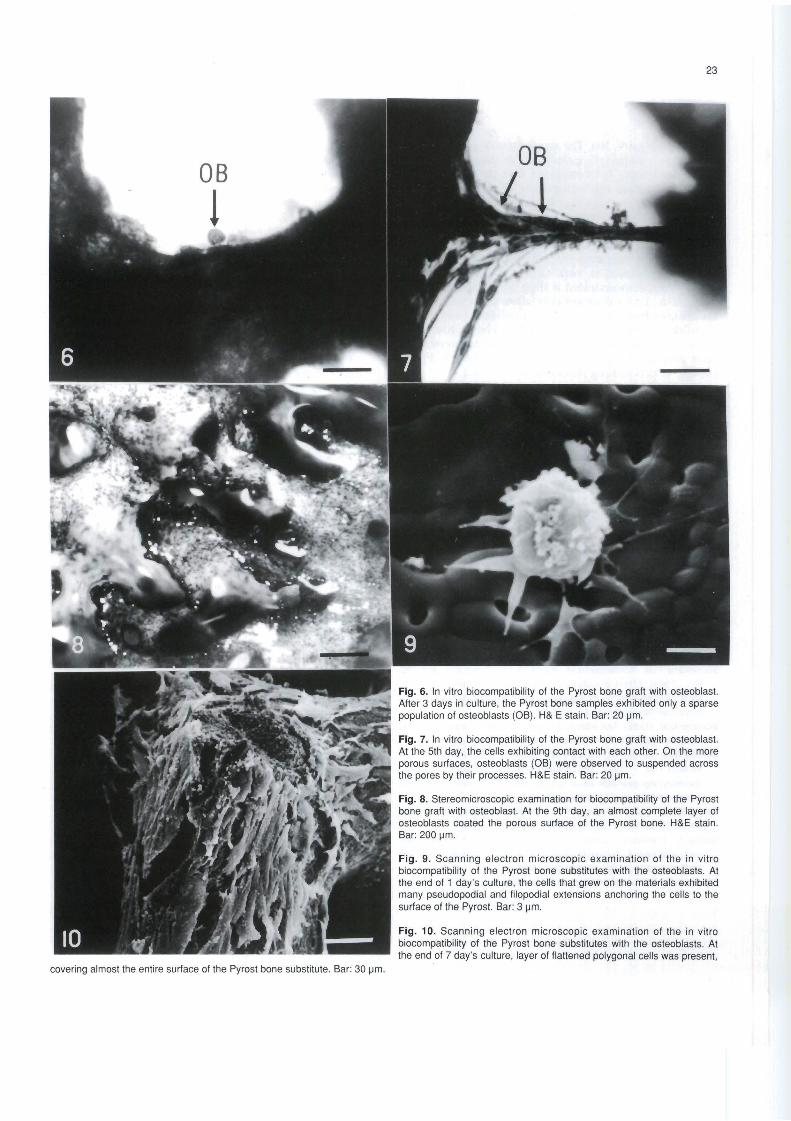
covering almost the entire surface of the Pyrost bone substitute. Bar: 30 j.Jm.
23
Fig. 6. In vitro biocompatibility of the Pyrost bone graft with osteoblast. After 3 days in culture, the Pyrost bone samples exhibited only a sparse population of osteoblasts (OB) . H& E stain. Bar: 20 j.Jm .
Fig. 7. In vitro biocompatibility of the Pyrost bone graft with osteoblast. At the 5th day, the cells exhibiting contact with each other. On the more porous surfaces, osteoblasts (OB) were observed to suspended across the pores by their processes. H&E stain. Bar: 20 j.Jm.
Fig . 8. Stereomicroscopic examination for biocompatibility of the Pyrost bone graft with osteoblast. At the 9th day, an almost complete layer of osteoblasts coated the porous surface of the Pyrost bone. H&E stain . Bar: 200 j.Jm.
Fig. 9. Scanning electron microscopic examination of the in vitro biocompatibility of the Pyrost bone substitutes with the osteoblasts. At the end of 1 day's culture, the cells that grew on the materials exhibited many pseudopodial and filopodial extensions anchoring the cells to the surface of the Pyrost. Bar: 3 j.Jm.
Fig. 10. Scanning electron microscopic examination of the in vitro biocompatibility of the Pyrost bone substitutes with the osteoblasts. At the end of 7 day's culture, layer of flattened polygonal cells was present,
24
Biocompatibility and osteoconductivity of Pyrost
substitute (Figs . 9, 10). The attachment of osteoblasts on the Pyrost materials exhibited numerous ce llul ar processes (Figs. 7,9) suggests that the cells tend to attach regardless the topography of the materials . It suggested that porosity may be important to allow fluid c irculation , bone ingrowth and mechanical stabi lity at the implantation site (Brunette, 1988). Such observations are in acco rd to those of Begley et al. (\993), and suggested that migratory osteoblasts adhere to any firm substrate presented in vitro (Jones and Boyde, 1979) . The Pyrost demonstrated a high biocompatibility to osteoblasts, i.e. , it does not only allow the attachment of the osteob lasts but al so the proliferation of the osteobJasts (Mats uda et aI., 1995) . The Pyrost bone substitute is poss ible to behave as a delivery system for bone growth . However, the mechanism whereby Pyrost implant affects healing remained to be solved.
Conclusion
In this study, Pyrost was used as a substitute after harvesting au toge nous ili ac bone graft. The resu lt showed that Pyrost bone obv ious ly exerts an intense stimu lus on osteo- regeneration and in-vitro study also showed good biocompatibility to the osteoblas ts. We consider Pyrost to be an alternate to the conventional preserved allografts that is occasionally necessary.
Acknowledgements. The authors sincerely thank the National Science Council (ROC) for their financial support of this research.
References
Begley C.T. , Doherty M.J., Kankey D.P. and Wilson D.J. (1993). The culture of human osteoblasts growth upon bone graft substitutes. Bone 14, 661-667.
Boonekamp P.M., Kekkelman JW., Hamilton JW., Cohn D.V. and Jilka R.L. (1984). Effect of culture on the hormone responsiveness of bone cells isolated by an improved sequential digestion procedure. Proc. Kon. Acad . Wet. B. 87, 371-384.
Brunette D.M. (1988) . The effects of implant surface topography on the behavior of cells. Int. J. Oral Maxillo!. Implants 3, 231-246.
Doherty M.J. , Schlag G., Schwarz N., Mollan R.A.B., Nolan P.C. and Wilson D.J . (1994). Biocompatibility of xe nogeneic bone , commercially available coral, a bioceramic and tissue sealant for human osteoblasts. Biomaterials 15, 601-608.
Doty S.B. and Schofield B.H. (1976). Enzyme histochemistry of bone and cartilage cells. Prog. Histochem. Cytochem. 8, 1-38.
Dutting A. , Thomas W., Lorenz H. and Holzt A. (1988). Komplikationen nach autologer Knochentransplantation am Entnahmeort. Zeitschrift fur Ortkopadie und ihre Grenzgebiete 126, 44-47.
Glowacki J. , Kaban I.B., Murray J.E. and Mulliken J.B. (1981). The
application of the biological principle of induced osteogenesis for craniofacial defects. Lancet 1, 956-962.
Gregorie M., Orly I. and Menanteau J. (1990). The influence of calcium phosphate biomaterials on human bone cell activities. An in vitro approach. J. Biomed. Mater. Res. 24,165-177.
Grob D. (1988) . Probleme bei autologer Knochentransplantation .
UnSalichirurgiet 89, 339-345. Grundel R.E., Chapman M.W., Yee T. and Moore D.C. (1991 ).
Autogeneic bone marrow and porous biphasic calcium phosphate ceramic for segmental bone defects in the canine ulna. Clin . Orthop. 266, 244-258.
Guillemin G., Patat J.L. , Fournie J. and Chetail M. (1987) . The use of coral as a bone garft substitute. J. Biomed. Mater. Res. 21 , 557-567.
Jones S.J . and Boyde A. (1979) . Colonization of various natural substrates by osteoblasts in vitro. Scanning Electr. Microsc. II, 529-
639. Karaca I. , Turker M. and Akbay C. (1994). Experimental investigation of
bone regeneration using Pyrost in animals . J. Nihon Univ. School
Dentistry 36, 95-101. Katthagen B.D. (1986). Bone regeneration with bone substitutes : an
animal study. Springer. Berlin , Heidelberg, New York. pp 29-50. Katthagen B.D. and Mittelmeier H. (1983) . Vergleichende
Tierexperimentelle Untersuchungen uber die induktive Knochenregeneration mit pyrolisiertem enteiweiBtem
Knochenimplant. Vortrag Dtsch Ges Plas Whst Chir, Gie Ben. Katthagen B.D. and Mittelmeier H. (1984). Experimental animal
investigation of bone regeneration with collagen apatite. Arch . Orthop. Trauma Surg. 103, 291 .
Kitsugi T. , Yamamuro T., Takeuchi H. and Ono M. (1988). Bonding behavior of three types of hydroxyapatite with different sintering temperatures implanted in bone. Clin. Orthop. 234, 280-290.
Kotani S., Yamamuro T., Nakamura T., Kitsugi T., Fujita Y., Kawanabe K. and Kokubo T. (1992). Enhancement of bone bonding to bioactive ceramic by demineralized bone powder. Clin. Orthop. 278, 226-234.
Matsuda T. and Daves J.E. (1987). The in vitro response of osteoblasts to bioactive glass. Biomaterials 8, 275-284.
Matsuda M., Kita S., Tatekawa M., Ohtsubo S. and Tsuyama K. (1995). Scanning electron and light microscopic observations on the healing process after sintered bone implantation in rats. Histol. Histopathol. 10, 673-679.
Morscher E. (1982). Operative therapie benigner und semimaligner Knochentumoren . Langenbecks Archiv fur Chirurgie 358.
Recker R. R. (1983). Bone histomorphometry . Techniques and interpretation. CRC Press. Boca Raton. Florida. pp 13-37.
Sautier J., Nefussi J., Boulekbache H. and Forest N. (1990). In vitro bone formation on coral granules. In Vitro Cell Dev. BioI. 26, 1079-1085.
Urist M.R., O'Connor B.T. and Burwell R.G. (1994) . Bone grafts , derivatives and substitutes. Butterworth-Heinemann Ltd. Linacre House, Jordan Hill. Oxford. pp 220-234.
Accepted May 20, 1996