biochimica et biophysica acta - university of hawaiiyzuo/documents/1-zhang-2011.pdf · h. zhang et...
TRANSCRIPT
Biochimica et Biophysica Acta 1808 (2011) 1832–1842
Contents lists available at ScienceDirect
Biochimica et Biophysica Acta
j ourna l homepage: www.e lsev ie r.com/ locate /bbamem
Comparative study of clinical pulmonary surfactants using atomic force microscopy
Hong Zhang a,b, Qihui Fan a, Yi E. Wang a, Charles R. Neal c, Yi Y. Zuo a,⁎a Department of Mechanical Engineering, University of Hawaii at Manoa, Honolulu, HI 96822, USAb Department of Respiratory Medicine, Peking University First Hospital, Beijing 100034, Chinac Department of Pediatrics, John A. Burns School of Medicine, University of Hawaii, Honolulu, HI 96826, USA
⁎ Corresponding author at: 2540 Dole St, Holmes HallTel.: +1 808 956 9650; fax: +1 808 956 2373.
E-mail address: [email protected] (Y.Y. Zuo).
0005-2736/$ – see front matter © 2011 Elsevier B.V. Adoi:10.1016/j.bbamem.2011.03.006
a b s t r a c t
a r t i c l e i n f oArticle history:Received 23 December 2010Received in revised form 21 February 2011Accepted 14 March 2011Available online 23 March 2011
Keywords:Surfactant replacement therapyDipalmitoyl phosphatidylcholineCholesterolDomain formationSurface tensionMonolayer
Clinical pulmonary surfactant is routinely used to treat premature newbornswith respiratory distress syndrome,and has shown great potential in alleviating a number of neonatal and adult respiratory diseases. Despiteextensive study of chemical composition, surface activity, and clinical performance of various surfactantpreparations, a direct comparison of surfactant films is still lacking. In this study, we use atomic forcemicroscopyto characterize and compare four animal-derived clinical surfactants currently used throughout the world, i.e.,Survanta, Curosurf, Infasurf and BLES. These modified-natural surfactants are further compared to dipalmitoylphosphatidylcholine (DPPC), a synthetic model surfactant of DPPC:palmitoyl-oleoyl phosphatidylglycerol(POPG) (7:3), and endogenous bovine natural surfactant. Atomic forcemicroscopy reveals significant differencesin the lateral structure and molecular organization of these surfactant preparations. These differences arediscussed in terms of DPPC and cholesterol contents. We conclude that all animal-derived clinical surfactantsassume a similar structure of multilayers of fluid phospholipids closely attached to an interfacial monolayerenriched in DPPC, at physiologically relevant surface pressures. This study provides the first comprehensivesurvey of the lateral structure of clinical surfactants at various surface pressures. Itmay have clinical implicationson future application and development of surfactant preparations.
302, Honolulu, HI 96822, USA.
ll rights reserved.
© 2011 Elsevier B.V. All rights reserved.
1. Introduction
The use of clinical pulmonary surfactant began three decades agosince Fujiwara et al. first reported a successful trial of surfactant therapyin a small groupof premature infantsusing amodifiednatural surfactantextracted from bovine lungs [1]. Since then, surfactant replacementtherapy has become the standard therapeutic intervention to treatrespiratory distress syndrome (RDS) in preterm infants [2]. With its useover the past three decades, it is estimated that surfactant therapy alonecontributes to a 6% reduction in infantmortality in the United States [3].
A number of clinical surfactant preparations have been developedworldwide. Based on the surfactant protein content, these preparationsare generally divided into the first-generation protein-free syntheticsurfactants, the new generation synthetic surfactants that containsimplified peptides or recombinant surfactant protein analogs, and themodified natural surfactants derived from animal sources [3–5]. Amongthese preparations, protein-free synthetic surfactants have faded awaypartly due to their relatively poor biophysical properties but morebecause of suboptimal clinical performance; while peptide-containingsynthetic surfactants, although promising, are still under development[3].
Being the only surfactant preparations used in current clinicalpractice, animal-derived modified-natural surfactants have beenextensively studied. Meta-analyses and retrospective reviews on thecomparison between modified-natural surfactants and syntheticsurfactants [6–10], comparison among different modified-naturalsurfactants [11,12], and comparison of surfactant administrationregimes [13,14], are well documented. Compared to the systematicstudy of these surfactant preparations in clinical trials, there are few invitro studies that compare their biochemical and biophysical properties[15–17]. Direct characterization and comparison of surfactant films atthe microscale and nanoscale are still lacking.
Application of microscopic and surface spectroscopic techniques tothe studyof pulmonary surfactantshas revolutionizedourunderstandingof these preparations in the last decade [5,18,19]. Direct film imagingwith fluorescencemicroscopy, scanning probemicroscopy, and time-of-flight secondary ion mass spectrometry (ToF-SIMS) has revealedphospholipid phase separation, phospholipid–protein interaction, andlocalized chemical composition of pulmonary surfactant films [5,18,19].This information significantly complements the conventional in vitroassessment of clinical surfactants, and is especially crucial for mecha-nistic study.
Among thedifferentbiophysicochemical characterization techniques,atomic force microscopy (AFM) has been proven to be an ideal imagingtechnique and sensitive probing tool for studying pulmonary surfac-tant films [5,18–21]. AFM is superior to conventional fluorescencemicroscopy by permitting submicron resolution and eliminating the
1833H. Zhang et al. / Biochimica et Biophysica Acta 1808 (2011) 1832–1842
use of fluorescence dyes. It also requires no staining procedure, whichis requisite in electron microscopy. Moreover, AFM can detect not onlytwo-dimensional but also three-dimensional topographic featuresand hence is capable of studying bothmonolayered andmultilayeredsurfactant films.
In the present study, we report the first comprehensive comparisonoffilmstructure forallmajor animal-derivedclinical surfactants currentlyused throughout the world, including Survanta (Abbott Laboratories,North Chicago, IL, USA), Curosurf (Chiesi Farmaceutici, Parma, Italy),Infasurf (ONY Inc., Amherst, NY, USA), and BLES (BLES Biochemicals,London, ON, Canada). Survanta and Infasurf are licensed in the USA.Survanta is also licensed in Japan under the trade name of Surfacten(Tokyo Tanabe, Tokyo, Japan). Curosurf is licensed in Europe and theUSA(Cornerstone Therapeutics, Cary, NC, USA). BLES is licensed mainly inCanada. To gain a better understanding of the surfactant composition–structure correlation, we also include in the comparison pure dipalmi-toyl phosphatidylcholine (DPPC), DPPC:palmitoyl-oleoyl phosphatidyl-glycerol (POPG) (7:3) as a simple protein-free model system, andendogenous bovine natural surfactant (BNS)without organic extractiontopreserve theactual in vivo surfactant compositions, includingsurfactantprotein A (SP-A).
2. Materials and methods
2.1. Materials
DPPC (16:0/16:0 PC) andPOPG(16:0/18:1PG)were purchased fromAvanti Polar Lipids (Alabaster, AL, USA) and used without furtherpurification. Both DPPC and POPGwere dissolved in chloroform to formstock solutions at 1 mg/mL. A simple protein-free model surfactant wasprepared bymixingDPPC and POPG at aweight ratio of 7 to 3. In spite ofbeing a very simple model system, DPPC:POPG (7:3) contains bothzwitterionic (PC) and anionic (PG) headgroups, and both disaturated(dipalmitoyl) and unsaturated (palmitoyl-oleoyl) acyl chains. It hasbeen proven to be a simple yet effective model to represent somebiophysical properties of natural surfactants [5].
Bovine natural surfactant (BNS) was obtained from bronchopul-monary lavage of freshly slaughtered cattle with a saline/magnesiumchloride/calcium chloride solution and isolated by density gradientcentrifugation [22]. Without organic extraction, BNS preserves mostcomponents of the endogenous surfactant, including the hydrophilicSP-A. The phospholipid concentration of the original stock suspen-sion was determined to be ~16 mg/mL by the phosphorus assay. BNSwas stored frozen. At the day of experiment it was diluted to 5 mgphospholipids/mL using a saline buffer of 0.9% NaCl, 1.5 mM CaCl2,and 2.5 mM HEPES, adjusted to pH 7.0.
Curosurf, Infasurf, and BLES were donated by the pharmaceuticalcompanies and Survanta was obtained from the Newborn Special CareUnit at Kapi'olani Medical Center for Women and Children. Theseclinical preparations are designated modified-natural surfactants asthey undergo organic extraction during the manufacture, whichremoves the hydrophilic protein (SP-A) and in some cases reduces thecontent of hydrophobic proteins (SP-B/C) [15]. Additional proceduresare involved in the manufacture of Survanta, Curosurf and BLES toremove/reduce neutral lipids, mainly cholesterol. Survanta is furthersupplemented with synthetic DPPC, palmitic acid and tripalmitin. Thedetailed chemical compositions of these four clinical surfactants havebeen well-documented [4,5,15–17,22–24] and summarized in Table 1together with synthetic surfactants and BNS, in the order of approxi-mately decreasing DPPC content. It should be noted that these valuesare obtained from the manufacturers and from the literature, andbecause in some cases different methodologies have been applied,direct comparison cannot necessarily be made in all cases. All fourclinical surfactants were extracted by chloroform-methanol usinga methodmodified fromBligh andDyer [25]. The chloroform-methanolextracts were dried under a nitrogen stream and re-dissolved in
chloroform to a final concentration of 1 mg/mL. All stock solutionswerestored at−20 °C until use.
All solvents used were HPLC grade. The water used was Milli-Qultrapure water (Millipore, Billerica, MA) which has a resistivity higherthan 18 MΩ-cm at room temperature.
2.2. Methods
2.2.1. Langmuir–Blodgett troughSpreading, compression and Langmuir–Blodgett (LB) transfer of
surfactant films were conductedwith a LB trough (KSVNima, Coventry,UK) at room temperature (20±1 °C). This trough is equipped with twoDelrin barriers to minimize film leakage [26]. The trough contains a~160 mL subphase and has a large operational surface area of ~300 cm2,which overcomes the pressure restriction imposed by a smaller trough[20,21].
2.2.1.1. Film spreading. All films were prepared by spreading sampleson ultrapure water. Our previous studies showed that spreading onbuffer instead of pure water caused no detectable differences in thecompression isotherms [20,21]. Films were spread by depositing tinydroplets of samples uniformly throughout the air–water interfaceusing a 10 μL microsyringe. Synthetic (DPPC and DPPC:POPG) andclinical surfactant preparations (Survanta, Curosurf, Infasurf and BLES)were spread from 1 mg/mL chloroform-extracted solutions, while BNSwas spread from a 5 mg/mL aqueous suspension. Our previous studieshave demonstrated that spreading a clinical surfactant (BLES) from achloroform-extracted solution or an aqueous suspension dose notaffect the compression isotherm and film structure [20,21]. All initialspreading increased surface pressure (π) to 1–3 mN/m. The volumeof spread samples varies from 20 to 30 μL for different surfactantpreparations. After spreading, allfilmswere left undisturbed for 10 minto allow equilibrium and evaporation of solvent.
2.2.1.2. Film compression. All spread films were compressed at a rateof 20 cm2/min, namely 0.1% initial surface area per second. Duringcompression, surface pressure–area (π–A) isotherms were recorded.The trough surface area (cm2) rather than the absolute moleculararea (Å2/molecule) was used to express the compression isotherms.This is due to the difficulty of controlling the exact amount of surfactantmolecules at the air–water interface (thus the accurate molecular area)when the films were spread from aqueous media (as to BNS) [20,21],and the difficulty of actually estimating the molecular mass of differentclinical surfactant preparations (as to Survanta, Curosurf, Infasurfand BLES). The use of surface area also facilitates the comparison ofcompression isotherms of different surfactant preparations.
2.2.1.3. Film transfer. For atomic force microscopy imaging, surfactantfilms at the air–water interfacewere transferred to the surface of freshlycleaved mica using the LB technique. Surfactant films at controlledconstant π were deposited onto the mica surface by elevating thepreviously submergedmica vertically through the air–water interface ata rate of 1 mm/min. Deposited films were scanned by AFM within2 hours of deposition. Aging of LB films in air over this time period isconsidered to have negligible effects on film structure [20,21].
2.2.2. Atomic force microscopy (AFM)Topographical images of LB samples were obtained using an Innova
AFM (Bruker, Santa Barbara, CA). Samples were scanned in air. Eachsample was characterized at multiple locations with various scan areasto ensure the detection of representative structures. Both contact modeand tappingmodewere used. The different scanmodes gave equivalentresults. A silicon nitride cantilever with a spring constant of 0.12 N/mand a nominal tip radius of 2 nmwasused in contactmode, and a siliconprobe with a resonance frequency of 300 kHz and a spring constantof 40 N/m was used in tapping mode. Scan parameters, such as the

Table 1Lipid and protein compositions of synthetic, modified-natural, and natural surfactants used in this study [4,5,15–17,22–24].a
Generic name DPPC DPPC:POPG(7:3) Beractant Poractant alfa Calfactant BLES BNS
Trade name – – SURVANTA® CUROSURF® INFASURF® BLES® –
Source Synthetic Synthetic Bovine lung mince Porcine lung mince Calf lung lavage Bovine lung lavage Bovine lung lavagePhospholipids 100 100 84 99 91 96 85
PC/DPPC 100/100 70/70 71/50 69/47 79/43 77/41 69/36PG 0 30 2.4 1.2 4.5 13 10PE 0 0 3.4 4.5–7.4 2.8 2.6 3.0PI+PS 0 0 1.3 4.5–8.4 4.0 1.0 2.0LPC 0 0 1.5 1.0–7.0 b 1.0 0.9 0.2SM 0 0 3.4 1.8–7.9 0.8 1.4 2.0
Neutral LipidsCholesterol 0 0 b 0.2 0 5–8 2–3 5–8Free fatty acids 0 0 5.8–14 n/a 0.64 n/a 0.25
Hydrophilic Proteins (SP-A) 0 0 0 0 0 0 8.0Hydrophobic proteins 0 0 0.94 1.1 1.6–2.2 2.0 2.0
SP-B 0 0 0.04 0.4 0.9 0.5 1.0SP-C 0 0 0.9 0.7 0.7–1.3 1.5 1.0
BLES: Bovine lipid extract surfactant; BNS: bovine natural surfactant; PC: phosphatidylcholine; DPPC: dipalmitoyl phosphatidylcholine; POPG: palmitoyl-oleoyl phosphatidylglycerol;PG: phosphatidylglycerol; PS: phosphatidylserine; PE: phosphatidylethanol; PI: phosphatidylinositol; LPC: lysophosphatidylcholine; SM: Sphingomyelin; SP: surfactant protein.
a Data shown in this table represent weight percentage of each composition with respect to the total mass of pulmonary surfactant.
1834 H. Zhang et al. / Biochimica et Biophysica Acta 1808 (2011) 1832–1842
deflection setpoint, proportional–integral–derivative (PID) gains, andscan rate, were optimized in such a way that the lowest force andhighest gains possiblewere used to scan the sample [27]. Analysis of theAFM images, such as determination of relative height of the surfacetopography, was carried out by Nanoscope software (ver. 7.30). ImageJ(National Institutes of Health, Bethesda, MD) was used to quantify areafractions of condensed domains.
70
80
Region IV
2.2.3. StatisticsFor each surfactant, LB sample preparation was repeated for at
least 3 times at each surface pressure. Multiple AFM images weretaken for each sample at each surface pressure. All data are expressedas mean±SD (nN5 unless otherwise indicated). Group differenceswere analyzed by one-way ANOVA using OriginPro8.0. A Pb0.05 wasconsidered statistically significant.
0 50 100 150 200 250 300
0
10
20
30
40
50
60 Region III
Region II
Sur
face
pre
ssur
e (m
N/m
)
Surface area (cm2)
DPPC DPPC:POPG (7:3) Survanta Curosurf Infasurf BLES BNS
Region I
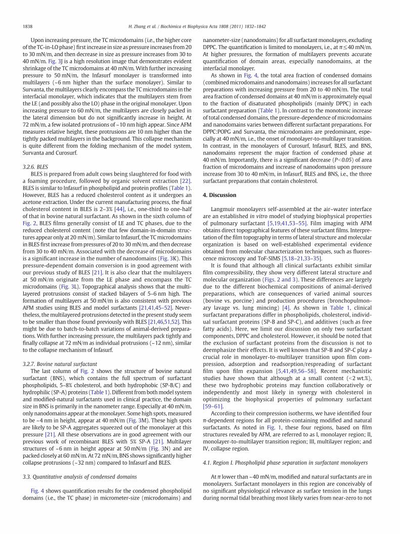
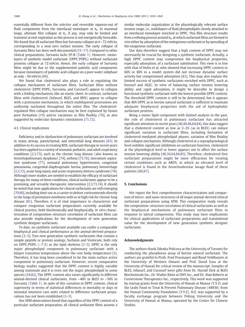
Fig. 1. Comparison of typical compression isotherms of various synthetic, modified-natural, and natural surfactant films at room temperature. The two protein-freesynthetic systems are pure DPPC and DPPC:POPG (7:3). The four clinical modified-natural surfactants are Survanta, Curosurf, Infasurf, and BLES. The native naturalsurfactant for comparison is bovine natural surfactant (BNS). All pulmonary surfactantswere spread as monolayers to an initial surface pressure (π) of 1–3 mN/m prior tocompression. Surfactant films were compressed at an identical rate of 20 cm2/min untilfilm collapse. Four pressure-dependent regions are detected for the compressionisotherms of protein-containing modified and natural surfactants. These are: Region I.Monolayer region at π≤40 mN/m; Region II. Monolayer-to-multilayer transition regionat 40bπb50 mN/m; Region III. Multilayer region at π≥50 mN/m; and Region IV.Collapse region at 72 mN/m for all films but Survanta, which collapses at 62 mN/m. Thestructural nature of these four regions is revealed in Fig. 2 by AFM.
3. Results
3.1. Comparison of compression isotherms
Fig. 1 compares the typical compression isotherms of DPPC, DPPC:POPG (7:3), Survanta, Curosurf, Infasurf, BLES, and BNS. First, compres-sion isotherms of all modified-natural surfactants and BNS feature aplateau region at π 40–50 mN/m. In contrast to these protein-containingsurfactants, pure DPPC shows a well-characterized isotherm with aphase transition plateau at 3–5 mN/m [28,29], and DPPC:POPGshows no apparent plateau region. Due to the existence of the plateau,compression isotherms of modified-natural and natural surfactants canbe separated into four regions, as indicated in Fig. 1. In the regions beforeand after reaching the plateau, surfactant films have a significantlylower film compressibility (i.e., less area reduction to increase π) thanthe plateau region. The fourth region refers to the film collapse plateauat which the surfactant films reach their maximum π. The molecularnatureof these four regions is discussedbelow in combinationwithAFMobservations. Second, the compression isotherms shift, after passingthe plateau region, from right to left in the order of DPPC, DPPC:POPG,Survanta, Curosurf, Infasurf, BLES, and BNS. This order in generalagrees with the rank of decreasing DPPC content in each preparation(see Table 1). Third, all surfactant films but Survanta collapse at π of~72 mN/m, corresponding to near-zero surface tension at roomtemperature. Survanta, on the other hand, collapses at ~62 mN/m.
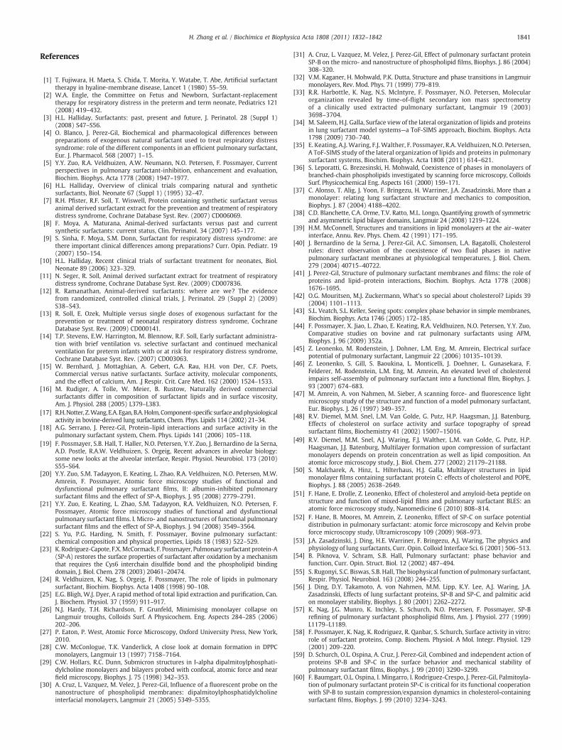
3.2. Comparison of film structures
Fig. 2 is a compilation of AFM images comparingmicro- and nano-structures of different surfactant films obtained at increasing π. Allsurfactants, with the exception of DPPC, were studied at 20, 30, 40,50, 60 mN/m, and the pressure at which the films collapse. Incomparison, DPPC was studied at 2.8, 4.0, 10, 30, 60 and 72 mN/m.These characteristic pressures were selected to cover the complete

Fig. 2. Comparison of characteristic AFM topographic images of various synthetic, modified-natural, and natural surfactant films at increasing surface pressure (π). All surfactants butDPPC were studied at a series of π of 20, 30, 40, 50, 60 mN/m, and the π at which the film collapses, i.e., 72 mN/m for DPPC:POPG, Curosurf, Infasurf, BLES, and BNS, and 62 mN/m forSurvanta. In comparison, DPPC was studied at π of 2.8, 4.0, 10, 30, 60 and 72 mN/m. These characteristic pressures were selected to cover the complete and detailed evolution of eachsurfactant film under compression, i.e., before, during and after the plateau region in the compression isotherm (Fig. 1). All AFM images were obtained with the same scanparameters, i.e., Setpoint=1 V; PID Gains=3/2/0; and Scan rate=1 Hz. The AFM scan area was 50×50 μm for all images. For the purpose of comparison among differentsurfactants, the full z-range was set to be 5 nm for all images at π≤40 mN/m, and 20 nm for πN40 mN/m (with the only exception being DPPC at 60 mN/m, the z-range was reducedto 5 nm to demonstrate topographic features). Relative height of critical structures is pointed by arrows. Critical lateral structures indicated by rectangular boxes are shown in high-resolution images in Fig. 3.
1835H. Zhang et al. / Biochimica et Biophysica Acta 1808 (2011) 1832–1842
and detailed evolution of each surfactant film under compression.For the purpose of comparison, all AFM images were obtained withthe same scan parameters, i.e., Setpoint=1 V; PID Gains=3/2/0;and Scan rate=1 Hz. All images have the same scan area of 50×50 μm.The full z-range is 5 nm for all images at π≤40 mN/m (i.e., beforereaching the plateau), and 20 nm for all images at πN40 mN/m (i.e.,after passing the plateau). The only exception is the DPPC image at60 mN/m, which has a full z-range of 5 nm to demonstrate shallowfeatures.
Fig. 3 shows selectedheight profiles, three-dimensional surfaceplots,and high-resolution AFM images, corresponding to regions indicated byboxes in Fig. 2. These images have different scan areas and z-ranges,adjusted for optimum presentation of detailed film structures.
3.2.1. DPPCAs shown in the first column of Fig. 2, pure DPPC remains a
monolayer up to collapse at 72 mN/m. At a very low pressure of2.8 mN/m, the DPPC monolayer is in a fluid-like liquid-expanded (LE)phase in which the phospholipid molecules have a low packingdensity and the fatty acid chains remain largely disordered and fluid[28,29]. At 4.0 mN/m, the DPPC monolayer shows well-defined chiralmicrodomains and small nanodomains [5,28–31]. The domain forma-tion indicates coexistence of the LE phase with a more ordered and
rigid tilted-condensed (TC) phase, which extends ~1 nm beyond theLE phase [5,21,32]. Note that when discussing phospholipid phasebehavior we adopt the nomenclature proposed by Kaganer et al. [32],who suggest the use of TC phase to replace the commonly used liquid-condensed (LC) phase. The line tracing in Fig. 3A illustrates heights ofmicrodomains and nanodomains, relative to the continuous LE phase.Both microdomains and nanodomains have the same height, indicat-ing they are both in the TC phase [29–31]. The plateau shown in theDPPC isotherm at 3–5 mN/m therefore indicates a first-order phasetransition [28,32]. After passing the phase transition plateau, the DPPCmonolayer at 10 and 30 mN/m is primarily in a homogenous TC phase,thus with a very low film compressibility. When pressure is increasedto 60 mN/m, a number of small “peaks” of only 0.4 nm high (indicatedby the arrow) appear uniformly in the monolayer, indicating onset ofmonolayer destabilization under the extreme lateral compression. At72 mN/m, the DPPC monolayer collapses completely, as indicated bythe formation of a film collapse plateau in the isotherm and bilayerstacks (Fig. 3B) on top of the monolayer.
3.2.2. DPPC:POPG (7:3)The second column of Fig. 2 shows the film structure of the protein-
free binary model system DPPC:POPG (7:3). At 20 mN/m, the DPPC:POPG monolayer shows phospholipid phase separation. Lateral

1836 H. Zhang et al. / Biochimica et Biophysica Acta 1808 (2011) 1832–1842
chemical analysis in previous ToF-SIMS experiments proved that the TCdomains consist of DPPC and the surrounding LE phase contains lessordered POPG [33–35]. In addition to the circular domains, there arestripes with the same height as the TC domains and organized in thedirection perpendicular to the dippingdirection. Similar phenomenon is
also found for Curosurf. It appears that these stripes are not due to anAFM tip artifact as they remain unchanged as varying scan direction,rate, and force, or using different scanmodes. These might be due to anartifact of LB transfer inwhich substrate-mediated condensation occursduring or after the transfer [36]. However, the reason why this artifact

20 30 40
Survanta20 30 40
0
10
20
30
40
50
60
70
80
Are
a fr
actio
n of
con
dens
ed d
omai
ns (
%)
DPPC:POPG20 30 40
Curosurf20 30 40
Infasurf
*
20 30 40
Nanodomains Microdomains
BLES20 30 40
**
BNSπ(mN/m)
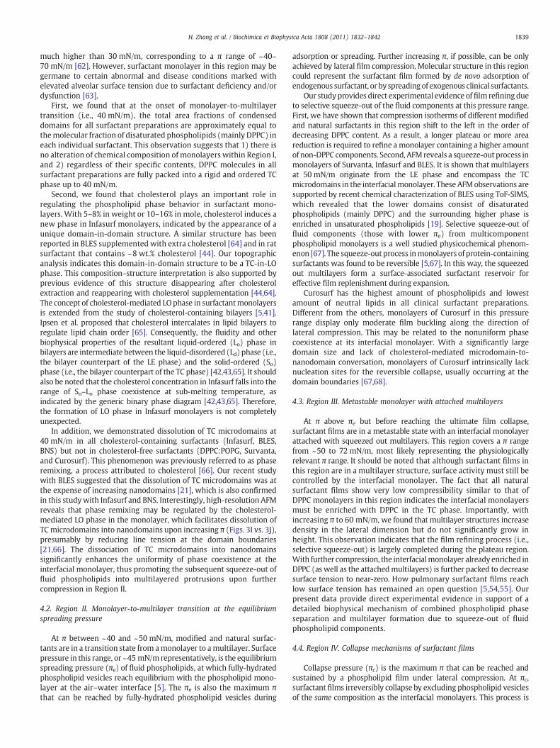
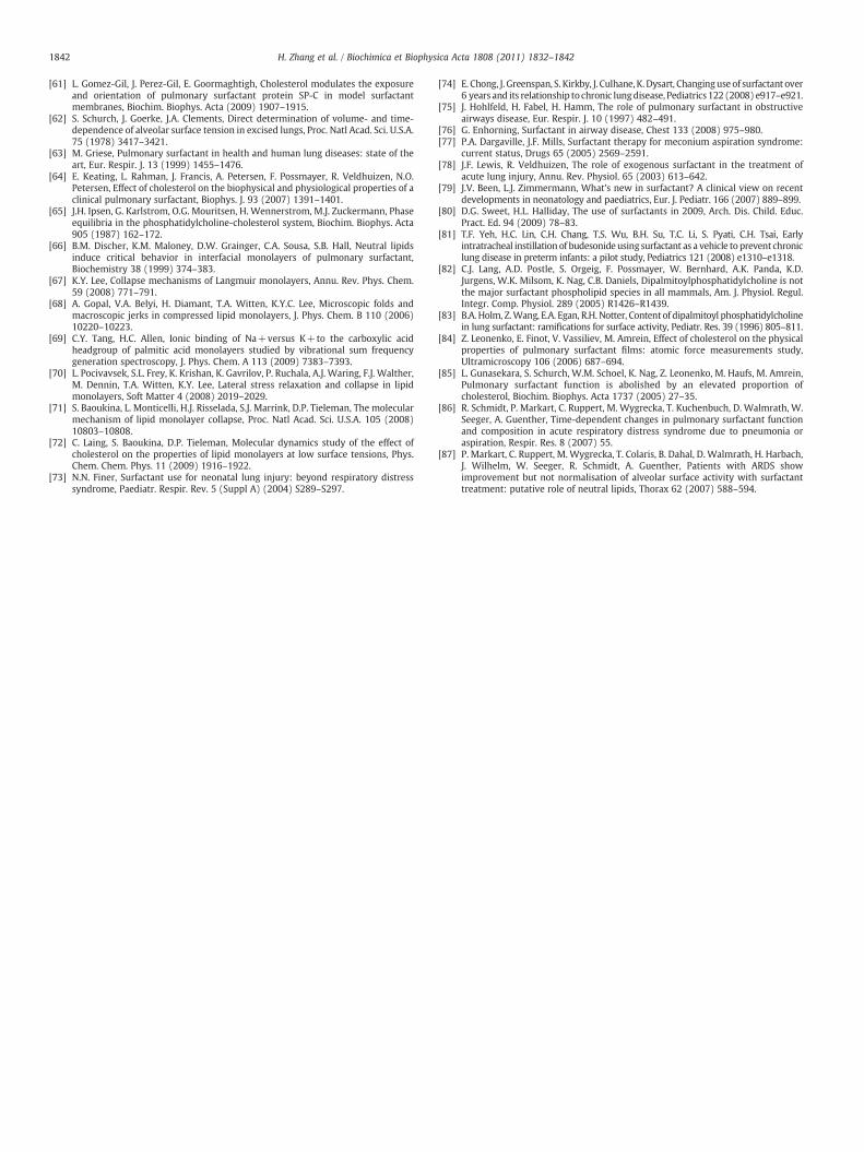
Fig. 4. Quantification results of monolayer coverage of tilted-condensed (TC) domainsupon film compression. Monolayers of DPPC:POPG (7:3), Survanta, Curosurf, Infasurf,BLES, and BNS were quantified at surface pressures (π) of 20, 30, and 40 mN/m. At eachpressure, area fractions of microdomains, nanodomains, and total TC domains (sum ofmicro- and nano-domains) were quantified with image analysis. *Pb0.05 fordifferences between area fractions of microdomains at 30 and 40 mN/m, for Infasurf,BLES, and BNS.
1837H. Zhang et al. / Biochimica et Biophysica Acta 1808 (2011) 1832–1842
seems to be less detectable for the other surfactant preparations isunknown.
With pressure increase to 30 mN/m, the TC domains grow in size. At40 mN/m, the TC domains become less detectable in the topographicimage. A closer look at the film (Fig. 3C) confirms that the TC domainsare packed together to form a somewhat continuous phasewith “holes”of ~0.5 nm in depth, likely due to the trapped LE phase. It appears thatfurther increasing pressure to 50 mN/m induces formation of isolatedbilayer structures (~3 nminheight, aspointed by thearrow) attached tothe interfacial monolayer, due to localized film collapse. The multilayerstructures grow in the lateral dimension but not significantly in height(~4 nm as pointed) with further increasing pressure to 60 mN/m. Thefilm eventually collapses at 72 mN/m,where patterns of film folding areclearly observed along the direction of film compression (Fig. 3D).
3.2.3. SurvantaSurvanta is prepared fromminced bovine lung tissue, extractedwith
chloroform-methanol, and further purified by precipitation with ethylacetate. These purification procedures result in a loss of cholesteroland a reduction in SP-B [15,17]. Due to its relatively poor capacity inreducing surface tension, Survanta is further supplemented withsynthetic DPPC, palmitic acid and tripalmitin. As shown in the thirdcolumn of Fig. 2, Survanta monolayers at 20 mN/m present a clearTC-LE phase separation, indicated by formation of round-shaped TCdomains mainly in the micrometer size. The area coverage of theTC phase increases continuously with increasing pressure up to40 mN/m (Fig. 4). At 50 mN/m, i.e., immediately past the plateau inthe compression isotherm, the original monolayer is transformedinto multilayers. A surface plot at 50 mN/m (Fig. 3E) demonstratesthat a matrix of multilayers (about two bilayers high relative to thesurface monolayer) is formed surrounding the TC domains in theinterfacial monolayer. Similar structure of Survanta has been reportedby another group [37]. This film structure strongly implies that themultilayersmust be initiated from the LEphase in theoriginal surfactantmonolayers. As π is increased to 60 mN/m, themultilayersmainly growin the lateral dimension but not in the altitudinal direction (Fig. 3F);while the TCdomains in the interfacialmonolayer are further packed. At62 mN/m, the Survanta film permanently collapses and forms localizedfolding structures in arbitrary orientations, which differs from thewhole-film, orientational folding of DPPC:POPG film at its collapsepressure.
3.2.4. CurosurfCurosurf is prepared fromminced porcine lung tissue. It is depleted
of cholesterol by undergoing gel-chromatography to remove all neutrallipids during manufacture [4]. As shown in the fourth column of Fig. 2,the Curosurf monolayer at 20 mN/m exhibits significantly fewer, butlarger, noncircular domains, compared to Survanta, presumablyindicating a higher film viscosity [38,39]. With increasing π, the domainshape becomes more ramified. Nanodomains with the same height ofmicrodomains tend to line up horizontally to form stripes (Fig. 3G). Aspressure is increased to 50 mN/m, the Curosurf film displays moderate
Fig. 3. High-resolution AFM images, as indicated by rectangular boxes in Fig. 2, demonstratiheight profile shows the surface topography along the line tracing indicated in the AFM imagand worm-like nanodomains, both ~1 nm higher than the surrounding liquid-expended (Lstacks ejected from the interfacial monolayer at the collapse pressure. C. Monolayer of DPPC:phase with “holes” of LE phase ~0.5 nm lower. D. Surface plot (three-dimensional topografolding along the direction of lateral compression. E. Surface plot of Survanta film at 50 mN/mfrom the surrounding LE phase. F. Survanta film at 60 mN/m, illustrating an increasing multisignificantly, compared to 50 mN/m. G. Curosurf monolayer at 40 mN/m shows a single micheight profile. H. Curosurf film at 50 mN/m, showing a single TCmicrodomain with moderatemonolayer at 20 mN/m shows the cholesterol-mediated liquid-ordered (LO) phase and aphospholipid phase. The lipid chain order of the LO phase is intermediate between the TCheight). J. Infasurf monolayer at 40 mN/m, demonstrating a single TC-in-LO domain with an i30 mN/m). K. BLES monolayer at 40 mN/m, showing clearly a decrease of microdomains imicrodomains traced out by multilayers formed from the surrounding LE phase. M. BNS mospots, likely SP-A aggregates squeezed out of the monolayer at this pressure. N. BNS film at
(only ~1.5 nmhigh) buckling along the direction of lateral compression(Fig. 3H). At 60 mN/m and even 72 mN/m where the film ultimatelycollapses, the multilayer structures only grow slightly to ~4 nm high,indicating very limited film collapse and high film stability.
3.2.5. InfasurfInfasurf is prepared from lung lavage of newborn calves by
centrifugation and organic solvent extraction, and it contains all ofthe hydrophobic components of natural surfactant. Compared toSurvanta and Curosurf, Infasurf has a high cholesterol content(Table 1). It is well known that cholesterol has a profound influenceon the phase behavior of phospholipid monolayers and membranes[5,40–43]. Depending on its ratio to phospholipids in monolayers,cholesterol can selectively partition into TC phase phospholipiddomains and induce a new liquid-ordered (LO) phase, whose degreeof lipid chain order is intermediate between the LE and TC phases[5,40–43]. As shown in the fifth column of Fig. 2, all three phases(i.e., LE, LO, and TC) are detected in the Infasurf monolayer. As aconsequence of partitioning into the TC domains, the high cholesterolcontent in Infasurf results in a unique domain-in-domain (TC-in-LO)structure. In Fig. 3I, scanning from left to right, one observes thebackground LEphase, amicroscale TCdomain 0.8 nmhigher than the LEphase, a LO phase 0.2 nm lower than the TC phase, and nanoscale TCdomains 0.8 nm higher than the surrounding LE phase. These heightvariations are in good agreement with the relative chain order of thesethree phases.
ng detailed structural features of surfactant films. A. DPPC monolayer at 4.0 mN/m. Thee. It shows that the tilted-condensed (TC) phase consists of both leaf-like microdomainsE) phase. B. Collapsed DPPC monolayer at 72 mN/m, showing the formation of bilayerPOPG (7:3) at 40 mN/m, showing that the condensed domains merge into a continuousphic image) of DPPC:POPG film at the collapse pressure (i.e., 72 mN/m), showing film. It shows that the TC microdomains are traced out by higher multilayers that originatelayer density in the lateral dimension while the height of multilayers does not increaserodomain with lines of nanodomains, as indicated by the intensive fluctuations in thefilm buckling, indicated by vertical lines of collapse sites of only ~1.5 nm high. I. Infasurfunique TC-in-LO structure as a consequence of cholesterol partitioning into the TCand LE phases, as indicated by height differences detected by AFM (i.e., TCNLONLE inncreasing number of nanodomains and evident shrink of the TC core (2.5 μm vs. 5 μm atn size and increase of nanodomains in number. L. BLES film at 50 mN/m, showing TCnolayer at 40 mN/m, showing the detailed morphology of nanodomains and some high50 mN/m, showing detailed multilayer structure.
1838 H. Zhang et al. / Biochimica et Biophysica Acta 1808 (2011) 1832–1842
Upon increasing pressure, the TCmicrodomains (i.e., the higher coreof theTC-in-LOphase)first increase in size aspressure increases from20to 30 mN/m, and then decrease in size as pressure increases from 30 to40 mN/m. Fig. 3J is a high resolution image that demonstrates evidentshrinkage of the TCmicrodomains at 40 mN/m.With further increasingpressure to 50 mN/m, the Infasurf monolayer is transformed intomultilayers (~6 nm higher than the surface monolayer). Similar toSurvanta, themultilayers clearly encompass the TCmicrodomains in theinterfacial monolayer, which indicates that the multilayers stem fromthe LE (and possibly also the LO) phase in the original monolayer. Uponincreasing pressure to 60 mN/m, the multilayers are closely packed inthe lateral dimension but do not significantly increase in height. At72 mN/m, a few isolated protrusions of ~10 nmhigh appear. Since AFMmeasures relative height, these protrusions are 10 nm higher than thetightly packed multilayers in the background. This collapse mechanismis quite different from the folding mechanism of the model system,Survanta and Curosurf.
3.2.6. BLESBLES is prepared from adult cows being slaughtered for food with
a foaming procedure, followed by organic solvent extraction [22].BLES is similar to Infasurf in phospholipid and protein profiles (Table 1).However, BLES has a reduced cholesterol content as it undergoes anacetone extraction. Under the current manufacturing process, the finalcholesterol content in BLES is 2–3% [44], i.e., one-third to one-halfof that in bovine natural surfactant. As shown in the sixth column ofFig. 2, BLES films generally consist of LE and TC phases, due to thereduced cholesterol content (note that few domain-in-domain struc-tures appear only at 20 mN/m). Similar to Infasurf, the TCmicrodomainsin BLESfirst increase frompressures of 20 to 30 mN/m, and then decreasefrom 30 to 40 mN/m. Associated with the decrease of microdomainsis a significant increase in the number of nanodomains (Fig. 3K). Thispressure-dependent domain conversion is in good agreement withour previous study of BLES [21]. It is also clear that the multilayersat 50 mN/m originate from the LE phase and encompass the TCmicrodomains (Fig. 3L). Topographical analysis shows that the multi-layered protrusions consist of stacked bilayers of 5–6 nm high. Theformation of multilayers at 50 mN/m is also consistent with previousAFM studies using BLES and model surfactants [21,41,45–52]. Never-theless, themultilayered protrusionsdetected in thepresent study seemto be smaller than those found previously with BLES [21,46,51,52]. Thismight be due to batch-to-batch variations of animal-derived prepara-tions. With further increasing pressure, the multilayers pack tightly andfinally collapse at 72 mN/m as individual protrusions (~12 nm), similarto the collapse mechanism of Infasurf.
3.2.7. Bovine natural surfactantThe last column of Fig. 2 shows the structure of bovine natural
surfactant (BNS), which contains the full spectrum of surfactantphospholipids, 5–8% cholesterol, and both hydrophobic (SP-B/C) andhydrophilic (SP-A) proteins (Table 1).Different frombothmodel systemand modified-natural surfactants used in clinical practice, the domainsize in BNS is primarily in the nanometer range. Especially at 40 mN/m,only nanodomains appear at themonolayer. Somehigh spots,measuredto be ~4 nm in height, appear at 40 mN/m (Fig. 3M). These high spotsare likely to be SP-A aggregates squeezed out of the monolayer at thispressure [21]. All these observations are in good agreement with ourprevious work of recombinant BLES with 5% SP-A [21]. Multilayerstructures of ~6 nm in height appear at 50 mN/m (Fig. 3N) and arepacked closely at 60 mN/m.At72 mN/m,BNS shows significantlyhighercollapse protrusions (~32 nm) compared to Infasurf and BLES.
3.3. Quantitative analysis of condensed domains
Fig. 4 shows quantification results for the condensed phospholipiddomains (i.e., the TC phase) in micrometer-size (microdomains) and
nanometer-size (nanodomains) for all surfactantmonolayers, excludingDPPC. The quantification is limited to monolayers, i.e., at π≤40 mN/m.At higher pressures, the formation of multilayers prevents accuratequantification of domain areas, especially nanodomains, at theinterfacial monolayer.
As shown in Fig. 4, the total area fraction of condensed domains(combinedmicrodomainsandnanodomains) increases for all surfactantpreparations with increasing pressure from 20 to 40 mN/m. The totalarea fraction of condensed domains at 40 mN/m is approximately equalto the fraction of disaturated phospholipids (mainly DPPC) in eachsurfactant preparation (Table 1). In contrast to the monotonic increaseof total condensed domains, the pressure-dependence ofmicrodomainsand nanodomains varies between different surfactant preparations. ForDPPC:POPG and Survanta, the microdomains are predominant, espe-cially at 40 mN/m, i.e., the onset of monolayer-to-multilayer transition.In contrast, in the monolayers of Curosurf, Infasurf, BLES, and BNS,nanodomains represent the major fraction of condensed phase at40 mN/m. Importantly, there is a significant decrease (Pb0.05) of areafraction of microdomains and increase of nanodomains upon pressureincrease from 30 to 40 mN/m, in Infasurf, BLES and BNS, i.e., the threesurfactant preparations that contain cholesterol.
4. Discussion
Langmuir monolayers self-assembled at the air–water interfaceare an established in vitro model of studying biophysical propertiesof pulmonary surfactant [5,19,41,53–55]. Film imaging with AFMobtains direct topographical features of these surfactant films. Interpre-tationof the film topography in terms of lateral structure andmolecularorganization is based on well-established experimental evidenceobtained from molecular characterization techniques, such as fluores-cence microscopy and ToF-SIMS [5,18–21,33–35].
It is found that although all clinical surfactants exhibit similarfilm compressibility, they show very different lateral structure andmolecular organization (Figs. 2 and 3). These differences are largelydue to the different biochemical compositions of animal-derivedpreparations, which are consequences of varied animal sources(bovine vs. porcine) and production procedures (bronchopulmon-ary lavage vs. lung mincing) [4]. As shown in Table 1, clinicalsurfactant preparations differ in phospholipids, cholesterol, individ-ual surfactant proteins (SP-B and SP-C), and additives (such as freefatty acids). Here, we limit our discussion on only two surfactantcomponents, DPPC and cholesterol. However, it should be noted thatthe exclusion of surfactant proteins from the discussion is not todeemphasize their effects. It is well known that SP-B and SP-C play acrucial role in monolayer-to-multilayer transition upon film com-pression, adsorption and readsorption/respreading of surfactantfilm upon film expansion [5,41,49,56–58]. Recent mechanisticstudies have shown that although at a small content (b2 wt.%),these two hydrophobic proteins may function collaboratively orindependently and most likely in synergy with cholesterol inoptimizing the biophysical properties of pulmonary surfactant[59–61].
According to their compression isotherms, we have identified fourπ-dependent regions for all protein-containing modified and naturalsurfactants. As noted in Fig. 1, these four regions, based on filmstructures revealed by AFM, are referred to as I, monolayer region; II,monolayer-to-multilayer transition region; III, multilayer region; andIV, collapse region.
4.1. Region I. Phospholipid phase separation in surfactant monolayers
At π lower than ~40 mN/m,modified and natural surfactants are inmonolayers. Surfactant monolayers in this region are conceivably ofno significant physiological relevance as surface tension in the lungsduring normal tidal breathing most likely varies from near-zero to not
1839H. Zhang et al. / Biochimica et Biophysica Acta 1808 (2011) 1832–1842
much higher than 30 mN/m, corresponding to a π range of ~40–70 mN/m [62]. However, surfactant monolayer in this region may begermane to certain abnormal and disease conditions marked withelevated alveolar surface tension due to surfactant deficiency and/ordysfunction [63].
First, we found that at the onset of monolayer-to-multilayertransition (i.e., 40 mN/m), the total area fractions of condenseddomains for all surfactant preparations are approximately equal tothemolecular fraction of disaturated phospholipids (mainly DPPC) ineach individual surfactant. This observation suggests that 1) there isno alteration of chemical composition of monolayers within Region I,and 2) regardless of their specific contents, DPPC molecules in allsurfactant preparations are fully packed into a rigid and ordered TCphase up to 40 mN/m.
Second, we found that cholesterol plays an important role inregulating the phospholipid phase behavior in surfactant mono-layers. With 5–8% in weight or 10–16% in mole, cholesterol induces anew phase in Infasurf monolayers, indicated by the appearance of aunique domain-in-domain structure. A similar structure has beenreported in BLES supplemented with extra cholesterol [64] and in ratsurfactant that contains ~8 wt.% cholesterol [44]. Our topographicanalysis indicates this domain-in-domain structure to be a TC-in-LOphase. This composition–structure interpretation is also supported byprevious evidence of this structure disappearing after cholesterolextraction and reappearing with cholesterol supplementation [44,64].The concept of cholesterol-mediated LO phase in surfactantmonolayersis extended from the study of cholesterol-containing bilayers [5,41].Ipsen et al. proposed that cholesterol intercalates in lipid bilayers toregulate lipid chain order [65]. Consequently, the fluidity and otherbiophysical properties of the resultant liquid-ordered (Lo) phase inbilayers are intermediate between the liquid-disordered (Ld) phase (i.e.,the bilayer counterpart of the LE phase) and the solid-ordered (So)phase (i.e., the bilayer counterpart of the TC phase) [42,43,65]. It shouldalso be noted that the cholesterol concentration in Infasurf falls into therange of So–Lo phase coexistence at sub-melting temperature, asindicated by the generic binary phase diagram [42,43,65]. Therefore,the formation of LO phase in Infasurf monolayers is not completelyunexpected.
In addition, we demonstrated dissolution of TC microdomains at40 mN/m in all cholesterol-containing surfactants (Infasurf, BLES,BNS) but not in cholesterol-free surfactants (DPPC:POPG, Survanta,and Curosurf). This phenomenon was previously referred to as phaseremixing, a process attributed to cholesterol [66]. Our recent studywith BLES suggested that the dissolution of TC microdomains was atthe expense of increasing nanodomains [21], which is also confirmedin this studywith Infasurf and BNS. Interestingly, high-resolution AFMreveals that phase remixing may be regulated by the cholesterol-mediated LO phase in the monolayer, which facilitates dissolution ofTCmicrodomains into nanodomains upon increasing π (Figs. 3I vs. 3J),presumably by reducing line tension at the domain boundaries[21,66]. The dissociation of TC microdomains into nanodomainssignificantly enhances the uniformity of phase coexistence at theinterfacial monolayer, thus promoting the subsequent squeeze-out offluid phospholipids into multilayered protrusions upon furthercompression in Region II.
4.2. Region II. Monolayer-to-multilayer transition at the equilibriumspreading pressure
At π between ~40 and ~50 mN/m, modified and natural surfac-tants are in a transition state from amonolayer to amultilayer. Surfacepressure in this range, or ~45 mN/m representatively, is the equilibriumspreading pressure (πe) of fluid phospholipids, at which fully-hydratedphospholipid vesicles reach equilibrium with the phospholipid mono-layer at the air–water interface [5]. The πe is also the maximum πthat can be reached by fully-hydrated phospholipid vesicles during
adsorption or spreading. Further increasing π, if possible, can be onlyachieved by lateral film compression. Molecular structure in this regioncould represent the surfactant film formed by de novo adsorption ofendogenous surfactant, orbyspreadingof exogenous clinical surfactants.
Our studyprovides direct experimental evidence offilm refining dueto selective squeeze-out of the fluid components at this pressure range.First, we have shown that compression isotherms of different modifiedand natural surfactants in this region shift to the left in the order ofdecreasing DPPC content. As a result, a longer plateau or more areareduction is required to refine amonolayer containing a higher amountof non-DPPC components. Second, AFMreveals a squeeze-out process inmonolayers of Survanta, Infasurf and BLES. It is shown that multilayersat 50 mN/m originate from the LE phase and encompass the TCmicrodomains in the interfacialmonolayer. These AFMobservations aresupported by recent chemical characterization of BLES using ToF-SIMS,which revealed that the lower domains consist of disaturatedphospholipids (mainly DPPC) and the surrounding higher phase isenriched in unsaturated phospholipids [19]. Selective squeeze-out offluid components (those with lower πe) from multicomponentphospholipid monolayers is a well studied physicochemical phenom-enon [67]. The squeeze-out process inmonolayers of protein-containingsurfactants was found to be reversible [5,67]. In this way, the squeezedout multilayers form a surface-associated surfactant reservoir foreffective film replenishment during expansion.
Curosurf has the highest amount of phospholipids and lowestamount of neutral lipids in all clinical surfactant preparations.Different from the others, monolayers of Curosurf in this pressurerange display only moderate film buckling along the direction oflateral compression. This may be related to the nonuniform phasecoexistence at its interfacial monolayer. With a significantly largedomain size and lack of cholesterol-mediated microdomain-to-nanodomain conversation, monolayers of Curosurf intrinsically lacknucleation sites for the reversible collapse, usually occurring at thedomain boundaries [67,68].
4.3. Region III. Metastable monolayer with attached multilayers
At π above πe but before reaching the ultimate film collapse,surfactant films are in a metastable state with an interfacial monolayerattached with squeezed out multilayers. This region covers a π rangefrom ~50 to 72 mN/m, most likely representing the physiologicallyrelevant π range. It should be noted that although surfactant films inthis region are in a multilayer structure, surface activity must still becontrolled by the interfacial monolayer. The fact that all naturalsurfactant films show very low compressibility similar to that ofDPPC monolayers in this region indicates the interfacial monolayersmust be enriched with DPPC in the TC phase. Importantly, withincreasing π to 60 mN/m, we found that multilayer structures increasedensity in the lateral dimension but do not significantly grow inheight. This observation indicates that the film refining process (i.e.,selective squeeze-out) is largely completed during the plateau region.With further compression, the interfacialmonolayer already enriched inDPPC (as well as the attachedmultilayers) is further packed to decreasesurface tension to near-zero. How pulmonary surfactant films reachlow surface tension has remained an open question [5,54,55]. Ourpresent data provide direct experimental evidence in support of adetailed biophysical mechanism of combined phospholipid phaseseparation and multilayer formation due to squeeze-out of fluidphospholipid components.
4.4. Region IV. Collapse mechanisms of surfactant films
Collapse pressure (πc) is the maximum π that can be reached andsustained by a phospholipid film under lateral compression. At πc,surfactant films irreversibly collapse by excluding phospholipid vesiclesof the same composition as the interfacial monolayers. This process is
1840 H. Zhang et al. / Biochimica et Biophysica Acta 1808 (2011) 1832–1842
essentially different from the selective and reversible squeeze-out offluid components from the interfacial monolayer at πe. In mammallungs, ultimate film collapse at πc, if any, may only be limited andtransient at end-expiration as this process is not energetically favorable.We found that all surfactant films but Survanta collapse at π ~72 mN/m,corresponding to a near-zero surface tension. The early collapse ofSurvanta films has been well-documented [15–17]. Compared to otherclinical preparations, Survanta lacks SP-B (Table 1). However, mono-layers of synthetic model surfactant (DPPC:POPG) without surfactantproteins collapse at 72 mN/m. Hence, the early collapse of Survantafilms might be due to the supplemented palmitic acid componentsbecause monolayers of palmitic acid collapse on a pure water subphaseat only ~50 mN/m [69].
We found that cholesterol also plays a role in regulating thecollapse mechanism of surfactant films. Surfactant films withoutcholesterol (DPPC:POPG, Survanta, and Curosurf) appear to collapsewith a folding mechanism, like an elastic sheet. In contrast, surfactantfilms with cholesterol (Infasurf, BLES, and BNS) appear to collapsewith a protrusion mechanism, in which multilayered protrusions areuniformly nucleated throughout the entire film. The cholesterol-regulated film collapse mechanism may be best explained by fusionpore formation [19] and/or variations in film fluidity [70], as alsosuggested by molecular dynamics simulations [71,72].
4.5. Clinical implications
Deficiency and/or dysfunction of pulmonary surfactant are involvedin many airway, parenchymal, and interstitial lung diseases [63]. Inaddition to its success in treating RDS, surfactant therapy in recent yearshas been applied to a variety of neonatal, pediatric, and adult respiratoryconditions [2,3,73], such as the chronic lung disease (also known asbronchopulmonary dysplasia) [74], asthma [75,76], meconium aspira-tion syndrome [77], neonatal pulmonary hypertension, congenitalpneumonia, congenital diaphragmatic hernia, pulmonary hemorrhage[2,3,73], acute lung injury and acute respiratory distress syndrome [78].Althoughmore studies are needed to establish the efficacy of surfactanttherapy for many of these conditions, clinical surfactants appear to be apromising and versatile therapeutic intervention [2,3,73,74]. It shouldbe noted that new applications for clinical surfactants are still emerging[79,80], including their use as a carrier to deliver corticosteroids directlyto the lungs of premature newbornswith or at high risk for chronic lungdisease [81]. Therefore, it is of vital importance to characterize andcompare exogenous surfactant preparations currently available forclinical practice, both biochemically and biophysically. In vitro charac-terization of composition–structure correlation of surfactant films canalso provide implications for the development of new generationsynthetic designer surfactants.
To date, no synthetic surfactant available can confer a comparablebiophysical and clinical performance as the animal-derived prepara-tions [3–5]. Two new generation synthetic surfactants that contain asimple peptide or protein analogs, Surfaxin and Venticute, both relyon DPPC:POPG (~7:3) as the lipid skeleton [3–5]. DPPC is the onlymajor phospholipid component in pulmonary surfactant with abilayer transition temperature above the core body temperature [5].Therefore, it has long been considered to be the main surface activecomponent in pulmonary surfactant. However, recent comparativebiology studies suggested that the DPPC content is highly variableamong mammals and it is even not the major phospholipid in somespecies [24,82]. The DPPC content also varies significantly in differentanimal-derived clinical surfactants, from ~40% in BLES to ~50% inSurvanta (Table 1). In spite of this variation in DPPC content, clinicalsuperiority in terms of statistical differences in mortality or days inneonatal intensive care units related to difference surfactant prepa-rations has not been established [3–5].
Our AFM observations found that regardless of the DPPC content of aparticular surfactant preparation, all clinical surfactant films assume a
similar molecular organization at the physiologically relevant surfacetension range, i.e., multilayers of fluid phospholipids closely attached toan interfacial monolayer enriched in DPPC. This film structure resultsfroma refiningprocess aroundπe, atwhichsurfactantfilmsare formed invivoeither by adsorptionof theendogenous surfactantor by spreadingofthe exogenous surfactant.
Our data therefore suggest that a high content of DPPC may notnecessarily be crucial for designing a synthetic surfactant. Actually, ahigh DPPC content may compromise the biophysical properties,especially adsorption, of a surfactant substitution. This view is in linewith that of Holm et al. who showed that increasing DPPC content to60% or 80% in a model system did not increase dynamic surfaceactivity but compromised adsorption [83]. This may also explain thelimited success of synthetic surfactants enriched with DPPC, such asExosurf and ALEC. In view of balancing surface tension loweringability and rapid adsorption, it might be desirable to design afunctional synthetic surfactant with the lowest possible DPPC content.The threshold DPPC content is still to be determined, but it appearsthat 40% DPPC as in bovine natural surfactant is sufficient to maintainadequate biophysical properties with the aid of hydrophobicsurfactant proteins.
Being a minor lipid component with limited analysis in the past,the role of cholesterol in pulmonary surfactant has attractedsignificant attention in recent years [40,44,46,64,84]. Our data suggestthat a cholesterol content as low as 2–3% (as in BLES) can inducesignificant variation in surfactant films, including formation ofcholesterol-mediated phospholipid phases, variation of film fluidityand collapsemechanism. Although cholesterol at a supraphysiologicallevel exhibits significant inhibition on surfactant function, cholesterolat the physiological level or lower appears not to affect the surfacetension lowering ability [46,50,64,84,85]. Therefore, cholesterol-freesurfactant preparations might be more efficacious for treatingcertain conditions such as ARDS, in which an elevated level ofcholesterol is found in the bronchoalveolar lavage fluid of thesepatients [86,87].
5. Conclusions
We report the first comprehensive characterization and compar-ison of micro- and nano-structures of all major animal-derived clinicalsurfactant preparations using AFM. This comparative study revealsthe composition–structure correlation of clinical surfactants as well asthe biophysical mechanisms of pulmonary surfactant films inresponse to lateral compression. This study may have implicationsfor clinical applications of surfactant preparations and translationalvalue for the development of new generation synthetic designersurfactants.
Acknowledgments
The authors thank Zdenka Policova at the University of Toronto forconducting the phosphorus assay of bovine natural surfactant. Theauthors are grateful to Profs. Fred Possmayer and Ruud Veldhuizen atthe University of Western Ontario and Prof. David Easa at theUniversity of Hawaii for critical review of the manuscript. Samples ofBLES, Infasurf, and Curosurf were gifts from Dr. Harold Dick at BLESBiochemicals Inc., Dr. Walter Klein at ONY Inc., and Dr. Alan Roberts atCornerstone Therapeutics Inc., respectively. This work was supportedby startup grants from the University of Hawaii at Manoa (Y.Y.Z) andthe Leahi Fund to Treat & Prevent Pulmonary Disease (44936) fromthe Hawaii Community Foundation (Y.Y.Z). H.Z. was supported by afaculty exchange program between Peking University and theUniversity of Hawaii at Manoa, operated by the Center for ChineseStudies.
1841H. Zhang et al. / Biochimica et Biophysica Acta 1808 (2011) 1832–1842
References
[1] T. Fujiwara, H. Maeta, S. Chida, T. Morita, Y. Watabe, T. Abe, Artificial surfactanttherapy in hyaline-membrane disease, Lancet 1 (1980) 55–59.
[2] W.A. Engle, the Committee on Fetus and Newborn, Surfactant-replacementtherapy for respiratory distress in the preterm and term neonate, Pediatrics 121(2008) 419–432.
[3] H.L. Halliday, Surfactants: past, present and future, J. Perinatol. 28 (Suppl 1)(2008) S47–S56.
[4] O. Blanco, J. Perez-Gil, Biochemical and pharmacological differences betweenpreparations of exogenous natural surfactant used to treat respiratory distresssyndrome: role of the different components in an efficient pulmonary surfactant,Eur. J. Pharmacol. 568 (2007) 1–15.
[5] Y.Y. Zuo, R.A. Veldhuizen, A.W. Neumann, N.O. Petersen, F. Possmayer, Currentperspectives in pulmonary surfactant-inhibition, enhancement and evaluation,Biochim. Biophys. Acta 1778 (2008) 1947–1977.
[6] H.L. Halliday, Overview of clinical trials comparing natural and syntheticsurfactants, Biol. Neonate 67 (Suppl 1) (1995) 32–47.
[7] R.H. Pfister, R.F. Soll, T. Wiswell, Protein containing synthetic surfactant versusanimal derived surfactant extract for the prevention and treatment of respiratorydistress syndrome, Cochrane Database Syst. Rev. (2007) CD006069.
[8] F. Moya, A. Maturana, Animal-derived surfactants versus past and currentsynthetic surfactants: current status, Clin. Perinatol. 34 (2007) 145–177.
[9] S. Sinha, F. Moya, S.M. Donn, Surfactant for respiratory distress syndrome: arethere important clinical differences among preparations? Curr. Opin. Pediatr. 19(2007) 150–154.
[10] H.L. Halliday, Recent clinical trials of surfactant treatment for neonates, Biol.Neonate 89 (2006) 323–329.
[11] N. Seger, R. Soll, Animal derived surfactant extract for treatment of respiratorydistress syndrome, Cochrane Database Syst. Rev. (2009) CD007836.
[12] R. Ramanathan, Animal-derived surfactants: where are we? The evidencefrom randomized, controlled clinical trials, J. Perinatol. 29 (Suppl 2) (2009)S38–S43.
[13] R. Soll, E. Ozek, Multiple versus single doses of exogenous surfactant for theprevention or treatment of neonatal respiratory distress syndrome, CochraneDatabase Syst. Rev. (2009) CD000141.
[14] T.P. Stevens, E.W. Harrington, M. Blennow, R.F. Soll, Early surfactant administra-tion with brief ventilation vs. selective surfactant and continued mechanicalventilation for preterm infants with or at risk for respiratory distress syndrome,Cochrane Database Syst. Rev. (2007) CD003063.
[15] W. Bernhard, J. Mottaghian, A. Gebert, G.A. Rau, H.H. von Der, C.F. Poets,Commercial versus native surfactants. Surface activity, molecular components,and the effect of calcium, Am. .J Respir. Crit. Care Med. 162 (2000) 1524–1533.
[16] M. Rudiger, A. Tolle, W. Meier, B. Rustow, Naturally derived commercialsurfactants differ in composition of surfactant lipids and in surface viscosity,Am. J. Physiol. 288 (2005) L379–L383.
[17] R.H.Notter, Z.Wang,E.A.Egan,B.A.Holm,Component-specific surfaceandphysiologicalactivity in bovine-derived lung surfactants, Chem. Phys. Lipids 114 (2002) 21–34.
[18] A.G. Serrano, J. Perez-Gil, Protein–lipid interactions and surface activity in thepulmonary surfactant system, Chem. Phys. Lipids 141 (2006) 105–118.
[19] F. Possmayer, S.B. Hall, T. Haller, N.O. Petersen, Y.Y. Zuo, J. Bernardino de la Serna,A.D. Postle, R.A.W. Veldhuizen, S. Orgeig, Recent advances in alveolar biology:some new looks at the alveolar interface, Respir. Physiol. Neurobiol. 173 (2010)S55–S64.
[20] Y.Y. Zuo, S.M. Tadayyon, E. Keating, L. Zhao, R.A. Veldhuizen, N.O. Petersen, M.W.Amrein, F. Possmayer, Atomic force microscopy studies of functional anddysfunctional pulmonary surfactant films, II: albumin-inhibited pulmonarysurfactant films and the effect of SP-A, Biophys. J. 95 (2008) 2779–2791.
[21] Y.Y. Zuo, E. Keating, L. Zhao, S.M. Tadayyon, R.A. Veldhuizen, N.O. Petersen, F.Possmayer, Atomic force microscopy studies of functional and dysfunctionalpulmonary surfactant films. I. Micro- and nanostructures of functional pulmonarysurfactant films and the effect of SP-A, Biophys. J. 94 (2008) 3549–3564.
[22] S. Yu, P.G. Harding, N. Smith, F. Possmayer, Bovine pulmonary surfactant:chemical composition and physical properties, Lipids 18 (1983) 522–529.
[23] K. Rodriguez-Capote, F.X.McCormack, F. Possmayer, Pulmonary surfactant protein-A(SP-A) restores the surface properties of surfactant after oxidation by a mechanismthat requires the Cys6 interchain disulfide bond and the phospholipid bindingdomain, J. Biol. Chem. 278 (2003) 20461–20474.
[24] R. Veldhuizen, K. Nag, S. Orgeig, F. Possmayer, The role of lipids in pulmonarysurfactant, Biochim. Biophys. Acta 1408 (1998) 90–108.
[25] E.G. Bligh, W.J. Dyer, A rapid method of total lipid extraction and purification, Can.J. Biochem. Physiol. 37 (1959) 911–917.
[26] N.J. Hardy, T.H. Richardson, F. Grunfeld, Minimising monolayer collapse onLangmuir troughs, Colloids Surf. A Physicochem. Eng. Aspects 284–285 (2006)202–206.
[27] P. Eaton, P. West, Atomic Force Microscopy, Oxford University Press, New York,2010.
[28] C.W. McConlogue, T.K. Vanderlick, A close look at domain formation in DPPCmonolayers, Langmuir 13 (1997) 7158–7164.
[29] C.W. Hollars, R.C. Dunn, Submicron structures in I-alpha dipalmitoylphosphati-dylcholine monolayers and bilayers probed with confocal, atomic force and nearfield microscopy, Biophys. J. 75 (1998) 342–353.
[30] A. Cruz, L. Vazquez, M. Velez, J. Perez-Gil, Influence of a fluorescent probe on thenanostructure of phospholipid membranes: dipalmitoylphosphatidylcholineinterfacial monolayers, Langmuir 21 (2005) 5349–5355.
[31] A. Cruz, L. Vazquez, M. Velez, J. Perez-Gil, Effect of pulmonary surfactant proteinSP-B on the micro- and nanostructure of phospholipid films, Biophys. J. 86 (2004)308–320.
[32] V.M. Kaganer, H. Mohwald, P.K. Dutta, Structure and phase transitions in Langmuirmonolayers, Rev. Mod. Phys. 71 (1999) 779–819.
[33] R.R. Harbottle, K. Nag, N.S. McIntyre, F. Possmayer, N.O. Petersen, Molecularorganization revealed by time-of-flight secondary ion mass spectrometryof a clinically used extracted pulmonary surfactant, Langmuir 19 (2003)3698–3704.
[34] M. Saleem, H.J. Galla, Surface view of the lateral organization of lipids and proteinsin lung surfactant model systems—a ToF-SIMS approach, Biochim. Biophys. Acta1798 (2009) 730–740.
[35] E. Keating, A.J. Waring, F.J. Walther, F. Possmayer, R.A. Veldhuizen, N.O. Petersen,A ToF-SIMS study of the lateral organization of lipids and proteins in pulmonarysurfactant systems, Biochim. Biophys. Acta 1808 (2011) 614–621.
[36] S. Leporatti, G. Brezesinski, H. Mohwald, Coexistence of phases in monolayers ofbranched-chain phospholipids investigated by scanning force microscopy, ColloidsSurf. Physicochemical Eng. Aspects 161 (2000) 159–171.
[37] C. Alonso, T. Alig, J. Yoon, F. Bringezu, H. Warriner, J.A. Zasadzinski, More than amonolayer: relating lung surfactant structure and mechanics to composition,Biophys. J. 87 (2004) 4188–4202.
[38] C.D. Blanchette, C.A. Orme, T.V. Ratto, M.L. Longo, Quantifying growth of symmetricand asymmetric lipid bilayer domains, Langmuir 24 (2008) 1219–1224.
[39] H.M. McConnell, Structures and transitions in lipid monolayers at the air–waterinterface, Annu. Rev. Phys. Chem. 42 (1991) 171–195.
[40] J. Bernardino de la Serna, J. Perez-Gil, A.C. Simonsen, L.A. Bagatolli, Cholesterolrules: direct observation of the coexistence of two fluid phases in nativepulmonary surfactant membranes at physiological temperatures, J. Biol. Chem.279 (2004) 40715–40722.
[41] J. Perez-Gil, Structure of pulmonary surfactant membranes and films: the role ofproteins and lipid–protein interactions, Biochim. Biophys. Acta 1778 (2008)1676–1695.
[42] O.G. Mouritsen, M.J. Zuckermann, What's so special about cholesterol? Lipids 39(2004) 1101–1113.
[43] S.L. Veatch, S.L. Keller, Seeing spots: complex phase behavior in simple membranes,Biochim. Biophys. Acta 1746 (2005) 172–185.
[44] F. Possmayer, X. Jiao, L. Zhao, E. Keating, R.A. Veldhuizen, N.O. Petersen, Y.Y. Zuo,Comparative studies on bovine and rat pulmonary surfactants using AFM,Biophys. J. 96 (2009) 352a.
[45] Z. Leonenko, M. Rodenstein, J. Dohner, L.M. Eng, M. Amrein, Electrical surfacepotential of pulmonary surfactant, Langmuir 22 (2006) 10135–10139.
[46] Z. Leonenko, S. Gill, S. Baoukina, L. Monticelli, J. Doehner, L. Gunasekara, F.Felderer, M. Rodenstein, L.M. Eng, M. Amrein, An elevated level of cholesterolimpairs self-assembly of pulmonary surfactant into a functional film, Biophys. J.93 (2007) 674–683.
[47] M. Amrein, A. von Nahmen, M. Sieber, A scanning force- and fluorescence lightmicroscopy study of the structure and function of a model pulmonary surfactant,Eur. Biophys. J. 26 (1997) 349–357.
[48] R.V. Diemel, M.M. Snel, L.M. Van Golde, G. Putz, H.P. Haagsman, J.J. Batenburg,Effects of cholesterol on surface activity and surface topography of spreadsurfactant films, Biochemistry 41 (2002) 15007–15016.
[49] R.V. Diemel, M.M. Snel, A.J. Waring, F.J. Walther, L.M. van Golde, G. Putz, H.P.Haagsman, J.J. Batenburg, Multilayer formation upon compression of surfactantmonolayers depends on protein concentration as well as lipid composition. Anatomic force microscopy study, J. Biol. Chem. 277 (2002) 21179–21188.
[50] S. Malcharek, A. Hinz, L. Hilterhaus, H.J. Galla, Multilayer structures in lipidmonolayer films containing surfactant protein C: effects of cholesterol and POPE,Biophys. J. 88 (2005) 2638–2649.
[51] F. Hane, E. Drolle, Z. Leonenko, Effect of cholesterol and amyloid-beta peptide onstructure and function of mixed-lipid films and pulmonary surfactant BLES: anatomic force microscopy study, Nanomedicine 6 (2010) 808–814.
[52] F. Hane, B. Moores, M. Amrein, Z. Leonenko, Effect of SP-C on surface potentialdistribution in pulmonary surfactant: atomic force microscopy and Kelvin probeforce microscopy study, Ultramicroscopy 109 (2009) 968–973.
[53] J.A. Zasadzinski, J. Ding, H.E. Warriner, F. Bringezu, A.J. Waring, The physics andphysiology of lung surfactants, Curr. Opin. Colloid Interface Sci. 6 (2001) 506–513.
[54] B. Piknova, V. Schram, S.B. Hall, Pulmonary surfactant: phase behavior andfunction, Curr. Opin. Struct. Biol. 12 (2002) 487–494.
[55] S. Rugonyi, S.C. Biswas, S.B. Hall, The biophysical function of pulmonary surfactant,Respir. Physiol. Neurobiol. 163 (2008) 244–255.
[56] J. Ding, D.Y. Takamoto, A. von Nahmen, M.M. Lipp, K.Y. Lee, A.J. Waring, J.A.Zasadzinski, Effects of lung surfactant proteins, SP-B and SP-C, and palmitic acidon monolayer stability, Biophys. J. 80 (2001) 2262–2272.
[57] K. Nag, J.G. Munro, K. Inchley, S. Schurch, N.O. Petersen, F. Possmayer, SP-Brefining of pulmonary surfactant phospholipid films, Am. J. Physiol. 277 (1999)L1179–L1189.
[58] F. Possmayer, K. Nag, K. Rodriguez, R. Qanbar, S. Schurch, Surface activity in vitro:role of surfactant proteins, Comp. Biochem. Physiol. A Mol. Integr. Physiol. 129(2001) 209–220.
[59] D. Schurch, O.L. Ospina, A. Cruz, J. Perez-Gil, Combined and independent action ofproteins SP-B and SP-C in the surface behavior and mechanical stability ofpulmonary surfactant films, Biophys. J. 99 (2010) 3290–3299.
[60] F. Baumgart, O.L. Ospina, I. Mingarro, I. Rodriguez-Crespo, J. Perez-Gil, Palmitoyla-tion of pulmonary surfactant protein SP-C is critical for its functional cooperationwith SP-B to sustain compression/expansion dynamics in cholesterol-containingsurfactant films, Biophys. J. 99 (2010) 3234–3243.
1842 H. Zhang et al. / Biochimica et Biophysica Acta 1808 (2011) 1832–1842
[61] L. Gomez-Gil, J. Perez-Gil, E. Goormaghtigh, Cholesterol modulates the exposureand orientation of pulmonary surfactant protein SP-C in model surfactantmembranes, Biochim. Biophys. Acta (2009) 1907–1915.
[62] S. Schurch, J. Goerke, J.A. Clements, Direct determination of volume- and time-dependence of alveolar surface tension in excised lungs, Proc. Natl Acad. Sci. U.S.A.75 (1978) 3417–3421.
[63] M. Griese, Pulmonary surfactant in health and human lung diseases: state of theart, Eur. Respir. J. 13 (1999) 1455–1476.
[64] E. Keating, L. Rahman, J. Francis, A. Petersen, F. Possmayer, R. Veldhuizen, N.O.Petersen, Effect of cholesterol on the biophysical and physiological properties of aclinical pulmonary surfactant, Biophys. J. 93 (2007) 1391–1401.
[65] J.H. Ipsen, G. Karlstrom, O.G. Mouritsen, H. Wennerstrom, M.J. Zuckermann, Phaseequilibria in the phosphatidylcholine-cholesterol system, Biochim. Biophys. Acta905 (1987) 162–172.
[66] B.M. Discher, K.M. Maloney, D.W. Grainger, C.A. Sousa, S.B. Hall, Neutral lipidsinduce critical behavior in interfacial monolayers of pulmonary surfactant,Biochemistry 38 (1999) 374–383.
[67] K.Y. Lee, Collapse mechanisms of Langmuir monolayers, Annu. Rev. Phys. Chem.59 (2008) 771–791.
[68] A. Gopal, V.A. Belyi, H. Diamant, T.A. Witten, K.Y.C. Lee, Microscopic folds andmacroscopic jerks in compressed lipid monolayers, J. Phys. Chem. B 110 (2006)10220–10223.
[69] C.Y. Tang, H.C. Allen, Ionic binding of Na+versus K+to the carboxylic acidheadgroup of palmitic acid monolayers studied by vibrational sum frequencygeneration spectroscopy, J. Phys. Chem. A 113 (2009) 7383–7393.
[70] L. Pocivavsek, S.L. Frey, K. Krishan, K. Gavrilov, P. Ruchala, A.J. Waring, F.J. Walther,M. Dennin, T.A. Witten, K.Y. Lee, Lateral stress relaxation and collapse in lipidmonolayers, Soft Matter 4 (2008) 2019–2029.
[71] S. Baoukina, L. Monticelli, H.J. Risselada, S.J. Marrink, D.P. Tieleman, The molecularmechanism of lipid monolayer collapse, Proc. Natl Acad. Sci. U.S.A. 105 (2008)10803–10808.
[72] C. Laing, S. Baoukina, D.P. Tieleman, Molecular dynamics study of the effect ofcholesterol on the properties of lipid monolayers at low surface tensions, Phys.Chem. Chem. Phys. 11 (2009) 1916–1922.
[73] N.N. Finer, Surfactant use for neonatal lung injury: beyond respiratory distresssyndrome, Paediatr. Respir. Rev. 5 (Suppl A) (2004) S289–S297.
[74] E. Chong, J. Greenspan, S. Kirkby, J. Culhane,K.Dysart, Changinguse of surfactant over6 years and its relationship to chronic lungdisease, Pediatrics122 (2008) e917–e921.
[75] J. Hohlfeld, H. Fabel, H. Hamm, The role of pulmonary surfactant in obstructiveairways disease, Eur. Respir. J. 10 (1997) 482–491.
[76] G. Enhorning, Surfactant in airway disease, Chest 133 (2008) 975–980.[77] P.A. Dargaville, J.F. Mills, Surfactant therapy for meconium aspiration syndrome:
current status, Drugs 65 (2005) 2569–2591.[78] J.F. Lewis, R. Veldhuizen, The role of exogenous surfactant in the treatment of
acute lung injury, Annu. Rev. Physiol. 65 (2003) 613–642.[79] J.V. Been, L.J. Zimmermann, What's new in surfactant? A clinical view on recent
developments in neonatology and paediatrics, Eur. J. Pediatr. 166 (2007) 889–899.[80] D.G. Sweet, H.L. Halliday, The use of surfactants in 2009, Arch. Dis. Child. Educ.
Pract. Ed. 94 (2009) 78–83.[81] T.F. Yeh, H.C. Lin, C.H. Chang, T.S. Wu, B.H. Su, T.C. Li, S. Pyati, C.H. Tsai, Early
intratracheal instillation of budesonideusing surfactant as a vehicle to prevent chroniclung disease in preterm infants: a pilot study, Pediatrics 121 (2008) e1310–e1318.
[82] C.J. Lang, A.D. Postle, S. Orgeig, F. Possmayer, W. Bernhard, A.K. Panda, K.D.Jurgens, W.K. Milsom, K. Nag, C.B. Daniels, Dipalmitoylphosphatidylcholine is notthe major surfactant phospholipid species in all mammals, Am. J. Physiol. Regul.Integr. Comp. Physiol. 289 (2005) R1426–R1439.
[83] B.A. Holm, Z.Wang, E.A. Egan, R.H. Notter, Content of dipalmitoyl phosphatidylcholinein lung surfactant: ramifications for surface activity, Pediatr. Res. 39 (1996) 805–811.
[84] Z. Leonenko, E. Finot, V. Vassiliev, M. Amrein, Effect of cholesterol on the physicalproperties of pulmonary surfactant films: atomic force measurements study,Ultramicroscopy 106 (2006) 687–694.
[85] L. Gunasekara, S. Schurch, W.M. Schoel, K. Nag, Z. Leonenko, M. Haufs, M. Amrein,Pulmonary surfactant function is abolished by an elevated proportion ofcholesterol, Biochim. Biophys. Acta 1737 (2005) 27–35.
[86] R. Schmidt, P. Markart, C. Ruppert, M. Wygrecka, T. Kuchenbuch, D. Walmrath, W.Seeger, A. Guenther, Time-dependent changes in pulmonary surfactant functionand composition in acute respiratory distress syndrome due to pneumonia oraspiration, Respir. Res. 8 (2007) 55.
[87] P. Markart, C. Ruppert, M. Wygrecka, T. Colaris, B. Dahal, D. Walmrath, H. Harbach,J. Wilhelm, W. Seeger, R. Schmidt, A. Guenther, Patients with ARDS showimprovement but not normalisation of alveolar surface activity with surfactanttreatment: putative role of neutral lipids, Thorax 62 (2007) 588–594.