biochimica et biophysica acta - uclouvain ·...
TRANSCRIPT
Biochimica et Biophysica Acta 1788 (2009) 1832–1840
Contents lists available at ScienceDirect
Biochimica et Biophysica Acta
j ourna l homepage: www.e lsev ie r.com/ locate /bbamem
Interactions of oritavancin, a new lipoglycopeptide derived from vancomycin, withphospholipid bilayers: Effect on membrane permeability and nanoscale lipidmembrane organization
Oscar Domenech a, Grégory Francius b, Paul M. Tulkens a, Françoise Van Bambeke a,Yves Dufrêne b, Marie-Paule Mingeot-Leclercq a,⁎a Université catholique de Louvain, Faculté de Médecine, Louvain Drug Research Institute, Unité de pharmacologie cellulaire et moléculaire, UCL 73.70, B-1200 Bruxelles, Belgiumb Université catholique de Louvain, Faculté d'ingénierie biologique, agronomique et environnementale, Unité de chimie des interfaces, B-1348 Louvain-la-Neuve, Belgium
⁎ Corresponding author. Tel.: +32 2 764 73 74; fax: +E-mail address: [email protected]
0005-2736/$ – see front matter © 2009 Elsevier B.V. Adoi:10.1016/j.bbamem.2009.05.003
a b s t r a c t
a r t i c l e i n f oArticle history:Received 30 January 2009Received in revised form 25 April 2009Accepted 5 May 2009Available online 18 May 2009
Keywords:VancomycinOritavancinPOPEPOPGPOPCCardiolipinCalcein releaseAFMMembrane permeability
Antibiotics acting on bacterial membranes are receiving increasing attention because of widespreadresistance to agents acting on other targets and of potentially improved bactericidal effects. Oritavancin is aamphiphilic derivative of vancomycin showing fast and extensive killing activities against multi-resistant(including vancomycin insusceptible) Gram-positive organisms with no marked toxicity towards eukaryoticcells. We have undertaken to characterize the interactions of oritavancin with phospholipid bilayers, usingliposomes (LUV) and supported bilayers made of cardiolipin (CL) or phosphatidylglycerol (POPG) andphosphatidylethanolamine (POPE), all abundant in Gram-positive organisms. Changes in membranepermeability were followed by the release of calcein entrapped in liposomes at self-quenchingconcentrations, and changes in nanoscale lipid organization examined by Atomic Force Microscopy (AFM).Oritavancin caused a fast (b5 min) and complete (N95%) release of calcein from CL:POPE liposomes, and aslower but still substantial (50% in 60 min) release from POPG:POPE liposomes, which was (i) concentration-dependent (0–600 nM; [microbiologically meaningful concentrations]); (ii) enhanced by an increase inPOPG:POPE ratio, and decreased when replacing POPG by DPPG. AFM of CL:POPE supported bilayers showedthat oritavancin (84 nM) caused a remodeling of the lipid domains combined with a redisposition of the drugand degradation of the borders. In all the above studies, vancomycin was without a significant effect at5.5 μM. Electrostatic interactions, together with lipid curvature, lipid polymorphism as well of fluidity play acritical role for the permeabilization of lipid bilayer and changes in lipid organization induced by oritavancin.
© 2009 Elsevier B.V. All rights reserved.
1. Introduction
Biological membranes act as semi-permeable barriers to allow theinside environment of cells or organelles to differ from that outsidemedium, and mediate cell communications and interactions. In Gram-positive bacteria, thepericellularmembrane is surroundedbya thick cellwall of peptidoglycan that confers mechanical resistance to externalosmotic and other physical stress. Both the pericellular membrane andthe cell wall of peptidoglycan are essential for cell survival, and,therefore, are privileged targets in antibacterial chemotherapy. Anti-biotics that act on the membrane of Gram-positive organisms includedaptomycin and nisin. Daptomycin is a lipopeptide approved for clinicaluse which, after complexation with Ca+, forms an oligomeric assemblyexerting a detergent-like effect towards bilayers with a high content innegatively-chargedheadgroups, such as those found in bacteria, causingleakage of cytosolic contents [1]. Nisin kills bacteria by forming a pore
32 2 764 73 73.(M.-P. Mingeot-Leclercq).
ll rights reserved.
complex together with lipid II [2]. Antibiotics acting on the formation ofthe bacterial cell wall include the β-lactams, which act primarily byinhibition of the transpeptidases (or penicillin-binding proteins andimpair the reticulation of the peptidoglycan [3] and the glycopeptides(vancomycin, teicoplanin) that act by binding to the D-Ala–D-Alasequence of the pentapeptide of lipid II, thereby preventing thetranspeptidation and transglycosylation reactions in the late stages ofpeptidoglycan synthesis [4]. Both approaches, however, have theirlimitations, with the most striking examples of the emergence ofresistance of a major medical importance being shown by Staphylococ-cus aureus. This pathogenhas indeed acquiredmechanisms of resistanceto β-lactams (under the form of the so-called methicillin-resistantorganisms that produce a transpeptidase [PBP 2a] with decreasedaffinity for β-lactams [5], to glycopeptides (with the emergence of theso-called glycopeptide-intermediate S. aureus characterized by athickened cell wall, and also of fully resistant organisms in which theterminal D-Ala is replaced by Ser or a lactate [6], and to daptomycin (byalteration of the biosynthetic pathway of phosphatidylglycerol and/orby acquisition of a thickened cell wall (see [7] for review).

1833O. Domenech et al. / Biochimica et Biophysica Acta 1788 (2009) 1832–1840
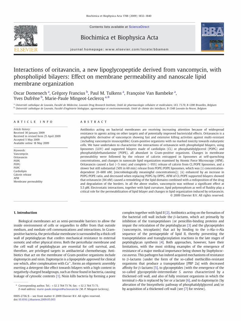
Chemical modification of the glycopeptides, however, has provideda new class of derivatives called collectively lipoglycopeptides whichshow improved activity against such multi-resistant strains (see [8]for review). Among them, oritavancin (LY333328) is the most activeagainst vancomycin-resistant organisms [7]. Compared to vancomy-cin, oritavancin possesses a 4-(4-chlorophenyl)-phenylmethyl sidechain [9,10], synthetically added to the natural product precursor,chloroeremomycin, which differs from vancomycin by the presence ofa 4-epi-vancosamine sugar (Fig. 1). Oritavancin, as well as anotherlipoglycopeptide, telavancin, is considerably more rapidly bactericidalthan vancomycin, suggesting a novelmode of action that could involvemembrane destabilization [11–13]. This mechanism ought to bespecific of bacteria since both compounds are devoid of markedtoxicities towards eukaryotic cells and have been successfully broughtto clinical applications in humans [7,11].
Because interactions of lipoglycopeptides in general, and of orita-vancin in particular, with model membranes is still largely unexplored,we attempted to further characterize them using a multidisciplinaryapproach but taking care of using conditions that mimicked those underwhich the drug exerts its antibacterial effects. As maintenance of apermeability barrier is the central property of biological membranes, wefirst compared the ability of oritavancin and vancomycin to permeabilizelipid bilayers. The results were correlated with real-time observations ofthe interactions of both molecules with lipids at the nanoscale levelusing Atomic Force Microscopy (AFM) [14]. Since the bacterialmembrane is rich in acidic phospholipids [15], we used liposomes andsupported bilayers made of cardiolipin (CL) or phosphatidylglycerol(PG) and phosphatidylethanolamine (PE). PG is the predominantphospholipid in the membranes of S. aureus [16], and cardiolipin isalso found at high levels in these bacteria [17] as well several otherGram-positive organisms [15,18]. While being more abundant in Gram-negative bacteria, PE is nevertheless present in S. aureus [19,20].
2. Materials and methods
2.1. Materials
Beef heart cardiolipin (CL; Disodium Salt; purity N99%), and 1-palmitoyl-2-oleoyl-sn-glycero-3-phosphoglycerol (POPG), 1-palmi-
Fig. 1. Structural formulae of vancomycin and oritavancin. Key differences are (i) the chlorohydrophobic character to the molecule, and (ii) the amino group present in the additional ecalculated log P and log D (at pH 7) of vancomycin are –1.44 and –4.70, and those of oritav
toyl-2-palmitoyl-sn-glycero-3-phosphoglycerol (DPPG), 1-palmitoyl-2-oleoyl-sn-glycero-3-phosphocholine (POPC), 1-palmitoyl-2-oleoyl-sn-glycero-3-phosphoethanol-amine (POPE) were purchased fromAvanti Polar Lipids (Alabaster, AL) and used without further purifica-tion. Calcein was purchased from Sigma-Aldrich (St. Louis, MO) andwas purified as described previously [21]. Briefly, calcein wasdissolved in 6 N NaOH and subjected to size-exclusion chromato-graphy through a SephadexR LH-20 column. The final concentration ofcalcein solution in 20 mM Tris–HCl was 73 mM with an osmolarity of434 mOsm/kg (measured by the freezing point technique, using amodel 3C2 Advanced Cryomatic Osmometer [Advanced Instruments,Needham Heights, MA]). Oritavancin diphosphate powder wassupplied by Targanta Therapeutics, Cambridge, MA, as microbiologicalstandard for in vitro investigations. It was dissolved in water contain-ing 0.002% polysorbate-80 (vol:vol). Vancomycin was obtained fromGlaxoSmithKline s.a., Genval, Belgium as the product registered forparenteral use in humans and complying with the provisions of theEuropean Pharmacopeia.
2.2. Large unilamellar vesicle preparation
Lipids were dissolved in CHCl3:CH3OH (2:1, v/v) and mixed toobtain the desired composition in a round flask. The solvent wasevaporated using a Rotavapor system (model R-210, Buchi Labortech-nik AG, Flawil, Switzwerland). Dried films were maintained underreduced pressure overnight and thereafter hydratedwith 20mMTris–HCl,150mMNaCl, 20mMCaCl2 2H20, pH 7.4 for AFM studies andwiththe purified calcein for permeability experiments. Large UnilamellarVesicles (LUV) were obtained after 5 cycles of freeze/thawing and 10cycles of extrusion in a 10 mL Thermobarrel Extruder (LipexBiomembranes, Vancouver, Canada) under a nitrogen pressure of 10bars through two polycarbonate filters of 100 nm or 400 nm pore sizefor permeability and AFM studies, respectively (Nucleopore, CostarCorporation, Badhoevedorp, The Netherlands). Non-entrapped calceinwas removed using minicolumn centrifugation [21]. The size andpolydispersity of liposome suspensions (Table 1) were monitored byquasi-elastic light scattering with a Zetasizer Nano SZ (MalvernInstruments, Worcestershire, UK). Lipid concentration on the liposo-mal suspensions was measured by phosphorous quantification [22].
-biphenyl methylene moiety (block arrow; absent in vancomycin) that confers a morepi-vancosamine moiety (thin arrow) that contributes to its amphipathic character. Theancin 4.10 and –3.43.

Table 1Diameter and polydispersity index of liposomes used for permeability studies(measured by quasi-elastic light scattering).
Composition Diameter (nm) Polydispersity index
POPC:POPE (4:8, mol:mol) 114.1 0.172POPG:POPE: (4:8, mol:mol) 109.9 0.110CL:POPE: (2:8, mol:mol) 127.6 0.145DPPG:POPE: (4:8, mol:mol) 103.1 0.079
Fig. 2. Release of calcein from liposomes (5 μM phospholipids) made of CL:POPE (2:8molar ratio; upright triangles) or POPG:POPE (4:8 molar ratio; upside down triangles)upon exposure at 37°C to 600 nMoritavancin (ORI; filled symbols) or vancomycin (VAN;open symbols). (A) 0–60 min exposure time; (B) 0–200 s exposure time (CL:POPEliposomes only). The ordinate shows the percentage of calcein released compared towhat was observed after addition of 2% Triton X-100. Each value is the mean of threeindependent experimental determinations ±SEM (when not visible, the bars aresmaller than the symbols). For oritavancin, the graph shows the functions obtained byfitting a one phase exponential association function to the data by non-linear regression.
1834 O. Domenech et al. / Biochimica et Biophysica Acta 1788 (2009) 1832–1840
2.3. Calcein release
The liposome total phospholipid concentration was adjusted to afinal concentration of 5 μM with isoosmotic buffer (20 mM Tris–HCl,200 mMNaCl, pH 7.4). After overnight storage at 4 °C, liposomes wereexposed to the drug under study at 37 °C at the desired concentrationand for the suitable timewith continuous stirring and protection fromlight. Leakage of entrapped calcein was monitored by the increase influorescence of the samples, resulting from the dilution and release ofself-quenching of this tracer [23]. All fluorescence determinationswere performed on an LS 55 fluorescence spectrophotometer (Perkin-Elmer Ltd., Beaconsfield, UK) using λexc and λem of 476 nm and512 nm, respectively and slits fixed at 3 nm. The percentage of calceinreleased under the influence of drug was defined as [(Ft−Fcontr)/(Ftot−Fcontr)]×100, where Ft is the fluorescence signal measured attime t in the presence of drug, Fcontr is the fluorescence signalmeasured at the same time in the absence of drug, and Ftot is the totalfluorescence signal obtained after complete disruption of liposomesby Triton X-100 at a final concentration of 2% (checked by quasi-elasticlight spectroscopy) [21].
2.4. Atomic force microscopy
Before each experiment, the contact mode cell was extensivelywashed with ethanol and water. Mica squares (0.25 cm2) were gluedonto a steel disc, cleaned carefully with water before use and cleavedto obtain a flat and uniform surface. Immediately, an aliquot of 50 μL ofvesicles (CL:POPE [2:8] or POPE only; adjusted to 250 μM in 20 mMTris–HCl, 150 mM NaCl, 20 mM CaCl2 2H20, pH 7.4), was deposited onthe mica surface and incubated for 1 h at room temperature. Thesample was thereafter washed with buffer (20 mM Tris–HCl, 150 mMNaCl, pH 7.4) to eliminate non-adsorbed vesicles. AFM contact modeimages in liquid were obtained using a Nanoscope IV Multimode AFM(Veeco Metrology Group, Santa Barbara, CA) with triangular Si3N4
cantilevers (Microlevers, Veeco Metrology Group, Santa Barbara, CA.)with a nominal spring constant of 0.01 N m−1. The instrument wasequipped with a “J” scanner (120 μm). To minimize the applied forceon the sample the set point was continuously adjusted duringimaging. Images were acquired at 90° scan angle with a scan rate of2 Hz. All images were processed using the Veeco software.
3. Results
3.1. Membrane permeability
The effect of oritavancin and vancomycin at a fixed concentration(600 nM) on the permeability of liposomes made of CL:POPE (2:8) orPOPG:POPE (4:8) (so as to obtain the same amount of negativecharges) is shown in Fig. 2A. Oritavancin caused a release of calceinthat was almost immediate (less than 5 min) and complete whenadded to CL:POPE liposomes. The release of calcein was slower buteventually reached about 50% within 60 min when oritavancin wasadded to POPG:POPE liposomes (maintaining oritavancin in contactwith these liposomes for up to 8 h caused about 60% calcein release).Vancomycin was almost without effect on both CL:POPE and POPG:POPE liposomes. To obtain more information about the true rate ofrelease of calcein from CL:POPE liposomes upon exposure to
oritavancin, experiments were repeated using shorter periods ofcontact (0–200 s). Results presented in Fig. 2B show that 50% calceinrelease was obtained within about 50 s. Vancomycin was withoutconstant effect under these conditions.
In a second series of experiments, we examined the effect oforitavancin concentration on the permeabilization of CL:POPE andPOPG:POPE vesicles. The concentration range chosen (0–600 nM)wasmeant to attain the values reported as causing bacterial death of S.aureus in vitro (0.12–1 mg/L [66–550 nM], depending upon assayconditions [24,25]. The results, as seen after 8 h of contact (to ensuremaximal effects) are presented in Fig. 3. Oritavancin caused aconcentration-dependent release of calcein that reached a valueclose to 98% at 600 nM when tested against CL:POPE liposomes, and60% for POPG:POPE liposomes. Vancomycin was without significanteffect over the same range of concentrations (data not shown).
The experiments reported so far did not address the question as towhether the permeabilization effect seenwith oritavancinwas relatedto a bilayer charge per se or to hydrophobic interactions between thislipoglycopeptide and membranes. We, therefore, used POPG:POPEand POPC:POPE vesicles and examined the effect of changing the ratiobetween POPG and POPE and between POPC and POPE on the ability oforitavancin to cause calcein release. The results presented in Fig. 4show that an increase of the POPG to POPE ratio from 0.5 to 0.75 wasassociated with an increase of calcein release, above which only a

Fig. 4. Release of calcein from liposomes (5 μM phospholipids) made of POPG:POPE(triangles) or POPC:POPE (squares) of variable composition (see abscissa) uponexposure at 37 °C and for 8 h to 600 nM (1.08mg/L) oritavancin. The ordinate shows thepercentage of calcein released compared to what was observed after addition of 2%Triton X-100. Each value is themean of three independent experimental determinations±SEM (when not visible, the bars are smaller than the symbols).
Fig. 3. Release of calcein from liposomes (5 μM phospholipids) made of CL:POPE (2:8molar ratio; upright triangles) or POPG:POPE (4:8 molar ratio; upside downtriangles) upon exposure at 37 °C and for 8 h to increasing concentrations oforitavancin (0–600 nM; 0–1.08 mg/L). The ordinate shows the percentage of calceinreleased compared to what was observed after addition of 2% Triton X-100. Eachvalue is the mean of three independent experimental determinations ±SEM (whennot visible, the bars are smaller than the symbols).
1835O. Domenech et al. / Biochimica et Biophysica Acta 1788 (2009) 1832–1840
more modest increase was seen. Conversely, the extent of release ofcalcein, which was substantial when using liposomes with a lowPOPC:POPE ratio (0.5) was decreased when the POPC:POPE ratio wasincreased to 0.75 or more. The influence of the nature of the fatty acidchain was also investigated by replacing POPG by DPPG in liposomeswhile leaving the ratio PG:PE identical (4:8). This caused a drasticreduction of the permeabilization effect of oritavancin that droppedfrom 60% to 24% (measured after 8 h of contact with 600 nMoritavancin).
3.2. Atomic force microscopy
To go further in the study of the interaction of oritavancin and lipidbilayers and especially to characterize at the nanoscale, the effect ofthis antibiotic on membrane organization, we prepared supportedlipid bilayers made of POPE only, or of CL:POPE (2:8). The lattercomposition has been selected in view to the larger permeabilizingeffect induced by oritavancin on CL:POPE (2:8) as compared to POPG:POPE (4:8). Supported lipid bilayers made of POPG:POPEwere alreadystudied by AFM for some of us [26] showing no lipid separationbetween POPE and POPG. Supported bilayers of POPE and CL:POPEwere prepared by fusion of unilamellar vesicles on mica, and theirorganization was imaged by AFM. Preliminary experiments assessedthe stability of POPE and CL:POPE bilayers during consecutivescanning in buffer. Successive images of the same bilayer locationwere recorded but this did not cause any significant change of thesurface morphology, indicating that the bilayer was stable in theseconditions for at least 3 h.
3.2.1. POPE bilayersFig. 5A shows a typical topographic image of a POPE bilayer
supported on mica recorded in buffer solution where the darkestregion is the mica surface while the brightest region is the surface ofthe supported planar bilayer. The step height with respect to the micasurface was 5.24 nm (n=50), with a roughness (Ra) mean value of0.07 nm (n=25). Fig. 5B shows a typical image of the same bilayerafter 1 h exposure to vancomycin (5.5 μM [8 mg/L]), a concentrationresulting in a bactericidal effect towards S. aureus in vitro [27,28]. Thedrug induced an erosion in the unprotected edges, border and holes, ofthe supported planar bilayer, but did not substantially change itsthickness, the step height being 4.90 nm (n=50) with a roughness of0.12 nm (n=25). When the experiment was performed in thepresence of oritavancin (84 nM [0.15 mg/L], a concentration at whichoritavancin becomes bactericidal in vitro [25], the drug induced anerosion of the bilayer together with a decrease of the height down to
4.00 nm (Fig. 5D) vs. 4.80 nm [n=50] for control (Fig. 5C) withoutchange in roughness (0.09 nm [n=25]). These results suggest,therefore, that vancomycin had no major effect on the supportedplanar bilayer of pure POPE whereas oritavancin induced a slighterosion and thinning of the bilayer (Table 2).
3.2.2. CL:POPE (2:8) bilayersIn control supported bilayers, two laterally segregated domains
were observed. The step height between the uncovered mica, and theunderlying domain was 4.55 nm (n=50) with a Ra value of 0.17 nm(n=25) (Fig. 6A). Several smaller domains were seen which were1.03 nm (n=50) taller than the underlying domain with a mean Ravalue of 0.09 nm (n=25). These domains were clearly seen in thecross section supported below as a protruding planar structure on themore extended domain.
After 1 h of contact with vancomycin (5.5 μM [8 mg/L]), thefrontiers of the more extended lipid domain were eroded (Fig. 6B).From section analysis, the more extended domain presented a heightof 3.79 nm (n=50) from the mica surface with a mean Ra value of0.15 nm (n=25). In parallel, reorganisation and/or disappearance ofseveral small lipid domains was observed with solubilisation andafterwards spreading of the material on the mica surface. Moreover,new domains appeared which were 0.75 nm higher than theunderlying domain with a mean Ra value of 0.07 nm (n=25).When the supported bilayers were exposed to oritavancin to a finalconcentration of 84 nM (0.15 mg/L) for 1 h, a completely differenteffect was seen compared to vancomycin. While the control supportedbilayers (Fig. 6C) showed two segregated domains of 4.57 nm and5.26 nm heights and 0.08 nm and 0.14 nm roughness, respectively,oritavancin (Fig. 6D) caused a remodelling of the lipid domaincombined with a redisposition of the drug as well as a degradation ofthe unprotected borders of the supported bilayer. Thus, oritavancininduced a decrease of the height from 4.57 nm to 4.10 nm (n=50)with a roughness of 0.15 nm. This underlying domain was covered bytwo other domains. The first onewas located at 0.48 nm (n=50) witha roughness of 0.13 nmwhereas the second one, more heterogeneous,ranged from 0.51 to 1.40 nm (n=25).
4. Discussion
The bacterial envelope has long been recognized as a critical targetfor antibiotics, with β-lactams and glycopeptides—both directedagainst the peptidoglycan—being the most widely used today inclinical practice to fight Gram-positive bacteria and S. aureus inparticular. In contrast, antibiotics acting on the lipidmembrane part of

Fig. 5. Nanoscale membrane activity of vancomycin (top) or oritavancin (bottom) on POPE supported bilayer. AFM height images (5 μm×5 μm; z-scale: 20 nm) of POPE supportedplanar bilayer recorded prior (0 min) (A; C) or after exposure for 1 h to vancomycin (5.5 μM [8 mg/L]) (B) or oritavancin (84 nM [0.15 mg/L]) (D). Vertical cross-sections were takenalong the position indicated by the continuous line. Upper domain and mica are indicated by black and white stars, respectively.
1836 O. Domenech et al. / Biochimica et Biophysica Acta 1788 (2009) 1832–1840
the bacterial membrane have for long remained largely unused ashuman medicines, mainly because of lack of specificity resulting in aglobally less favorable toxicological profile. The emergence of wide-spread resistance to β-lactams and the slow but inescapable loss ofpotency of glycopeptides has, however, directed again the attention tomembrane-acting antibiotics, as these may potentially offer novelmodes of action coupled with powerful antibacterial activities [7].Among these, lipoglycopeptides attracted increasing attention sincethese have now been successfully developed clinically [29,30] and arebeing considered for approval for the treatment of multi-resistantGram-positive organisms.
For lipoglycopeptides, differences in activities relative to vanco-mycin do not relate to the aglycone part of these molecules, i.e. in theD-Ala–D-Ala binding sites, since these are similar with affinities
essentially identical among all of them [31]. However, the presence ofa lipophilic tail confers the ability to lipoglycopeptides to interact withmembranes. This has been shown for telavancin, a drug with anoverall structure and antibacterial properties similar to those oforitavancin, for which 25% only of the antibiotic is recovered in thepeptidoglycan while 75% remains associated the protoplast uponseparation of these two constituents of the bacterial envelope (vs.N90% bound to the peptidoglycan and b10% to the protoplasts forvancomycin) [12]. The novel mode of action proposed involvesdissipation of membrane potential and membrane permeabilization[13], and has been related to loss of membrane integrity favored byinteraction of the drug with lipid II (the molecule that carries the D-Ala–D-Ala terminated pentapeptide from the cytosolic to the extra-cellular face of the bacterial membrane). Similarly, ongoing studies

Table 2Height and roughness values in supported phospholipid bilayers of POPE (top) and CL:POPE (2:8; mol:mol) (bottom).
Height±SD (nm) Roughness (nm)
Vancomycin Before 5.24±0.19 0.07After 4.90±0.20 0.12
Oritavancin Before 4.80±0.20 0.09After 4.00±0.15 0.09
Domain height±SD (nm) Domain roughness (nm)
Low Intermediate High Low Intermediate High
Vancomycin Before – 4.55±0.20 5.58±0.16 – 0.17 0.09After – 3.79±0.30 4.54±0.11 – 0.15 0.07
Oritavancin Before 4.57±0.15 5.26±0.20 – 0.08 0.14After 4.10 ± 0.30 4.58±0.07 4.61–5.50 0.15 0.13 –
1837O. Domenech et al. / Biochimica et Biophysica Acta 1788 (2009) 1832–1840
have now described the depolarization of membranes of vancomycinnon-susceptible S. aureus following incubation with oritavancin (G.A.McKay, S. Beaulieu, T.R. Parr Jr, G. Moeck, in Program and Abstracts ofthe 48th Annual Interscience Conference on Antimicrobial Agents andChemotherapy, Washington DC (2008) abstract no. C1-4182; [13]).
In the present work, and using calcein as a validated probe for thestudy of lipid membrane permeabilization [21,23], and AFM toinvestigate the effect of the lipoglycopeptide at the nanoscale, weshowed that oritavancin induced membrane permeabilizationtogether with changes in the lipid membrane organization indomains. It is, however, important to remember that the two modelsused, liposomes and supported lipid bilayers, are different, notablyregarding the curvature of the bilayer and the ability of the bothleaflets of lipids to be in contact with aqueous phases. The effectsinduced by oritavancin are observed in the absence of any binding to aD-Ala–D-Ala motif, indicating that alteration of membrane integrity isprobably not due to the aglycone part of the molecule, since its iscommon to that of vancomycin, which exerts no effect in our models.
The mechanisms responsible for membrane permeabilization arecomplex, but four main mechanisms have been proposed, namely (i)the barrel-stave channel, i.e. a bundle of membrane-spanning helicesaligned with the polar side chains oriented towards the center [32,33](ii) the carpet-like mechanism where a monolayer of surface-lyingpeptides covers the membrane surface [34], (iii) the toroidal model[35,36], and (iv) the detergent-like peptidemodel [37]. Our study doesnot provide direct evidence for either of thesemodels,which should bethe subject of future investigations. Especially the ability of oritavancinto insert in the outer monolayer of the bilayer, leading to anasymmetric increase in lateral pressure that tends to bend the bilayerand induce a transient disruption of the membrane [37] has to beexamined. Moreover, the formation of rich clusters in the membraneand destabilization of the lipid membrane by formation of mixedmicelles, could be determined.
If the permeabilization clearly depends on drug structure, the lipidcomposition, and therefore the physical properties of membranes, canalso play a critical role for permeabilization. This is clearly reflected bythe ranking found for the ability of oritavancin to permeabilize lipidbilayers (CL:POPENPOPC:POPENPOPG:POPENDPPG:POPE). Fourparameters could be critical in this respect.
First, one feature that distinguishes the membrane of prokaryoticorganisms from that of eukaryotic organisms is that only the formerharbor negatively-charged lipids in the outer leaflet of the plasmamembrane. So a large proportion of negatively-charged lipids,especially POPG, is generally considered critical for the selectivity ofaction of antibacterial peptides and lipopeptides [1]. Recently,however, this point has been questioned. Some amphipathic α-helicalpeptide analogues derived from Helicobacter pylori [38] had strongantibacterial activity despite weak binding to phosphatidylglycerol.Similarly, homologous antimicrobial oligomers mimicking hostdefense peptides are not sensitive to charge [39]. Regardingoritavancin, clearly, the presence of negatively-charged lipids is not
the only parameter required to induce the maximal membranepermeabilization since a higher permeabilizing effect has beenobserved on bilayers of POPC:POPE as compared to POPG:POPE. Thisis in agreement with other studies performed on plantaricin A, a 26-residue peptide pheromone, for which leakage of calcein from SOPC:POPG vesicles is less extensive than from SOPC vesicles [40]. Similarly,pardaxin, a membrane-lysing peptide originally isolated from the fishPardachirus marmoratus, also shows a lower ability to disrupt POPG:POPE as compared to homogenous POPC bilayers [41].
Second, besides electrostatic interactions, the ability of phospho-lipids to form H bonds could also be a major parameter governingtheir interaction with glycopeptides. Indeed, POPG:POPE bilayersshow a high propensity to interact through hydrogen bonds withphosphate and carbonyl oxygen atoms, as well as a much denserpacking of chain atoms in the near-the-interface regions of thehydrophobic core, as compared to POPC bilayers [42]. Moreover, POPGis less hydrated and shows a low probability of gauche conformationin the beginning of the β-chain as compared to POPC [43]. This maycontribute to disturb packing of atoms in the near-the-interfaceregions of the POPC bilayer core, creating defects and explaining whyoritavancin showed greater permeabilizing effect towards POPC:POPE(4:8) than POPG:POPE (4:8).
Third, the propensity of CL and PE to induce negative curvature andadopt hexagonal phase [44,45] may also contribute to favor thepermeabilization induced by oritavancin. At a molecular level, thepolar headgroup of PE is characterized by a smaller diameter than thehydrocarbon chain region in the fluid phase. This results in amolecularshape of a truncated cone and an intrinsic propensity of PE bilayers toform surfaces with a negative curvature, leading to the formation ofnon-lamellar phases. Formation of hexagonal phase can be related tohigh compressibility in fluid phase [46], to an ability to easily adapt tohighly irregular surfaces [47], and to provide the lateral force for anadequate stress profile [46,48]. This could thus explain why, when thePOPE content is increased in liposomes containing POPC:POPE, anenhancement of calcein release is observed. In CL:POPE liposomes, therapid and important effect of oritavancin on membrane permeabilitycould result from a local reduction in electrostatic repulsion betweencardiolipin molecules, thus imposing negative curvature strain on themembrane. In comparison to CL:POPE, the lower permeabilizing effectof oritavancin on POPG:POPE liposomes, could be related to the factthat PG preferentially adopts a cylindrical molecular shape and formsflat bilayer, even at high temperatures.
Fourth, the unsaturated nature of the bacterial phospholipidconfers fluidity to the membrane and possibly augments the insertionof exogeneous compounds like antimicrobial peptides [49] or theglycopeptides themselves [50]. The higher release of calcein fromliposomes of POPG:POPE: as compared to DPPG:POPE could resultfrom this.
AFM, a now well-established technique for imaging supportedlipid bilayers at nanometer resolution [14] has also provided us withuseful information on the changes brought by oritavancin on bilayer

Fig. 6. Nanoscale membrane activity of vancomycin (top) or oritavancin (bottom) on CL:POPE (2:8) supported bilayer. AFM height images (5 μm×5 μm; z-scale: 20 nm) of CL:POPEsupported planar bilayer recorded prior (0min) (A; C) or after exposure for 1 h to vancomycin (5.5 μM [8mg/L]) (B) or oritavancin (84 nM [0.15 mg/L]) (D). Vertical cross-sections weretakenalongtheposition indicatedby thecontinuous line.Upperdomainandmicaare indicatedbyblackandwhite stars, respectively.White arrows showeddomainsenriched incardiolipin.
1838 O. Domenech et al. / Biochimica et Biophysica Acta 1788 (2009) 1832–1840
organization. Previous studies performed by mixing CL and POPE atthe air–water interface, and comparing the values of the excess energyof mixing, showed that the composition chosen (CL:POPE at 2:8 molarratio) was the most stable [51]. In addition to the lateral phaseseparation due to the differences in molecular structure, POPE and CLcan simultaneously undergo a thermal transition at the same time[52], explaining the occurrence of the three domains in a binarysystem observed by AFM. Since increasing the amount of cardiolipinresulted in an increase of the area occupied by the upper domains, ithas been assumed that the lower domain is mainly formed by POPEwhereas the upper domain is mainly formed by cardiolipin [52].
We show here that oritavancin did not modify the height of asupported bilayer of POPE, but decreased the step height between the
mica surface and the surface of the more extended domain in the CL:POPE bilayer. In a supported bilayer of pure POPE, the PE headgroupscan form hydrogen bonds between adjacentmolecules [53] conferringrigidity to the surface, while in a CL:POPE mixed supported bilayer,cardiolipin molecules can disturb the formation of the hydrogenbounds network [51]. Thus, oritavancin could interact with CL, whichtends to form non-lamellar phases, promoting a negative curvature inthe region of the domain and destroying its integrity (detergent-likepeptide model) [37].
Our studies were performed under conditions that are pertinent tothe antibacterial activity of oritavancin, in that we (i) used bilayersthat, like bacterial membranes, are rich in both a negatively-chargedlipid like phosphatidylglycerol and/or cardiolipin and phosphatidy-

1839O. Domenech et al. / Biochimica et Biophysica Acta 1788 (2009) 1832–1840
lethanolamine [16,17,19,20]; (ii) performed all observations at con-centrations of oritavancin that are similar to those at whichbactericidal effects can be demonstrated in vitro towards bothvancomycin-susceptible and vancomycin-resistant S. aureus or otherGram-positive organisms [24,54,55]. These ranges of concentrationsare also those reached in serum during conventional administration ofthe drug to humans [55]. All together, the data, therefore, stronglysuggest that membrane effects are responsible for the increasedpotency of oritavancin as compared to vancomycin (and its activityagainst vancomycin-resistant organisms). Moreover, recent studieshave demonstrated that oritavancin induced rapid bactericidal activityagainst both exponential phase and stationary-phase inocula of S.aureus in relation with membrane depolarization and membranepermeabilization [13]. It remains, however, that membrane-destabi-lizing effects of oritavancin, if critical for its antibacterial activity, mustbe largely specific to prokaryotic cells since cellular experiments witheukaryotic cells failed to markedly affect the cell viability (assessed bymeasuring the release of a cytosolic enzyme, the lactate deshydro-genase) of macrophages incubated with concentrations of up to25 mg/L, i.e. 200-fold the minimal inhibitory concentration observedin vitro for the least susceptible bacteria [25]. The explanationprobably lies in the difference in membrane composition and morespecifically, in the lack of significant amounts of POPG or CL in thepericellular membrane of eukaryotic cells.
An important aspect underlined by our studies is that only weakalterations in membrane permeability or bilayer morphology wereseen with large concentrations of vancomycin. These cause killing ofGram-positive organisms in vitro, but through a slow-acting mechan-ism unrelated to direct membrane destabilization but, most likely, tothe activation of autolytic enzyme systems [56] through signaltransduction (via a death signal peptide) common to vancomycinand β-lactams [57,58].
Other hypotheses, however, could be suggested. If oritavancin andvancomycin are comparable inhibitors of transglycosylation, orita-vancin is also a potent inhibitor of transpeptidation [59]. Moreover, inaddition to the binding to the primary site D-Ala–D-Ala, sequestrationof lipid II and inhibition of transglycosylation, the hydrophobicbiphenyl moiety and components of the aglycone structure oforitavancin could form a secondary binding site for pentaglycylsegments [59,60] due to the biphenyl group that contributes to anenlargement of the drug hydrophobic surface area [61]. Also, the factthat all effects can be seen at very low concentrations is of directimportance. Indeed, oritavancin has been shown to dimerize easily,and this, together with its membrane anchoring properties, has beenproposed as an important determinant in its superior activity tovancomycin by favoring its interaction with the D-Ala–D-Ala motif[62]. Dimerization is not only critical for peptide recognition but alsofor membrane destabilization as it has been shown for antibacterials,such as cathelicidin [63] or magainin [64]. Oligomerization couldenhance the kinetics and thermodynamic stabilities of the complexformed [65] and affect the rate of association/dissociation to/fromlipids [66]. Lastly, a hydrophobic effect could also be important butwith some degree of specificity, as no beneficial effect is observedwhen an aliphatic chain was introduced to the E-ring of vancomycin[67].
In conclusion, our work has advanced the characterization of theinteraction between oritavancin, a novel lipoglycopeptide endowedwith potent antimicrobial activity, and phospholipid bilayers. Ourobservations indicate that lipid composition is a critical parameterin this context with a clear role played by cardiolipin. This effect isprobably related to changes in the local physico-chemical propertiesof the membranes, such as the surface charge, ability to form Hbonds, curvature, and fluidity. Future biological and pharmaceuticalresearch may help in developing and expanding these observationson what may constitute essential parts of antimicrobial armamen-tarium in the future.
Acknowledgements
Y.F.D. is ResearchAssociate and F.VB. Senior ResearchAssociate of theBelgian Fonds de la Recherche Scientifique (F.R.S.-FNRS). This work wassupported by the Région wallonne (NANOMEMB), the F.R.S.-FNRS(grant no.1.5.236.08 F), the Fonds de la Recherche ScientifiqueMédicale(F.R.S.M.; grantsno. 2.4.601.06 and3.4.597.06), theUniversité catholiquede Louvain (Fonds Spéciaux de Recherche and Actions de RechercheConcertées), andwith a grant-in-aid fromTarganta Therapeutics , a fullyowned subsidiary of the Medicines Company, Parsippany, NJ.
References
[1] S.K. Straus, R.E. Hancock, Mode of action of the new antibiotic for Gram-positivepathogens daptomycin: comparison with cationic antimicrobial peptides andlipopeptides, Biochim. Biophys. Acta 1758 (2006) 1215–1223.
[2] K. Christ, I. Wiedemann, U. Bakowsky, H.G. Sahl, G. Bendas, The role of lipid II inmembrane binding of and pore formation by nisin analyzed by two combinedbiosensor techniques, Biochim. Biophys. Acta 1768 (2007) 694–704.
[3] J.M. Ghuysen, Serine beta-lactamases and penicillin-binding proteins, Annu. Rev.Microbiol. 45 (1991) 37–67.
[4] P.E. Reynolds, Structure, biochemistry and mechanism of action of glycopeptideantibiotics, Eur. J. Clin. Microbiol. Infect. Dis. 8 (1989) 943–950.
[5] H.F. Chambers, Methicillin-resistant staphylococci, Clin. Microbiol. Rev. 1 (1988)173–186.
[6] L.B. Rice, Antimicrobial resistance in gram-positive bacteria, Am. J. Med. 119(2006) S11–S19.
[7] F. Van Bambeke, M.P. Mingeot-Leclercq, M.J. Struelens, P.M. Tulkens, The bacterialenvelope as a target for novel anti-MRSA antibiotics, Trends Pharmacol. Sci. 29(2008) 124–134.
[8] F. Van Bambeke, Glycopeptides and glycodepsipeptides in clinical development: acomparative review of their antibacterial spectrum, pharmacokinetics and clinicalefficacy, Curr. Opin. Investig. Drugs 7 (2006) 740–749.
[9] N.E. Allen, T.I. Nicas, Mechanism of action of oritavancin and related glycopeptideantibiotics, FEMS Microbiol. Rev. 26 (2003) 511–532.
[10] R.D. Cooper, N.J. Snyder, M.J. Zweifel, M.A. Staszak, S.C. Wilkie, T.I. Nicas, et al.,Reductive alkylation of glycopeptide antibiotics: synthesis and antibacterialactivity, J. Antibiot.(Tokyo) 49 (1996) 575–581.
[11] F. Van Bambeke, Glycopeptides in clinical development: pharmacological profileand clinical perspectives, Curr. Opin. Pharmacol. 4 (2004) 471–478.
[12] D.L. Higgins, R. Chang, D.V. Debabov, J. Leung, T.Wu, K.M. Krause, et al., Telavancin,a multifunctional lipoglycopeptide, disrupts both cell wall synthesis and cellmembrane integrity in methicillin-resistant Staphylococcus aureus, Antimicrob.Agents Chemother. 49 (2005) 1127–1134.
[13] A. Belley, E. Neesham-Grenon, G. McKay, F.F. Arhin, R. Harris, T. Beveridge, et al.,Oritavancin kills stationary-phase and biofilm Staphylococcus aureus in vitro,Antimicrob. Agents Chemother 53 (2008) 918–925.
[14] Y.F. Dufrene, Towards nanomicrobiology using atomic force microscopy, Nat.Rev.Microbiol. 6 (2008) 674–680.
[15] M.C. Trombe, M.A. Laneelle, G. Laneelle, Lipid composition of aminopterin-resistant and sensitive strains of Streptococcus pneumoniae. Effect of aminopterininhibition, Biochim. Biophys. Acta 574 (1979) 290–300.
[16] W. Dowhan, Molecular basis for membrane phospholipid diversity: why are thereso many lipids? Annu. Rev. Biochem. 66 (1997) 199–232.
[17] P.R. Beining, E. Huff, B. Prescott, T.S. Theodore, Characterization of the lipids ofmesosomal vesicles and plasma membranes from Staphylococcus aureus, J.Bacteriol. 121 (1975) 137–143.
[18] M. Cohen, C. Panos, Membrane lipid composition of Streptococcus pyogenes andderived L form, Biochemistry 5 (1966) 2385–2392.
[19] S.A. Short, D.C. White, Metabolism of the glycosyl diglycerides and phosphati-dylglucose of Staphylococcus aureus, J. Bacteriol. 104 (1970) 126–132.
[20] R.F. Epand, P.B. Savage, R.M. Epand, Bacterial lipid composition and theantimicrobial efficacy of cationic steroid compounds (ceragenins), Biochim.Biophys. Acta 1768 (2007) 2500–2509.
[21] F. Van Bambeke, M.P. Mingeot-Leclercq, A. Schanck, R. Brasseur, P.M. Tulkens,Alterations in membrane permeability induced by aminoglycoside antibiotics:studies on liposomes and cultured cells, Eur. J. Pharmacol. 247 (1993) 155–168.
[22] G.R. Bartlett, Colorimetric assay methods for free and phosphorylated glycericacids, J. Biol. Chem. 234 (1959) 469–471.
[23] J.N. Weinstein, S. Yoshikami, P. Henkart, R. Blumenthal, W.A. Hagins, Liposome-cell interaction: transfer and intracellular release of a trapped fluorescent marker,Science 195 (1977) 489–492.
[24] C. Seral, F. Van Bambeke, P.M. Tulkens, Quantitative analysis of gentamicin,azithromycin, telithromycin, ciprofloxacin, moxifloxacin, and oritavancin(LY333328) activities against intracellular Staphylococcus aureus in mouse J774macrophages, Antimicrob. Agents Chemother. 47 (2003) 2283–2292.
[25] F.F. Arhin, K. Tomfohrde, D.C. Draghi, M. Aranza, T.R. Parr, D.F. Sahm, et al., Newlydefined in vitro quality control ranges for oritavancin broth microdilution testingand impact of variation in testing parameters, Diagn. Microbiol. Infect. Dis. 62(2008) 92–95.
[26] O. Domenech, S. Merino-Montero, MT. Montero, J. Hernandez-Borrell, Surfaceplanar bilayers of phospholipids used in protein membrane reconstitution: anatomic force microscopy study, Colloids Surf., B Biointerfaces 47 (2006) 102–106.

1840 O. Domenech et al. / Biochimica et Biophysica Acta 1788 (2009) 1832–1840
[27] M. Barcia-Macay, S. Lemaire, M.P. Mingeot-Leclercq, P.M. Tulkens, F. Van Bambeke,Evaluation of the extracellular and intracellular activities (human THP-1macrophages) of telavancin versus vancomycin against methicillin-susceptible,methicillin-resistant, vancomycin-intermediate and vancomycin-resistant Sta-phylococcus aureus, J. Antimicrob. Chemother. 58 (2006) 1177–1184.
[28] M. Barcia-Macay, C. Seral, M.P. Mingeot-Leclercq, P.M. Tulkens, F. Van Bambeke,Pharmacodynamic evaluation of the intracellular activities of antibiotics againstStaphylococcus aureus in a model of THP-1 macrophages, Antimicrob. AgentsChemother. 50 (2006) 841–851.
[29] E.C. Nannini, M.E. Stryjewski, A new lipoglycopeptide: telavancin, Expert Opin.Pharmacother. 9 (2008) 2197–2207.
[30] G. Poulakou, H. Giamarellou, Oritavancin: a new promising agent in the treatmentof infections due to Gram-positive pathogens, Expert. Opin. Investig. Drugs 17(2008) 225–243.
[31] J.C. Barna, D.H. Williams, The structure and mode of action of glycopeptideantibiotics of the vancomycin group, Annu. Rev. Microbiol. 38 (1984) 339–357.
[32] M.P. Mingeot-Leclercq, X. Gallet, C. Flore, F. Van Bambeke, J. Peuvot, R. Brasseur,Experimental and conformational analyses of interactions between butenafineand lipids, Antimicrob. Agents Chemother. 45 (2001) 3347–3354.
[33] B. Bechinger, Structure and functions of channel-forming peptides: magainins,cecropins, melittin and alamethici, J. Membr. Biol. 156 (1997) 197–211.
[34] G. Fimland, L. Johnsen, B. Dalhus, J. Nissen-Meyer, Pediocin-like antimicrobialpeptides (class IIa bacteriocins) and their immunity proteins: biosynthesis,structure, and mode of action, J. Pept. Sci. 11 (2005) 688–696.
[35] F. Porcelli, R. Verardi, L. Shi, K.A. Henzler-Wildman, A. Ramamoorthy, G. Veglia,NMR structure of the cathelicidin-derived human antimicrobial peptide LL-37 indodecylphosphocholine micelles, Biochemistry 47 (2008) 5565–5572.
[36] S. Campagna, N. Saint, G. Molle, A. Aumelas, Structure and mechanism of action ofthe antimicrobial peptide piscidin, Biochemistry 46 (2007) 1771–1778.
[37] H. Heerklotz, J. Seelig, Leakage and lysis of lipid membranes induced by thelipopeptide surfactin, Eur. Biophys. J. 36 (2007) 305–314.
[38] S.C. Park, M.H. Kim, M.A. Hossain, S.Y. Shin, Y. Kim, L. Stella, et al., Amphipathicalpha-helical peptide, HP (2-20), and its analogues derived from Helicobacterpylori: pore formation mechanism in various lipid compositions, Biochim.Biophys. Acta 1778 (2008) 229–241.
[39] A. Som, G.N. Tew, Influence of lipid composition on membrane activity ofantimicrobial phenylene ethynylene oligomers, J. Phys. Chem. B 112 (2008)3495–3502.
[40] H. Zhao, R. Sood, A. Jutila, S. Bose, G. Fimland, J. Nissen-Meyer, et al., Interaction ofthe antimicrobial peptide pheromone Plantaricin A with model membranes:implications for a novel mechanism of action, Biochim. Biophys. Acta 1758 (2006)1461–1474.
[41] K.J. Hallock, D.K. Lee, J. Omnaas, H.I. Mosberg, A. Ramamoorthy, Membranecomposition determines pardaxin's mechanism of lipid bilayer disruption,Biophys. J. 83 (2002) 1004–1013.
[42] K. Murzyn, T. Rog, M. Pasenkiewicz-Gierula, Phosphatidylethanolamine–phos-phatidylglycerol bilayer as a model of the inner bacterial membrane, Biophys. J. 88(2005) 1091–1103.
[43] T. Rog, K. Murzyn, R. Gurbiel, Y. Takaoka, A. Kusumi, M. Pasenkiewicz-Gierula,Effects of phospholipid unsaturation on the bilayer nonpolar region: a molecularsimulation study, J. Lipid Res. 45 (2004) 326–336.
[44] M.J. Hope, D.C Walker, P.R. Cullis, Ca2+ and pH induced fusion of smallunilamellar vesicles consisting of phosphatidylethanolamine and negativelycharged phospholipids: a freeze fracture study, Biochem. Biophys. Res. Commun.110 (1983) 15–22.
[45] S.B. Farren, P.R. Cullis, Polymorphism of phosphatidylglycerol–phosphatidyletha-nolamine model membrane systems: a 31p NMR study, Biochem. Biophys. Res.Commun. 97 (1980) 182–191.
[46] R.M. Epand, R. Bottega, Determination of the phase behaviour of phosphatidy-lethanolamine admixed with other lipids and the effects of calcium chloride:implications for protein kinase C regulation, Biochim. Biophys. Acta 944 (1988)144–154.
[47] P.K. Fyfe, K.E. McAuley, A.W. Roszak, N.W. Isaacs, R.J. Cogdell, M.R. Jones, Probingthe interface between membrane proteins and membrane lipids by X-raycrystallography, Trends Biochem. Sci. 26 (2001) 106–112.
[48] M. Rappolt, A. Hickel, F. Bringezu, K. Lohner, Mechanism of the lamellar/inversehexagonal phase transition examined by high resolution X-ray diffraction,Biophys. J. 84 (2003) 3111–3122.
[49] J.L. Ding, P. Li, B. Ho, The sushi peptides: structural characterization and mode ofaction against Gram-negative bacteria, Cell Mol. Life Sci. 65 (2008) 1202–1219.
[50] M. Rakotomanga, M. Saint-Pierre-Chazalet, P.M. Loiseau, Alteration of fatty acidand sterol metabolism in miltefosine-resistant Leishmania donovani promasti-gotes and consequences for drug-membrane interactions, Antimicrob. AgentsChemother. 49 (2005) 2677–2686.
[51] O. Domenech, F. Sanz, M.T. Montero, J. Hernandez-Borrell, Thermodynamic andstructural study of the main phospholipid components comprising the mitochon-drial inner membrane, Biochim. Biophys. Acta 1758 (2006) 213–221.
[52] O. Domenech, A. Morros, M.E. Cabanas, M.T. Montero, J. Hernandez-Borrell,Thermal response of domains in cardiolipin content bilayers, Ultramicroscopy 107(2007) 943–947.
[53] D.B. Kell, On the functional proton current pathway of electron transportphosphorylation, an electrodic view, Biochim. Biophys. Acta 549 (1979) 55–99.
[54] G. Lin, K. Credito, L.M. Ednie, P.C. Appelbaum, Antistaphylococcal activity ofdalbavancin, an experimental glycopeptides, Antimicrob. Agents Chemother. 49(2005) 770–772.
[55] S.M. Bhavnani, J.A. Passarell, J.S. Owen, J.S. Loutit, S.B. Porter, P.G. Ambrose,Pharmacokinetic–pharmacodynamic relationships describing the efficacy oforitavancin in patients with Staphylococcus aureus bacteremia, Antimicrob. AgentsChemother. 50 (2006) 994–1000.
[56] D.J. Krogstad, A.R. Pargwette, Defective killing of enterococci: a common propertyof antimicrobial agents acting on the cell wall, Antimicrob. Agents Chemother. 17(1980) 965–968.
[57] R. Novak, B. Henriques, E. Charpentier, S. Normark, E. Tuomanen, Emergence ofvancomycin tolerance in Streptococcus pneumoniae, Nature 399 (1999) 590–593.
[58] R. Novak, E. Charpentier, J.S. Braun, E. Tuomanen, Signal transduction by a deathsignal peptide: uncovering the mechanism of bacterial killing by penicillin, Mol.Cell 5 (2000) 49–57.
[59] S.J. Kim, L. Cegelski, D. Stueber, M. Singh, E. Dietrich, K.S. Tanaka, et al., Oritavancinexhibits dual mode of action to inhibit cell-wall biosynthesis in Staphylococcusaureus, J. Mol. Biol. 377 (2008) 281–293.
[60] S.J. Kim, L. Cegelski, M. Preobrazhenskaya, J. Schaefer, Structures of Staphylococcusaureus cell-wall complexes with vancomycin, eremomycin, and chloroeremomy-cin derivatives by 13C(92) and 15N(93) rotational-echo double resonance,Biochemistry 45 (2006) 5235–5250.
[61] M.A. Cooper, D.H. Williams, Binding of glycopeptide antibiotics to a model of avancomycin-resistant bacterium, Chem. Biol. 6 (1999) 891–899.
[62] D.A. Beauregard, A.J. Maguire, D.H. Williams, P.E. Reynolds, Semiquantitation ofcooperativity in binding of vancomycin-group antibiotics to vancomycin-susceptibleand-resistant organisms, Antimicrob. Agents Chemother. 41 (1997) 2418–2423.
[63] M. Scocchi, I. Zelezetsky, M. Benincasa, R. Gennaro, A. Mazzoli, A. Tossi, Structuralaspects and biological properties of the cathelicidin PMAP-36, FEBS J. 272 (2005)4398–4406.
[64] C.E. Dempsey, S. Ueno, M.B. Avison, Enhanced membrane permeabilization andantibacterial activity of a disulfide-dimerizedmagainin analogue, Biochemistry 42(2003) 402–409.
[65] C. Lehmann, G. Bunkoczi, L. Vertesy, GM Sheldrick, Structures of glycopeptideantibiotics with peptides that model bacterial cell-wall precursors, J. Mol. Biol. 318(2002) 723–732.
[66] DM. Cooper, AJ Crossthwaite, Higher-order organization and regulation ofadenylyl cyclases, Trends Pharmacol. Sci. 27 (2006) 426–431.
[67] Y. Jia, E. Gonzalez-Zamora, N. Ma, Z. Liu, M. Bois-Choussy, A. Malabarba, et al.,Identification of synthetic compounds active against VRE: the role of the lipidatedaminoglucose and the structure of glycopeptide binding pocket, Bioorg. Med.Chem. Lett. 15 (2005) 4594–4599.