biochemical persistence in thyroid cancer: is there anything to worry about?
TRANSCRIPT
ORIGINAL ARTICLE
Biochemical persistence in thyroid cancer: is there anythingto worry about?
Pitoia Fabian • Abelleira Erika • Tala Hernan •
Bueno Fernanda • Urciuoli Carolina •
Cross Graciela
Received: 3 September 2013 / Accepted: 19 October 2013
� Springer Science+Business Media New York 2013
Abstract To evaluate the outcome of differentiated thy-
roid cancer (DTC) patients with biochemical persistence of
disease (BP) after initial treatment (total thyroidectomy
with or without lymph node dissection (LND) and thyroid
remnant ablation). BP was defined as suppressed thyro-
globulin (Tg) levels \1 ng/ml and rhTSH-stimulated thy-
roglobulin (St-Tg) [1ng/ml, with no evidence of structural
disease. Structural persistence/recurrence (SPR): clinically
identifiable disease. We reviewed 278 records of DTC
patients. Tg-Ab positive patients (n = 73) were excluded
and 32 were included in the analysis (median age 45 years,
range 18–77 years); risk of recurrence ATA was: low in
38 %, Intermediate in 47 %, and high in 15 % of patients.
All subjects had Tg levels \1 ng/ml under thyroid hor-
mone therapy. Patients were divided into three groups:
Group 1: St-Tg 1–2 ng/ml, n = 6; Group 2: St-Tg 2–10 ng/
ml, n = 17; Group 3: St-Tg [ 10 ng/ml, n = 9. In 5/32
(16 %) patients, SPR was observed after a median follow-
up of 6 years (range 2–23 years). In Group 1: all patients
were considered with no evidence of disease after a median
follow-up of 2 years (range 1–2.5 years). In Group 2:
13/17 (76.5 %) patients continued with only a BP after a
median follow-up of 4 years (range 2–10 years) and 4/17
(23.5 %) patients with intermediate risk of recurrence had a
structural persistence (lymph nodes metastasis) diagnosed
between 1 and 3.5 years after initial assessment. Following
LND, all of them remained with BP after a median of
2 years (range 1.5–5 years). In Group 3: 8/9 (89 %)
patients had BP after a median follow-up of 7 years (range
2–23 years) and 1/9 (11 %) had a SPR diagnosed
28 months after initial assessment, LND was indicated but
he continued with BP, 5 years after the second surgery.
Most patients with DTC and BP present an indolent course
of the disease. In these patients the diagnosis of the
structural recurrence did not change the outcome because
all of them continued with BP.
Keywords Thyroid � Cancer � Risk of recurrence �Biochemical � Thyroglobulin
Introduction
The follow-up of patients with papillary and follicular
thyroid carcinoma after total thyroidectomy and radioio-
dine remnant ablation (RRA) is mainly based on serum
thyroglobulin (Tg) level assessment and neck ultrasonog-
raphy (US) [1, 2]. By using the American Thyroid Asso-
ciation (ATA) and Latin American Thyroid Association
(LATS) risk of recurrence prognostic systems to risk
stratify patients treated with total thyroidectomy and RRA
at a single thyroid cancer specialty center, we have recently
confirmed the utility of the ATA system and for the first
time, demonstrated the clinical utility of the LATS system
[3]. Thus, the ATA risk stratification system has now been
validated in cohorts of differentiated thyroid cancer
patients in Argentina [3], Brazil [4], Italy [5], and New
York [6] confirming its clinical applicability across a wide
spectrum of patients and health care systems.
Approximately 20 % of patients who are clinically with
no evidence of disease and undetectable Tg under thyroid
P. Fabian (&) � A. Erika � B. Fernanda � U. Carolina �C. Graciela
Division of Endocrinologıa, Hospital de Clınicas, University of
Buenos Aires, Cordoba 2351, 5th floor, Buenos Aires, Argentina
e-mail: [email protected]
T. Hernan
Clınica Alemana de Santiago, Universidad del Desarrollo,
Santiago, Chile
123
Endocrine
DOI 10.1007/s12020-013-0097-6
hormone therapy—whatever the risk of recurrence—will
present Tg levels above 2 ng/ml after recombinant human
thyrotropin (rhTSH) or thyroid hormone withdrawal
(THW) [7–13]. Structural persistence/recurrence (SPR)
may be identified on imaging studies in about 1/3 of these
patients [7–13]. But the clinical significance of minimally
detectable serum Tg levels is still unclear, especially when
this situation occurs after TSH stimulation [3–6].
Therefore, the aim of the present study was to evaluate
the outcome of patients with differentiated thyroid cancer
in medium/long-term follow-up who, after initial treatment
(total thyroidectomy with or without neck dissection and
RRA) have been diagnosed with biochemical persistence.
Biochemical persistence (BP) was defined as suppressed
Tg levels \1 ng/ml and rhTSH-stimulated Tg (St-
Tg) [1 ng/ml without the evidence of structural disease
found on ultrasound (US) and fine needle aspiration biopsy
(FNAB) and/or other imaging modalities.
Materials and methods
Patients
We retrospectively reviewed our database containing 535
file records of patients with DTC who had been followed up
from January 2001 to December 2012. We included patients
treated with total thyroidectomy (either with or without
lymph node dissection, n = 278) and RRA in whom
response to initial therapy was assessed after 8–15 months
with a diagnostic whole body scan according to the risk of
recurrence, and neck ultrasound together with rhTSH-St-Tg
measurement. Patients should have had at least 24 months of
follow-up after this initial response to therapy assessment
and St-Tg should have been performed always after rhTSH
administration [1]. From 535 DTC patients evaluated at our
center, 257 were excluded because of the following criteria:
156 had a follow-up less than 1 year, 73 had detectable anti-
thyroglobulin antibody (Tg-Ab), 16 were older than 80 years
at diagnosis and follow-up less than 16 months, and 12 were
treated with hemithyroidectomy without RRA. With these
criteria, 278 DTC patients were included in the study.
Patients were classified according to the ATA risk of recur-
rence classification [1].
RRA protocol
Our ablation protocol used fixed radioiodine activities
based on the extent of initial disease. Patients typically
received 3.70 GBq (100 mCi) 131I for Low risk (ATA)
disease, 5.55 GBq (150 mCi) for Intermediate risk (ATA)
disease, and 7.40 GBq (200 mCi) for T4 and M1 patients.
A low iodine diet was prescribed from 1 week before
radioiodine administration through 2 days afterward. One
hundred and seventy one patients were prepared after THW
and the remaining 107 patients were ablated after the use of
rhTSH. Patients prepared on THW received radioiodine
after at least 3 weeks without thyroid hormone from the
time of thyroidectomy reaching all of them TSH levels
above 50 mIU/l.
Patients prepared with rhTSH (Thyrogen �—Genzyme,
A Sanofi Company) received radioiodine on day 3 after
two consecutive intramuscularly 0.9 mg doses of rhTSH.
Post-therapy whole body scan (WBS) was performed
5–7 days after therapeutic RAI administration.
Clinical management during follow-up
After this initial approach, patients were followed-up
according to the ATA guidelines. Clinical status in
response to initial therapy was assessed using rhTSH-
stimulated Tg and neck US in all patients including diag-
nostic WBS in intermediate and high-risk patients
(150 MBq [4 mCi] activity). St-Tg and WBS were per-
formed 8–15 (mean 12 ± 3) months after ablation. Neck
US using an 11 MHz linear array transducer was per-
formed every 6 months after ablation. Patients with stim-
ulated or unstimulated Tg [ 1 ng/ml, suspicious neck US
findings, or both during follow-up underwent morpholog-
ical or functional imaging or both, including computed
tomography (CT) (n = 56 [20 %]) or 18-fluorodeoxyglu-
cose positron emission tomography (FDG-PET) (n = 27
[10 %]). All ultrasonographically suspicious nodules
C1 cm in diameter underwent fine needle aspiration with
measurement of Tg in the aspirate.
After ablation, all patients were kept on a suppressed
TSH level until February 2006 when thyroid hormone
therapy was adjusted in all patients according to the ATA
recommendations for each risk of recurrence group [3].
Subsequent follow-up was assessed by using unstimulated
Tg levels and neck US every 6 months in all patients. Those
patients with BP [St-Tg [ 1 ng/ml at the first assessment
(8–15 months)] were followed-up as follows: for the low risk
patients a new St-Tg were performed every 11–28 months;
for the intermediate and high risk patients a St-Tg was asso-
ciated to a diagnostic WBS every 14–26 months.
Biochemical persistence
BP was defined as suppressed Tg levels \1 ng/ml and St-
Tg [ 1 ng/ml at the first assessment (8–15 months) after
initial treatment, with no evidence of structural disease.
According to the St-Tg level, we arbitrarily divided
these patients into three groups: Group 1: St-Tg levels
between 1 and 2 ng/ml (all low risk ATA patients), n = 6;
Group 2: St-Tg levels between 2 and 10 ng/ml, n = 17
Endocrine
123
(6 low risk, 10 intermediate risk, 1 high risk); Group 3: St-Tg
levels [ 10 ng/ml, n = 9 (5 intermediate risk, 4 high risk).
The endpoint of the study was the assessment of the
clinical status at time of last follow-up after initial
assessment [median 6 years (range 2–23 years)]. Patients
with persistent disease at the time of last follow-up were
classified as either, biochemical (suppressed and/or stim-
ulated Tg [ 1 ng/ml in the absence of structural disease) or
structural persistent/recurrent disease (lymph node metas-
tasis confirmed by fine-needle aspiration biopsy with
positive cytology, and/or distant metastasis confirmed by
biopsy and/or imaging).
Those patients with low or intermediate risk received
only the first RAI dose (remnant ablation), and the 5
patients with high risk received a second RAI dose (3 due
to a T4 tumor and 2 due to a diffuse lung metastasis,
median cumulative RAI therapy of 300 mCi, 6–12 months
after remnant ablation).
Thyroglobulin and TgAb measurement
Samples for Tg and TgAb measurement were performed on
the day of radioiodine administration. Tg and TgAb levels
were assessed in one of two reference laboratories from
Argentina using one of two commercial immunometric
assays; the same laboratory and assay were used through-
out a patient’s follow-up. Tg assays comprised the Elecsys
Tg Electrochemiluminescence Immunoassay (Roche
Diagnostics GmbH, Mannheim, Germany), which has a
functional sensitivity of 0.5 lg/l, or the Immulite 2000 Tg
Chemiluminiscence Assay (Siemens Corp., Los Angeles,
CA, USA), with a functional sensitivity of 0.9 lg/l. TgAb
assays comprised the Elecsys Anti-Tg Electrochemilumi-
nescence Immunoassay (RSR Ltd., Pentwyn, Cardiff, UK),
or the Immulite 2000 Anti-TG Ab chemiluminescent im-
munometric assay method (Siemens). For both TgAb
assays, values [20 IU/ml were considered to be positive,
and to render Tg measurements non interpretable.
Statistical analysis
Data are expressed as mean ± SD and median (range).
Categorical comparisons were made using Chi-square
testing with the Fischer’s exact test when appropriate.
Analysis was performed using SPSS software (version
15.0.0: SPSS, Inc., Chicago, IL, USA). p values B 0.05
were considered to be statistically significant.
Results
From the 278 included subjects, 32 (11.5 %) had a bio-
chemical persistence as best response to initial treatment.
The median follow-up since the time of the first St-Tg was
6 years (range 2–23 years). As can be observed in Table 1,
the majority of patients were classic PTC (91 %), 81 %
were female and 85 % had a low or intermediate ATA risk
of recurrence.
Lymph node dissections had been performed in 56 % of
the patients (n = 18) as part of the initial treatment. From
these 18 patients, 14 (78 %) had ultimately confirmed
nodal involvement. It was N1a alone for 8 patients and
N1a?N1b for the remaining 6 subjects.
The diagnosis of SPR during the subsequent follow-up
was observed in 5/32 (16 %) patients.
In Group 1, six patients had initial St-Tg levels between
1.5 and 1.9 ng/ml; at the end of the follow-up (median
Table 1 Characteristics of the 32 patients with BP after initial
treatment
n = 32 patients
Sex
F/M 26 (81 %)/6 (19 %)
Median age (range) 45 years, range
18–77 years
Papillary thyroid cancer 32 (100 %)
Variant
Classic 29 (91 %)
Follicular 2 (6 %)
Tall cell 1(3 %)
Bilateral tumor 39 (23 %)
Multifocal tumor 48 (28 %)
Neck dissection performed 18 (56 %)
Absence of LN metastasis 4 (22 %)
Presence of LN Metastasis 14 (78 %)
Central 5 (36 %)
Central and lateral 13 (74 %)
No neck dissection 14 (44 %)
M1 patients (lungs diffuse uptake) 2 (6 %)
ATA risk of recurrence
Low 12 (38 %)
Intermediate 15 (47 %)
High 5 (15 %)
Median radioiodine activity for RA
(mCi)
150 (range 100–150 mCi)
Median cumulative activity (mCi) 150 (range 100–400 mCi)
Clinical status at final follow-up
NED 6 (19 %)
Biochemical persistent disease 21 (65 %)
Persistent/recurrent disease 5 (16 %)
Follow-up in months (median (range) 6 years (range 2–23 years)
M male, F female, AJCC American Joint Committee On Cancer 7th
Edition, ATA American Thyroid Association, LN lymph node, M1
systemic metastatic disease, RA remnant ablation, mCi miliCuries
Endocrine
123
2 years (range 1.8–2.5 years), St-Tg levels became unde-
tectable in all of them and they were considered with no
evidence of disease.
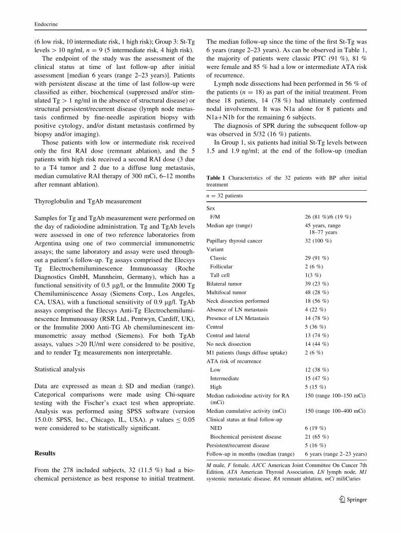
In Group 2 (Fig. 1), 13/17 patients (76.5 %) continued
with only a BP after a median follow-up of 4 years (range
2–10 years). Four patients with intermediate risk of
recurrence from these 17 subjects of Group 2 (23.5 %) had
a structural persistence (lymph nodes metastasis) diagnosed
between 1 and 3.5 years after initial assessment. Neck
lymph node metastases in these four subjects were clini-
cally detected by US/FNAB and were subsequently con-
firmed by lymph node dissection (1–4 affected lymph
nodes from 15 to 43 lymph nodes obtained in the LN
dissection).
All the four patients remained with biochemically per-
sistent disease once the surgical procedures were done,
after a median post re-surgical interventions follow-up of
2 years (range 1.5–5 years).
Considering the outcome of St-Tg levels in the Group 2:
seven patients (41 %) had a mild decrease in the stimu-
lated-Tg levels (from a mean of 7 ± 2 ng/ml to a mean of
3 ± 1.7 ng/ml, p = 0.04), 8 (47 %) had a stable stimulated
Tg levels, and the remaining two patients (12 %) had a
slight increase (from a mean of 4 ± 2 ng/ml to a mean of
5 ± 1.7 ng/ml, p = 0.4) (Table 2).
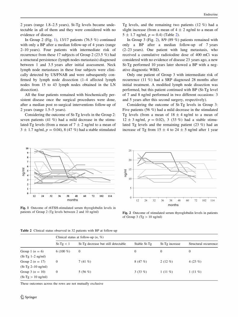
In Group 3 (Fig. 2), 8/9 (89 %) patients remained with
only a BP after a median follow-up of 7 years
(2–23 years). One patient with lung metastasis, who
received a cumulative radioiodine dose of 400 mCi was
considered with no evidence of disease 23 years ago, a new
St-Tg performed 10 years later showed a BP with a neg-
ative diagnostic WBD.
Only one patient of Group 3 with intermediate risk of
recurrence (11 %) had a SRP diagnosed 28 months after
initial treatment. A modified lymph node dissection was
performed, but this patient continued with BP (St-Tg level
of 7 and 8 ng/ml performed in two different occasions: 3
and 5 years after this second surgery, respectively).
Considering the outcome of St-Tg levels in Group 3:
Five patients (56 %) had a mild decrease in the stimulated
Tg levels (from a mean of 18 ± 4 ng/ml to a mean of
12 ± 3 ng/ml, p = 0.02), 3 (33 %) had a stable stimu-
lated Tg levels and the remaining patient (23 %) had an
increase of Tg from 15 ± 4 to 24 ± 5 ng/ml after 1 year
0
1
2
3
4
5
6
7
8
9
10
12 24 32 36 38 48 60 72 102 114
ng/m
l
months
Fig. 1 Outcome of rhTSH-stimulated serum thyroglobulin levels in
patients of Group 2 (Tg levels between 2 and 10 ng/ml)
0
5
10
15
20
25
12 24 32 36 38 48 60 72 102 114
ng/m
l
months
Fig. 2 Outcome of stimulated serum thyroglobulin levels in patients
of Group 3 (Tg [ 10 ng/ml)
Table 2 Clinical status observed in 32 patients with BP at follow-up
Clinical status at follow-up (n, %)
St-Tg \ 1 St-Tg decrease but still detectable Stable St-Tg St-Tg increase Structural recurrence
Group 1 (n = 6)
(St-Tg 1–2 ng/ml)
6 (100 %) 0 0 0 0
Group 2 (n = 17)
(St-Tg 2–10 ng/ml)
0 7 (41 %) 8 (47 %) 2 (12 %) 4 (23 %)
Group 3 (n = 10)
(St-Tg [ 10 ng/ml)
0 5 (56 %) 3 (33 %) 1 (11 %) 1 (11 %)
These outcomes across the rows are not mutually exclusive
Endocrine
123
of follow-up. This was the patient who was diagnosed
with the SPR.
Discussion
Thyroglobulin is a specific tumor marker when total thy-
roidectomy is performed and remnant ablation is indicated.
Most differentiated thyroid cancer cells synthesize Tg,
although there may be differences in the molecular con-
formation of this tumor-derived Tg [14]. The presence of
detectable Tg after total thyroidectomy and remnant abla-
tion may indicate one of the following three situations: the
presence of normal thyroid remnant not completely ablated
after the first radioiodine dose, persistence, or recurrence of
the disease [1, 2]. The likelihood of finding structural
disease is usually associated to the serum Tg level: the
higher the Tg level, the larger the risk [14]. Patients with
rhTSH-Tg levels \2 ng/ml are rarely associated to struc-
tural or progressive disease, while patients with rhTSH-
Tg [2.0 ng/ml are more likely to have structural persistent
disease [7–14].
When Tg is detectable—either on suppression or after
TSH stimulus—and no structural findings are detected on
neck US and/or other imaging methods, patients may be
classified as having BP. It has been shown that 11–19 % of
ATA low risk patients, 21–22 % of ATA intermediate risk
patients, and 16–18 % of ATA high-risk patients had bio-
chemical persistent disease during follow-up [3–6]. Usu-
ally, the ATA high risk patients are less likely to have BP
than the ATA low and intermediate risk groups because the
ATA high risk group has a lot more structural persistent
disease and is less likely to have an excellent response [3–
6]. Also, very few high risk patients achieve a suppressed
Tg \1 ng/ml, and therefore would have been excluded
from this study.
In this retrospective study, we refer specifically to those
patients who had this situation at their first control during
follow-up, in which response to therapy was assessed. We
used strict criteria to define biochemical persistence:
rhTSH-stimulated Tg [ 1 ng/ml, absence of TgAb, and
absence of any evidence of structural disease on neck US
and/or any other imaging study. St-Tg was always per-
formed after rhTSH stimulation, which homogenized the
evaluation of the results. Our definition of BP includes
what in other studies has been defined as acceptable
response to therapy (St-Tg between 1.0 and 10 ng/ml and
non specific or small finding on imaging) and biochemical
incomplete response to therapy (St-Tg [ 10 ng/ml with no
structural disease) [3–6]. The aim of our study was to
evaluate the clinical outcome and rhTSH-Tg trend of these
patients followed with repeated St-Tg, neck US and other
imaging methods.
By using these criteria, all patients who had the first St-
Tg between 1–2 ng/ml had an undetectable St-Tg during
their follow-up (n = 6) and all patients with St-Tg [ 2 ng/
ml persisted with detectable rhTSH-Tg during their follow-
up. However 12 (37 %) had a decrease in mean St-Tg
levels (Table 2).
Several previous studies have also demonstrated that
many patients with a BP have a gradual decline in serum
Tg levels over time without additional therapy [4, 15–24].
As it was previously postulated, it is probable that the
outcome of these patients from this ‘‘incomplete or
acceptable’’ response to therapy to a status of no evidence
of disease could be related to the simply passage of time
[4]. In this cited study, a 100 % 5-year survival and 34 %
of spontaneous resolution of the non-stimulated-Tg values
over time was observed [4].
Therefore, if serum Tg levels can continue to decline for
many years after RRA, then an early assessment of
response to therapy or new St-Tg assessments during fol-
low-up could lead to excessive evaluations and treatments
in patients with low-level Tg values that are likely to
resolve or continue without changes over time without
additional therapies [24]. That is why many authors are
now recommending postponing the moment to define the
response of initial treatment by the end of the second year
[24, 25].
It was also recently reported that the doubling time of
Tg measured under thyroid hormone therapy could be a
potent dynamic factor to predict survival, distant metasta-
sis, and loco-regional recurrence in patients with papillary
thyroid cancer [26]. Even though this investigation was
performed with Tg levels measured under thyrotropin
suppression, the data can probably be applied to the trend
in rhTSH-Tg levels over time [26]. The most important
information extrapolated to our study is that patients with
BP with a still detectable but declining Tg over time have a
very good prognosis in terms on survival and recurrence.
On the other hand, patients with stable or slowly increasing
Tg levels, possibly have a less favorable but still a good
prognosis in terms on survival and recurrence.
We conclude that clinical outcomes in patients with
BP after initial treatment are very good; only 16 % of
patients with BP developed structurally identifiable dis-
ease over the first 4 years of follow-up. Regarding St-Tg
over time, 19 % of patients had undetectable stimulated-
Tg and 65 % continued to have persistently abnormal
stimulated-Tg values without structural correlate. The
detection of overt clinical disease and subsequent surgi-
cal therapeutic intervention in our patients did not
change the outcome, because all of them continued with
biochemical persistence.
Conflict of interest None.
Endocrine
123
References
1. D.S. Cooper, G.M. Doherty, B.R. Haugen, R.T. Kloos, S.L. Lee,
S.J. Mandel, E.L. Mazzaferri, B. McIver, F. Pacini, M. Schlum-
berger, S.I. Sherman, D.L. Steward, R.M. Tuttle, Thyroid 19,
1167 (2009)
2. F. Pitoia, L. Ward, N. Wohllk, C. Friguglietti, E. Tomimori, A.
Gauna, R. Camargo, M. Vaisman, R. Harach, F. Munizaga, S.
Corigliano, E. Pretell, H. Niepomnizcze, Arq. Bras. Endocrinol.
Metabol. 53, 884 (2009)
3. F. Pitoia, F. Bueno, C. Urciuoli, E. Abelleira, G. Cross, R.M.
Tuttle, Thyroid (2013). doi:10.1089/thy.2013.0011
4. F. Vaisman, D. Momesso, D.A. Bulzico, C.H. Pessoa, F. Dias, R.
Corbo, M. Vaisman, R.M. Tuttle, Clin. Endocrinol. (Oxf). 77,
132 (2012)
5. M.G. Castagna, F. Maino, C. Cipri, V. Belardini, A. Theodoro-
poulou, G. Cevenini, F. Pacini, Eur. J. Endocrinol. 165, 441
(2011)
6. R.M. Tuttle, H. Tala, J. Shah, R. Leboeuf, R. Ghossein, M. Go-
nen, M. Brokhin, G. Omry, J.A. Fagin, A. Shaha, Thyroid 20,
1341 (2010)
7. B.R. Haugen, E.C. Ridgway, B.A. McLaughlin, M.T. McDer-
mott, Thyroid 12, 37 (2002)
8. E.L. Mazzaferri, R.T. Kloos, J. Clin. Endocrinol. Metab. 87, 1490
(2002)
9. R.J. Robbins, J.T. Chon, M. Fleisher, S.M. Larson, R.M. Tuttle, J.
Clin. Endocrinol. Metab. 87, 3242 (2002)
10. L. Wartofsky, Thyroid 12, 583 (2002)
11. A. David, A. Blotta, M. Bondanelli, R. Rossi, E. Roti, L.E.
Braverman, L. Busutti, E.C. degli Uberti, J. Nucl. Med. 42, 1470
(2001)
12. F. Pacini, E. Molinaro, F. Lippi, M.G. Castagna, L. Agate, C.
Ceccarelli, D. Taddei, R. Elisei, M. Capezzone, A. Pinchera, J.
Clin. Endocrinol. Metab. 86, 5686 (2001)
13. B.R. Haugen, F. Pacini, C. Reiners, M. Schlumberger, P.W.
Ladenson, S.I. Sherman, D.S. Cooper, K.E. Graham, L.E.
Braverman, M.C. Skarulis, T.F. Davies, L.J. DeGroot, E.L.
Mazzaferri, G.H. Daniels, D.S. Ross, M. Luster, M.H. Samuels,
D.V. Becker, H.R. Maxon 3rd, R.R. Cavalieri, C.A. Spencer, K.
McEllin, B.D. Weintraub, E.C. Ridgway, J. Clin. Endocrinol.
Metab. 84, 3877 (1999)
14. R. Schulz, H. Bethauser, L. Stempka, B. Heilig, A. Moll, M.
Hufner, Eur. J. Clin. Invest. 19, 459 (1989)
15. A.S. Alzahrani, G. Mohamed, A. Al Shammary, S. Aldasouqi, S.
Abdal Salam, M. Shoukri, J. Endocrinol. Invest. 28, 540 (2005)
16. E. Baudin, C. Do Cao, A.F. Cailleux, S. Leboulleux, J.P. Trav-
agli, M. Schlumberger, J. Clin. Endocrinol. Metab. 88, 1107
(2003)
17. J. Biko, C. Reiners, M.C. Kreissl, F.A. Verburg, Y. Demidchik,
V. Drozd, Eur. J. Nucl. Med. Mol. Imaging 38, 123 (2011)
18. E.G. Black, M.C. Sheppard, R. Hoffenberg, Clin. Endocrinol.
(Oxf). 27, 115 (1987)
19. A.L. Gutierrez Cardo, J.R. Rodriguez, I. Borrego Dorado, E.
Navarro Gonzalez, J.L. Tirado, R. Vazquez Albertino, Rev. Esp.
Med. Nucl. 26, 138 (2007)
20. S.H. Huang, P.W. Wang, Y.E. Huang, F.F. Chou, R.T. Liu, S.C.
Tung, J.F. Chen, M.C. Kuo, J.R. Hsieh, H.H. Hsieh, Thyroid 16,
1273 (2006)
21. W.G. Kim, J.S. Ryu, E.Y. Kim, J.H. Lee, J.H. Baek, J.H. Yoon,
S.J. Hong, E.S. Kim, T.Y. Kim, W.B. Kim, Y.K. Shong, J. Clin.
Endocrinol. Metab. 95, 1169 (2011)
22. M. Ozata, S. Suzuki, T. Miyamoto, R.T. Liu, F. Fierro-Renoy,
L.J. De Groot, J. Clin. Endocrinol. Metab. 79, 98 (1994)
23. F. Vaisman, H. Tala, R. Grewal, Thyroid 21, 1317–1322 (2011)
24. R.P. Padovani, E. Robenshtok, M. Brokhin, R.M. Tuttle, Thyroid
22, 778 (2012)
25. H. Tala, R.M. Tuttle, Clin. Oncol. 22, 419 (2010)
26. A. Miyauchi, T. Kudo, A. Miya, K. Kobayashi, Y. Ito, Y. Ta-
kamura, T. Higashiyama, M. Fukushima, M. Kihara, H. Inoue, C.
Tomoda, T. Yabuta, H. Masuoka, Thyroid 21, 707 (2011)
Endocrine
123