biochemical investigation of the bacteriophage protein ... · biochemical investigation of the...
TRANSCRIPT
Biochemical Investigation of the Bacteriophage Protein HK97 gp74
by
Serisha Moodley
A thesis submitted in conformity with the requirements
for the degree of Master of Science
Graduate Department of Chemistry
University of Toronto
© Copyright by Serisha Moodley 2010
ii
Biochemical Investigation of the Bacteriophage Protein HK97 gp74
Serisha Moodley
Master of Science
Graduate Department of Chemistry
University of Toronto
2010
Abstract
Bacteriophages are viruses that infect and propagate within bacteria by making use of
the host’s biosynthetic machinery. With a global population of 1031
, phages pose a
significant influence on microbial populations. Studies of bacteriophage proteins can
elucidate the influence that bacteriophages play on the evolution of bacteria, as well as,
providing the basis for the use of phage proteins as possible therapeutics and bioengineering
solutions.
This study aims to investigate the structural and functional role of the HK97 phage
protein gp74. Sequence alignments indicate that gp74 is related to homing HNH
endonucleases. Homing endonucleases are predominantly double-stranded DNases,
suggesting that gp74 mediates integration of phage genes into the host genome or may target
foreign phage DNA. DNA digestion experiments with gp74 reveals that gp74 mediates non-
specific double-stranded cleavage of lambda phage DNA and single strand cleavage of
plasmid DNA. Our initial work demonstrates that HK97 gp74 is an HNH endonuclease.
iii
Acknowledgements
I owe my deepest gratitude to my family, who without their support this thesis would
not have been possible. For the innumerable trips to and from campus and their saintly
patience when I seemed to experience temporary insanity, I am deeply touched by the love
they have shown me throughout this past year. To my mom and dad, thank you for your
encouragement and for giving me the confidence to continue my studies. To my brother,
thank you for always making me smile and for your continuing support. And to my Bruno
for always being a source of love and silliness.
It has been an honour to study under the supervision of Prof. Voula Kanelis and
whose encouragement, guidance and support from the initial to the final level enabled me to
develop an understanding of biochemical research principles. I would like to thank Dr.
Svetlana Tzvetkova for being a remarkable mentor and friend in my undergraduate research
education. It is also a pleasure to thank my lab colleagues, Naila Ahmed, Lynn Ikeda, Leen
Ghozlani, Elvin DeAraujo and Dennis Guo for their help and friendship.
I am also grateful to our collaborators, Prof. Alan Davidson and Karen Maxwell for
their counsel and support. And for the use of their lab equipment and guidance, I would like
to thank Prof. Scott Prosser, Prof. Peter Macdonald, Prof. Patrick Gunning, Prof. George
Espie and Prof. Jumi Shin.
iv
Table of Contents
1. Introduction
1.1 What are bacteriophages 1
1.2 Bacteriophage Impact on Bacterial Species 8
1.3 Bacteriophage HK97 11
1.4 Homing Endonucleases 15
1.5 Biophysical Tools
1.5.1 NMR Spectroscopy 25
1.5.2 Circular Dichroism Spectroscopy 27
2. Materials & Methods
2.1 Structure Based Sequence Alignment 29
2.2 Expression of HK97 gp74 30
2.3 N15
labeled HK97 gp74 Protein Expression 32
2.4 Imobilized Metal Affinity Chromatography Purification of HK97 gp74 33
2.5 Expression & Purification of TEV Protease 34
2.6 Removal of the 6xHis tag from HK97 gp74 35
2.7 Size Exclusion Chromatography of HK97 gp74 36
2.8 Determination of HK97 gp74 Protein Concentration 36
2.9 Preparation of the NMR Sample 39
2.10 Biophysical Tools
2.10.1 NMR Studies: 15
N-1H correlation spectrum (HSQC) 39
2.10.2 Structural Characterization of HK97 gp74 by Circular Dichroism 40
v
2.11 Substrates for DNA Cleavage
2.11.1 Plasmid DNA 41
2.11.2 Phage DNA 41
2.11.3 Purification of Single Stranded DNA 42
2.12 Tests for Endonuclease Activity
2.12.1 DNA Cleavage Assay 43
2.12.2 Reporter Methods for DNA Cleavage Assay
a) Absorbance Assay 45
b) Agarose Gel Electrophoresis 46
2.13 Reporter Methods for DNA Binding
2.13.1 UV-Vis Metal Binding Assay 47
2.13.2 Metal Binding Experiments by NMR Titration of HK97 gp74 48
2.13.3 Tryptophan Fluorescence Spectroscopy 48
2.14 Mutant Primer and QuikChange Mutagenesis 49
3. Results
3.1 BlastP Search of HK97 gp74 Protein Sequence 52
3.2 Structure Based Sequence Alignment 53
3.3 Vector Map of HK97 gp74-pET-15b 54
3.4 Expression & Purification of 6xHis-HK97 gp74
3.4.1 Ni2+
Affinity Chromatography Purification of 6xHis-HK97 gp74 56
3.4.2 Analysis of TEV Protease Cleavage of 6xHis-HK97 gp74 59
3.5 Size Exclusion Chromatography of HK97 gp74 60
vi
3.6 Analysis of Endonuclease Activity
3.6.1 HK97 gp74-Mediated Digestion of Plasmid DNA 62
3.6.2 HK97 gp74-Mediated Digestion of Phage DNA 68
3.6.3 pH Dependence of HK97 gp74 Activity 71
3.6.4 Metal-Ion Stoichiometry & HK97 gp74 Activity 74
3.7 Structural Characterization of HK97 gp74 by NMR Spectroscopy 77
3.8 Analysis of HK97 gp74 Metal Binding
3.8.1 Metal Binding Titration Experiments using NMR Spectroscopy 80
3.8.2 Metal Binding Studies using Tryptophan Fluorescence 81
4. Discussion and Conclusions
4.1 HK97 gp74 84
4.2 Role of HK97 gp74 HNH Endonucleases 85
4.3 Future Biochemical & Biophysical Studies 87
4.4 Future Applications of HK97 gp74 93
4.5 Conclusions 95
5. References 97
6. Appendix 1
6.1 Sample Calculations of Amino Acid Analysis 101
vii
List of Figures
Figure 1: Schematic drawing of the lytic and lysogenic life cycles of bacteriophages 3
Figure 2: Schematic diagram of Caudovirales bacteriophage morphology 4
Figure 3: Schematic drawing of bacteriophage structure 6
Figure 4: Genome map showing the organization of the bacteriophage λ genome 7
Figure 5: Genome map of bacteriophage HK97 14
Figure 6: Representative diagram of the homing mechanism 16
Figure 7: Structures of homing endonucleases 19
Figure 8: Ribbon diagram showing the structure of the I-HmuI-DNA complex 20
Figure 9: Sequence and ribbon diagram of the colicin E9 HNH motif 22
Figure 10: Diagram of the DNA cleavage mechanism of I-HmuI 24
Figure 11: Structure-based sequence alignment of HK97 gp74 with colicin E9 54
Figure 12: Vector map of pET-15b-HK97 gp74 55
Figure 13: SDS-PAGE gel of 6xHis-HK97 gp74 Ni2+
affinity protein purification 57
Figure 14: SDS-PAGE gel of 6xHis-HK97 gp74 protein elution samples 58
Figure 15: SDS-PAGE gel of 6xHis-HK97 gp74 cleaved with TEV protease 59
Figure 16: Size exclusion chromatography of HK97 gp74 61
Figure 17: SDS-PAGE gel of size exclusion chromatography purified HK97 gp74 62
Figure 18: HK97 gp74 cleaves pUC-18 plasmid DNA 64
Figure 19: SDS-PAGE gel of HK97 gp74 in reducing and non-reducing conditions 65
Figure 20: HK97 gp74 cleaves pBluescript (SK+) plasmid DNA 67
Figure 21: HK97 gp74 cleaves λ phage DNA 69
Figure 22: HK97 gp74 cleavage of λ phage DNA with Zn2+
70
viii
Figure 23: pH Dependence of HK97 gp74 cleavage of λ phage DNA
72
Figure 24: CD spectra of HK97 gp74 74
Figure 25: Stoichiometric assay of HK97 gp74 cleavage of λ phage DNA 75
Figure 26: UV-Vis assay of HK97 gp74 cleavage of λ phage DNA 76
Figure 27: Agarose gel of UV-Vis assay of HK97 gp74 cleavage of λ phage DNA 77
Figure 28: 15
N-1H correlation spectrum of HK97 gp74 79
Figure 29: 15
N-1H correlation spectra of apo-HK97 gp74 and Zn
2+ bound HK97 gp74 81
Figure 30: Tryptophan fluorescence spectra of metal-bound HK97 gp74 83
Figure 31: Agarose gel of pET-15b- H82A-HK97 gp74 PCR reactions 91
ix
List of Tables
Table 1: Summary of the data obtained from amino acid analysis 101
x
List of Appendices
6.1 Sample Calculations of Amino Acid Analysis 101
xi
List of Abbreviations
DNA deoxyribonucleic acid
RNA ribonucleic acid
HK97 Hong Kong 97th
strain
Gp74 gene product 74
ROS reactive oxygen species
BLAST basic local alignment search tool
ORF open reading frame
hnRNA heteronuclear ribonucleic acid
mRNA messenger ribonucleic acid
NMR nuclear magnetic resonance
PDB protein data bank
TEV tobacco etch virus
LB Luria-Bertani
OD optical density
PMSF phenylmethanesulfonylfluoride
IMAC immobilized metal affinity chromatography
BSA bovine serum albumin
DSS 2,2-dimethyl-2-silapentane-5-sulfonic acid
HSQC heteronuclear single quantum coherence
DTT dithiothreitol
HEPES 4-(2-hydroxyethyl)-1-piperazineethanesulfonic acid
TE tris-EDTA
xii
EDTA ethylenediaminetetraacetic acid
CD circular dichroism
dNTP deoxyribonucleotide triphosphate
PCR polymerase chain reaction
MCS multiple cloning site
SDS-PAGE sodium dodecyl sulfate polyacrylamide gel electrophoresis
UV ultraviolet
SEC size exlusion chromatography
ppm parts per million
DMD Duchenne muscular dystrophy
DCA deoxycholic acid
1
1. Introduction
1.1 What are Bacteriophages?
Bacteriophages are true viruses and were first observed in 1915 by the English
microbiologist, F.W. Twort and Canadian microbiologist F. d’Herelle.1 Bacteriophages were
later defined by Adams as autonomous, obligate, intracellular parasites that infect, grow and
multiply within bacteria by making use of some or all of the host’s biosynthetic machinery.1
Bacteriophages are a diverse group of organisms that significantly influence bacterial
ecology.2 Like other organisms, bacteria are susceptible to infection by an assortment of
viruses or virus-like particles.1 Research in the field of microbial viruses was driven by early
interest in the potential use of bacteriophages in treating bacterial diseases. Phage therapy
was later abandoned after the discovery of antibiotics. However, research into
bacteriophages has increased significantly since the 1940s when M. Delbruck and colleagues
demonstrated that bacteriophages reproduce in a “one step” growth mechanism in contrast to
the exponential growth of cellular organisms like bacteria.3 One step growth is described as
a growth pattern where virions within a host undergo a “burst” or sudden growth period, in
which viruses are released, followed by a latent period in which no viruses are reproduced or
released.3 More recently, interest in bacteriophage research has been rekindled due to the
emergence of antibiotic resistant strains of bacteria and the search for alternative means to
treat human diseases.4
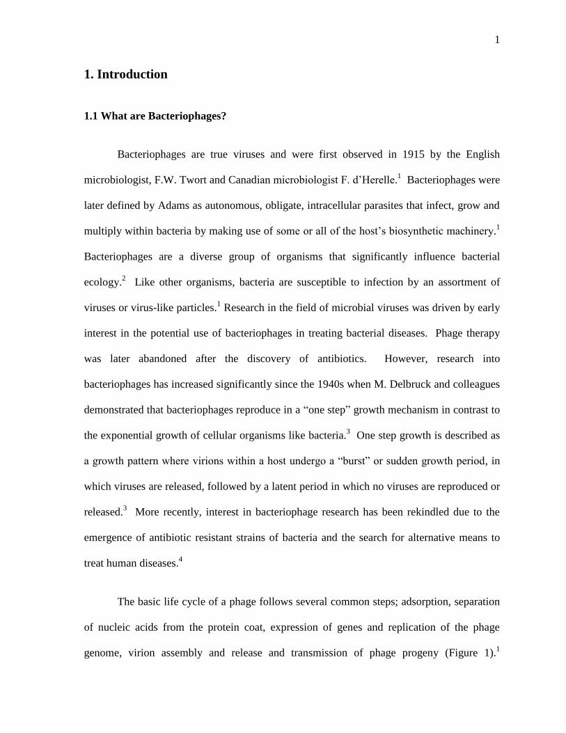
The basic life cycle of a phage follows several common steps; adsorption, separation
of nucleic acids from the protein coat, expression of genes and replication of the phage
genome, virion assembly and release and transmission of phage progeny (Figure 1).1
2
Bacteriophages undergo two different replicative cycles. In the infective or lytic cycle, the
genome or nucleic acid component of the bacteriophage is injected into the host bacterial
cell.5,6
Subsequently, the host cell machinery transcribes and translates phage genes, leading
to the production of whole phage particles. Whole phage particles accumulate within the
host cell and release virus-encoded lytic enzymes, such as lysins and holins, that cause host
bacterial cell lysis.1 In the lysogenic cycle, the injected phage genome is incorporated into
the bacterial cell genome.7,8
The phage genes can remain dormant until induced by a
response or they can be transcribed along with bacterial genes.1 In fact, many genes
identified through sequencing of bacterial genomes were originally phage genes.9 Phage
genes incorporated into the bacterial genome can then be transcribed and translated to
produce phage particles, which are released out of the host cell by lysis. Bacteriophages that
undergo only the lytic life cycle are referred to as virulent, whereas, phages that undergo
both of these life cycles or just the lysogenic life cycle are referred to as temperate.1
3
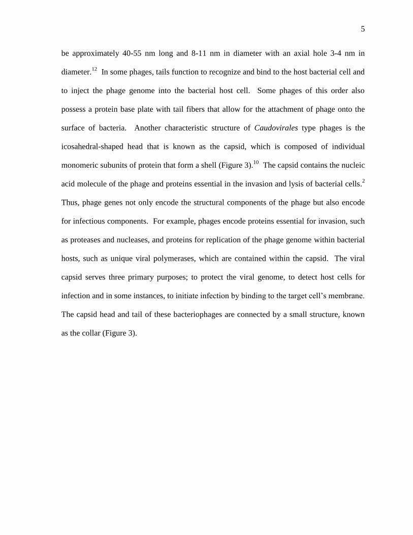
Figure 1: Schematic drawing of the lytic and lysogenic life cycles of bacteriophages.
Lysogenic life cycles allow the integration of bacteriophage genomes into host bacterial
genomes, thus, forming a lysogen or prophage.8 The prophage is replicated along with the
bacterium’s genome during each cell division and remains integrated until a lytic signal
initiates the dissimilation of the of the phage genome.1 The independent phage genome
forms a circular molecule that can be replicated and transcribed.1 The lytic life cycle allows
for the prophage expression, replication and production of phage progeny, which are released
via the lysis of the host bacterium.1
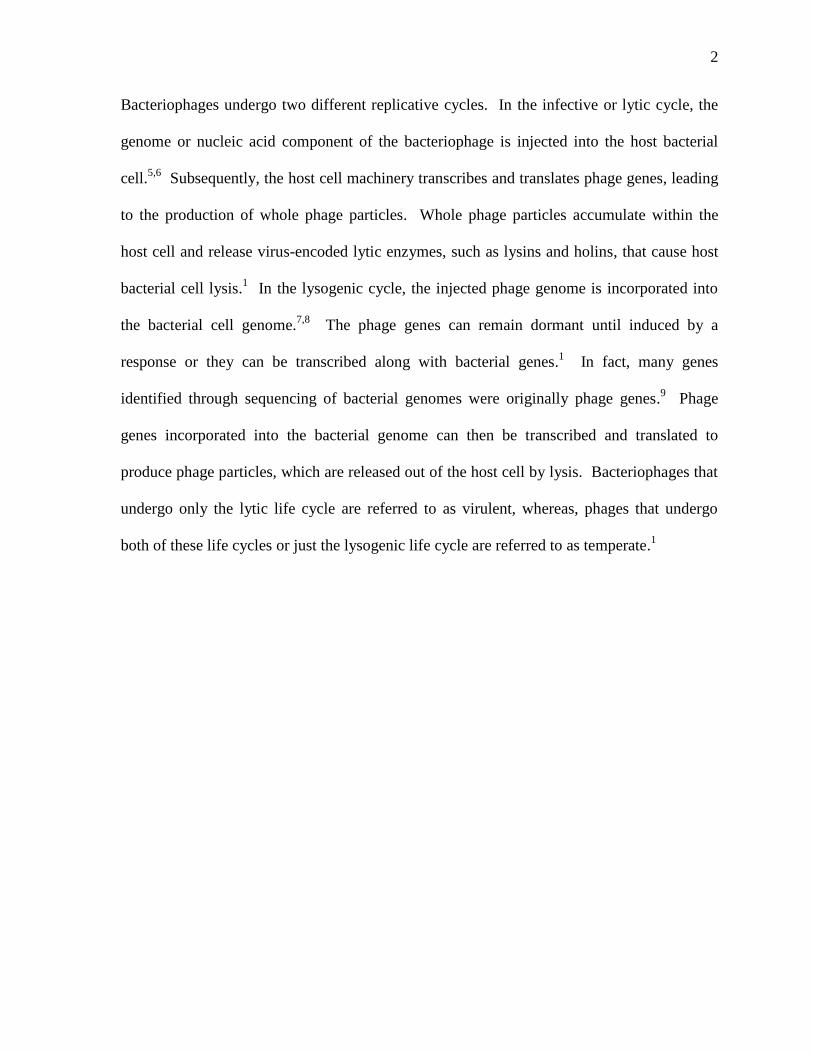
Phages are much smaller in comparison to the bacteria they infect and are usually
between twenty and two hundred nanometers in size.5 The most common bacteriophages
belong to the Caudovirales order and comprise approximately 96 % of the entire population
of bacteriophages.9 Also known as tailed viruses, the Caudovirales order comprises three
families of bacteriophages, including the myoviridae (bacteriophages with contractile tails),
siphoviridae (bacteriophages characterized with long, non-contractile tails) and the
podoviridae (or bacteriophages with short, non-contractile tails) (Figure 2).10
4
Figure 2: Schematic diagram of Caudovirales bacteriophage morphology.10
Approximately
96 % of all bacteriophages belong to the double-stranded DNA (dsDNA) tailed phages,
known as the Caudovirales.9 Caudovirales is an order of viruses that are characterized by
dsDNA genomes and an icosahedral head.2 The Caudovirales order is comprised of three
families of bacteriophages, including the myoviridae, siphoviridae and the podoviridae.10
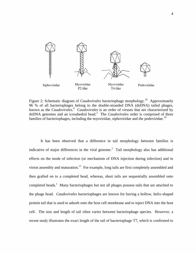
It has been observed that a difference in tail morphology between families is
indicative of major differences in the viral genome.1 Tail morphology also has additional
effects on the mode of infection (or mechanism of DNA injection during infection) and in
virion assembly and maturation.11
For example, long tails are first completely assembled and
then grafted on to a completed head, whereas, short tails are sequentially assembled onto
completed heads.1 Many bacteriophages but not all phages possess tails that are attached to
the phage head. Caudovirales bacteriophages are known for having a hollow, helix-shaped
protein tail that is used to adsorb onto the host cell membrane and to inject DNA into the host
cell. The size and length of tail often varies between bacteriophage species. However, a
recent study illustrates the exact length of the tail of bacteriophage T7, which is confirmed to
5
be approximately 40-55 nm long and 8-11 nm in diameter with an axial hole 3-4 nm in
diameter.12
In some phages, tails function to recognize and bind to the host bacterial cell and
to inject the phage genome into the bacterial host cell. Some phages of this order also
possess a protein base plate with tail fibers that allow for the attachment of phage onto the
surface of bacteria. Another characteristic structure of Caudovirales type phages is the
icosahedral-shaped head that is known as the capsid, which is composed of individual
monomeric subunits of protein that form a shell (Figure 3).10
The capsid contains the nucleic
acid molecule of the phage and proteins essential in the invasion and lysis of bacterial cells.2
Thus, phage genes not only encode the structural components of the phage but also encode
for infectious components. For example, phages encode proteins essential for invasion, such
as proteases and nucleases, and proteins for replication of the phage genome within bacterial
hosts, such as unique viral polymerases, which are contained within the capsid. The viral
capsid serves three primary purposes; to protect the viral genome, to detect host cells for
infection and in some instances, to initiate infection by binding to the target cell’s membrane.
The capsid head and tail of these bacteriophages are connected by a small structure, known
as the collar (Figure 3).
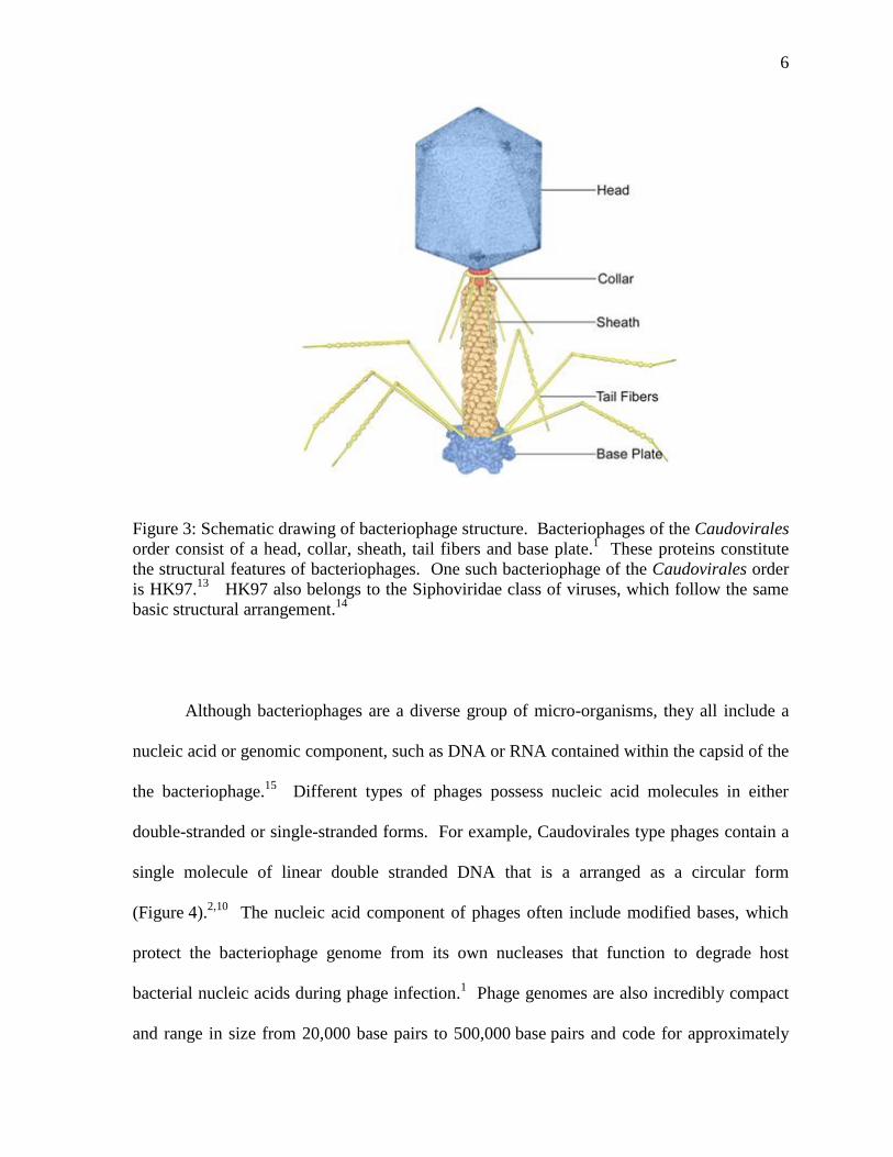
6
Figure 3: Schematic drawing of bacteriophage structure. Bacteriophages of the Caudovirales
order consist of a head, collar, sheath, tail fibers and base plate.1 These proteins constitute
the structural features of bacteriophages. One such bacteriophage of the Caudovirales order
is HK97.13
HK97 also belongs to the Siphoviridae class of viruses, which follow the same
basic structural arrangement.14
Although bacteriophages are a diverse group of micro-organisms, they all include a
nucleic acid or genomic component, such as DNA or RNA contained within the capsid of the
the bacteriophage.15
Different types of phages possess nucleic acid molecules in either
double-stranded or single-stranded forms. For example, Caudovirales type phages contain a
single molecule of linear double stranded DNA that is a arranged as a circular form
(Figure 4).2,10
The nucleic acid component of phages often include modified bases, which
protect the bacteriophage genome from its own nucleases that function to degrade host
bacterial nucleic acids during phage infection.1 Phage genomes are also incredibly compact
and range in size from 20,000 base pairs to 500,000 base pairs and code for approximately
7
3-5 average-sized gene products (approximately 150-500 amino acid residues in length) in
simple phages and approximately 100 gene products in more complex phages.5,16
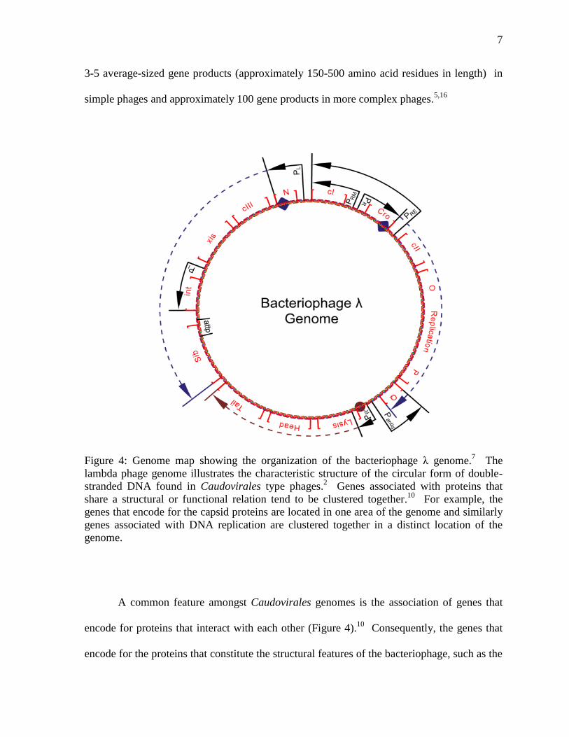
Figure 4: Genome map showing the organization of the bacteriophage λ genome.7 The
lambda phage genome illustrates the characteristic structure of the circular form of double-
stranded DNA found in Caudovirales type phages.2 Genes associated with proteins that
share a structural or functional relation tend to be clustered together.10
For example, the
genes that encode for the capsid proteins are located in one area of the genome and similarly
genes associated with DNA replication are clustered together in a distinct location of the
genome.
A common feature amongst Caudovirales genomes is the association of genes that
encode for proteins that interact with each other (Figure 4).10
Consequently, the genes that
encode for the proteins that constitute the structural features of the bacteriophage, such as the
8
capsid, neck and tail or proteins that interact with each other to accomplish specific cellular
functions, such as DNA replication, tend to be clustered together. For example, the
capsomere protein, which comprises the identical morphological subunits of the protein coat
or capsid is located in the same region of the genome as the scaffolding protein (required for
DNA packaging) and the portal protein (which forms a hole that allows the passage of DNA
during packaging and forms the junction between the phage head and tail protein to allow for
DNA ejection).14
In some instances, an entire set of structural genes are grouped and
transcribed together from a single promoter.
1.2 Bacteriophage Impact on Bacterial Species
As the most abundant and most rapidly reproducing biologically active organism on
Earth, with a global population of 1031
, bacteriophages pose a significant influence on
microbial populations.17
These small organisms impact the species distribution, nutrient
cycling, food network and population density of bacteria. Phage ecology is the study of the
interaction of bacteriophages with other organisms and the environment. Phage community
ecology has allowed for the observation and characterization of the interactions between
phages and bacteria to determine predator-prey interactions and to understand the co-
evolution of bacteriophages and bacteria.18
Some of the more typical relationships between
bacteria and phages include mutualism (where bacteria harbor phages via lysogenic
conversion and these phages may in turn kill related bacterial competitors) or parasitization
(or predation of particular species) of bacteria.
9
Bacteriophages are highly diverse and have been found in all ecosystems colonized
by bacterial populations, such as aquatic, soil, and gastrointestinal locales.18
Phage
population density generally increases with the productivity of the ecosystem. For example,
in marine environments, phage population is highest in coastal areas, due to the complexity
and abundance of the natural flora and fauna, and lowest in the deep sea.19
Bacteriophage
abundance is also higher in fresh water systems as compared to marine systems, and in
shallow depths, as in lake ecosystems, as compared to deeper environments. However, this
is also dependent on oxygen availability as lower depths are more likely to have anoxic
conditions. Bacteriophages tolerate a vast range of environmental conditions and resources,
including temperature, hydrostatic pressure, radiation, oxygen, pH and host availability. For
example, temperature is an important environmental factor for phage survival and most
phages have been found to function at temperatures between 15 °C and 42 °C.18
Bacteriophages also affect the microenvironment of bacteria, where phages use their hosts as
a source of energy and matter. Phages seize control of bacterial biosynthetic processes in
order to synthesize viral macromolecules that are used for the formation of viral particles.
Thus, bacterial hosts serve as the bioreactor or factory for the production of phages.
Many phages develop a symbiotic interaction with their bacterial hosts via lysogeny
that involves the integration of the phage genome into the host’s replicon.20
These phages
can exist as prophages, which are a form of the phage genome that can be inserted and
replicated with the host bacterial genome. These silent infections can then be inherited by
daughter cells and induced by an environmental or stress response to activate biosynthesis of
phage particles and cause host lysis.19
Bacterial genomes have been shown to consist of
3 % - 10 % of prophage-encoded genes and often carry on average three prophages.7,9,20
In
10
Baltic Sea bacterial isolates, the overall frequency of prophage particles was 28 % within
bacterial genomes.18
It has been suggested that these prophage inserted genes are the major
contributors to genomic diversity among bacterial species. In fact many sequenced genes
from bacterial strains originally thought to be bacterial are continually being identified as
phage genes. For example, the bacterial strain, Escherichia coli 0157:H7 has a genome
containing 18 prophage elements, which accounts for approximately 16 % of its total
genome.21
The prophage element contains a gene for the production of shiga toxin, which
inhibits protein synthesis within target cells by N-glycosidase activity that cleaves ribosomal
RNA.22
Thus, inclusion of the prophage element bestows an extremely pathogenic effect on
the bacteria. It has also been proposed that the presence of prophage genes may confer
defensive advantages to bacteria by protecting against infection by other phages and
increasing the pathogenicity or virulence of the host organism through the use of toxins, such
as pyocins, colicins and anaredoxins.23,24
Pyocins are similar to bacteriocins, which are
proteinaceous toxins that are produced by bacteria to inhibit the growth of related bacterial
strains, and resemble bacteriophage tail like structures.25
Colicins are bacteriocins and act as
nucleases to degrade DNA and RNA or cause the formation of pores in the cell membrane of
other bacteria, thereby lysing the cell.26
Anaredoxins function as oxidoreductases to form
reactive oxygen species (ROS) that are highly cytotoxic to bacteria.27
Prophage genes also
supply specific fitness factors, such as proteins that allow for the uptake and use of different
nutrients or nutrient biosynthetic pathways that increase the host’s selective advantage in a
particular system.20
Some prophage genes are able to promote host fitness through the use of
five different mechanisms. Prophages can function as transposons and lead to reorganization
or replication of genes.28
Prophages can interrupt genes causing silencing of non-essential
11
gene functions or can offer immunity to related phage infections or destroy related phages.1
Prophages also offer bactericidal factors and can cause lysis of related nearby strains of
bacteria.1 Lastly, prophage genes can introduce new fitness factors by conversion or
transduction.1 Thus, bacteriophages are not just dangerous molecular machines that cause
bacterial cell mortality but also function as key components to bacterial survival.
Understanding the molecular details by which phages control bacterial survival may help
elucidate novel therapeutics against bacterial infections.
1.3 Bacteriophage HK97
The bacteriophage HK97 was originally isolated in Hong Kong and was the ninety-
seventh strain characterized amongst a series of related viruses, hence the HK97
nomenclature.13,14
HK97 belongs to the Siphoviridae class of Caudovirales bacteriophages
(Figure 2) and is a temperate bacteriophage of Escherichia coli bacteria, meaning that it has
the ability to display both a lytic and a lysogenic life cycle after infection of E. coli cells. A
lysogenic life cycle allows bacteriophage HK97 to integrate its genome into the host E. coli
bacterium’s genome, thus, becoming a lysogen.8,9,10
The lytic life cycle permits prophage
expression, replication and production of phage progeny, which are released via the lysis of
the host bacterium (Figure 1).7,8
HK97 is also a lambdoid phage, meaning that this bacteriophage belongs to a group
of closely related phages that are similar in virion morphology and function to that of lambda
phage. Lambdoid phages also exclusively infect Escherichia coli and are thus, often referred
12
to as coliphages.7 Like other phages in its family, HK97 is composed of a capsid, which
forms an icosahedral, concatenated chain mail-like structure using repeating units of the
protein gp5, and an adaptor that connects the head to a long, non-contractile tail that
functions to adsorb onto E. coli cell membranes and as the channel through which DNA is
ejected from the head into the host bacterium.13
HK97 has a genome of 39.7 kB and a total of 61 protein coding genes.14
Like other
bacteriophages, the HK97 genome appears to cluster genes that are related by structure or
function. HK97 is a relatively new phage species, and as such some of the proteins have yet
to be determined. However, it is hypothesized that HK97 shares many of the same genes as
the lambdoid phages. Starting at gene 1 of the HK97 genome, the first set of known genes
(from gene 1 to 28) encode for the structural proteins that compose the head, tail and adaptor
components of the phage (Figure 5). In the lambdoid phages, genes A-F code for phage head
genes and genes J-Z code for phage tail genes.14
Genes encoding the proteins integrase,
excisionase, and recombinase are located following the structural genes. Integrase,
excisionase and recombinase are involved in lysogeny, which requires the integration of
phage DNA into the host genome and excision, as well as reconstruction of phage genomes
from the recombinant DNA of host genomes after induction of the lytic cycle.2,14
The next
sets of genes involve the transcription of phage genes. For example, the transcription
activator protein is encoded by cII, which is located near the gene cIII, which encodes a
binding protein that protects the transcription activator protein.29
Located to the left of the
transcription activator are the transcription inhibitor genes, cI and cro.14
Next, the genes O
and P are known to be involved in DNA replication followed by the genes that have been
identified as lytic cycle repressor proteins.14,29
The subsequent genes are involved in DNA
13
repair mechanisms. For example, the lambdoid phage RusA homolog also encodes for a
resolvase that ligates nucleic acid fragments at Holliday junctions, which are formed by
genetic recombination.30
The last genes of the HK97 genome are involved in lysis of host
bacterial cells and it is known that the S gene encodes a holin protein that creates pores
through which the R endolysin protein, which degrades the bacterial peptidoglycan wall, is
released.31
The very last gene of the HK97 genome encodes the protein gp74. However, the
functions of various genes of the HK97 bacterophage, including the gene gp74, have yet to
be determined.
14
Figure 5: Genome map of bacteriophage HK97. Genes are represented by red outlined
arrows. The gene size and location are represented by the length and position that the arrow
spans and is indicated in kilobase pairs on a ruler below the gene. Known genes are
identified as their gene number and letter designation and are named after homologous
lambda phage genes. Unknown genes are only identified by their order number in the
genome and their putative function or homology is identified in blue type above the genes.
The profound impact of bacteriophages on the ecology and evolution of bacterial
species has resulted in incredible interest in the field of phage biology.20
This study aims to
15
investigate the structural and possible functional role of the protein gp74 of the
bacteriophage HK97. At the beginning of this project there was no known function of the
bacteriophage protein HK97 gp74 and as of today there has yet to be a clear identity of the
role that this protein plays in the life cycle of HK97 or in the infection of host E. coli
bacterial cells. This thesis describes our study into the biochemical function of HK97 gp74.
1.4 Homing Endonucleases
Early in the project we conducted a search of proteins with similar sequences to
HK97 gp74 using the basic local alignment search tool (BLAST).32
The BLAST search
indicated that HK97 gp74 is a possible homing HNH endonuclease. Homing is a transfer
mechanism by which mobile genetic carriers or intervening sequences are integrated into
recipient homologous alleles that lack this gene sequence.28,33,34
Homing processes were first
described for group I introns of the budding yeast Saccharomyces cerevisiae, which was
found to contain a genetic marker that encoded for an endonuclease in an open reading frame
in a 1.1 kB intron.28
Homing endonucleases initiate transfer of intron and intein elements by
generating double-stranded breaks in alleles that are homologous to the endonuclease gene
and lack the intron or intein element containing the homing endonuclease gene (Figure 6).35
Homologous recombination, as a result of cellular repair mechanisms, at the double-stranded
DNA break leads to the transfer of the homing endonuclease-containing intron or intein,
thereby causing the proliferation of the homing endonuclease gene within the genome.
Consequently, homing endonuclease genes are inherited in a non-Mendelian manner and
tend to be the dominant allele when incorporated into the genome, resulting in efficient
16
proliferation of the gene and gene product in prokaryotic and viral genomes. The integration
of transferred intervening sequences containing the homing endonuclease gene has been
observed between DNA regions and across biological kingdoms.28
Homing endonucleases
found in phage genomes display a greater diversity in homing mechanisms. For example, it
has been observed that some endonucleases of phage origin generate single strand nicks in
comparison to double-stranded digestion.28
Figure 6: Representative diagram of the homing mechanism. Homing is the mechanism by
which introns or inteins are integrated into recipient alleles that lack this gene sequence.28
Homing endonucleases cause double-stranded breaks in alleles that are homologous to the
endonuclease gene and lack the intron or intein element containing the homing endonuclease
gene.33
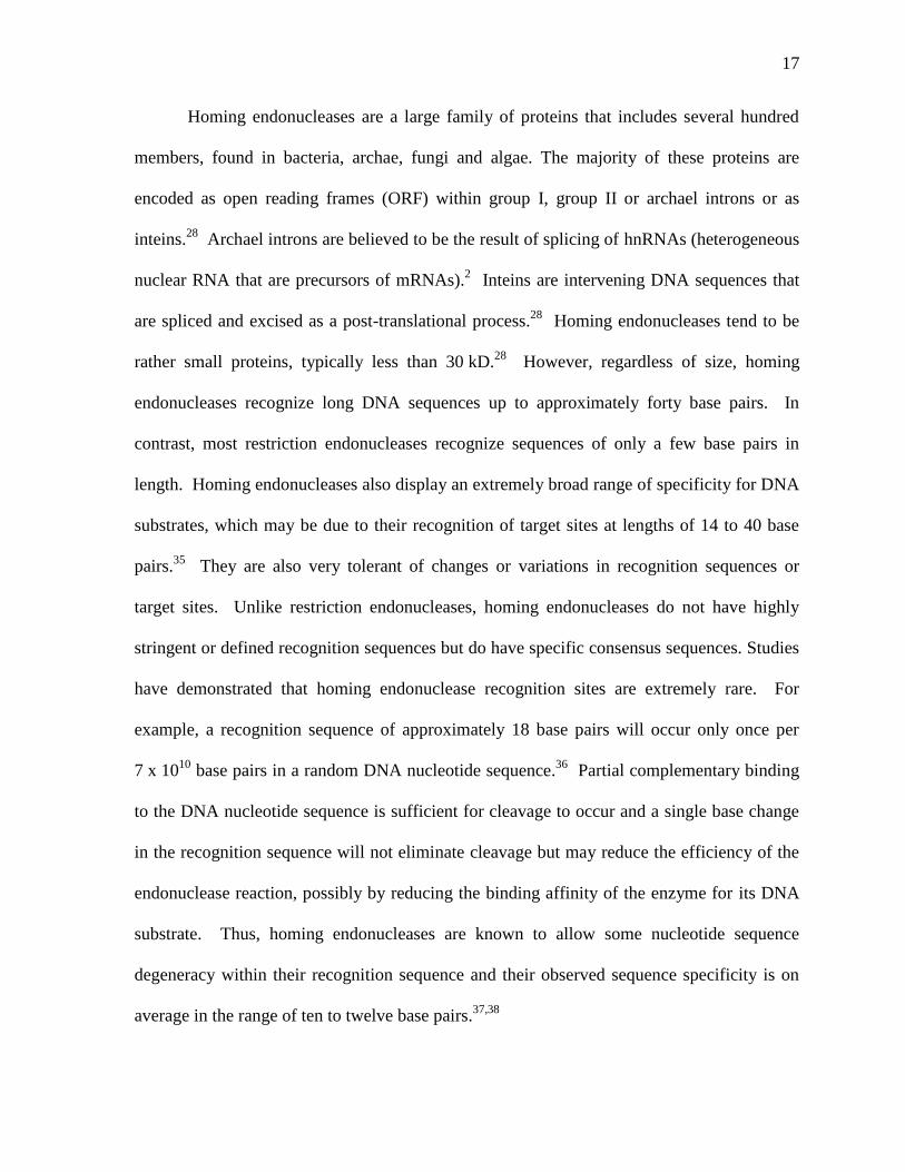
Homologous recombination at the double-stranded DNA break leads to the transfer
of the homing endonuclease-containing intron or intein, thereby causing the proliferation of
the homing endonuclease gene within the genome.28,33,34
17
Homing endonucleases are a large family of proteins that includes several hundred
members, found in bacteria, archae, fungi and algae. The majority of these proteins are
encoded as open reading frames (ORF) within group I, group II or archael introns or as
inteins.28
Archael introns are believed to be the result of splicing of hnRNAs (heterogeneous
nuclear RNA that are precursors of mRNAs).2 Inteins are intervening DNA sequences that
are spliced and excised as a post-translational process.28
Homing endonucleases tend to be
rather small proteins, typically less than 30 kD.28
However, regardless of size, homing
endonucleases recognize long DNA sequences up to approximately forty base pairs. In
contrast, most restriction endonucleases recognize sequences of only a few base pairs in
length. Homing endonucleases also display an extremely broad range of specificity for DNA
substrates, which may be due to their recognition of target sites at lengths of 14 to 40 base
pairs.35
They are also very tolerant of changes or variations in recognition sequences or
target sites. Unlike restriction endonucleases, homing endonucleases do not have highly
stringent or defined recognition sequences but do have specific consensus sequences. Studies
have demonstrated that homing endonuclease recognition sites are extremely rare. For
example, a recognition sequence of approximately 18 base pairs will occur only once per
7 x 10¹⁰
base pairs in a random DNA nucleotide sequence.36
Partial complementary binding
to the DNA nucleotide sequence is sufficient for cleavage to occur and a single base change
in the recognition sequence will not eliminate cleavage but may reduce the efficiency of the
endonuclease reaction, possibly by reducing the binding affinity of the enzyme for its DNA
substrate. Thus, homing endonucleases are known to allow some nucleotide sequence
degeneracy within their recognition sequence and their observed sequence specificity is on
average in the range of ten to twelve base pairs.37,38
18
There are four main families of homing endonucleases, each of which are named for
the conserved residues associated with their nuclease domain, including the LAGLIDADG,
the His-Cys box, HNH enzymes and GIY-YIG family of enzymes (Figure 7).28
Sequence
data indicates that HK97 gp74 belongs to the HNH class of homing endonucleases, which
are named for two conserved histidine residues and an invariant asparagine residue. In some
HNH endonucleases, such as the bacterial colicins, the first histidine and the asparagine
residue are thought to be involved in DNA cleavage while the second hisitidine is involved
in divalent metal ion binding.28
19
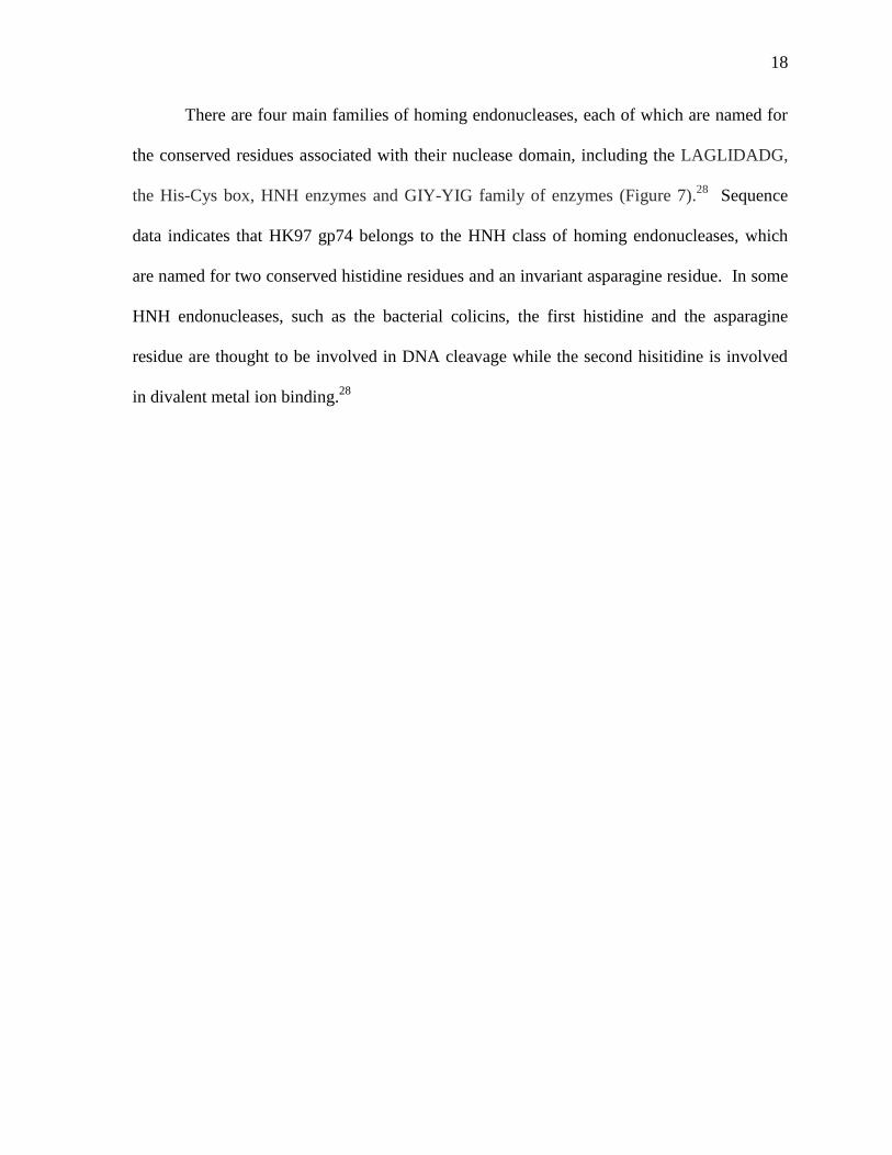
Figure 7: Structures of homing endonucleases.28
There are four families that comprise the
homing endonucleases, which include the LAGLIDADG, the His-Cys box, HNH enzymes
and GIY-YIG family of enzymes.28
This diagram shows the representative structures of
enzymes that belong to the four families of the homing endonucleases bound to DNA. The
enzymes shown in the diagram are I-CreI (a LAGLIDADG enzyme), I-PpoI (a His-Cys box
enzyme), I-HmuI (a HNH enzyme) and I-TevI (a GIY-YIG enzyme).28
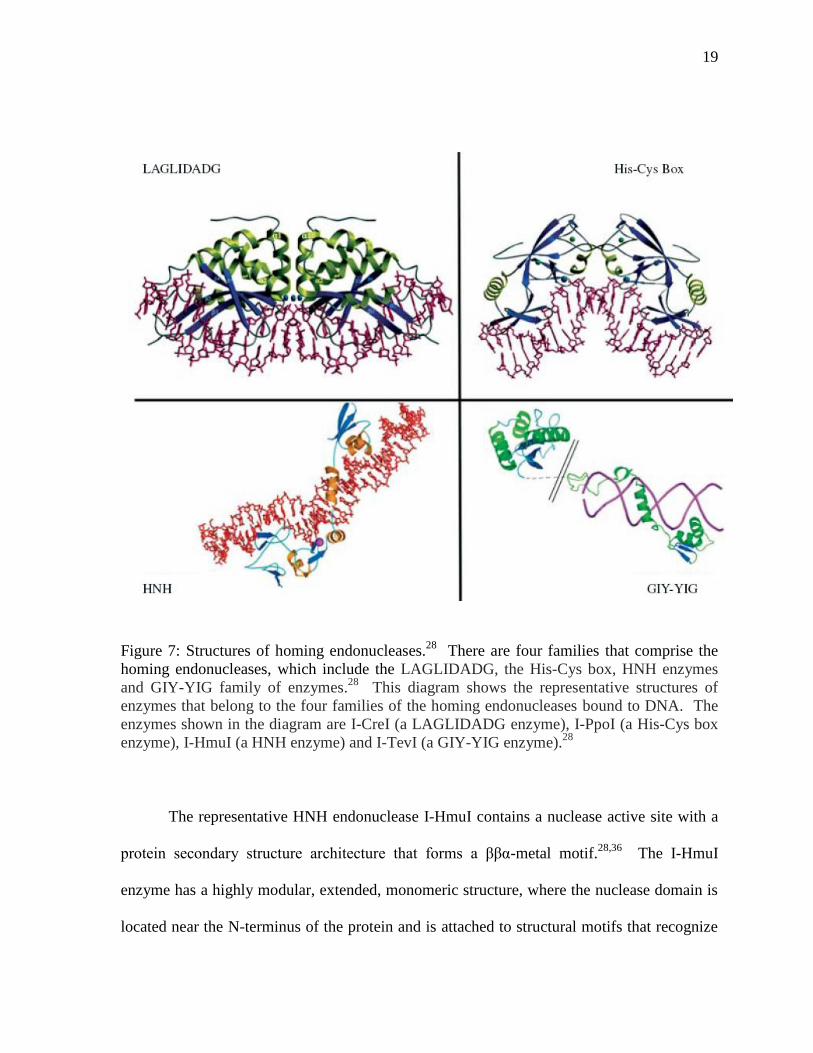
The representative HNH endonuclease I-HmuI contains a nuclease active site with a
protein secondary structure architecture that forms a ββα-metal motif.28,36
The I-HmuI
enzyme has a highly modular, extended, monomeric structure, where the nuclease domain is
located near the N-terminus of the protein and is attached to structural motifs that recognize
20
DNA substrates for binding (Figure 8).28
By structural analysis of I-HmuI, it is believed that
the HNH motif or ββα-metal active site binds to and spans approximately 25 base pairs of the
minor groove of DNA and contacts the DNA phosphate and 3’ hydroxyl group of the scissile
phosphate.36
Furthermore, I-HmuI exists as a monomer and binds only a single metal ion,
which is coordinated by a conserved asparagine from the HNH motif and an additional
aspartate residue. These residues, Asn-96 and Asp-74 and a non-bridging oxygen from the
scissile phosphate group of the DNA bind the divalent metal manganese at the I-HmuI HNH
endonuclease active site.28
Figure 8: Ribbon diagram showing the structure of the I-HmuI-DNA complex.28
The
enzyme I-HmuI (green) has a highly modular, extended, monomeric structure.36
The HNH
motif spans approximately 25 base pairs of the minor groove of DNA (blue) and contacts the
DNA phosphate and 3’ hydroxyl group of the scissile phosphate via a divalent metal
coordination centre (red).36
21
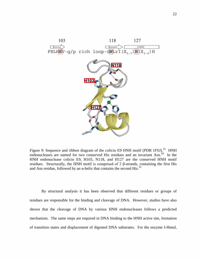
The HNH endonuclease motif of the non-specific bacterial colicins has also been
shown to be highly similar to the structure of I-HmuI. The HNH motif is comprised of 2 β-
strands, containing the first histidine and asparagine residue, followed by an α-helix that
contains the second histidine (Figure 9). In the colicin HNH motif, the first histidine and
aparagine are involved in catalysis, and the second histidine is involved in divalent metal ion
binding. In colicin E9, the first conserved His-103 interacts with the minor groove of the
DNA and makes contact with the DNA backbone during catalysis.28
Investigation of colicins
have revealed that binding and cleavage of DNA at these HNH domains involve a single,
bound divalent cation, such as zinc or magnesium, to coordinate the phosphate and 3’ leaving
group of DNA, which are susceptible to cleavage. The residues His-102 and His-127, which
is a conserved HNH motif residue, of colicin E9 bind the divalent metal at the HNH
endonuclease active site.35
While it has been observed that the position and stabilization of the complexed
divalent metal ion at the HNH motif of several related endonucleases is similar, the chemical
identities of the metal ions, coordination and interaction at the active site of the divalent
metal ions with their metal binding residues differ significantly between HNH
endonucleases. The chemical identity of the divalent metal ion that binds to the HNH active
site varies considerably and the range of metals include magnesium, zinc, cobalt, manganese,
nickel, strontium and calcium.39
Coordination of the metal ions also varies between HNH
endonucleases and studies have shown that the metal ion can be coordinated by one
asparagine and one aspartic acid residue, like the I-HmuI enzyme (Figure 8) or two histidine
residues, as in the bacterial E9 colicin (Figure 9).28
The differences observed in metal
binding may correlate with differences in metal specificity.
22
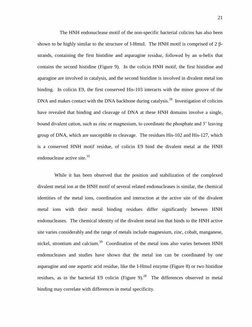
Figure 9: Sequence and ribbon diagram of the colicin E9 HNH motif (PDB 1FSJ).35
HNH
endonucleases are named for two conserved His residues and an invariant Asn.28
In the
HNH endonuclease colicin E9, H103, N118, and H127 are the conserved HNH motif
residues. Structurally, the HNH motif is comprised of 2 β-strands, containing the first His
and Asn residue, followed by an α-helix that contains the second His.35
By structural analysis it has been observed that different residues or groups of
residues are responsible for the binding and cleavage of DNA. However, studies have also
shown that the cleavage of DNA by various HNH endonucleases follows a predicted
mechanism. The same steps are required in DNA binding to the HNH active site, formation
of transition states and displacement of digested DNA substrates. For the enzyme I-HmuI,
23
the N-terminus of a β-strand of the HNH motif binds the major groove of the DNA substrate
and an α-helix binds the minor groove and lastly, the C-terminal of the HNH motif of I-HmuI
binds the opposite end of the DNA at its major groove (Figure 8).28,36
The enzyme appears
to straddle the phosphate backbone of the DNA substrate twice, once at the cleavage site and
again at the 3’ end of the target site.
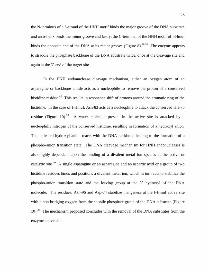
In the HNH endonuclease cleavage mechanism, either an oxygen atom of an
asparagine or backbone amide acts as a nucleophile to remove the proton of a conserved
histidine residue.40
This results in resonance shift of protons around the aromatic ring of the
histidine. In the case of I-HmuI, Asn-83 acts as a nucleophile to attack the conserved His-75
residue (Figure 10).28
A water molecule present in the active site is attacked by a
nucleophilic nitrogen of the conserved histidine, resulting in formation of a hydroxyl anion.
The activated hydroxyl anion reacts with the DNA backbone leading to the formation of a
phospho-anion transition state. The DNA cleavage mechanism for HNH endonucleases is
also highly dependent upon the binding of a divalent metal ion species at the active or
catalytic site.40
A single asparagine or an asparagine and an aspartic acid or a group of two
histidine residues binds and positions a divalent metal ion, which in turn acts to stabilize the
phospho-anion transition state and the leaving group at the 3’ hydroxyl of the DNA
molecule. The residues, Asn-96 and Asp-74 stabilize manganese at the I-HmuI active site
with a non-bridging oxygen from the scissile phosphate group of the DNA substrate (Figure
10).36
The mechanism proposed concludes with the removal of the DNA substrates from the
enzyme active site.
24
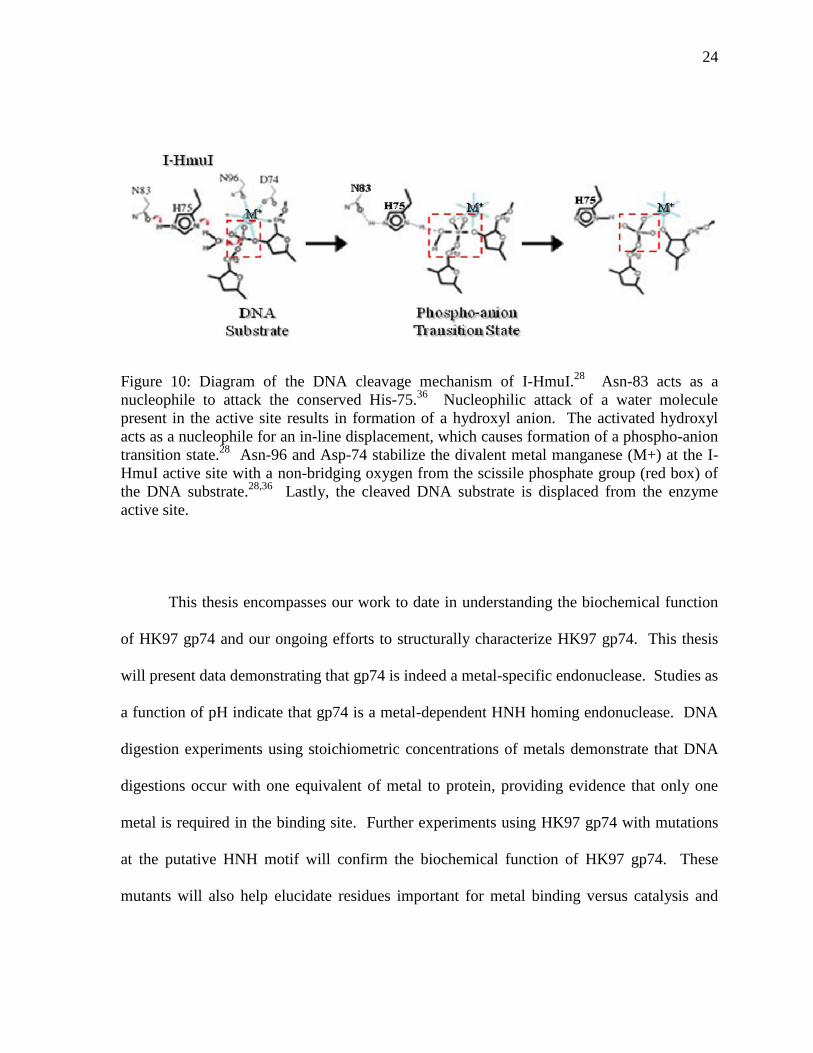
Figure 10: Diagram of the DNA cleavage mechanism of I-HmuI.28
Asn-83 acts as a
nucleophile to attack the conserved His-75.36
Nucleophilic attack of a water molecule
present in the active site results in formation of a hydroxyl anion. The activated hydroxyl
acts as a nucleophile for an in-line displacement, which causes formation of a phospho-anion
transition state.28
Asn-96 and Asp-74 stabilize the divalent metal manganese (M+) at the I-
HmuI active site with a non-bridging oxygen from the scissile phosphate group (red box) of
the DNA substrate.28,36
Lastly, the cleaved DNA substrate is displaced from the enzyme
active site.
This thesis encompasses our work to date in understanding the biochemical function
of HK97 gp74 and our ongoing efforts to structurally characterize HK97 gp74. This thesis
will present data demonstrating that gp74 is indeed a metal-specific endonuclease. Studies as
a function of pH indicate that gp74 is a metal-dependent HNH homing endonuclease. DNA
digestion experiments using stoichiometric concentrations of metals demonstrate that DNA
digestions occur with one equivalent of metal to protein, providing evidence that only one
metal is required in the binding site. Further experiments using HK97 gp74 with mutations
at the putative HNH motif will confirm the biochemical function of HK97 gp74. These
mutants will also help elucidate residues important for metal binding versus catalysis and
25
may provide a mechanism to study a ternary complex (of protein-metal-DNA) by NMR
spectroscopy.
1.5 Biophysical Tools
1.5.1 NMR Spectroscopy
Nuclear magnetic resonance (NMR) spectroscopy is an analytical technique that
exploits the behavior of magnetically active atomic nuclei to provide information about the
structure, kinetic characteristics, and interactions of molecules.41
NMR spectroscopy has
been used to study the structure, dynamics and interactions of proteins in solution at atom-
specific resolution.42
NMR resonances have four basic properties: intensity, resonance
frequency (chemical shift), splitting and line width.43
The intensity or peak height is
measured as a volume and is proportional to the concentrations of nuclei.43
Thus, a less
concentrated sample will give a weak signal and increasing the concentration of a sample
increases the signal strength. The height of each peak in a frequency domain spectrum is
also dependent on the molecular weight and dynamics of a biological compound.41
The
chemical shift is the resonant frequency of a nucleus, such as 1H,
15N,
13C and
31P, which are
biologically relevant isotopes that occur in DNA, proteins and lipids.41
The chemical shift is
a relative scale measured in parts per million (ppm) that compares all signals in a spectrum to
the signal from a calibration or reference compound. Several factors affect chemical shift
and include the local electronic environment, the electronegativity of attached groups and the
spatial proximity of compounds. Splitting is called spin-spin coupling, which is described by
26
the coupling constant J (measured in Hz) and occurs as a close group of two or more
resonances that correspond to a single nucleus.43
Splitting is due to the interaction of spins
through chemical bonds and is caused by the induction of magnetic fields from the
interaction of the spins of nuclei and bonding electrons.43
In an HSQC experiment, coupling
of 1H and
15N is eliminated during the experiment. The line width (at half height) is related
to the T2 relaxation time.41
T2 relaxation time is dependent on molecular weight and on the
motions of a molecule within an applied field.41
For example, a small molecule that tumbles
quickly has a long T2 and gives a narrow, sharp peak, whereas, larger molecules tumble
slowly and have short T2 relaxation times that give broad peaks.
One of the more common types of NMR experiments is the 15
N-1H heteronuclear
single quantum coherence (HSQC) correlation experiment. A HSQC experiment correlates
the resonant frequency of the amide proton and the resonant frequency of the directly
attached amide nitrogen.41
HSQC experiments give information about the conformation of a
protein. Proteins in an unfolded conformation are comprised of residues that are at a distance
from each other and are exposed to the solvent. These residues experience the same
chemical and electronic environment. In a 15
N-1H HSQC, this results in resonances with
very similar 1H chemical shifts, where the
15N chemical shift is more dependent on amino
acid type). The HSQC can also give insights into the dynamic behavior of molecules.
Differential peak heights, as seen for HK97 gp74 indicate differential dynamics in different
regions of the protein (see section 3.7). A future goal for this project is to elucidate the NMR
solution structure of HK97 gp74, and also use NMR titration experiments to obtain
information on metal binding
27
1.5.2 Circular Dichroism Spectroscopy
Circular dichroism is an analytical and spectroscopic technique that rapidly
determines the secondary structure, folding characteristics and binding properties of proteins.
CD arises from the differential absorption of left-handed and right-handed circularly
polarized light by chiral molecules.44
As biological molecules, DNA and protein are ideal
candidates for CD spectroscopy because they exhibit both dextrorotary and levorotary
components and are optically active. A far-UV CD spectrum can illustrate important
secondary structure characteristics, such as the extent of α-helix, β-sheet, β-turn or random
coil conformation.45
A near-UV CD spectrum, resulting from the absorption of aromatic
residues (phenylalanines, tyrosines and tryptophans) and cysteine disulfide bridges, provides
information about the tertiary structure of proteins. CD is an efficient analytical tool that
requires smaller concentrations of protein, unlike NMR and X-ray crystallography, and can
be used in conjunction with a large range of solvent conditions, varying temperature, pH and
various cofactors and ligands.
Future applications of the techniques mentioned and the resultant data may lead to the
development of phage-based approaches to combat bacterial diseases. Understanding the
function of phage proteins, such as HK97 gp74, will also help elucidate the molecular basis
of the variability in bacterial populations, which is critical for fighting bacterial infections
and diseases. Moreover, better understanding of HK97 gp74 can enable its use as a possible
homologous recombination system to allow for the integration of genes that, for example,
recover deletion mutations that are involved in the development of diseases, such as
28
Duchenne muscular dystrophy. Homing endonucleases, thus, offer a new perspective on
gene therapy for many diseases.
29
2. Materials and Methods
2.1 Structure Based Sequence Alignment
To gain insights into the function of HK97 gp74, the protein sequence of HK97 gp74
was subjected to the basic local alignment search tool (BLAST).32
The BLAST program
utilizes a sequence comparison algorithm that is optimized for speed to search different
databases for the optimal sequence alignments to a specific search query, such as a protein or
DNA sequence.32
The full amino acid sequence of HK97 gp74 was used in the search. The
BLAST program analyses the resultant output data for the best possible matches to the query
and ranks related protein matches based on two variables, the bit score and E-value. A list of
highly similar sequences are compiled by the program, along with a diagram indicating the
most closely related family of proteins as determined by the conserved amino acid residues
and sequences present in the protein compared to similar proteins. One of the top protein
BLAST matches to HK97 gp74 is colicin E9.
Based on our BLAST search results, where one of the top search hits was the HNH
endonuclease colicin E9, a structure-based sequence alignment of HK97 gp74 and colicin E9
was made. A structure-based sequence alignment of HK97 gp74 and colicin E9 was
performed using ClustalW. ClustalW is a multiple sequence alignment program that is used
to compare the relatedness or conservation between protein sequences.46
A structure-based
sequence alignment uses the structural information of a protein to construct a better
alignment, in which the gaps are located in loops and not in the secondary structural
elements, such as α-helices and β-sheets. To align the sequences based on the secondary
structural characteristics of colicin E9, the secondary structure information file was used
30
from the coordinates in Protein Data Bank (PDB). Some manual alignment was necessary.
The final alignment is shown in Figure 11.
2.2 Expression of HK97 gp74
A sample of pET-15b plasmid vector containing the gene coding for HK97 gp74
(pET-15b-HK97 gp74) was obtained from the laboratory of Karen Maxwell, Structural
Genomics Consortium. The plasmid, pET-15b-HK97 gp74 encodes gp74 as a fusion protein
with a six histidine residue tag (6xHis-tag). A recognition site specific for the tobacco etch
virus (TEV) protease is located between the 6xHis-tag and HK97 gp74 sequence. The TEV
site aids in removal of the 6xHis-tag. The plasmid has an ampicillin resistance marker.
E. coli BL21 Star (DE3) cells were transformed with the pET-15b-HK97 gp74
plasmid and grown on LB agar plates containing 100 μg/ml ampicillin, overnight at 37°C. A
negative control was included for all transformations that consisted of plating cells without
DNA on LB agar plates and for the liquid cultures that consisted of 5 ml of LB media, both
of which contained 100 g/ml ampicillin. The next day, a single colony was used to
inoculate a 5 ml LB culture containing ampicillin (100 g/ml) and grown at 37 °C until an
OD600 of 0.6. Approximately 50 l of this culture was used to inoculate a 100 ml LB culture,
with ampicillin, which was incubated at a temperature of 25 °C overnight (approximately
17 hours) with very slow shaking (100 rpm for the overnight compared with 250 rpm for the
day culture). The optical density at 600 nm (OD600 nm) of the overnight cultures was 0.899.
The cultures were then centrifuged at 3,000 rpm (1,811xg) at 21°C for 20 minutes. The
31
100 ml overnight cultures were grown slowly to allow the cells to grow through log phase
but to not reach stationary phase, which often leads to poor protein expression, likely because
cells have lost the plasmid. Ampicillin was used as a selective agent during growth, since the
plasmid vector contains an ampicillin resistant gene. Ampicillin is hydrolyzed by the
enzyme β-lactamase, which is produced by the bacteria carrying the plasmids with the
ampicillin resistance gene.47
In bacterial cells that do not contain the plasmid vector, no β-
lactamase is produced and ampicillin acts as a competitive inhibitor to the bacterial protein,
transpeptidase. Transpeptidase is a bacterial enzyme that forms cross links in peptidoglycan
chains in order to form rigid cell walls. The antibiotic ampicillin binds to the transpeptidase
enzyme by forming a stable complex and inhibits the transpeptidase enzyme activity.48
Inhibition of transpeptidase leads to lysis of the bacterial cell. β-lactamase produced by the
transformed cells is secreted into the growth medium. Therefore, the elevated levels of β-
lactamase in solution can hydrolyze most of the ampicillin in the culture medium, thereby,
removing the selective pressure and resulting in the proliferation of cells that do not have the
plasmid of interest. This can then also result in low levels of protein expression.
Consequently, overnight cultures were centrifuged to remove secreted β-lactamase and the
pellet was resuspended in fresh growth media with fresh ampicillin. The 1 L cultures were
incubated at a temperature of 37 °C in a shaking incubator (250 rpm) until an OD600 nm of 0.6
(mid-log phase) was reached. Protein synthesis was induced with 1 mM of isopropyl β-D-1-
thiogalactopyranoside (IPTG). The cultures were incubated for an additional 3 hours at
37 °C with shaking at 250 rpm. The cells were collected by centrifugation at 5,000 rpm
(2,800xg) at 4 °C for 15 minutes. The cell pellets were stored at -20 °C for future protein
purification.
32
2.3 N15
labeled HK97 gp74 Protein Expression
The pET-15b-6xHis-HK97 gp74 plasmid DNA was transformed into E. coli
BL21Star (DE3) cells and grown on LB agar plates containing 100 g/ml ampicillin media,
overnight at a temperature of 37°C. Negative controls were performed as mentioned before.
The next day, a single colony was used to inoculate a 5 ml LB media containing ampicillin
(100 g/ml). The culture was incubated at 37°C with shaking at 250 rpm for three to four
hours during the day. Approximately 100 l of this culture was used to inoculate a 200 ml
M9 minimal media, with ampicillin, so that the initial OD600 nm was 0.001 and the 200 ml M9
minimal media was incubated at a temperature of 30 °C overnight (approximately 17 hours)
with shaking at 250 rpm. The optical density at 600 nm (OD600 nm) of the overnight cultures
was approximately 1.0. The cultures were then centrifuged at 2,500 rpm (1,258xg) at 4 °C
for 30 minutes. The bacterial pellet was resuspended in 15 ml of M9 minimal media
containing 6 g/L Na2HPO4 7H2O, 3 g/L KH2PO4 and 0.5 g/L NaCl at a pH of 7.5, 1 mM
MgSO4, 500 g/ml d-biotin, 500 g/ml thiamine-HCl, 1 µM ZnSO4, 0.1 mM CaCl2, 4 g/L
glucose, 1 g/L 14
NH4Cl (or 15
NH4Cl, as required) and 100 µg/ml ampicillin. The
resuspended cells were used to inoculate 1 L M9 minimal media cultures containing 14
N-
NH4Cl (or 15
N-NH4Cl, as required). The 1 L cultures were incubated at 37 °C with shaking
at 250 rpm until an OD600 nm of 0.6 to 0.7 was reached and the temperature was dropped to
30 °C. The growth was monitored until an OD600 nm of 0.7 to 0.8 was reached and the
temperature was dropped to 25 °C and finally when the OD600 nm reached 0.8, the
temperature was dropped to 16 °C and protein synthesis was induced with 1 mM of IPTG
and the cultures were incubated overnight with shaking at 250 rpm. In M9 minimal media,
induction of HK97 gp74 at 16 °C, overnight results in a more soluble protein than induction
33
at 37 °C for 3 hours. Cell cultures were centrifuged at 6,000 rpm (3,381xg) at 4 °C for 15
minutes to pellet the cells, which are stored at -20 °C.
2.4 Immobilized Metal Affinity Chromatography (IMAC) Purification of HK97 gp74
The 6xHis-HK97 gp74 was purified to homogeneity using standard immobilized
nickel metal affinity chromatography procedures. The pellet from our 2 L M9 culture was
resuspended in 30 ml of 20 mM tris Cl-, pH 7.9, 150 mM NaCl, 2 mM β-mercaptoethanol,
5 mM imidazole, 1 mM phenylmethylsulphonyl fluoride (PMSF), 5 mM benzamidine and
5 mM n-caproic acid. Imidazole is an aromatic, heterocyclic compound that is similar in
structure to the cyclic ring found in histidine and acts as a molecular mimic.
Phenylmethylsulphonyl fluoride (PMSF) and benzamidine are serine protease inhibitors.49,50
N-caproic acid is a lysine analogue that inhibits carboxypeptidases.51
To lyse the bacterial
cells, 1 mg/ml lysozyme and 2 mg/ml deoxycholic acid (DCA) was added for cell lysis,
along with a small amount of DNase. Lysozyme is an enzyme that catalyzes the hydrolysis
of 1,4-β-linkages between N-acetylmuramic acid and N-acetyl-D-glucosamine residues in
bacterial peptidoglycan cell walls.6 DCA is a mild anionic detergent that solubilises cellular
and membrane components for lysis of the bacterial cell membrane. DNase was also added
to degrade or cleave any contaminating DNA that would increase the viscosity of the lysate
and would interfere with protein purification. The cells were placed on ice and lysed by brief
sonication using 1 minute intervals, consisting of 20 seconds of sonication followed by a rest
period and repeated 4-6 times. Sonication is the use of sound energy to disrupt particles and
intermolecular interactions and also shears any remaining genomic DNA. Lysed cells were
34
centrifuged at 13,000 rpm (9,464 xg) at 4 °C for 30 minutes. Centrifugation pelleted any
large macromolecular complexes, such as the lysed cell membrane and insoluble proteins.
The pellets were resuspended in the lysis buffer described above and the sonication and
centrifuguation steps were repeated. The supernatants from these two cell lysis steps were
combined and applied to a 3 ml Ni2+
column that was pre-equilibrated with 20 mM tris Cl-,
pH 7.9, 500 mM NaCl, 20 mM imidazole, and 2 mM β-mercaptoethanol. Non-specifically
bound proteins were washed with 30 ml of the equilibration buffer. The 6xHis-HK97 gp74
was eluted with 20 mM tris Cl-, pH 7.9, 500 mM NaCl, and 400 mM imidazole in 3 ml
fractions. In addition, 5 mM -mercaptoethanol was added to the elution buffer, since -
mercaptoethanol reduces disulfide bonds. There are four cysteine residues in HK97 gp74.
After elution fractions of the 6xHis-HK97 gp74 protein were collected the protein expression
and the efficiency of our protein purification protocol was verified with SDS polyacrylamide
gel electrophoresis (SDS-PAGE).
2.5 Expression of TEV Protease
A sample of tobacco etch virus (TEV) protease DNA was obtained from the
laboratory of Karen Maxwell, Structural Genomics Consortium. TEV protease was
produced for removal of the six histidine residue (6xHis) tag, which is located at the amino
(N) terminus of our expressed 6xHis-HK97 gp74 fusion protein. The 6xHis-TEV protease
was expressed and purified by Ni2+
affinity chromatography using the same procedure as the
expression and purification of 6xHis-HK97 gp74 in LB growth media.
35
TEV protease protein elution samples were pooled and dialyzed in 120 mM tris Cl-,
pH 7.0, 50 mM NaCl at 4 °C, overnight. The 10 ml sample of TEV protease was then
extracted from the dialysis bag and 2 ml of 50 % glycerol was added to the sample and then
aliquoted into 1.5 ml eppendorf tubes for storage at -70 °C. Protein expression and the
efficiency of our protein purification protocol was verified with sodium dodecyl sulphate
polyacrylamide gel electrophoresis (SDS-PAGE).
2.6 Removal of the 6xHis tag from HK97 gp74
The HK97 gp74 protein elution samples were pooled and dialyzed in
50 mM phosphate, pH 7.0, 50 mM NaCl and 5 mM β-mercaptoethanol. Originally a higher
salt buffer was used (consisting of 150 mM NaCl) but it was observed that high salt results in
the precipitation of the HK97 gp74 protein out of solution during dialysis. TEV Protease
(1 mg TEV protease/ 40 mg protein) was added to the sample to cleave the 6xHis-tag off the
target protein during dialysis. Dithiothreitol (DTT) cannot be used as a reducing agent
because DTT inhibits the activity of TEV protease. SDS-PAGE was used to verify cleavage
of the 6xHis-tag from HK97 gp74.
The TEV-digested and dialyzed sample was concentrated using a centrifugation filter
(Millipore Ultra-15 Centrifugal Filter) with a molecular weight cut off of 3 kDa by
centrifugation at 2,500 rpm (1,258xg) at 4 °C in 20 minute stages. After each 20 minute
spin, the retentate was mixed gently using a pipette to avoid precipitation of the protein due
36
to the concentration gradient that builds up in the concentrator. This procedure was repeated
until a sample of 2 ml to 2.5 ml was obtained.
2.7 Size Exclusion Chromatography (SEC) Purification of HK97 gp74
The concentrated sample of the TEV protease-digested HK97 gp74 protein was
applied onto a 24 ml size exclusion column (Superdex 75 Pharmacia) with a bead size of
13 m that was pre-equilibrated with 50 mM Na2PO4, pH 7.0, 150 mM NaCl, 5 mM 6-
aminocaproic acid, 5mM benzamidine and 1 mM PMSF. Approximately 0.2 ml of the
concentrated TEV protease-digested HK97 gp74 protein sample was loaded and run through
the column at a flow rate of 0.5 ml/min. 0.5 ml fractions were collected. This procedure was
repeated 12 to 15 times to purify the entire sample of HK97 gp74.
2.8 Determination of HK97 gp74 Protein Concentration
Originally the concentration of HK97 gp74 was determined using the Biorad protein
assay, which is based on the Bradford method.52
The Bradford assay is a colorimetric,
analytical method that measures the concentration of proteins in solution and is based on the
absorbance shift (at 595 nm) of the acidic Coomassie Brilliant Blue G250 dye used in the
assay.52
A differential colour change of the dye in response to various concentrations of
protein is observed. The unbound dye is easily identified by its reddish-brown colour and
upon addition of protein, a protein: dye complex at basic and aromatic amino acid residues,
37
such as arginine, is formed. The Coomassie dye donates a single electron to the ionizable
groups of the protein.52
This enables the dye to bind covalently to the basic amino acids of
the protein being assayed, thus, stabilizing the blue Coomassie dye, which can be measured
by spectroscopic methods at an absorbance maximum of 595 nm. The absorbance at 595 nm
is directly proportional to the amount of bound dye and thus, to the amount of protein present
in the sample.
The Biorad assay kit contained the Coomassie Brilliant Blue G-250 reagent, which
was diluted with four parts distilled, deionized water. Ten dilutions of the bovine serum
albumin (BSA) protein standard were prepared with a range from 0 mg/ml to 50 mg/ml.
100 µL of each standard was added to 1 ml of the diluted dye and gently vortexed. The
samples were measured at an absorbance of 595 nm. Although BSA is commonly used as a
protein standard, it is not an ideal standard since proteins have different amino acid
compositions that react differently in a protein assay. Thus, the ideal condition would be to
use purified samples of the protein of interest as the standard. Approximately 10 µL of HK97
gp74 concentrated protein sample was added to 1 ml of the diluted dye and vortexed. The
sample was measured at an absorbance of 595 nm. Absorbance at 595 nm as a function of
BSA concentration was plotted and the concentration of HK97 gp74 was interpolated from
this data, where the actual HK97 gp74 concentration is ten times the interpolated
concentration. However, after concentration determination by amino acid analysis and an
absorbance of 280 nm, we observed that the Biorad assay was significantly different than the
consensus using the other two methods. The discrepancies in concentration may be due to
the use of BSA as a standard and that HK97 gp74 has more basic and aromatic residues.
Alternatively, HK97 gp74, as a smaller protein, is more easily disrupted and allows for the
38
exposure of hydrophobic pockets for formation of the protein: dye complex, thus, increasing
the absorbance at 595 nm.
Consequently, the concentration of HK97 gp74 sample was determined by
measuring the A280 nm of the protein in 8M urea.53
The concentration was calculated using
the Beer-Lambert Law, A = εcl, where ε describes the molar absorptivity of the protein in
urea. Because proteins are denatured in 8 M urea, and all amino acids are exposed to
solvent, the molar absorptivity can be approximated to be the sum of the individual values
for tryptophan (Trp), tyrosine (Tyr), and cysteine (Cys) in urea. HK97 gp74 contains 4 Trp,
2 Tyr, and 4 Cys residues. Therefore, the for HK97 gp74 in 8M urea is calculated as
follows: = (4)(5500 M-1
cm-1
) + (2)(1490 M-1
cm-1
) + (4)(125 M-1
cm-1
), which was
calculated to be 25,480 L·mol−1
·cm−1
.28
Rearrangment of the Beer-Lambert equation,
c = A/ l, allows for the determination of the concentration of HK97 gp74.
Amino acid analysis was used as a final validation of protein concentration. A
sample of HK97 gp74 was sent to the Amino Acid Analysis Facility at the Advanced Protein
Technology Centre, The Hospital for Sick Children. A report of the amount in picomoles of
each amino acid residue in the protein was obtained and the data was compared to the
sequence or number of each amino acid residue of HK97 gp74 to determine the
concentration of protein in the sample. See calculations in Appendix 1.
39
2.9 Preparation of the NMR Sample
The fractions containing the purified HK97 gp74 were pooled and concentrated in a
centrifugation filter (Millipore Ultra-15 Centrifugal Filter, with a molecular weight cut off of
3 kDa) by centrifugation at 2,500 rpm (1,258xg) at 4 °C in 20 minutes intervals. After each
20 minute spin, the retentate was mixed gently by pipetting. This procedure was repeated
until a sample of approximately 0.5 ml was obtained. The first NMR sample consisted of the
HK97 gp74 protein in a high salt buffer (containing 150 mM NaCl, 50 mM Na2PO4, pH 7.5,
5 mM 6-aminocaproic acid, 5 mM benzamidine and 1 mM PMSF), whereas, the other NMR
sample consisted of the HK97 gp74 protein in a low salt buffer (containing 50mM NaCl,
50 mM Na2PO4, pH 7.5, 5 mM 6-aminocaproic acid, 5 mM benzamidine and 1 mM PMSF).
The purified and concentrated 15
N-labeled HK97 gp74 was placed in an NMR tube.
50 μl of 99.9 % D2O and 5 μl of 100 mM DSS (2,2-dimethyl-2-silapentane-5-sulfonic acid)
were added to the sample. DSS is added to biological NMR samples in water as a calibration
standard. DSS has an easily identifiable proton resonance that is significantly further upfield
than any other resonance in proteins and nucleic acids.54
2.10 Biophysical Analysis of HK97 gp74
2.10.1 NMR Studies: 15
N-1H correlation spectrum (HSQC)
A 15
N-1H heteronuclear single quantum coherence (HSQC) correlation spectrum was
recorded for HK97 gp74 on a Varian Unity 600 at 25 °C or 15 °C, equipped with either a
40
triple resonance cryoprobe or a triple resonance room temperature probe. The 2D spectrum
correlates the resonant frequency of the amide proton and the resonant frequency of the
directly attached amide nitrogen.41
This experiment is most often used to determine protein
conformation (i.e. folded or non-folded) before other more complex experiments are
recorded, such as an experiment for resonance assignment or in structure determination. The
HSQC can also be used to screen for protein interactions or conformational changes in
conditions. We also used HSQC spectra to screen for ideal conditions for long term NMR
studies. Different salt conditions were tested since it was observed that at higher
concentrations of salt, HK97 gp74 precipitated out of solution.
2.10.2 Structural Characterization of HK97 gp74 by Circular Dichroism
In order to assess whether lower pH induces protein unfolding, which would affect
activity, circular dichroism (CD) was used to assess if there were any pH-dependent
structural changes. A 600 µl sample containing 2 µM HK97 gp74 in 20 mM HEPES, pH 5.0
to 8.0 was prepared, in the absence of either DTT or β-mercaptoethanol. A CD spectrum
was obtained after analysis of the samples using 5 scans per sample, measuring a range of
absorbance from 190 nm to 260 nm, where the absorbance was measured every 0.2 nm at
25 °C. A data file was retrieved and the absorbance was manipulated to plot intensity as
ellipticity (deg cm2/dmol). The data was plotted as ellipticity as a function of absorbance.
However, it was noted that HEPES buffer appears to behave very erratically and
produces a large portion of signal noise at 195 nm to 210 nm. Consequently, using 2 µM of
41
HK97 gp74 resulted in very poor intensity and samples of 10 µM, 20 µM and 40 µM were
analyzed. Comparison of the different concentrations proved that there was a shift in the
spectrum toward the right as the concentration increased. Consequently, our protein sample
was dialyzed in a 20 mM phosphate, 50 mM NaCl buffer at various pHs from 5.0 to 8.0. The
CD experiments were repeated using 2 µM HK97 gp74 in 20 mM phosphate, 50 mM NaCl
buffer at a pH of 5.0 to 8.0 and at 25 °C. A CD spectrum was obtained after analysis of the
samples using 5 scans per sample, measuring a range of absorbance from 195 nm to 260 nm,
where the absorbance was measured every 0.2 nm. Controls or “blanks” were run under the
same conditions containing 600 µl of 20 mM phosphate, 50 mM NaCl buffer at a pH of 5.0
to 8.0.
2.11 Substrates for DNA Cleavage
2.11.1 Plasmid DNA
The pUC-18 and pBluescript plasmid DNA was extracted from DH5α cells and purified
using a chromatography step (Sigma-Aldrich GenElute HP Plasmid MiniPrep).
2.11.2 Phage DNA
Lambda phage DNA was obtained commercially from NEB.
42
2.11.3 Purification of Single Stranded DNA
A sample of pBluescript (SK+) was transformed into DH5α cells and was grown
overnight at 37 °C on LB agar plates containing 100 g/ml ampicillin. A 50 ml LB media
containing 100 g/ml ampicillin and 108 pfu/ml R408 helper phage was inoculated with a
single colony of the overnight pBluescript (SK+) transformed DH5α cells and incubated at
37 °C with vigorous aeration for 16 to 24 hours. The cell culture was centrifuged at
2,500 rpm (1,258xg) at 4 °C for 20 minutes. The supernatant was decanted to a fresh tube
and 7.5 ml of a solution containing 20 % PEG-8000 and 2.5 M NaCl at pH 7.5 was added.
The phage particles were allowed to precipitate on ice or at 4 °C for 15 minutes to overnight
(for an increased yield). The sample was then centrifuged for 45 minutes at 14,000 rpm
(23,670xg) until a pellet formed. The supernatant was removed and the sample was
centrifuged again to remove all residual liquid. The pellet was resuspended by vortexing
vigorously in 4 ml of 0.3 M sodium acetate (pH 6.0) and 1 mM EDTA. The sample was
subjected to addition of one volume to sample of phenol-chloroform and centrifuged for
10 minutes at 4,000 rpm (3,584xg) to separate the organic phase from the aqueous phase.
The aqueous phase was transferred to a fresh tube and 4 ml of ethanol was added. The
sample was centrifuged for 10 minutes at 4,000 rpm (3,584xg). The ethanol was removed
and the DNA pellet was dried. The pellet was dissolved in 1 ml of TE buffer containing
20 mM tris Cl-, pH 7.5, 10 mM EDTA. The concentration of the DNA pellet was very low
and the sample was precipitated by adding 1 volume of 0.3 M sodium acetate at pH 5.2 and 2
volumes of ice cold 100 % ethanol to 1 volume of the DNA sample. The sample was mixed
and stored at -20 °C for at least 1 hour to precipitate the DNA. The precipitated DNA was
recovered by centrifugation at 15,000 rpm (5,040xg) for 15 minutes. The ethanol was
43
decanted and the pellet was washed twice with 70 % ethanol. The DNA pellet was air dried
and then resuspended in 100 µl of TE buffer. The resulting DNA samples ranged in
concentration from 2 µg/ml to 9 µg/ml.
2.12 Tests for Endonuclease Activity
2.12.1 DNA Cleavage Assays
A DNA cleavage assay was designed to test the digestion of pUC-18 plasmid DNA,
pBluescript (SK+) plasmid DNA and lambda phage DNA. Based on work with other
endonucleases, assays were performed using 10 µg/ml of DNA and 1.5 µg/ml of HK97 gp74
in 20 mM phosphate, pH 7.0, 50 mM NaCl. The assay varied the concentration of the
divalent metal ion (either Ni2+
, Mg2+
, Ca2+
, Zn2+
or Co2+
) from 10 mM to 40 mM, since HNH
endonuclease are known to require binding of a divalent metal for catalysis. Stock metal
solutions were prepared at a 0.1 M concentration in a 20 mM phosphate, pH 7.0, 50 mM
NaCl. Each reaction was prepared as a 0.5 ml sample and incubated at room temperature
(21°C) for several hours. Controls were performed to test the cleavage of DNA alone in
buffer, DNA with metal only or DNA in the presence of HK97 gp74 without metal. DNA
digestion was analyzed with a 1% agarose gel stained with SybrSafe® (Invitrogen). The
assay was repeated 3 to 5 times per divalent metal and substrate.
When analyzing digestion experiments with pUC-18 or pBluescript (SK+) plasmid
DNA, an EcoRI digested plasmid DNA sample was included to compare the migration
pattern of HK97 gp74-digested DNA to double stranded linear DNA. As mentioned above,
44
the assay was used to test a range of divalent metals, including magnesium, calcium, cobalt,
nickel and zinc. However, in assays containing zinc, cobalt, and high concentrations of
magnesium (5 mM), the formation of precipitate was observed when the metals were
dissolved in 20 mM phosphate, pH 7.0, 50 mM NaCl. The solubility of the divalent metals,
nickel, magnesium, zinc, cobalt and calcium were tested in a 50mM tris Cl-, pH 7.8 but a
significant amount of precipitation with zinc and cobalt were observed. Zinc formed a dense,
cloudy, insoluble precipitate in the buffer and cobalt formed a red, particulate precipitate.
Finally, the divalent metals were tested in 50 mM 4-(2-hydroxyethyl)-1-
piperazineethanesulfonic acid (HEPES), pH 7.0 and a reduction in precipitation was noted,
where zinc appeared to have little to no insoluble product and cobalt formed a red, particulate
fraction but appeared to be reduced in quantity. A reduction of buffer concentration, from
50 mM to 20 mM HEPES, resulted in higher divalent metal ion solubility. Therefore,
subsequent DNA cleavage assays were prepared with 20 mM HEPES, pH 7.0. All
experiments were repeated 3-5 times for all divalent metal ions and DNA substrates.
The concentration range of divalent metals was decreased to 1 mM, 2 mM, 5 mM and
10 mM compared to the previous assays that used 10 mM to 40 mM of divalent metal. The
concentration of pUC-18 DNA used in cleavage assays was increased to a maximum of 25
µg/ml, whereas, pBluescript (SK+) DNA concentration was increased to 20 µg/ml and
lambda phage DNA concentration was increased to 48 µg/ml. Only 2 µg/ml of single-
stranded pBluescript was used in assays since the yield was significantly lower than expected
at 2 µg/ml to 9 µg/ml of DNA. The concentration of HK97 gp74 was increased to 24 µg/ml
to test if the DNA could be cleaved to completion in a given amount of time. The
45
temperature of incubation was also increased to 37 °C from 21 °C to increase the reaction
rate of HK97 gp74 DNA cleavage.
Because there are two histidines involved in the reaction of HNH endonucleases and
if our protein is an HNH endonuclease, a change in cleavage activity with lower pH was
expected, thus, lambda phage DNA cleavage assays were performed at a pH range from 5 to
8. In another lambda phage DNA cleavage experiment, stoichiometric amounts of protein
and metal were tested to determine the stoichiometry of binding between HK97gp74 and the
divalent metal ion. In these reactions, one equivalent of protein is equal to 0.858 µM (or
11 µg/ml) and the divalent metal concentration was varied from 1 to 10 equivalents to
determine at what ratio cleavage was observed.
2.12.2 Reporter Methods for DNA Cleavage Assays
a) Absorbance at 260 nm
A predominantly common technique to test for endonuclease activity is
spectrophotometric assays. Such a spectrophotometric assay was first developed in 1950 by
M. Kunitz to measure DNase I activity. The assay measures the increase in the absorbance
of free bases of DNA at a wavelength of 260 nm (A260 nm) at a pH of 5.0 and a temperature of
25 °C.55
Thus, an increase in the A260 nm measurement indicates the degradation of a DNA
substrate. Furthermore, the rate of increase of the A260 nm measurement under the assay
conditions should produce a linear relationship, where the rate is proportional to the
concentration of active enzyme. This assay resulted in the definition of the Kunitz unit,
46
which is defined as the change in A260nm of 0.001/ minute/ ml at a pH of 5.0 and a
temperature of 25 °C. However, these conditions are often not optimal for many enzymes
and thus, the Kunitz assay has been modified commercially and in academic literature to
better study the activity of enzymes at different temperatures and pHs.
The assay was performed on pUC-18 and pBluescript (SK+) plasmid DNA in the
presence of metal and HK97 gp74. The A260 nm was measured every 10 minutes and the
activity of HK97 gp74 was stopped by the addition of 1 mM EDTA to each sample.
b) Agarose Gel Electrophoresis
DNA digestion was also monitored by agarose gel electrophoresis. Separation of
molecules and complexes by electrophoresis are based on size, charge and shape. DNA,
which is negatively charged, will migrate from the negative diode to the positive diode.
Smaller, more compact molecules, like super-coiled plasmids, are more likely to travel faster
through the gel, thereby, migrating to a position farther down the gel. Larger, more loosely
associated molecules, such as DNA plasmids that are cut on a single strand (nicked) and lose
their supercoiled structure are more likely to travel more slowly through the gel and reach a
higher position on the gel. Similarly, linear, double stranded DNA migrates to a higher
position on the gel because it is in a less compact form.
A 20 µl sample was removed from the reaction tube every 30 minutes for plasmid
DNA or one hour for phage DNA and enzyme activity of HK97 gp74 was stopped by the
addition of 25 mM EDTA to each sample. A sample was also taken prior to addition of
47
HK97 gp74 and just after addition of the enzyme. To analyze the digestion of DNA after a
2 to 8 hour incubation period, a 1 % agarose gel was run showing the DNA cleavage per half
hour or one hour for reactions that varied the metal concentration. Controls were run
alongside the reactions and tested digestion of the DNA alone, or DNA with either divalent
metal or HK97 gp74.
HK97 gp74 was analyzed by SDS-PAGE in either reducing or non-reducing
conditions. Two samples of HK97 gp74 protein were prepared, one containing β-
mercaptoethanol and another without any reducing agent. β-mercaptoethanol reduces
disulfide bonds formed between cysteine residues. Comparison of the SDS-PAGE migration
of the two samples allowed for analysis of HK97 gp74.
2.13 Reporter Methods for Metal Binding Experiments
2.13.1 UV-Vis Metal Binding Assay
Since reaction and stoichiometric conditions were determined for DNA cleavage of
lambda phage DNA, an assay was performed to test digestion at an absorbance of 260 nm.
In this protocol, we prepared a 500 µl reaction sample at varying equivalents of the divalent
metal, nickel. The absorbance at 260 nm was measured every 30 minutes and recorded.
Controls or “blanks” were used to control for any changes in absorbance due to the buffer or
of the metal and protein. Consequently, the first blank contained only 500 µl of
20 mM HEPES, pH 7.0 and the second blank contained 1 µM HK97 gp74, 1 µM nickel
sulfate and 20 mM HEPES, pH 7.0 buffer, which totaled a final volume of 500 µl.
48
2.13.2 Metal Binding Experiments by NMR Titration of HK97 gp74
Initially, metal binding experiments were done using NMR titrations. After recording
a 15
N-1H HSQC correlation spectrum for HK97 gp74, 0.5 mM of zinc sulfate in 20 mM
HEPES, pH 7.0 was added to the 0.5 mM HK97 gp74 sample. However, addition of metal at
a high concentration of protein caused immediate precipitation of most of the sample. A 15
N-
1H correlation spectrum for the metal bound HK97 gp74 was performed and recorded.
Since a highly concentrated sample of HK97 gp74 precipitated upon addition of
HK97 gp74, metal titrations using unlabelled HK97 gp74 protein samples were performed.
A 100 µM sample of HK97 gp74 and an equivalent (100 µM) of nickel sulfate or zinc
sulfate, in 20 mM HEPES, pH 7.0, was applied to a Pall Nanosep centrifuge filtering device
(with a molecular cut off of 3 kDa). The 500 µl sample was concentrated, at a speed of
2,000 rpm (112xg) at 4 °C, down to 50 µl or a concentration of 1 mM HK97 gp74 with an
equivalent concentration of divalent metal. Very little precipitate formed.
2.13.3 Tryptophan Fluorescence Spectroscopy
Metal binding was tested to correlate activity with metal dependence and to test the
HNH motif mutants. Furthermore, at high concentrations of protein required for NMR,
metal bound species aren’t soluble. Fluorescence spectroscopy was used to assess metal
binding. Since most of the intrinsic fluorescence emissions of a folded protein are due to
excitation of tryptophan residues and since tryptophan fluorescence is strongly influenced by
the chemical environment, metal binding should promote a change in the fluorescence
49
intensity.56
HK97 gp74 contains a total of four tryptophan residues; tryptophan 111 is
located directly adjacent to the metal binding histidine of the HNH motif and therefore, a
change in fluorescence intensity is expected. The other tryptophan residues are distributed
elsewhere in the protein. Thus, it is hypothesized that metal binding at the HNH motif may
result in significant and measurable changes in fluorescence.
100 µl reactions were prepared containing 0.5 µM HK97 gp74 in the presence of the
divalent metal Ni2+
at 1 µM, 3 µM and 5 µM in a 20 mM HEPES buffer, pH 7.0. A control
was performed and consisted of 100 µl of 0.5 µM HK97 gp74 in 20 mM HEPES, pH 7.0. A
Tecan Infinite M1000 Fluorescence Plate Reader (Gunning Group) was used to excite 60 µl
to 75 µl of each sample at a wavelength of 280 nm at a gain of 255, a frequency of excitation
of 400 Hz and a temperature of 24.4 °C to 24.6 °C, where the emission fluorescence intensity
was measured from 300 nm to 360 nm at step sizes of 1 nm.
2.14 Mutant Primer and QuikChange Mutagenesis
Mutagenesis at the putative HNH motif was performed to confirm that gp74 is an
HNH endonuclease and to determine which residues are essential for binding and cleavage of
DNA by HK97 gp74. Mutants were generated in which each HNH motif residue is changed
to alanine. Recall that the first histidine and asparagine are involved in catalysis and the
second histidine of the HNH motif is involved in divalent metal ion binding. Therefore,
single mutants may display differences in just metal binding or just catalysis. A triple mutant
50
that has all of the conserved residues of the HNH motif changed to alanine, should produce a
knockout or severe reduction in cleavage activity of HK97 gp74.
HK97 gp74 mutants were made using the Stratagene Quikchange®
Site Directed
Mutageneis Kit. Forward and reverse mutagenic oligonucleotide primers were designed that
contained the desired mutation for each of the conserved HNH motif amino acid sites. Each
of the primers were 45 bases in length, where the desired mutation was located in the middle
of the primer with 21 bases of correct sequence on either side of the alanine mutation. The
primers were also designed to have a GC content of 40 % or more to terminate in a C or G.
The primers were synthesized at the DNA Synthesis Facility at The Centre for Applied
Genomics, The Hospital for Sick Children and were obtained as dry samples. The pellets
were dissolved in 10 mM tris Cl-, pH 8.5 to a concentration of 100 µM and stored at -20 °C.
A set of PCR reactions were prepared as indicated in the Stratagene QuikChange Site
Directed Mutagenesis Kit Manual. A control reaction was prepared using 10x reaction
buffer, pWhitescript control plasmid, oligonucleotide primer #1 and #2, dNTP mix and
water. Sample reactions were prepared using 10x reaction buffer, varying the amount of
pET-15b-HK97 gp74 vector DNA from 5 ng, 10 ng, 25 ng and 50 ng, forward and reverse
oligonucleotide primers, dNTP mix and water. Pfu turbo DNA polymerase was added to
each of the PCR reactions, including the control. A PCR protocol was run where the first
phase was denaturation of DNA at 95 °C for 30 seconds, the second phase consisted of
denaturation of DNA at 95 °C for 30 seconds, followed by annealing at 55 °C for 1 minute
and extension at 68 °C for 12 minutes and 6 seconds (2 minutes per kilobase of vector
DNA). The second phase was repeated for 16 cycles.
51
The PCR reactions were electrophoresed on a 1 % agarose gel. The bands containing
the plasmid of interest were excised by a sterile surgical razor. The gel slice was weighed
and purified by Qiagen Qiaquick Gel Extraction Kit. Following purification, 1 µl (10 U/µl)
of Dpn I was added to the purified pET-15b-H82A HK97 gp74 DNA and incubated for 1
hour at 37 °C. The pET-15b-H82A HK97 gp74 plasmid was transformed into E. coli DH5α
cells and grown on LB agar plates containing 100 g/ml ampicillin, overnight at 37 °C.
Transformation of pET-15b-H82A-HK97 gp74 plasmid in E. coli strain DH5α cells
produced many clones, one of which was selected and grown in LB media containing 100
g/ml of ampicillin. The plasmid DNA was purified using Sigma-Aldrich GenElute HP
Plasmid MiniPrep. This DNA can now be used in future protein expression studies to
produce mutant versions of the HK97 gp74 protein, where the HNH motif can be studied
more extensively.
52
3. Results
3.1 BlastP Search of HK97 gp74 Protein Sequence
At the beginning of this study, there was no structural and functional data available
for the HK97 gp74 protein. Thus, we searched for proteins with similar sequences to HK97
gp74 using the basic local alignment search tool (BLAST). This program utilizes a sequence
comparison algorithm that is optimized for speed to search different databases for the
optimal local alignments to a specific search query, such as a protein or DNA sequence.32
A
protein-protein BLAST search indicated that HK97 gp74 is a possible bacterial homing
endonuclease. More specifically search results demonstrate that this gene product may
belong to the HNH-endonuclease family of enzymes that are conserved among bacteria and
viruses. HNH endonucleases are named for two conserved histidine residues and an
invariant asparagine residue. For example, the HNH endonuclease (Acidovorax
avenae subsp.) was one of the top search hits for HK97 gp74 having a bit score of 128 bits
and an E-value of 2 x 10-28
. The bit score is a value derived from the raw alignment score
(S), which is calculated as the sum of the scores for aligned position and gap scores. Gap
scores are calculated as the sum of the gap opening penalties and the gap extension costs. A
gap penalty results from the presence of a gap or consecutive number of spaces in a sequence
alignment. Gaps are caused by mutations, insertions or deletions in a sequence. The E-value
is the expectation value, which is defined as the number of different alignments with scores
that are equal to or better than the raw alignment score and that are expected to occur by
chance in a given database search. Thus, a lower E-value corresponds to a more significant
score.
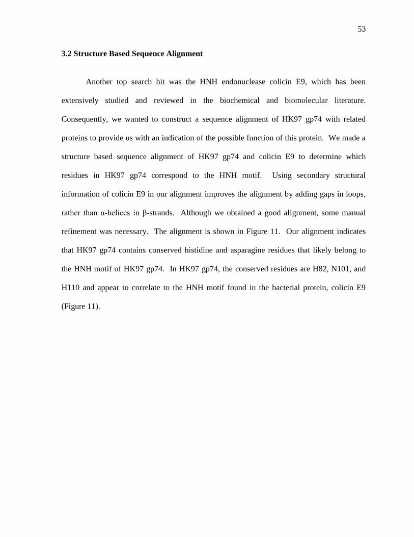
53
3.2 Structure Based Sequence Alignment
Another top search hit was the HNH endonuclease colicin E9, which has been
extensively studied and reviewed in the biochemical and biomolecular literature.
Consequently, we wanted to construct a sequence alignment of HK97 gp74 with related
proteins to provide us with an indication of the possible function of this protein. We made a
structure based sequence alignment of HK97 gp74 and colicin E9 to determine which
residues in HK97 gp74 correspond to the HNH motif. Using secondary structural
information of colicin E9 in our alignment improves the alignment by adding gaps in loops,
rather than α-helices in β-strands. Although we obtained a good alignment, some manual
refinement was necessary. The alignment is shown in Figure 11. Our alignment indicates
that HK97 gp74 contains conserved histidine and asparagine residues that likely belong to
the HNH motif of HK97 gp74. In HK97 gp74, the conserved residues are H82, N101, and
H110 and appear to correlate to the HNH motif found in the bacterial protein, colicin E9
(Figure 11).
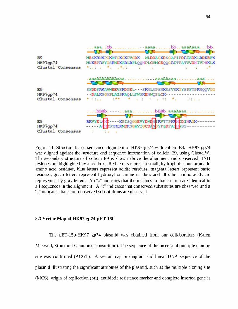
54
Figure 11: Structure-based sequence alignment of HK97 gp74 with colicin E9. HK97 gp74
was aligned against the structure and sequence information of colicin E9, using ClustalW.
The secondary structure of colicin E9 is shown above the alignment and conserved HNH
residues are highlighted by a red box. Red letters represent small, hydrophobic and aromatic
amino acid residues, blue letters represent acidic residues, magenta letters represent basic
residues, green letters represent hydroxyl or amine residues and all other amino acids are
represented by gray letters. An “*” indicates that the residues in that column are identical in
all sequences in the alignment. A “:” indicates that conserved substitutes are observed and a
“.” indicates that semi-conserved substitutions are observed.
3.3 Vector Map of HK97 gp74-pET-15b
The pET-15b-HK97 gp74 plasmid was obtained from our collaborators (Karen
Maxwell, Structural Genomics Consortium). The sequence of the insert and multiple cloning
site was confirmed (ACGT). A vector map or diagram and linear DNA sequence of the
plasmid illustrating the significant attributes of the plasmid, such as the multiple cloning site
(MCS), origin of replication (ori), antibiotic resistance marker and complete inserted gene is
55
shown in Figure 12. The plasmid vector, pET-15b contains an origin of replication, an
ampicillin resistance gene that acts as a selective agent, an abbreviated lac operon to control
protein expression by addition of the lactose analogue, IPTG, and a multiple cloning site,
which has various restriction enzyme recognition sites for insertion of a gene of interest. The
pET-15b plasmid vector used for protein biosynthesis includes the gene encoding a 6xHis
tagged HK97 gp74 bacteriophage protein with a tobacco etch virus (TEV) protease cleavage
site between the tag and HK97 gp74 protein (Figure 12).
Figure 12: Vector map of pET-15b-HK97 gp74.
56
3.4 Expression & Purification of 6xHis-HK97 gp74
3.4.1 Ni2+
Affinity Chromatography Purification of 6xHis-HK97 gp74
An SDS-PAGE gel was run for all purifications to verify the presence of our target
protein HK97 gp74, the size of the protein product, and the point of elution of the protein.
SDS-PAGE is a molecular technique used to separate and analyze proteins according to their
electrophoretic mobility, which is based on a protein’s size. The 6xHis-HK97 gp74 was
expressed in the soluble fraction in E. coli BL21 Star (DE3) cells and purified to
homogeneity using immobilized Ni2+
affinity chromatography. The basic principle of
affinity chromatography relies on the fact that the target protein to be purified must have
particular structural or chemical characteristics to allow for interaction of that molecule with
the resin and the addition of a structural or molecular mimic is required for the elution of the
target protein.6 Consequently, the HK97 gp74 protein was expressed as a fusion with an N-
terminal 6xHis tag, allowing the HK97 gp74 protein to be purified with Ni2+
affinity
chromatography. The protein was washed with 20 mM imidazole and eluted with 400 mM
imidazole. Imidazole is a molecular mimic that is an aromatic, heterocyclic compound
similar in structure to the cyclic ring found in histidine. The buffers contained 150 mM
NaCl to prevent the non-specific binding or interactions of proteins other than the 6xHis-
HK97 gp74 protein with the Ni2+
resin agarose beads. Several other components were
included in the buffers. β-mercaptoethanol (2 mM) was included because it reduces cysteine
disulphide bonds. Non-native disulfide bonds may result in protein precipitation or the loss
of activity. Protease inhibitors were also included, such as phenylmethanesulfonylfluoride
(150 μM) and benzamidine (5 mM), which are serine protease inhibitors and n-caproic acid,
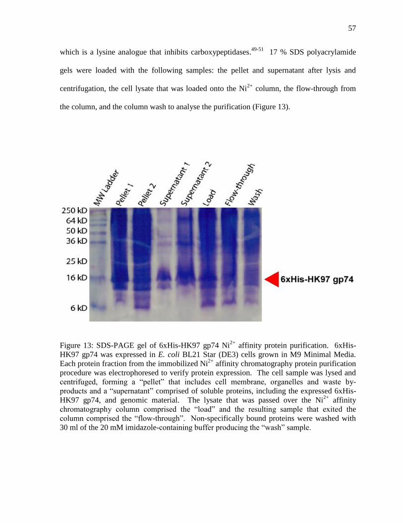
57
which is a lysine analogue that inhibits carboxypeptidases.49-51
17 % SDS polyacrylamide
gels were loaded with the following samples: the pellet and supernatant after lysis and
centrifugation, the cell lysate that was loaded onto the Ni2+
column, the flow-through from
the column, and the column wash to analyse the purification (Figure 13).
Figure 13: SDS-PAGE gel of 6xHis-HK97 gp74 Ni2+
affinity protein purification. 6xHis-
HK97 gp74 was expressed in E. coli BL21 Star (DE3) cells grown in M9 Minimal Media.
Each protein fraction from the immobilized Ni2+
affinity chromatography protein purification
procedure was electrophoresed to verify protein expression. The cell sample was lysed and
centrifuged, forming a “pellet” that includes cell membrane, organelles and waste by-
products and a “supernatant” comprised of soluble proteins, including the expressed 6xHis-
HK97 gp74, and genomic material. The lysate that was passed over the Ni2+
affinity
chromatography column comprised the “load” and the resulting sample that exited the
column comprised the “flow-through”. Non-specifically bound proteins were washed with
30 ml of the 20 mM imidazole-containing buffer producing the “wash” sample.
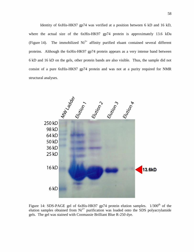
58
Identity of 6xHis-HK97 gp74 was verified at a position between 6 kD and 16 kD,
where the actual size of the 6xHis-HK97 gp74 protein is approximately 13.6 kDa
(Figure 14). The immobilized Ni2+
affinity purified eluant contained several different
proteins. Although the 6xHis-HK97 gp74 protein appears as a very intense band between
6 kD and 16 kD on the gels, other protein bands are also visible. Thus, the sample did not
consist of a pure 6xHis-HK97 gp74 protein and was not at a purity required for NMR
structural analyses.
Figure 14: SDS-PAGE gel of 6xHis-HK97 gp74 protein elution samples. 1/300th
of the
elution samples obtained from Ni2+
purification was loaded onto the SDS polyacrylamide
gels. The gel was stained with Coomassie Brilliant Blue R-250 dye.
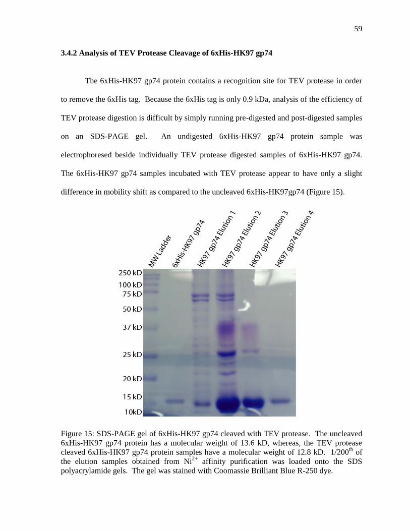
59
3.4.2 Analysis of TEV Protease Cleavage of 6xHis-HK97 gp74
The 6xHis-HK97 gp74 protein contains a recognition site for TEV protease in order
to remove the 6xHis tag. Because the 6xHis tag is only 0.9 kDa, analysis of the efficiency of
TEV protease digestion is difficult by simply running pre-digested and post-digested samples
on an SDS-PAGE gel. An undigested 6xHis-HK97 gp74 protein sample was
electrophoresed beside individually TEV protease digested samples of 6xHis-HK97 gp74.
The 6xHis-HK97 gp74 samples incubated with TEV protease appear to have only a slight
difference in mobility shift as compared to the uncleaved 6xHis-HK97gp74 (Figure 15).
Figure 15: SDS-PAGE gel of 6xHis-HK97 gp74 cleaved with TEV protease. The uncleaved
6xHis-HK97 gp74 protein has a molecular weight of 13.6 kD, whereas, the TEV protease
cleaved 6xHis-HK97 gp74 protein samples have a molecular weight of 12.8 kD. 1/200th
of
the elution samples obtained from Ni2+
affinity purification was loaded onto the SDS
polyacrylamide gels. The gel was stained with Coomassie Brilliant Blue R-250 dye.
60
3.5 Size Exclusion Chromatography of HK97 gp74
After dialysis and concomitant incubation of the 6xHis-HK97 gp74 protein with TEV
protease to remove the 6xHis tag, the protein sample was further purified using size
exclusion chromatography (SEC). In size exclusion chromatography, size separation of
molecules occurs via the principle that molecules of different sizes will move or elute
through the stationary phase (resin) at different rates. Each column is defined by a
permeation rate, which is the lowest molecular weight and smallest molecule that can
penetrate into the pores of the stationary phase completely.6 Thus, molecules larger than the
pores of the stationary phase will elute first, since they are not hindered by these porous
channels within the resin. Purification of HK97 gp74 from the 6xHis tag and the TEV
protease was accomplished with a 24 ml Superdex 75 column (GE Healthcare) composed of
cross-linked agarose and dextran with an average particle size of 13 µm and an optimum
separation range for proteins of 3,000 Da to 70,000 Da. This is an optimal separation range
for Ni2+
purified fractions of HK97 gp74, which contain proteins of high molecular weight in
addition to HK97 gp74. The size exclusion purification was monitored using UV
spectroscopy at an absorbance of 280 nm. Our A280 nm trace shows a large peak at 13.5 ml,
which corresponds to a protein with a monomeric molecular weight (Figure 16). SDS-PAGE
analysis verifies this peak migrates to a position between 6 kD and 16 kD, which is
indicative of HK97 gp74 with a molecular weight of 12.8 kDa (Figure 17). The size
exclusion purified HK97 gp74 sample was pure and contained no other proteins.
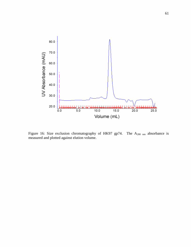
61
Figure 16: Size exclusion chromatography of HK97 gp74. The A280 nm absorbance is
measured and plotted against elution volume.
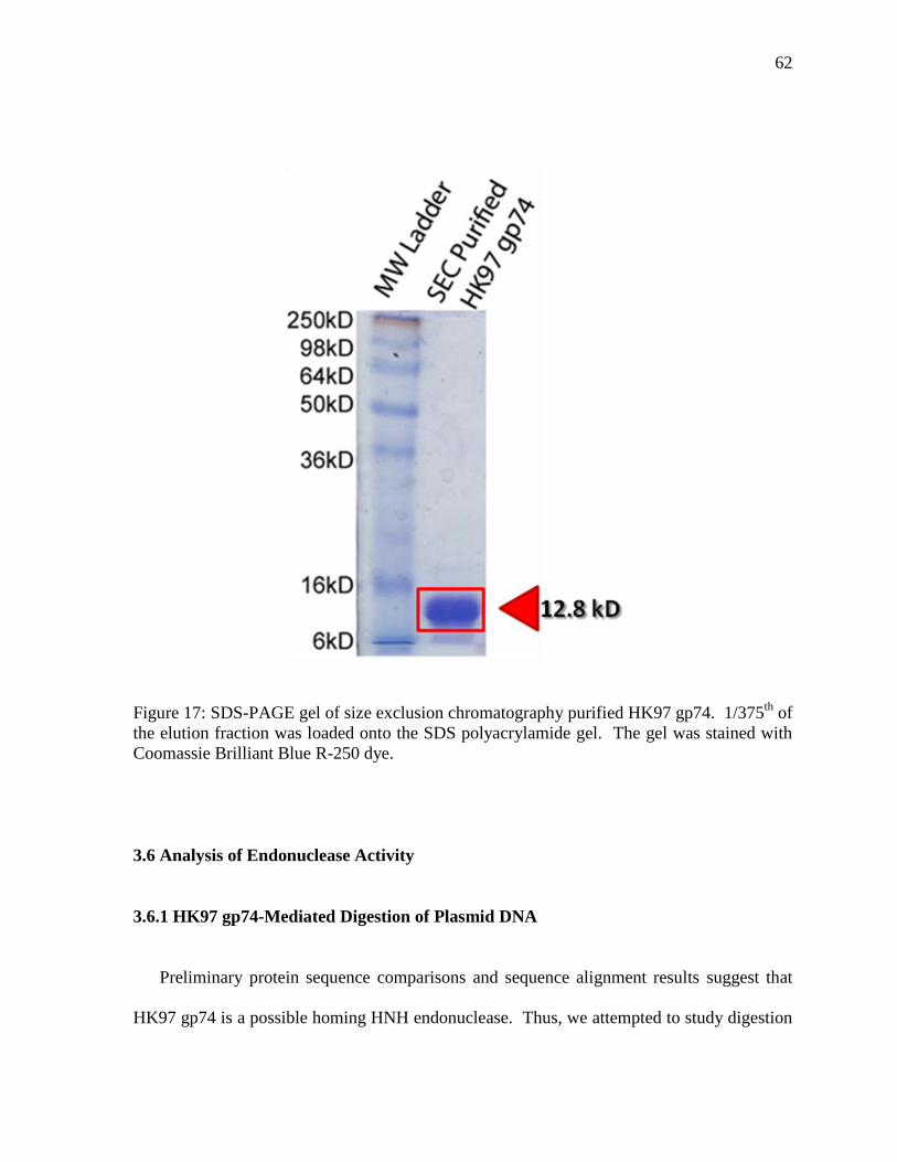
62
Figure 17: SDS-PAGE gel of size exclusion chromatography purified HK97 gp74. 1/375th
of
the elution fraction was loaded onto the SDS polyacrylamide gel. The gel was stained with
Coomassie Brilliant Blue R-250 dye.
3.6 Analysis of Endonuclease Activity
3.6.1 HK97 gp74-Mediated Digestion of Plasmid DNA
Preliminary protein sequence comparisons and sequence alignment results suggest that
HK97 gp74 is a possible homing HNH endonuclease. Thus, we attempted to study digestion
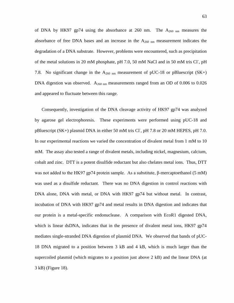
63
of DNA by HK97 gp74 using the absorbance at 260 nm. The A260 nm measures the
absorbance of free DNA bases and an increase in the A260 nm measurement indicates the
degradation of a DNA substrate. However, problems were encountered, such as precipitation
of the metal solutions in 20 mM phosphate, pH 7.0, 50 mM NaCl and in 50 mM tris Cl-, pH
7.8. No significant change in the A260 nm measurement of pUC-18 or pBluescript (SK+)
DNA digestion was observed. A260 nm measurements ranged from an OD of 0.006 to 0.026
and appeared to fluctuate between this range.
Consequently, investigation of the DNA cleavage activity of HK97 gp74 was analyzed
by agarose gel electrophoresis. These experiments were performed using pUC-18 and
pBluescript (SK+) plasmid DNA in either 50 mM tris Cl-, pH 7.8 or 20 mM HEPES, pH 7.0.
In our experimental reactions we varied the concentration of divalent metal from 1 mM to 10
mM. The assay also tested a range of divalent metals, including nickel, magnesium, calcium,
cobalt and zinc. DTT is a potent disulfide reductant but also chelates metal ions. Thus, DTT
was not added to the HK97 gp74 protein sample. As a substitute, β-mercaptoethanol (5 mM)
was used as a disulfide reductant. There was no DNA digestion in control reactions with
DNA alone, DNA with metal, or DNA with HK97 gp74 but without metal. In contrast,
incubation of DNA with HK97 gp74 and metal results in DNA digestion and indicates that
our protein is a metal-specific endonuclease. A comparison with EcoR1 digested DNA,
which is linear dsDNA, indicates that in the presence of divalent metal ions, HK97 gp74
mediates single-stranded DNA digestion of plasmid DNA. We observed that bands of pUC-
18 DNA migrated to a position between 3 kB and 4 kB, which is much larger than the
supercoiled plasmid (which migrates to a position just above 2 kB) and the linear DNA (at
3 kB) (Figure 18).
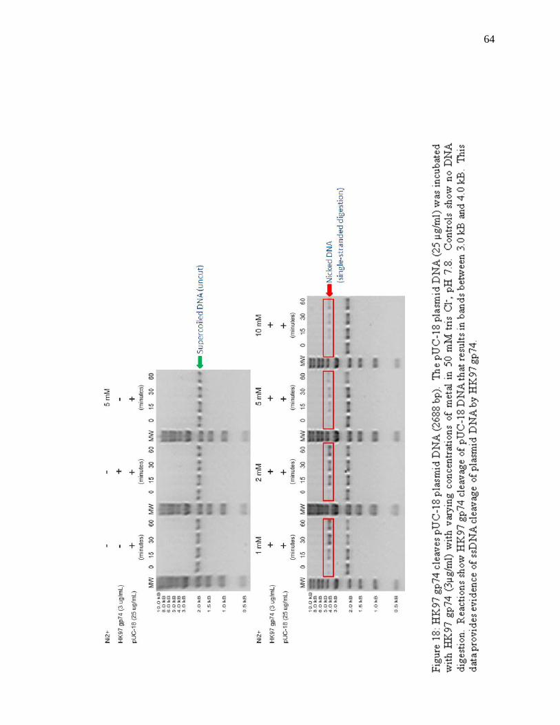
64
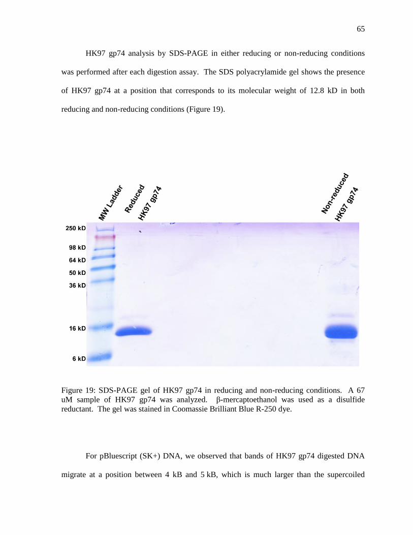
65
HK97 gp74 analysis by SDS-PAGE in either reducing or non-reducing conditions
was performed after each digestion assay. The SDS polyacrylamide gel shows the presence
of HK97 gp74 at a position that corresponds to its molecular weight of 12.8 kD in both
reducing and non-reducing conditions (Figure 19).
Figure 19: SDS-PAGE gel of HK97 gp74 in reducing and non-reducing conditions. A 67
uM sample of HK97 gp74 was analyzed. β-mercaptoethanol was used as a disulfide
reductant. The gel was stained in Coomassie Brilliant Blue R-250 dye.
For pBluescript (SK+) DNA, we observed that bands of HK97 gp74 digested DNA
migrate at a position between 4 kB and 5 kB, which is much larger than the supercoiled
66
phagemid (which migrates to a position just above 2 kB) and the linear DNA (at 3 kB)
(Figure 20). Thus, in the presence of divalent metal ions, HK97 gp74 also mediates single
strand cleavage of the pBluescript (SK+) phagemid DNA, similar to results observed for
pUC-18 plasmid DNA. Our results to date do not conclude whether single-stranded
digestion of plasmid DNA occurs at a specific site or multiple sites.
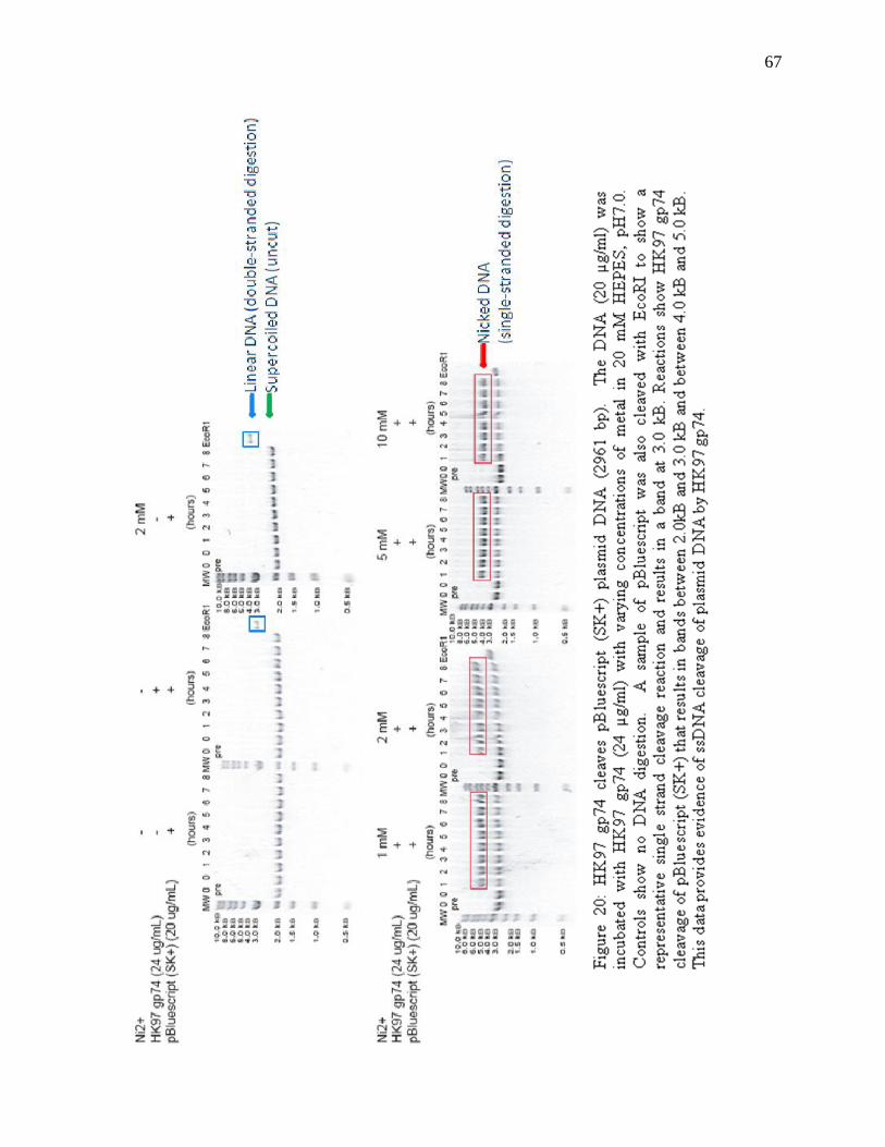
67
68
Thus, we attempted to generate ssDNA using pBluescript (SK+). However, we could
never view single-stranded DNA on an agarose gel, possibly due to low DNA
concentrations.
3.6.2 HK97 gp74-Mediated Digestion of Phage DNA
Since gp74 is a possible HNH endonuclease encoded by the bacteriophage HK97, we
tested the digestion of phage DNA by HK97 gp74. Upon incubation of lambda phage DNA
with HK97 gp74 and divalent metals, we observed a smear of DNA. This suggests that in
the presence of divalent metal ions (such as Ni2+
) HK97 gp74 mediates non-specific double-
stranded digestion of λ phage DNA (Figure 21). Control reactions indicate that both HK97
gp74 and metal are required for DNA digestion, consistent with our previous observations.
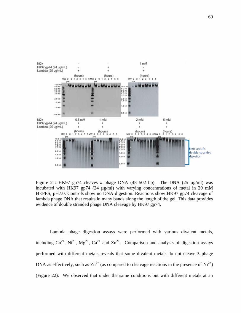
69
Figure 21: HK97 gp74 cleaves λ phage DNA (48 502 bp). The DNA (25 µg/ml) was
incubated with HK97 gp74 (24 µg/ml) with varying concentrations of metal in 20 mM
HEPES, pH7.0. Controls show no DNA digestion. Reactions show HK97 gp74 cleavage of
lambda phage DNA that results in many bands along the length of the gel. This data provides
evidence of double stranded phage DNA cleavage by HK97 gp74.
Lambda phage digestion assays were performed with various divalent metals,
including Co2+
, Ni2+
, Mg2+
, Ca2+
and Zn2+
. Comparison and analysis of digestion assays
performed with different metals reveals that some divalent metals do not cleave λ phage
DNA as effectively, such as Zn2+
(as compared to cleavage reactions in the presence of Ni2+
)
(Figure 22). We observed that under the same conditions but with different metals at an
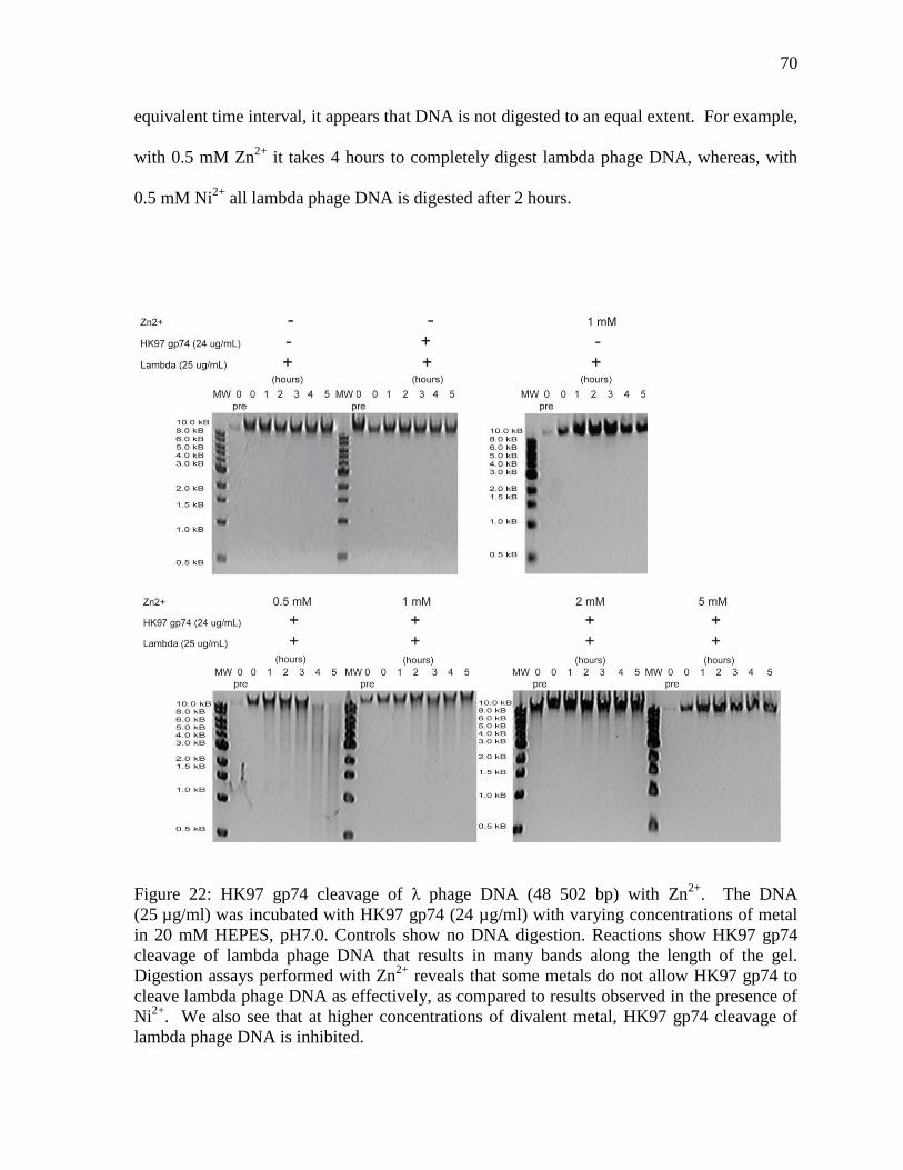
70
equivalent time interval, it appears that DNA is not digested to an equal extent. For example,
with 0.5 mM Zn2+
it takes 4 hours to completely digest lambda phage DNA, whereas, with
0.5 mM Ni2+
all lambda phage DNA is digested after 2 hours.
Figure 22: HK97 gp74 cleavage of λ phage DNA (48 502 bp) with Zn2+
. The DNA
(25 µg/ml) was incubated with HK97 gp74 (24 µg/ml) with varying concentrations of metal
in 20 mM HEPES, pH7.0. Controls show no DNA digestion. Reactions show HK97 gp74
cleavage of lambda phage DNA that results in many bands along the length of the gel.
Digestion assays performed with Zn2+
reveals that some metals do not allow HK97 gp74 to
cleave lambda phage DNA as effectively, as compared to results observed in the presence of
Ni2+
. We also see that at higher concentrations of divalent metal, HK97 gp74 cleavage of
lambda phage DNA is inhibited.
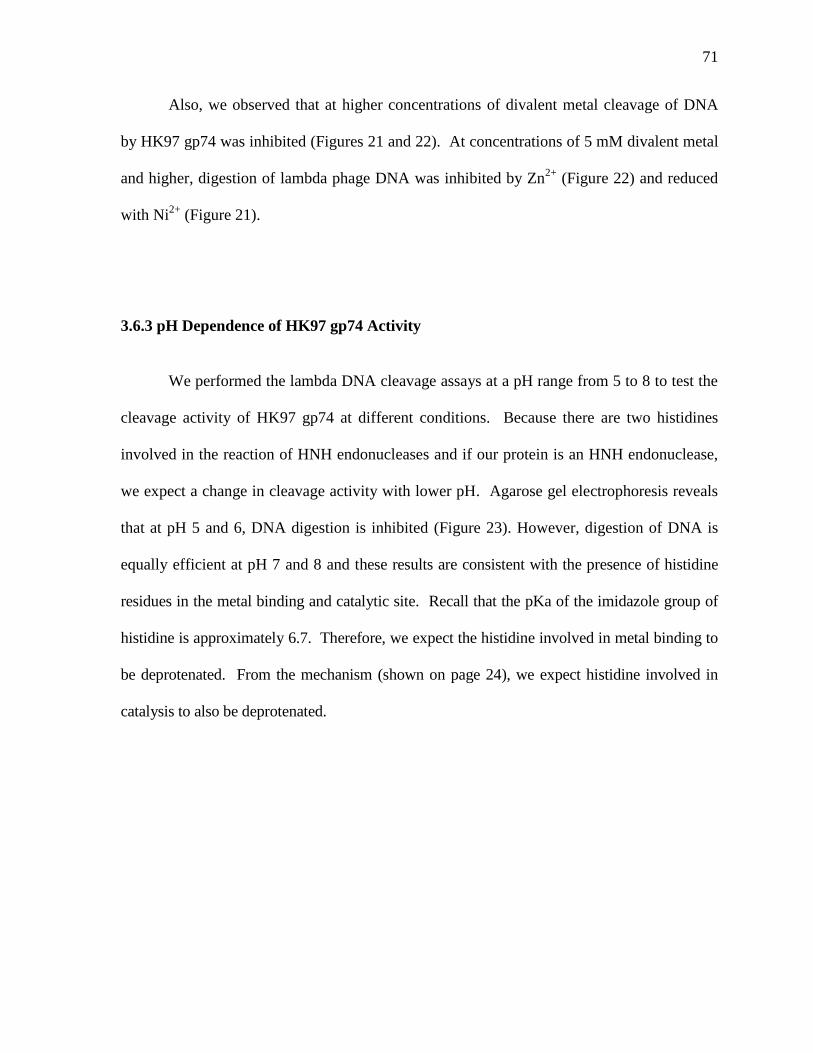
71
Also, we observed that at higher concentrations of divalent metal cleavage of DNA
by HK97 gp74 was inhibited (Figures 21 and 22). At concentrations of 5 mM divalent metal
and higher, digestion of lambda phage DNA was inhibited by Zn2+
(Figure 22) and reduced
with Ni2+
(Figure 21).
3.6.3 pH Dependence of HK97 gp74 Activity
We performed the lambda DNA cleavage assays at a pH range from 5 to 8 to test the
cleavage activity of HK97 gp74 at different conditions. Because there are two histidines
involved in the reaction of HNH endonucleases and if our protein is an HNH endonuclease,
we expect a change in cleavage activity with lower pH. Agarose gel electrophoresis reveals
that at pH 5 and 6, DNA digestion is inhibited (Figure 23). However, digestion of DNA is
equally efficient at pH 7 and 8 and these results are consistent with the presence of histidine
residues in the metal binding and catalytic site. Recall that the pKa of the imidazole group of
histidine is approximately 6.7. Therefore, we expect the histidine involved in metal binding to
be deprotenated. From the mechanism (shown on page 24), we expect histidine involved in
catalysis to also be deprotenated.
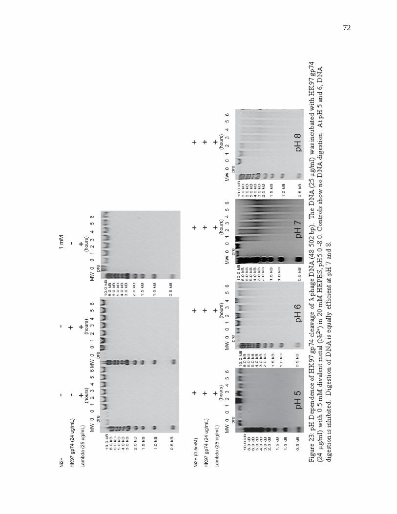
72
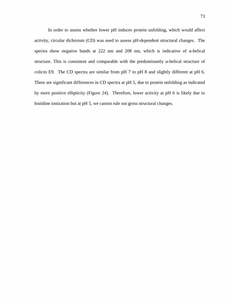
73
In order to assess whether lower pH induces protein unfolding, which would affect
activity, circular dichroism (CD) was used to assess pH-dependent structural changes. The
spectra show negative bands at 222 nm and 208 nm, which is indicative of α-helical
structure. This is consistent and comparable with the predominantly α-helical structure of
colicin E9. The CD spectra are similar from pH 7 to pH 8 and slightly different at pH 6.
There are significant differences in CD spectra at pH 5, due to protein unfolding as indicated
by more positive ellipticity (Figure 24). Therefore, lower activity at pH 6 is likely due to
histidine ionization but at pH 5, we cannot rule out gross structural changes.
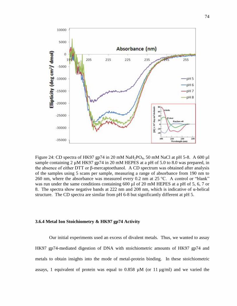
74
Figure 24: CD spectra of HK97 gp74 in 20 mM NaH2PO4, 50 mM NaCl at pH 5-8. A 600 µl
sample containing 2 µM HK97 gp74 in 20 mM HEPES at a pH of 5.0 to 8.0 was prepared, in
the absence of either DTT or β-mercaptoethanol. A CD spectrum was obtained after analysis
of the samples using 5 scans per sample, measuring a range of absorbance from 190 nm to
260 nm, where the absorbance was measured every 0.2 nm at 25 °C. A control or “blank”
was run under the same conditions containing 600 µl of 20 mM HEPES at a pH of 5, 6, 7 or
8. The spectra show negative bands at 222 nm and 208 nm, which is indicative of α-helical
structure. The CD spectra are similar from pH 6-8 but significantly different at pH 5.
3.6.4 Metal Ion Stoichiometry & HK97 gp74 Activity
Our initial experiments used an excess of divalent metals. Thus, we wanted to assay
HK97 gp74-mediated digestion of DNA with stoichiometric amounts of HK97 gp74 and
metals to obtain insights into the mode of metal-protein binding. In these stoichiometric
assays, 1 equivalent of protein was equal to 0.858 µM (or 11 µg/ml) and we varied the
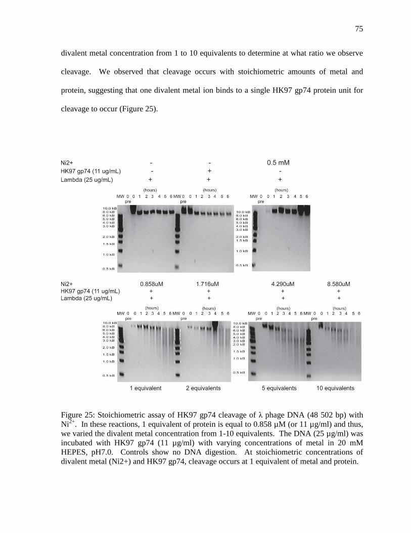
75
divalent metal concentration from 1 to 10 equivalents to determine at what ratio we observe
cleavage. We observed that cleavage occurs with stoichiometric amounts of metal and
protein, suggesting that one divalent metal ion binds to a single HK97 gp74 protein unit for
cleavage to occur (Figure 25).
Figure 25: Stoichiometric assay of HK97 gp74 cleavage of λ phage DNA (48 502 bp) with
Ni2+
. In these reactions, 1 equivalent of protein is equal to 0.858 µM (or 11 µg/ml) and thus,
we varied the divalent metal concentration from 1-10 equivalents. The DNA (25 µg/ml) was
incubated with HK97 gp74 (11 µg/ml) with varying concentrations of metal in 20 mM
HEPES, pH7.0. Controls show no DNA digestion. At stoichiometric concentrations of
divalent metal (Ni2+) and HK97 gp74, cleavage occurs at 1 equivalent of metal and protein.
76
The UV-vis metal binding assay was used to measure the increase in the absorbance
of free bases of DNA at a wavelength of 260 nm (A260 nm) due to DNA digestion by HK97
gp74 in the presence of stoichiometric amounts of metal. Like the A260 nm digestion assays,
we observed no significant change in A260 nm measurements over time. Furthermore, the
measurements appeared to decrease in absorbance in the absence of divalent metal ions and
fluctuated erratically as time passed with a difference in A260nm (OD) of approximately 0.05
from the lowest measurement recorded to the highest measurement for reactions that
included the presence of divalent metal (Figure 26).
Figure 26: UV-Vis assay of HK97 gp74 cleavage of λ phage DNA (48 502 bp) with Ni2+
. In
these reactions, 1 equivalent of protein is equal to 0.858 µM (or 11 µg/ml) and we varied the
divalent metal concentration from 0, 1, 5 and 10 equivalents. The DNA (25 µg/ml) was
incubated with HK97 gp74 (11 µg/ml) with varying concentrations of metal in 20 mM
HEPES, pH7.0. Controls were used to reduce any signal resulting from the buffer, protein or
metal. Digestion of DNA is marked by the increase in absorbance at a wavelength of 260 nm
due to the presence of free nucleotides. No significant changes in absorbance were observed
and the absorbance appears to rise and fall slightly as time progresses.
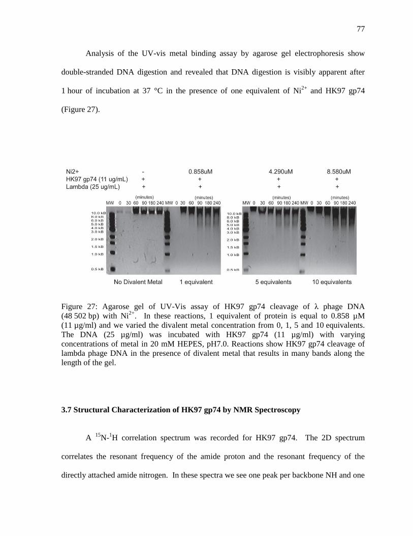
77
Analysis of the UV-vis metal binding assay by agarose gel electrophoresis show
double-stranded DNA digestion and revealed that DNA digestion is visibly apparent after
1 hour of incubation at 37 °C in the presence of one equivalent of Ni2+
and HK97 gp74
(Figure 27).
Figure 27: Agarose gel of UV-Vis assay of HK97 gp74 cleavage of λ phage DNA
(48 502 bp) with Ni2+
. In these reactions, 1 equivalent of protein is equal to 0.858 µM
(11 µg/ml) and we varied the divalent metal concentration from 0, 1, 5 and 10 equivalents.
The DNA (25 µg/ml) was incubated with HK97 gp74 (11 µg/ml) with varying
concentrations of metal in 20 mM HEPES, pH7.0. Reactions show HK97 gp74 cleavage of
lambda phage DNA in the presence of divalent metal that results in many bands along the
length of the gel.
3.7 Structural Characterization of HK97 gp74 by NMR Spectroscopy
A 15
N-1H correlation spectrum was recorded for HK97 gp74. The 2D spectrum
correlates the resonant frequency of the amide proton and the resonant frequency of the
directly attached amide nitrogen. In these spectra we see one peak per backbone NH and one
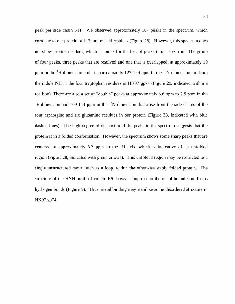
78
peak per side chain NH. We observed approximately 107 peaks in the spectrum, which
correlate to our protein of 113 amino acid residues (Figure 28). However, this spectrum does
not show proline residues, which accounts for the loss of peaks in our spectrum. The group
of four peaks, three peaks that are resolved and one that is overlapped, at approximately 10
ppm in the 1H dimension and at approximately 127-129 ppm in the
15N dimension are from
the indole NH in the four tryptophan residues in HK97 gp74 (Figure 28, indicated within a
red box). There are also a set of “double” peaks at approximately 6.6 ppm to 7.5 ppm in the
1H dimension and 109-114 ppm in the
15N dimension that arise from the side chains of the
four asparagine and six glutamine residues in our protein (Figure 28, indicated with blue
dashed lines). The high degree of dispersion of the peaks in the spectrum suggests that the
protein is in a folded conformation. However, the spectrum shows some sharp peaks that are
centered at approximately 8.2 ppm in the 1H axis, which is indicative of an unfolded
region (Figure 28, indicated with green arrows). This unfolded region may be restricted to a
single unstructured motif, such as a loop, within the otherwise stably folded protein. The
structure of the HNH motif of colicin E9 shows a loop that in the metal-bound state forms
hydrogen bonds (Figure 9). Thus, metal binding may stabilize some disordered structure in
HK97 gp74.
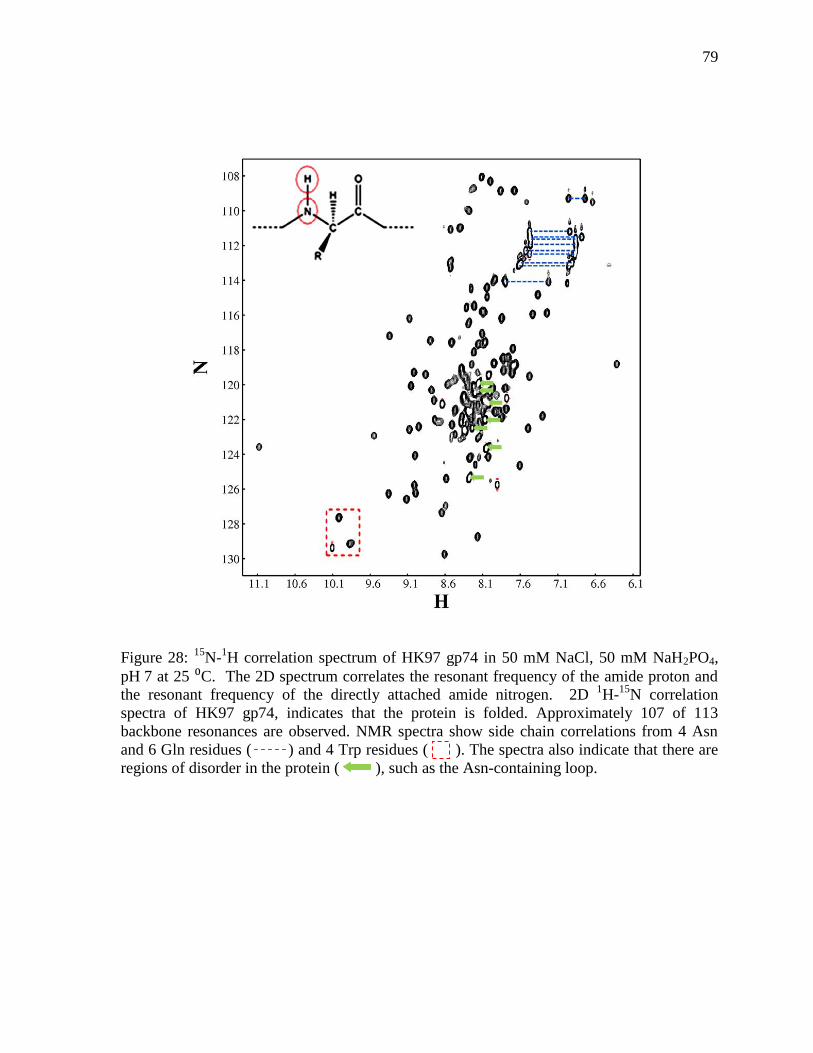
79
Figure 28: 15
N-1H correlation spectrum of HK97 gp74 in 50 mM NaCl, 50 mM NaH2PO4,
pH 7 at 25 ⁰C. The 2D spectrum correlates the resonant frequency of the amide proton and
the resonant frequency of the directly attached amide nitrogen. 2D 1H-
15N correlation
spectra of HK97 gp74, indicates that the protein is folded. Approximately 107 of 113
backbone resonances are observed. NMR spectra show side chain correlations from 4 Asn
and 6 Gln residues ( ) and 4 Trp residues ( ). The spectra also indicate that there are
regions of disorder in the protein ( ), such as the Asn-containing loop.
80
3.8 Analysis of HK97 gp74 Metal Binding
3.8.1 Metal Binding Titration Experiments using NMR Spectroscopy
Our results indicate that HK97 gp74 is a possible HNH homing endonuclease, which
binds and cleaves DNA in the presence of divalent metal cations. The structural or
conformational changes that occur as a result of divalent metal binding at the HNH motif or
in the global structure of HK97 gp74 were investigated. Addition of metal at a high
concentration of protein caused immediate precipitation of most of the sample. 15
N-1H
correlation spectra for the apo-HK97 gp74 and metal bound HK97 gp74 samples were
performed and recorded. However, comparison of the apo-HK97 gp74 15
N-1H correlation
spectrum and the metal-bound species of HK97 gp74 15
N-1H correlation spectrum showed no
change or shift of peaks in the spectra, likely because of precipitation of the metal-bound
species with only the apo-HK97 gp74 protein left in solution (Figure 29). Nonetheless, high
quality spectra of apo-HK97 gp74 indicate that our proposed structural studies are feasible.
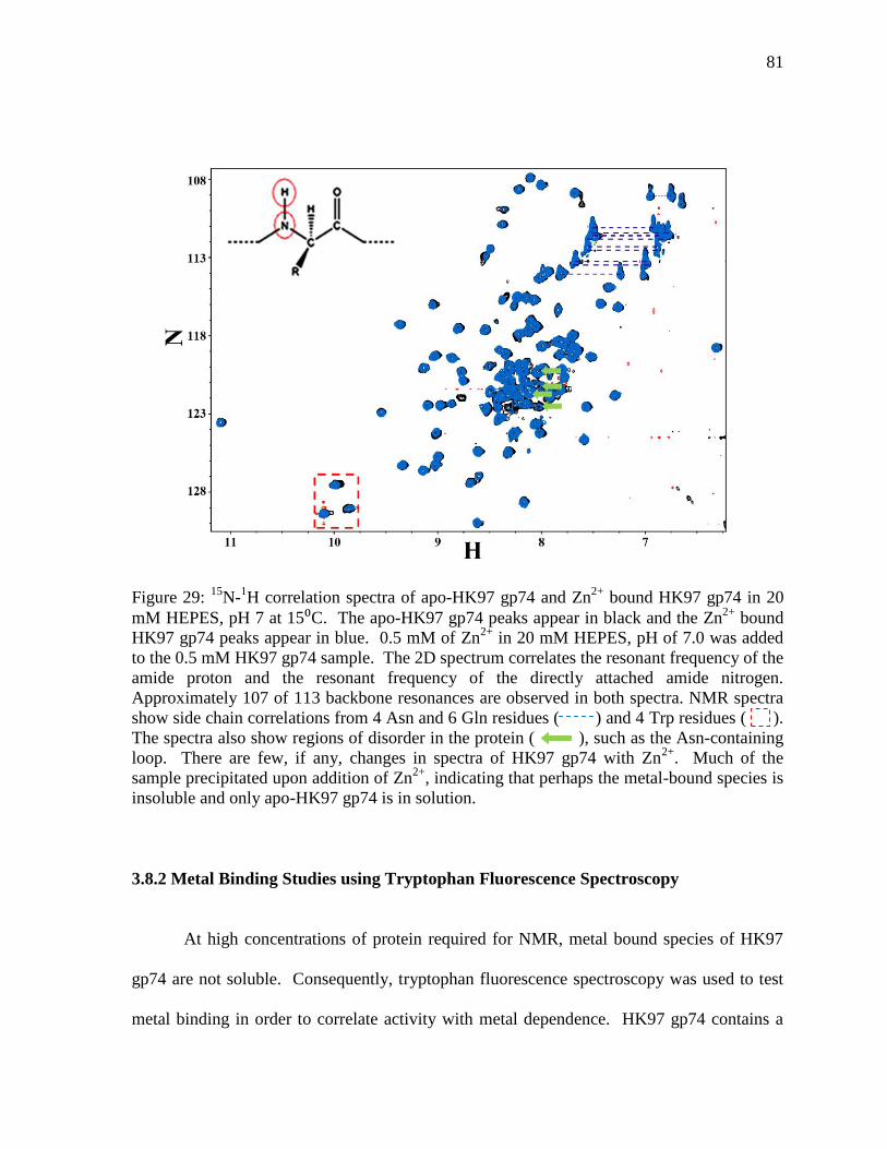
81
Figure 29: 15
N-1H correlation spectra of apo-HK97 gp74 and Zn
2+ bound HK97 gp74 in 20
mM HEPES, pH 7 at 15⁰C. The apo-HK97 gp74 peaks appear in black and the Zn2+
bound
HK97 gp74 peaks appear in blue. 0.5 mM of Zn2+
in 20 mM HEPES, pH of 7.0 was added
to the 0.5 mM HK97 gp74 sample. The 2D spectrum correlates the resonant frequency of the
amide proton and the resonant frequency of the directly attached amide nitrogen.
Approximately 107 of 113 backbone resonances are observed in both spectra. NMR spectra
show side chain correlations from 4 Asn and 6 Gln residues ( ) and 4 Trp residues ( ).
The spectra also show regions of disorder in the protein ( ), such as the Asn-containing
loop. There are few, if any, changes in spectra of HK97 gp74 with Zn2+
. Much of the
sample precipitated upon addition of Zn2+
, indicating that perhaps the metal-bound species is
insoluble and only apo-HK97 gp74 is in solution.
3.8.2 Metal Binding Studies using Tryptophan Fluorescence Spectroscopy
At high concentrations of protein required for NMR, metal bound species of HK97
gp74 are not soluble. Consequently, tryptophan fluorescence spectroscopy was used to test
metal binding in order to correlate activity with metal dependence. HK97 gp74 contains a
82
total of four tryptophan residues, where the residue, Trp-111, is located directly adjacent to
the metal binding histidine of the HNH motif. However, no conclusive evidence was
obtained to show that metal binding at the HNH motif promotes conformational changes.
Changes in tryptophan fluorescence of metal-bound HK97 gp74 were not significantly
different than changes in tryptophan fluorescence of the control, which contained the protein
in the absence of divalent metal (Figure 30). Furthermore, it is unclear why the tryptophan
fluorescence intensity of HK97 gp74 in the presence of 3 µM Ni2+
is lower than the
fluorescence intensity of HK97 gp74 in the presence of 1 µM Ni2+
. Changes to the protocol
(such as the concentration of protein or metal, temperature, buffer, controls, amount of
sample scanned, etc.) or equipment (such as the sample plate, scanning attributes, etc.) may
be required to obtain better tryptophan fluorescence intensity data in order to construct a
complete conclusion about the metal binding interactions of HK97 gp74.
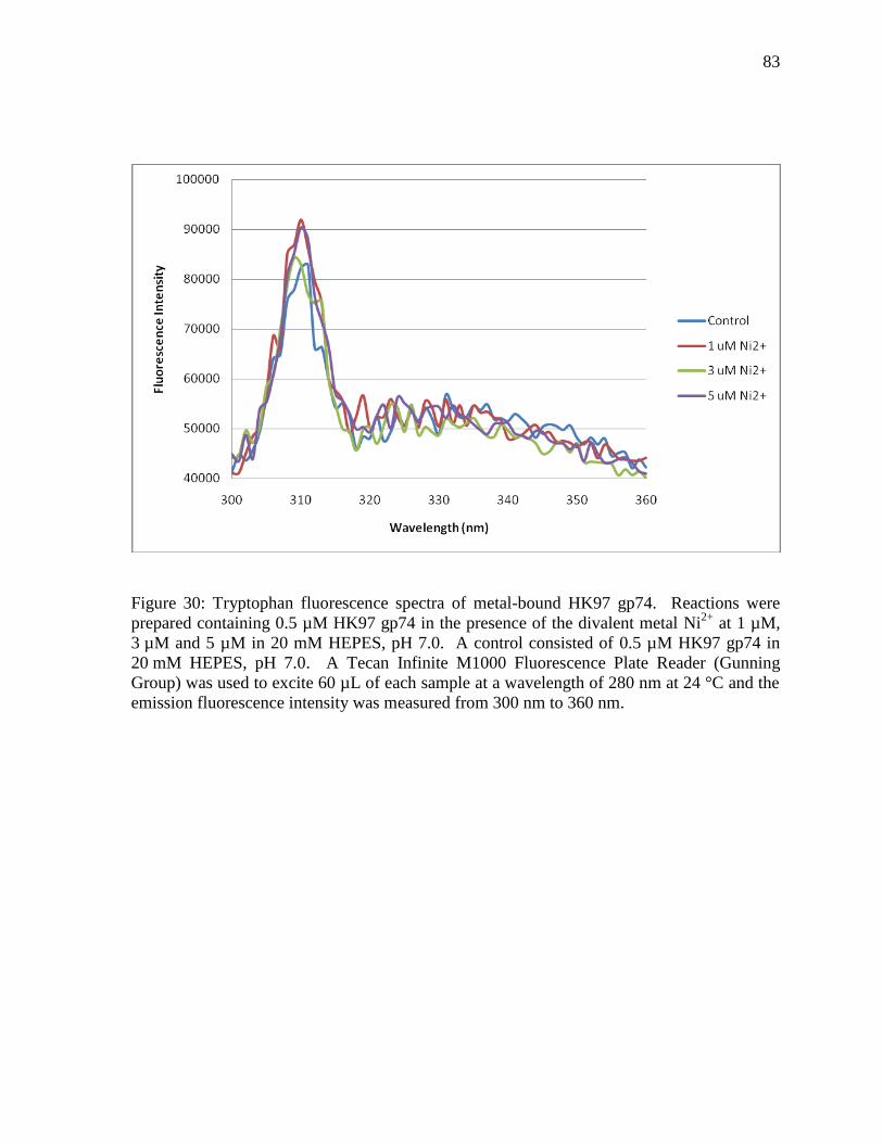
83
Figure 30: Tryptophan fluorescence spectra of metal-bound HK97 gp74. Reactions were
prepared containing 0.5 µM HK97 gp74 in the presence of the divalent metal Ni2+
at 1 µM,
3 µM and 5 µM in 20 mM HEPES, pH 7.0. A control consisted of 0.5 µM HK97 gp74 in
20 mM HEPES, pH 7.0. A Tecan Infinite M1000 Fluorescence Plate Reader (Gunning
Group) was used to excite 60 µL of each sample at a wavelength of 280 nm at 24 °C and the
emission fluorescence intensity was measured from 300 nm to 360 nm.
84
4. Discussion and Conclusions:
4.1 HK97 gp74
At the beginning of this project, the HK97 bacteriophage protein gp74 was
uncharacterized and previously had no known function or known structural characteristics.
The purpose of this study was to investigate the structural and possible functional role of the
HK97 bacteriophage protein gp74. Our preliminary search of proteins with similar
sequences to HK97 gp74 using the basic local alignment search tool (BLAST) indicated that
HK97 gp74 is a possible homing HNH endonuclease. Homing endonucleases bind double-
stranded DNA and cause double-stranded breaks in alleles that are homologous to the
endonuclease gene but lack the intron or intein element, which encodes the gene for the
homing endonuclease.33
There are four main families of homing endonucleases, each of which are named for
the conserved residues associated with their nuclease domain, which include the
LAGLIDADG, the His-Cys box, HNH enzymes and GIY-YIG family of enzymes.28
A top
BLAST search hit was the HNH endonuclease colicin E9, which has been extensively
studied and set the foundation of our functional investigations of HK97 gp74. Subsequent
structure-based sequence alignment and analysis provided us with evidence that HK97 gp74
contains conserved histidine and asparagine residues that likely compose the HNH motif of
HK97 gp74. In HK97 gp74, the conserved residues are H82, N101, and H110 and appear to
correlate to the HNH motif found in the bacterial protein, colicin E9 (Figure 11). Thus, basic
sequence alignment results obtained in this study suggest that HK97 gp74 protein belongs to
the HNH-endonuclease family of enzymes, which are conserved among bacteria and viruses.
85
The HNH class of homing endonucleases are named for two conserved histidine
residues and an invariant asparagine residue that comprise the HNH motif.35
This family is
known to include the pyocins, colicins and anaredoxins, which are bacterial proteins that
inhibit the growth of similar or related bacteria. For example, colicins are non-specific
bacterial HNH endonucleases and comparison to these family members may suggest that
HK97 gp74 may also play a role as a bacteriocin.
4.2 Role of HK97 gp74 HNH Endonucleases
Homing endonucleases are highly conserved in bacterial populations and have been
suggested to play a significant role in the homologous recombination of newly integrated
genes from one bacteria to another.11
However, very little is known as to the mechanism of
integration of the endonuclease gene among bacterial species and why this type of protein is
conserved among bacterial species. However, we hypothesize that the existence of an HNH
homing endonuclease in bacteriophages would provide a mechanism by which phage genes
are incorporated into the bacterial genome. It is known that phage invasion and genomic
integration through the lysogenic life cycle leads to the formation of prophage elements, and
this process may be a major contributor to genomic diversity amongst bacterial species.15
The HK97 gp74 protein may play a possible role in the phage lysogenic cycle. From the
gene map of the HK97 genome sequence, neighbouring genes to gp74 are involved in DNA
repair mechanisms and lysis of bacterial cells (Figure 5). The existence of genes involved in
lysis of bacterial cells and degradation of peptidoglycan, such as the S gene that encodes for
a holin protein and the R gene that encodes for an endolysin, further support the theory that
86
HK97 gp74 is involved in phage infection processes.31
Furthermore, gp69 is hypothesized to
be closely related to the RusA gene of lambda phage, which encodes for a resolvase that
ligates nucleic acid fragments at Holliday junctions.30
This offers evidence to support the
theory that HK97 gp74 is involved in the lysogenic cycle and more specifically that HNH
homing endonuclease activity of the HK97 gp74 protein would allow for the incorporation
and recombination of phage genes into the bacterial genome. Consequently, we suggest that
HK97 gp74 creates double stranded DNA breaks, thereby, initiating bacterial host cell repair
machinery that allows for homologous recombination at the cleaved DNA site to occur and
resulting in integration of the phage genome.
Furthermore, many phage endonucleases have been observed to target and cleave
other phage DNA. This strategy may be a result of competition between bacteriophage
species or mutualism with bacterial species to protect against infection and cell death. A
consequence of our study has lead us to suggest that HK97 gp74 may be responsible for the
digestion of foreign phage DNA that pose a competitive disadvantage to the integration and
proliferation of HK97 bacteriophage particles in a bacterial host and to protect the host cell
from invasion of foreign phage that may be virulent or bactericidal. Thus, HK97 gp74 is an
important target for study to understand the role of bacteriophages and their influence and
manipulation of bacterial populations. Future studies in biological function of HK97 gp74
may discriminate between these and other possibilities.
87
4.3 Future Biochemical & Biophysical Studies
The structural part of the HK97 bacteriophage protein gp74 project encompasses our
work to date in protein biochemistry to generate suitable samples and screen solution
conditions for our biochemical and NMR studies. A 15
N-1H correlation spectrum was
recorded for HK97 gp74 in the absence of metal and provided evidence that HK97 gp74 is in
a folded conformation. Future NMR studies involving the divalent metal binding of HK97
gp74 using Zn2+
or Ni2+
ions at stoichiometric concentrations will help to elucidate the
conformational changes associated with divalent metal binding at the HNH motif of HK97
gp74. Because samples of gp74 at high concentrations with metal precipitate, preparation of
metal-bound samples need to be investigated and may involve changing the buffer or adding
metal in dilute solutions and concentrating the metal-bound species to NMR concentrations.
We would also like to solve the solution structure of HK97 gp74 in order to gain insights into
the mechanism of DNA digestion. Resonance assignments will also be useful to study
metal-binding and DNA binding (provided that catalysis deficient mutants are amenable to
NMR studies).
Our biochemical and biophysical studies indicate that HK97 gp74 is a possible
bacterial homing HNH endonuclease. One of the future goals in this project is to identify
residues involved in DNA binding and those involved in DNA digestion. Our digestion
assays at different pH values suggest that protonation of one or both His residues decrease
DNA digestion. Protonation of the His residue involved in metal binding would likely
decrease the affinity for the metal, whereas protonation of the catalytic His would decrease
the catalytic efficiency of gp74. One of the first steps in the HNH endonuclease DNA
88
digestion mechanism is the abstraction of a proton from a water molecule by the catalytic His
(Figure 10 in Introduction 1.4 Homing Endonucleases), with the resulting hydroxide ion
attacking the phosphodiester DNA backbone.28
A protonated His residue would be less
efficient in this step.
In order to determine the protonation state of each His residue in the HNH motif at
physiological pH (pH 7), we need to know their specific pKas. NMR spectroscopy is an
ideal tool for measuring pKa values. Because we do not currently have resonance
assignments for HK97 gp74, we will need to specifically label our protein with 15
N-labeled
His residues. 15
N-1H HSQC spectra of
15N-His gp74 would consist of only 3 peaks. Titration
with acid would result in protonation of the His residues and concomitant chemical shift
changes, which can be used to determine the pKa values. A separate pH titration on
uniformly 15
N-labeled gp74 would need to be done to ensure that the protein is folded at
different pH values, and hence that the pKa values measured reflect changes in the
protonation state of the His residues and not global structural changes.
Changes in backbone 15
N-1H chemical shifts with His side chain protonation state
may not be large enough due to the distance of the His side chain from the backbone.
Further, backbone chemical shift changes may be influenced by ionization state of other
groups as the chemical shift dependence on through-space effects of titratable groups has a
1/r2 dependence and can be long range.
57,58 Thus, it would be advantageous to have side
chain probes for measuring pKa values. We could also do a similar experiment with gp74
that is uniformly labeled with 15
N that also contains 19
F-labeled His residues at the N
position. In this way, we could assess structural changes and changes in pKa values with one
89
sample. 2D 15
N-1H HSQC experiments would assess the folded state of the protein and 1D
19F spectra would probe protonation state. Perturbations of
19F to protein structure are
minimal and 19
F probes have a wide range of chemical shifts associated with different local
electronic environments in folded proteins. However, 19
F probes in His residues can shift the
pKa values by more than 2 units, which may affect the biochemical characteristics of the
protein. Thus, use of 19
F probes must be carefully analyzed.
We could also measure pKa values in His by using 1H-
15N heteronuclear multiple
bond correlation (HMBC) NMR experiments. HMBC experiments provide information
about weak proton-carbon or proton-nitrogen interactions.58
Thus, carbon-bound protons
that are two, three or four bonds away from the heteroatom of interest are observed. Thus,
these experiments require 15
N and 13
C labeled protein. In a 1H-
15N HMBC spectra of His
residues, the chemical shift of the 15
N resonance is correlated to the protonation state of the
imidazole nitrogen.58
Protonated nitrogens are observed at ~170 ppm, whereas, deprotenated
nitrogens are observed at ~250 ppm.59
In addition, we can also differentiate between two
neutral His tautomers using these experiments. Different spectral patterns are produced in
the 15
N-1H HMBC experiment due to different two-bond and three-bond
1H-
15N coupling
constants in each state.59
Thus, there are a number of different approaches that we can use to obtain pKa values
for His residues in gp74. In absence of backbone and side chain resonance assignments,
identification of specific resonances will require making His point mutants and recording
spectra to obtain the assignments. Nonetheless, NMR spectroscopy provides us with many
options to determine pKa values of the histidine imidazole groups involved in catalysis and
90
metal binding. These experiments are planned and will yield additional information
regarding the biochemical function and mechanism of gp74.
As stated above, future work will also include the protein biosynthesis of mutant
HK97 gp74 proteins. Mutant HK97 gp74 proteins will be designed to have alanine residues
in the place of the conserved residues of the putative HNH motif, to confirm that HK97 gp74
is an HNH endonuclease and to determine which residues are essential for binding and
cleavage of DNA by HK97 gp74. Mutagenesis studies are currently underway and the
mutant H82A-HK97 gp74 was designed and synthesized by PCR thermal cycling. Agarose
gel electrophoresis shows the appearance of the control product, in addition to, mutant HK97
gp74-pET-15b plasmid DNA that appears at 6 kB (Figure 31). The expected mutant pET-
15b-H82A-HK97 gp74 plasmid has a size of 6.1 kB. The mutant DNA will be used in future
protein expression studies to produce mutant versions of HK97 gp74, in which the conserved
residues of the HNH motif have been changed to alanine in order to study the decrease or
lack of activity of DNA digestion, as well as, divalent metal binding at the active site.
Furthermore, a triple mutant will be used to study the total inhibition or severe reduction in
cleavage activity. We hope to perform our DNA digestion assays to compare the wild type
activity to the mutant enzymes.
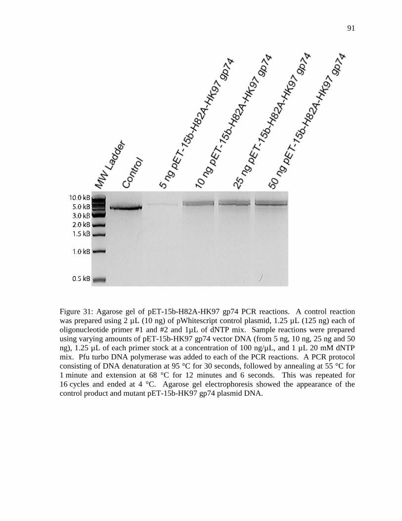
91
Figure 31: Agarose gel of pET-15b-H82A-HK97 gp74 PCR reactions. A control reaction
was prepared using 2 µL (10 ng) of pWhitescript control plasmid, 1.25 µL (125 ng) each of
oligonucleotide primer #1 and #2 and 1µL of dNTP mix. Sample reactions were prepared
using varying amounts of pET-15b-HK97 gp74 vector DNA (from 5 ng, 10 ng, 25 ng and 50
ng), 1.25 µL of each primer stock at a concentration of 100 ng/µL, and 1 µL 20 mM dNTP
mix. Pfu turbo DNA polymerase was added to each of the PCR reactions. A PCR protocol
consisting of DNA denaturation at 95 °C for 30 seconds, followed by annealing at 55 °C for
1 minute and extension at 68 °C for 12 minutes and 6 seconds. This was repeated for
16 cycles and ended at 4 °C. Agarose gel electrophoresis showed the appearance of the
control product and mutant pET-15b-HK97 gp74 plasmid DNA.
92
In order to determine the efficiency of cleavage by HK97 gp74, we would like to
compare the activity of gp74 cleavage against the activity of a control endonuclease. Colicin
E9 is an extensively studied HNH endonuclease that is closely related to HK97 gp74 (see
Figure 11 in Results 3.2 Structure Based Sequence Alignment). A BLAST search and our
structure-based sequence alignment highlight the similarities of HK97 gp74 and colicin E9,
particulary in the structural motif (Figure 11). Consequently, as an HNH endonuclease
that is structurally similar to HK97 gp74, the bacterial protein colicin E9 would be the best
choice as a control endonuclease to compare against the DNA cleavage activity of HK97
gp74. Colicin E9, like HK97 gp74, also shows a broad range of metal dependent DNA
digestion but catalytic activity is greatest in the presence of Ni2+
.35
Colicin E9 also displays
Michaelis-Menten kinetics and in the presence of Ni2+
and calf thymus DNA, the Km was
determined to be 0.93 M.60
Colicin E9 DNase is known to cleave DNA preferentially after
thymine residues of randomly designed DNA primers.35
The kinetics of DNA digestion by
HK97 gp74 and DNA specificity is currently not known, but these studies comprise future
experiments. In further tests, colicin E9 was also shown to mediate double stranded
digestion of supercoiled pUC-18 DNA, whereas our data indicate the HK97 gp74 mediates
only single-stranded digestion of supercoiled pUC-18 DNA (see Results 3.6.1 HK97 gp74-
Mediated Digestion of Plasmid DNA).60
Thus, a comparison of the enzymatic properties of
colicin E9 and HK97 gp74 will help determine features common to these HNH
endonucleases, but will also highlight differences which may determine the in vivo
specificity of these enzymes.
We would also like to study the affinity of HK97 gp74 for different divalent metals to
determine if different metal-binding affinity correlates with activity. We would like to use
93
tryptophan fluorescence for these experiments since HK97 gp74 has a Trp residue located
directly adjacent to the metal binding site of the HNH motif. Therefore, it is likely that the
tryptophan fluorescence spectra of free and bound protein differs.
Our future work will yield insights into the function of this protein and its role in the
HK97 phage. We would like to elucidate the significance of each amino acid residue in the
HNH motif and to determine which residue binds metal and which catalyzes activity. In
addition, we would like to determine the host or DNA specificity of HK97 gp74 using
different DNA sources.
4.4 Future Application of HK97 gp74
Understanding the HNH endonuclease function of phage proteins will also help
elucidate the molecular variability in bacterial populations, which is critical for fighting
bacterial infections and diseases. Future applications of the data obtained during
investigation of HK97 gp74 should demonstrate and lead to the development of phage-based
approaches to combat bacterial diseases.4 For example, like antibiotics, phage proteins can
target bacterial cells and through the introduction of bactericidal factors can cause bacterial
cell death. As an endonuclease, of which we do not know the host DNA substrate specificity
and activity, HK97 gp74 may in fact cleave bacterial genomes at many sites or can be
bioengineered to include recognition sequences from other nucleases in order to digest
bacterial genomes. This strategy may help to fight off bacterial infections or the spread of
bacterial diseases, such as pneumonia, which is the result of infection by pathogenic bacterial
94
species, like Streptococcus and Pseudomonas bacteria. Phage therapy may provide the
solutions required to combat some of the most challenging modern medical problems, such
as multidrug resistant bacteria.
Homing endonucleases may also offer a new perspective on gene therapy using phage
protein activity. Moreover, better understanding of HK97 gp74 can enable its use as a
possible homologous recombination system to allow for the integration of genes. For
example, a bioengineered vector could be constructed to recover deletion mutations that are
involved in the development of diseases, such as Duchenne muscular dystrophy. Duchenne
muscular dystrophy is a form of muscular dystrophy that is caused by a mutation in the DMD
gene, which encodes the protein dystrophin, a structural component of muscle tissue.61,62
HK97 gp74 as a homing endonuclease offers a mechanism for the integration of genes.
Bioengineering of the HK97 gp74 gene that incorporates recognition sequences and
functional alleles could act as a method to treat diseases that are caused by deleterious
mutant alleles. Release of the vector into a somatic cell would allow for expression of the
HK97 gp74 fusion protein. Recognition sequences would direct the protein to the correct
site in the genome. Endonuclease activity of HK97 gp74 at a target location would lead to
double-stranded DNA breaks, which would initiate DNA repair mechanisms. This
mechanism would allow for the integration of the vector into the cell genome by homologous
recombination, whereby the mutant allele would be replaced by a functional allele of the
same gene.
95
4.5 Conclusions
A protein-protein BLAST search and structure based sequence alignment provided
the first indication of protein function. We determined that the bacteriophage protein HK97
gp74 is related to the homing HNH endonuclease family of proteins. Functional studies
pertaining to the nuclease activity of HK97 gp74 with divalent metals provided us with the
first visible evidence to conclude that HK97 gp74 digests DNA. In the presence of divalent
metal ions, such as Ni2+
, HK97 gp74 mediates single-stranded cleavage of plasmid and
phagemid DNA. At this time, we hypothesize that gp74 may mediate integration of phage
genes into the host genome. Furthermore, cleavage assays involving phage DNA, in the
presence of divalent metal ions (such as Ni2+
), revealed that HK97 gp74 mediates non-
specific double-stranded digestion of λ phage DNA. Comparison of digestion assays
performed with different metals reveals that some divalent metals do not cleave λ phage
DNA as effectively, such as Zn2+
(as compared to cleavage reactions in the presence of Ni2+
).
Also, we observed that cleavage of DNA by HK97 gp74 is inhibited at higher concentrations
of divalent metal, which is comparable to results obtained for other small metal binding
proteins. Recent investigation of the stoichiometry of divalent metal binding to HK97 gp74
suggests that one divalent metal ion binds to a single HK97 gp74 protein unit for cleavage to
occur. Moreover, the pH dependent analysis of DNA digestion reveals that at pH 5 and 6,
DNA digestion is inhibited, whereas, digestion of DNA is equally efficient at pH 7 and 8.
Circular dichroism (CD) was used to assess pH-dependent structural changes, where the CD
spectra is similar from pH 7 to pH 8 and slightly different at pH 6, which may be due to
histidine ionization, and significantly different at pH 5, possibly due to protein unfolding.
96
Through the use of protein biosynthesis and analysis, we have shown that high levels
of HK97 gp74 can be solubly expressed in E. coli BL21 Star (DE3) cells in minimal M9
media, which is required for isotopic enrichment of proteins with 15
N nuclei for NMR
structural experiments. A 15
N-1H correlation spectrum was recorded for HK97 gp74 and
provided evidence that HK97 gp74 is in a folded conformation. The 15
N-1H correlation
spectrum shows some sharp peaks that are centered at approximately 8.2 ppm in the 1H axis,
which is indicative of an unfolded region, which may be a single unstructured motif, such as
a loop. The high quality of our spectra will enable further NMR experiments to characterize
the structure and function of HK97 gp74
Thus, we have obtained preliminary results that serve as the basis to understanding
the function and determining the solution structure of HK97 gp74. Investigation of the
protein HK97 gp74 has lead to the identification of a new endonuclease of bacteriophage
origin. Moreover, we have determined that HK97 gp74 requires a divalent metal cofactor for
digestion of DNA substrates. We have determined that the HK97 bacteriophage gp74
protein cleaves phage DNA non-specifically, which may suggest that HK97 gp74 targets
foreign phage DNA as a defensive mechanism to protect against competition by other phages
in the surrounding environment.
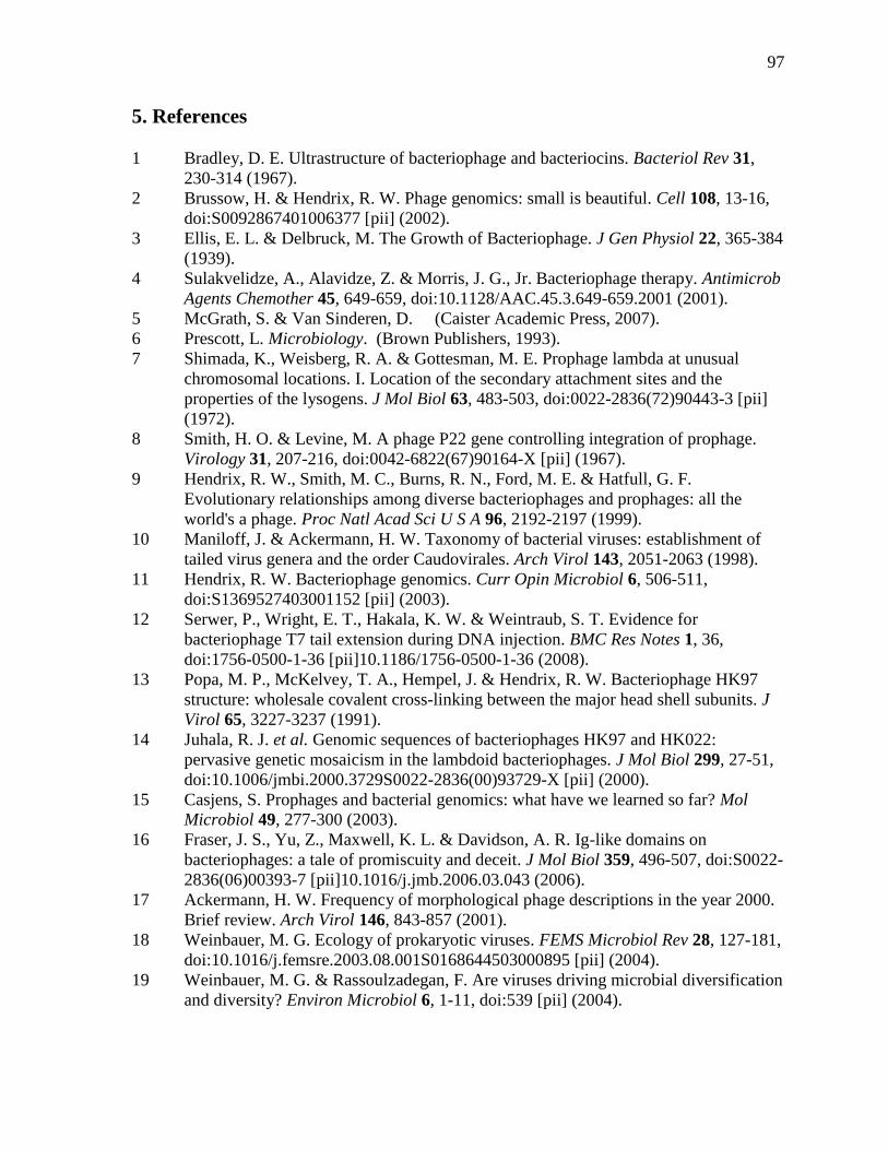
97
5. References
1 Bradley, D. E. Ultrastructure of bacteriophage and bacteriocins. Bacteriol Rev 31,
230-314 (1967).
2 Brussow, H. & Hendrix, R. W. Phage genomics: small is beautiful. Cell 108, 13-16,
doi:S0092867401006377 [pii] (2002).
3 Ellis, E. L. & Delbruck, M. The Growth of Bacteriophage. J Gen Physiol 22, 365-384
(1939).
4 Sulakvelidze, A., Alavidze, Z. & Morris, J. G., Jr. Bacteriophage therapy. Antimicrob
Agents Chemother 45, 649-659, doi:10.1128/AAC.45.3.649-659.2001 (2001).
5 McGrath, S. & Van Sinderen, D. (Caister Academic Press, 2007).
6 Prescott, L. Microbiology. (Brown Publishers, 1993).
7 Shimada, K., Weisberg, R. A. & Gottesman, M. E. Prophage lambda at unusual
chromosomal locations. I. Location of the secondary attachment sites and the
properties of the lysogens. J Mol Biol 63, 483-503, doi:0022-2836(72)90443-3 [pii]
(1972).
8 Smith, H. O. & Levine, M. A phage P22 gene controlling integration of prophage.
Virology 31, 207-216, doi:0042-6822(67)90164-X [pii] (1967).
9 Hendrix, R. W., Smith, M. C., Burns, R. N., Ford, M. E. & Hatfull, G. F.
Evolutionary relationships among diverse bacteriophages and prophages: all the
world's a phage. Proc Natl Acad Sci U S A 96, 2192-2197 (1999).
10 Maniloff, J. & Ackermann, H. W. Taxonomy of bacterial viruses: establishment of
tailed virus genera and the order Caudovirales. Arch Virol 143, 2051-2063 (1998).
11 Hendrix, R. W. Bacteriophage genomics. Curr Opin Microbiol 6, 506-511,
doi:S1369527403001152 [pii] (2003).
12 Serwer, P., Wright, E. T., Hakala, K. W. & Weintraub, S. T. Evidence for
bacteriophage T7 tail extension during DNA injection. BMC Res Notes 1, 36,
doi:1756-0500-1-36 [pii]10.1186/1756-0500-1-36 (2008).
13 Popa, M. P., McKelvey, T. A., Hempel, J. & Hendrix, R. W. Bacteriophage HK97
structure: wholesale covalent cross-linking between the major head shell subunits. J
Virol 65, 3227-3237 (1991).
14 Juhala, R. J. et al. Genomic sequences of bacteriophages HK97 and HK022:
pervasive genetic mosaicism in the lambdoid bacteriophages. J Mol Biol 299, 27-51,
doi:10.1006/jmbi.2000.3729S0022-2836(00)93729-X [pii] (2000).
15 Casjens, S. Prophages and bacterial genomics: what have we learned so far? Mol
Microbiol 49, 277-300 (2003).
16 Fraser, J. S., Yu, Z., Maxwell, K. L. & Davidson, A. R. Ig-like domains on
bacteriophages: a tale of promiscuity and deceit. J Mol Biol 359, 496-507, doi:S0022-
2836(06)00393-7 [pii]10.1016/j.jmb.2006.03.043 (2006).
17 Ackermann, H. W. Frequency of morphological phage descriptions in the year 2000.
Brief review. Arch Virol 146, 843-857 (2001).
18 Weinbauer, M. G. Ecology of prokaryotic viruses. FEMS Microbiol Rev 28, 127-181,
doi:10.1016/j.femsre.2003.08.001S0168644503000895 [pii] (2004).
19 Weinbauer, M. G. & Rassoulzadegan, F. Are viruses driving microbial diversification
and diversity? Environ Microbiol 6, 1-11, doi:539 [pii] (2004).
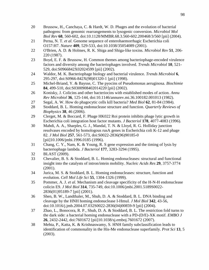
98
20 Brussow, H., Canchaya, C. & Hardt, W. D. Phages and the evolution of bacterial
pathogens: from genomic rearrangements to lysogenic conversion. Microbiol Mol
Biol Rev 68, 560-602, doi:10.1128/MMBR.68.3.560-602.200468/3/560 [pii] (2004).
21 Perna, N. T. et al. Genome sequence of enterohaemorrhagic Escherichia coli
O157:H7. Nature 409, 529-533, doi:10.1038/35054089 (2001).
22 O'Brien, A. D. & Holmes, R. K. Shiga and Shiga-like toxins. Microbiol Rev 51, 206-
220 (1987).
23 Boyd, E. F. & Brussow, H. Common themes among bacteriophage-encoded virulence
factors and diversity among the bacteriophages involved. Trends Microbiol 10, 521-
529, doi:S0966842X02024599 [pii] (2002).
24 Waldor, M. K. Bacteriophage biology and bacterial virulence. Trends Microbiol 6,
295-297, doi:S0966-842X(98)01320-1 [pii] (1998).
25 Michel-Briand, Y. & Baysse, C. The pyocins of Pseudomonas aeruginosa. Biochimie
84, 499-510, doi:S0300908402014220 [pii] (2002).
26 Konisky, J. Colicins and other bacteriocins with established modes of action. Annu
Rev Microbiol 36, 125-144, doi:10.1146/annurev.mi.36.100182.001013 (1982).
27 Segal, A. W. How do phagocytic cells kill bacteria? Med Biol 62, 81-84 (1984).
28 Stoddard, B. L. Homing endonuclease structure and function. Quarterly Reviews of
Biophysics 38, 46 (2006).
29 Clerget, M. & Boccard, F. Phage HK022 Roi protein inhibits phage lytic growth in
Escherichia coli integration host factor mutants. J Bacteriol 178, 4077-4083 (1996).
30 Mahdi, A. A., Sharples, G. J., Mandal, T. N. & Lloyd, R. G. Holliday junction
resolvases encoded by homologous rusA genes in Escherichia coli K-12 and phage
82. J Mol Biol 257, 561-573, doi:S0022-2836(96)90185-0
[pii]10.1006/jmbi.1996.0185 (1996).
31 Chang, C. Y., Nam, K. & Young, R. S gene expression and the timing of lysis by
bacteriophage lambda. J Bacteriol 177, 3283-3294 (1995).
32 BLAST (2009).
33 Chevalier, B. S. & Stoddard, B. L. Homing endonucleases: structural and functional
insight into the catalysts of intron/intein mobility. Nucleic Acids Res 29, 3757-3774
(2001).
34 Jurica, M. S. & Stoddard, B. L. Homing endonucleases: structure, function and
evolution. Cell Mol Life Sci 55, 1304-1326 (1999).
35 Pommer, A. J. et al. Mechanism and cleavage specificity of the H-N-H endonuclease
colicin E9. J Mol Biol 314, 735-749, doi:10.1006/jmbi.2001.5189S0022-
2836(01)95189-7 [pii] (2001).
36 Shen, B. W., Landthaler, M., Shub, D. A. & Stoddard, B. L. DNA binding and
cleavage by the HNH homing endonuclease I-HmuI. J Mol Biol 342, 43-56,
doi:10.1016/j.jmb.2004.07.032S0022-2836(04)00859-9 [pii] (2004).
37 Zhao, L., Bonocora, R. P., Shub, D. A. & Stoddard, B. L. The restriction fold turns to
the dark side: a bacterial homing endonuclease with a PD-(D/E)-XK motif. EMBO J
26, 2432-2442, doi:7601672 [pii]10.1038/sj.emboj.7601672 (2007).
38 Mehta, P., Katta, K. & Krishnaswamy, S. HNH family subclassification leads to
identification of commonality in the His-Me endonuclease superfamily. Prot Sci 13, 5
(2003).
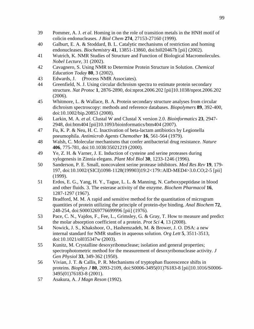
99
39 Pommer, A. J. et al. Homing in on the role of transition metals in the HNH motif of
colicin endonucleases. J Biol Chem 274, 27153-27160 (1999).
40 Galburt, E. A. & Stoddard, B. L. Catalytic mechanisms of restriction and homing
endonucleases. Biochemistry 41, 13851-13860, doi:bi020467h [pii] (2002).
41 Wutrich, K. NMR Studies of Structure and Function of Biological Macromolecules.
Nobel Lecture, 31 (2002).
42 Cavagnero, S. Using NMR to Determine Protein Structure in Solution. Chemical
Education Today 80, 3 (2002).
43 Edwards, J. (Process NMR Associates).
44 Greenfield, N. J. Using circular dichroism spectra to estimate protein secondary
structure. Nat Protoc 1, 2876-2890, doi:nprot.2006.202 [pii]10.1038/nprot.2006.202
(2006).
45 Whitmore, L. & Wallace, B. A. Protein secondary structure analyses from circular
dichroism spectroscopy: methods and reference databases. Biopolymers 89, 392-400,
doi:10.1002/bip.20853 (2008).
46 Larkin, M. A. et al. Clustal W and Clustal X version 2.0. Bioinformatics 23, 2947-
2948, doi:btm404 [pii]10.1093/bioinformatics/btm404 (2007).
47 Fu, K. P. & Neu, H. C. Inactivation of beta-lactam antibiotics by Legionella
pneumophila. Antimicrob Agents Chemother 16, 561-564 (1979).
48 Walsh, C. Molecular mechanisms that confer antibacterial drug resistance. Nature
406, 775-781, doi:10.1038/35021219 (2000).
49 Ye, Z. H. & Varner, J. E. Induction of cysteine and serine proteases during
xylogenesis in Zinnia elegans. Plant Mol Biol 30, 1233-1246 (1996).
50 Sanderson, P. E. Small, noncovalent serine protease inhibitors. Med Res Rev 19, 179-
197, doi:10.1002/(SICI)1098-1128(199903)19:2<179::AID-MED4>3.0.CO;2-5 [pii]
(1999).
51 Erdos, E. G., Yang, H. Y., Tague, L. L. & Manning, N. Carboxypeptidase in blood
and other fluids. 3. The esterase activity of the enzyme. Biochem Pharmacol 16,
1287-1297 (1967).
52 Bradford, M. M. A rapid and sensitive method for the quantitation of microgram
quantities of protein utilizing the principle of protein-dye binding. Anal Biochem 72,
248-254, doi:S0003269776699996 [pii] (1976).
53 Pace, C. N., Vajdos, F., Fee, L., Grimsley, G. & Gray, T. How to measure and predict
the molar absorption coefficient of a protein. Prot Sci 4, 13 (2008).
54 Nowick, J. S., Khakshoor, O., Hashemzadeh, M. & Brower, J. O. DSA: a new
internal standard for NMR studies in aqueous solution. Org Lett 5, 3511-3513,
doi:10.1021/ol035347w (2003).
55 Kunitz, M. Crystalline desoxyribonuclease; isolation and general properties;
spectrophotometric method for the measurement of desoxyribonuclease activity. J
Gen Physiol 33, 349-362 (1950).
56 Vivian, J. T. & Callis, P. R. Mechanisms of tryptophan fluorescence shifts in
proteins. Biophys J 80, 2093-2109, doi:S0006-3495(01)76183-8 [pii]10.1016/S0006-
3495(01)76183-8 (2001).
57 Asakura, A. J Magn Reson (1992).
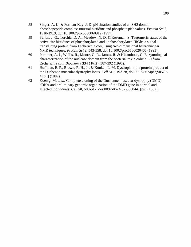
100
58 Singer, A. U. & Forman-Kay, J. D. pH titration studies of an SH2 domain-
phosphopeptide complex: unusual histidine and phosphate pKa values. Protein Sci 6,
1910-1919, doi:10.1002/pro.5560060912 (1997).
59 Pelton, J. G., Torchia, D. A., Meadow, N. D. & Roseman, S. Tautomeric states of the
active-site histidines of phosphorylated and unphosphorylated IIIGlc, a signal-
transducing protein from Escherichia coli, using two-dimensional heteronuclear
NMR techniques. Protein Sci 2, 543-558, doi:10.1002/pro.5560020406 (1993).
60 Pommer, A. J., Wallis, R., Moore, G. R., James, R. & Kleanthous, C. Enzymological
characterization of the nuclease domain from the bacterial toxin colicin E9 from
Escherichia coli. Biochem J 334 ( Pt 2), 387-392 (1998).
61 Hoffman, E. P., Brown, R. H., Jr. & Kunkel, L. M. Dystrophin: the protein product of
the Duchenne muscular dystrophy locus. Cell 51, 919-928, doi:0092-8674(87)90579-
4 [pii] (1987).
62 Koenig, M. et al. Complete cloning of the Duchenne muscular dystrophy (DMD)
cDNA and preliminary genomic organization of the DMD gene in normal and
affected individuals. Cell 50, 509-517, doi:0092-8674(87)90504-6 [pii] (1987).
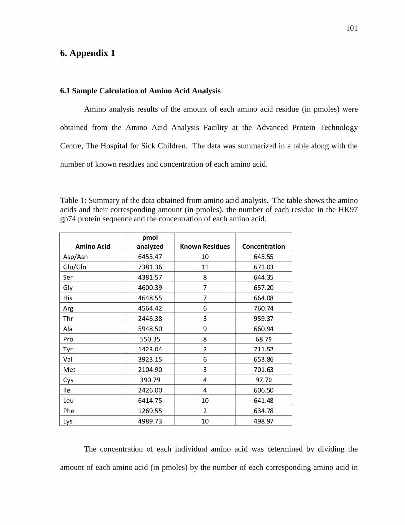
101
6. Appendix 1
6.1 Sample Calculation of Amino Acid Analysis
Amino analysis results of the amount of each amino acid residue (in pmoles) were
obtained from the Amino Acid Analysis Facility at the Advanced Protein Technology
Centre, The Hospital for Sick Children. The data was summarized in a table along with the
number of known residues and concentration of each amino acid.
Table 1: Summary of the data obtained from amino acid analysis. The table shows the amino
acids and their corresponding amount (in pmoles), the number of each residue in the HK97
gp74 protein sequence and the concentration of each amino acid.
Amino Acid pmol
analyzed Known Residues Concentration
Asp/Asn 6455.47 10 645.55
Glu/Gln 7381.36 11 671.03
Ser 4381.57 8 644.35
Gly 4600.39 7 657.20
His 4648.55 7 664.08
Arg 4564.42 6 760.74
Thr 2446.38 3 959.37
Ala 5948.50 9 660.94
Pro 550.35 8 68.79
Tyr 1423.04 2 711.52
Val 3923.15 6 653.86
Met 2104.90 3 701.63
Cys 390.79 4 97.70
Ile 2426.00 4 606.50
Leu 6414.75 10 641.48
Phe 1269.55 2 634.78
Lys 4989.73 10 498.97
The concentration of each individual amino acid was determined by dividing the
amount of each amino acid (in pmoles) by the number of each corresponding amino acid in
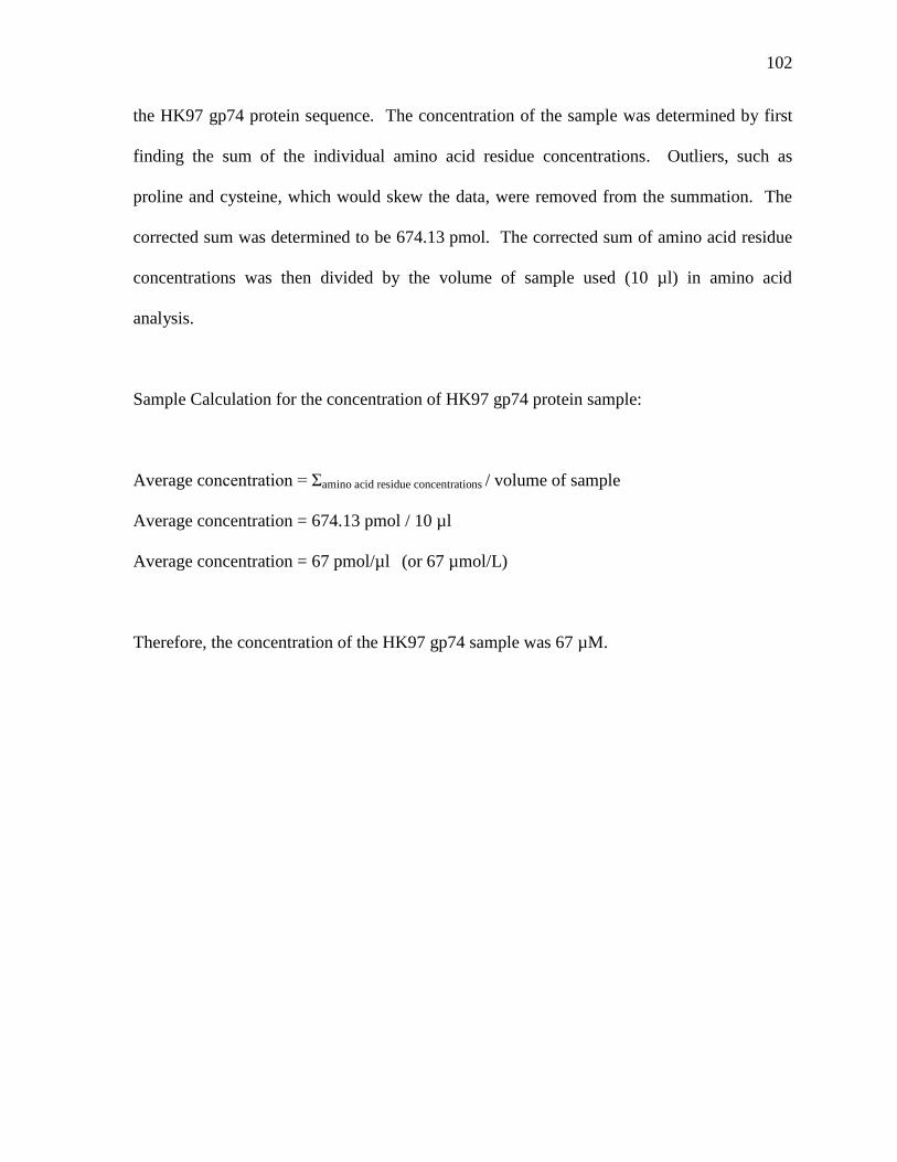
102
the HK97 gp74 protein sequence. The concentration of the sample was determined by first
finding the sum of the individual amino acid residue concentrations. Outliers, such as
proline and cysteine, which would skew the data, were removed from the summation. The
corrected sum was determined to be 674.13 pmol. The corrected sum of amino acid residue
concentrations was then divided by the volume of sample used (10 µl) in amino acid
analysis.
Sample Calculation for the concentration of HK97 gp74 protein sample:
Average concentration = Σamino acid residue concentrations / volume of sample
Average concentration = 674.13 pmol / 10 µl
Average concentration = 67 pmol/µl (or 67 µmol/L)
Therefore, the concentration of the HK97 gp74 sample was 67 µM.