bioanalytical applications of polyion-sensitive electrodes
TRANSCRIPT
Journal of Pharmaceutical and Biomedical Analysis19 (1999) 1–14
Bioanalytical applications of polyion-sensitive electrodes
S. Dai a, J.M. Esson a, O. Lutze a, N. Ramamurthy a, V.C. Yang b, M.E. Meyerhoff a,*a Department of Chemistry, The Uni6ersity of Michigan, Ann Arbor, MI, 48109-1055, USA
b College of Pharmacy, The Uni6ersity of Michigan, Ann Arbor, MI, 48109-1065, USA
Received 27 January 1998; received in revised form 18 May 1998; accepted 18 May 1998
Abstract
Recent developments in the design and bioanalytical applications of polyion-sensitive electrodes (PSEs) arereviewed. The general electrochemical principles governing the potentiometric response of such polymer membrane-based devices are summarized and new directions for the use of these novel sensors are detailed. These new directionsinclude basic fundamental studies aimed at determining the thermodynamics of polyion extraction into ionexchanger-doped polymeric membranes, new methods to quantitate the anticoagulant drug heparin in whole bloodvia titrations with polycationic protamine, selective assays of protease activities (and inhibitors of such activities)using natural and synthetic polyionic peptides as substrates, and novel homogeneous immunoassay schemes based onpotentiometric polyion detection. © 1999 Elsevier Science B.V. All rights reserved.
Keywords: Polyion-sensitive electrode; Protease assays; Non-separation immunoassay; Heparin determination
1. Introduction
Recently, we have found that polymeric mem-branes (e.g. plasticized poly(vinyl chloride)) dopedwith appropriate lipophilic ion-exchangers candisplay significant non-equilibrium potentiometricresponse (EMF) to biologically importantpolyionic species (e.g. heparin, protamine,polyphosphates, DNA, etc.) [1–11]. When assem-bled in typical ion-selective membrane electrodeconfigurations, the resulting sensors exhibit poten-
tiometric responses to sub-mM levels of polyionsin samples as complex as whole blood [2,8]. Suchresponse has been attributed to a highly selectivenon-equilibrium extraction of the polyion into themembrane phase via a cooperative ion-pairinginteraction with the lipophilic ion-exchangers[4,6]. This extraction process leads to a steady-state change in the phase boundary potential atthe membrane/sample interface. For example,membranes formulated with tridodecylmethylam-monium chloride (TDMAC), rapidly exchangechloride for certain polyanions, and thus exhibit avery large potentiometric response to such species(including porcine and beef lung heparins), withthe magnitude of response dependent on both thechain length and charge density of the polyanion
* Corresponding author. Tel.: +1 734 7635916; fax: +1734 6474865; e-mail: [email protected]
0731-7085/99/$ - see front matter © 1999 Elsevier Science B.V. All rights reserved.
PII S0731-7085(98)00134-4
S. Dai et al. / J. Pharm. Biomed. Anal. 19 (1999) 1–142
structure [6]. In contrast, polymer membranescontaining tetraphenylborate derivatives or di-nonylnaphthalene sulfonate sites (DNNS) re-spond potentiometrically to low levels ofpolycations, including the antidote of heparin,protamine, as well as synthetic peptides rich inarginine residues [7,10]. Such response is reversedwhen specific proteases cleave these polycationicpeptides into smaller fragments that are not fa-vorably extracted into the organic membranephase, providing a convenient electrochemicalmethod to detect protease activities [10].
Herein, we summarize results from our mostrecent fundamental and applied studies involvingthese new polyion sensitive electrodes (PSE). Abrief discussion of the mechanism of potentiomet-ric polyion response is provided and the roles ofpolyion molecular weight, lipophilicity and chargedensity on the magnitude of EMF response areassessed using different polyphosphate, poly(amino acid), and nucleic acid structures as testpolyanions. Various methods to use polyion sen-sors for the determination of heparin levels inblood are also reviewed, including a new pro-tamine titration method that functions effectivelyfor monitoring the concentrations of both conven-tional and low molecular weight heparin prepara-tions in whole blood (using a DNNS-basedpolymer membrane electrode as the end-point de-tector). Other novel bioanalytical applications ofPSE detection are discussed, including the abilityto monitor specific protease activities and in-hibitors, and the design of novel non-separationelectrochemical immunoassays for small hapten/drug molecules.
2. Fundamental studies
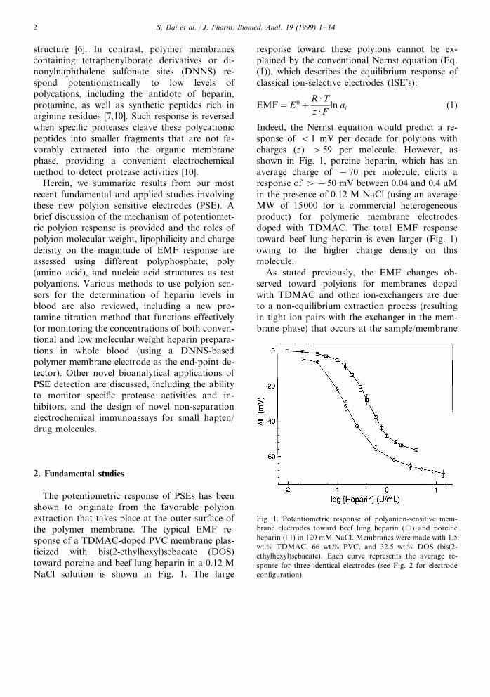
The potentiometric response of PSEs has beenshown to originate from the favorable polyionextraction that takes place at the outer surface ofthe polymer membrane. The typical EMF re-sponse of a TDMAC-doped PVC membrane plas-ticized with bis(2-ethylhexyl)sebacate (DOS)toward porcine and beef lung heparin in a 0.12 MNaCl solution is shown in Fig. 1. The large
response toward these polyions cannot be ex-plained by the conventional Nernst equation (Eq.(1)), which describes the equilibrium response ofclassical ion-selective electrodes (ISE’s):
EMF=E0+R ·Tz ·F
ln ai (1)
Indeed, the Nernst equation would predict a re-sponse of B1 mV per decade for polyions withcharges (z) \59 per molecule. However, asshown in Fig. 1, porcine heparin, which has anaverage charge of −70 per molecule, elicits aresponse of \−50 mV between 0.04 and 0.4 mMin the presence of 0.12 M NaCl (using an averageMW of 15000 for a commercial heterogeneousproduct) for polymeric membrane electrodesdoped with TDMAC. The total EMF responsetoward beef lung heparin is even larger (Fig. 1)owing to the higher charge density on thismolecule.
As stated previously, the EMF changes ob-served toward polyions for membranes dopedwith TDMAC and other ion-exchangers are dueto a non-equilibrium extraction process (resultingin tight ion pairs with the exchanger in the mem-brane phase) that occurs at the sample/membrane
Fig. 1. Potentiometric response of polyanion-sensitive mem-brane electrodes toward beef lung heparin (�) and porcineheparin ( ) in 120 mM NaCl. Membranes were made with 1.5wt.% TDMAC, 66 wt.% PVC, and 32.5 wt.% DOS (bis(2-ethylhexyl)sebacate). Each curve represents the average re-sponse for three identical electrodes (see Fig. 2 for electrodeconfiguration).
S. Dai et al. / J. Pharm. Biomed. Anal. 19 (1999) 1–14 3
interface [4,6]. At high concentrations of thepolyion, the response levels off (Fig. 1) as thesurface of the membrane achieves electrochemicalequilibrium with the polyion in the sample phase(i.e. all the chloride of TDMAC is replaced byheparin). However, at lower sample concentra-tions of the polyion, a steady-state situation ex-ists at the interface, where the flux of polyion tothe surface is equal to the flux of the polyion-ex-changer ion pairs into the bulk of the polymericmembrane. The change in EMF in this concen-tration regime is given by the following equation[4]:
DEMF =RTF
ln(1−zDadm
RTDmda
cpolyion) (2)
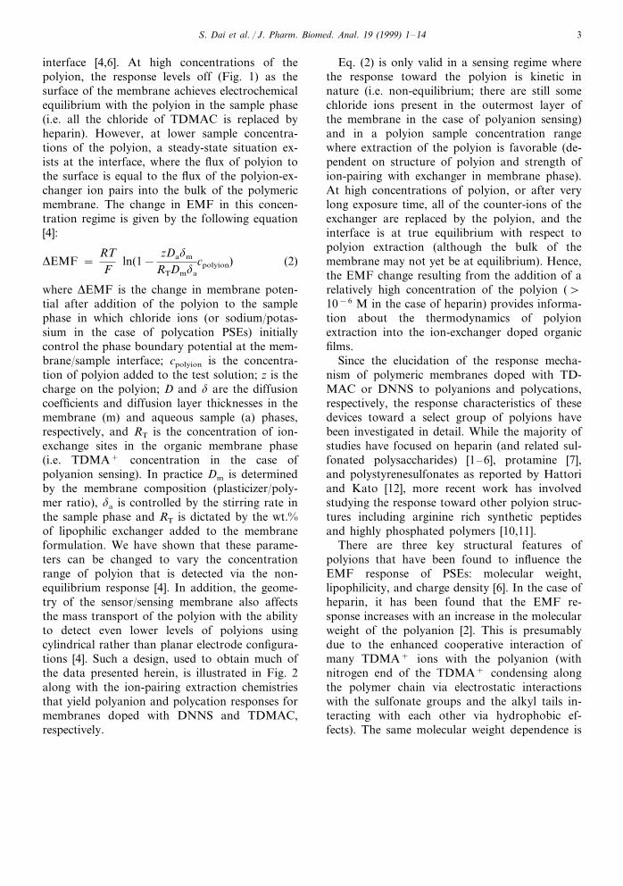
where DEMF is the change in membrane poten-tial after addition of the polyion to the samplephase in which chloride ions (or sodium/potas-sium in the case of polycation PSEs) initiallycontrol the phase boundary potential at the mem-brane/sample interface; cpolyion is the concentra-tion of polyion added to the test solution; z is thecharge on the polyion; D and d are the diffusioncoefficients and diffusion layer thicknesses in themembrane (m) and aqueous sample (a) phases,respectively, and RT is the concentration of ion-exchange sites in the organic membrane phase(i.e. TDMA+ concentration in the case ofpolyanion sensing). In practice Dm is determinedby the membrane composition (plasticizer/poly-mer ratio), da is controlled by the stirring rate inthe sample phase and RT is dictated by the wt.%of lipophilic exchanger added to the membraneformulation. We have shown that these parame-ters can be changed to vary the concentrationrange of polyion that is detected via the non-equilibrium response [4]. In addition, the geome-try of the sensor/sensing membrane also affectsthe mass transport of the polyion with the abilityto detect even lower levels of polyions usingcylindrical rather than planar electrode configura-tions [4]. Such a design, used to obtain much ofthe data presented herein, is illustrated in Fig. 2along with the ion-pairing extraction chemistriesthat yield polyanion and polycation responses formembranes doped with DNNS and TDMAC,respectively.
Eq. (2) is only valid in a sensing regime wherethe response toward the polyion is kinetic innature (i.e. non-equilibrium; there are still somechloride ions present in the outermost layer ofthe membrane in the case of polyanion sensing)and in a polyion sample concentration rangewhere extraction of the polyion is favorable (de-pendent on structure of polyion and strength ofion-pairing with exchanger in membrane phase).At high concentrations of polyion, or after verylong exposure time, all of the counter-ions of theexchanger are replaced by the polyion, and theinterface is at true equilibrium with respect topolyion extraction (although the bulk of themembrane may not yet be at equilibrium). Hence,the EMF change resulting from the addition of arelatively high concentration of the polyion (\10−6 M in the case of heparin) provides informa-tion about the thermodynamics of polyionextraction into the ion-exchanger doped organicfilms.
Since the elucidation of the response mecha-nism of polymeric membranes doped with TD-MAC or DNNS to polyanions and polycations,respectively, the response characteristics of thesedevices toward a select group of polyions havebeen investigated in detail. While the majority ofstudies have focused on heparin (and related sul-fonated polysaccharides) [1–6], protamine [7],and polystyrenesulfonates as reported by Hattoriand Kato [12], more recent work has involvedstudying the response toward other polyion struc-tures including arginine rich synthetic peptidesand highly phosphated polymers [10,11].
There are three key structural features ofpolyions that have been found to influence theEMF response of PSEs: molecular weight,lipophilicity, and charge density [6]. In the case ofheparin, it has been found that the EMF re-sponse increases with an increase in the molecularweight of the polyanion [2]. This is presumablydue to the enhanced cooperative interaction ofmany TDMA+ ions with the polyanion (withnitrogen end of the TDMA+ condensing alongthe polymer chain via electrostatic interactionswith the sulfonate groups and the alkyl tails in-teracting with each other via hydrophobic ef-fects). The same molecular weight dependence is
S. Dai et al. / J. Pharm. Biomed. Anal. 19 (1999) 1–144
Fig. 2. Schematic of simple cylindrical PSE configuration, as well as an expanded view of the extraction and ion-pairing reactionsof (A) polycations and (B) polyanions into and within the organic membrane phases of such devices.
also observed in the case of polyanions containingphosphate groups. As shown in Table 1, TD-MAC-based PSEs elicit larger equilibrium poten-tial changes to a 66-mer polyphosphate(−86.691.1 mV) compared to a 5-mer
polyphosphate (−62.292.0 mV), and to a 12-mer DNA oligomer (−32.793.9 mV) comparedto an 8-mer DNA oligomer (−5.592.2 mV).This trend has also been observed for peptidehomopolymers containing lysine, aspartic acid,
S. Dai et al. / J. Pharm. Biomed. Anal. 19 (1999) 1–14 5
and glutamic acid. By comparing the EMF re-sponses of two different membranes, one dopedwith TDMAC and the other with the ion-ex-changer tetradodecylammonium chloride (TDAC)(which does not ion-pair strongly with polyanionsdue to steric hindrance), the free energy of ion-pairing in the membrane phase can be estimatedaccording to Eq. (3)[6].
Dg IP0 = −{DEMF(TDAC)−DEMF(TDMAC)}F
+RT ln[R−] (3)
Table 1 reports that the apparent free energy ofion-pairing per charge increases from −7.2390.21 kJ mol−1 for the 8-mer DNA oligomer to−9.1490.39 kJ mol−1 for the 12-mer DNAoligomer. A schematic representation of the ion-pairing interaction of polyions with TDMAC andDNNS that occur in the sensing polymer mem-branes of PSEs is illustrated in Fig. 2.
Charge density is another aspect of polyionstructure that influences the magnitude of EMFresponse observed. It has been shown in the caseof sulfonated polysaccharides that the EMF re-sponse toward heparins is much greater than thatobserved for dermatan sulfate and various car-rageenans [6], species that are structurally similarbut possess less total sulfonate groups per linearsegment of carbohydrate polymer. For phosphate-based polyanions, three different polyanions thathave the same number of charged groups, butdifferent spacing between the charges, have alsobeen examined (pentaphosphate, diadenosine pen-taphosphate (a linear polyphosphate with twoadenosine units at either end) and pentaadenylicacid). Pentaphosphate was found to elicit a non-equilibrium potential change of −16.890.1 mVat 0.63 mg ml−1 in 10 mM NaCl, while 10 mgml−1 of diadenosine polyphosphate was requiredto yield a comparable response (−15.690.9mV). No potential change occurred when theelectrode was exposed to pentaadenylic acid,which possesses the lowest charge density. Thus,polyions with higher charge density will elicitlarger EMF changes.
Lastly, the lipophilicity of the polyion structurealso plays a significant role in the magnitude ofthe potentiometric response observed. For exam-ple, the EMF response of a TDMAC-based mem-brane to two DNA oligomers with the samecharge density, but different lipophilicity, has re-cently been compared [11]. The 9-merpolyadenylic acid elicits a non-equilibrium poten-tial change of −8.491.3 mV at a concentrationof 3.8 mg ml−1, while no response is seen for theless lipophilic 9-mer polythymidylic acid at thesame concentration. Similarly, earlier work withsynthetic arginine rich synthetic peptides demon-strated that the total equilibrium EMF responseof DNNS-based membranes toward these struc-tures is enhanced when the peptides contain thelipophilic amino acids phenylalanine and leucinewithin their structure [10].
Knowing the effects of polyion structural char-acteristics on the magnitude of the EMF changestoward such species is extremely valuable in thedevelopment of new bioanalytical methods basedon PSE detection. For example, as described be-
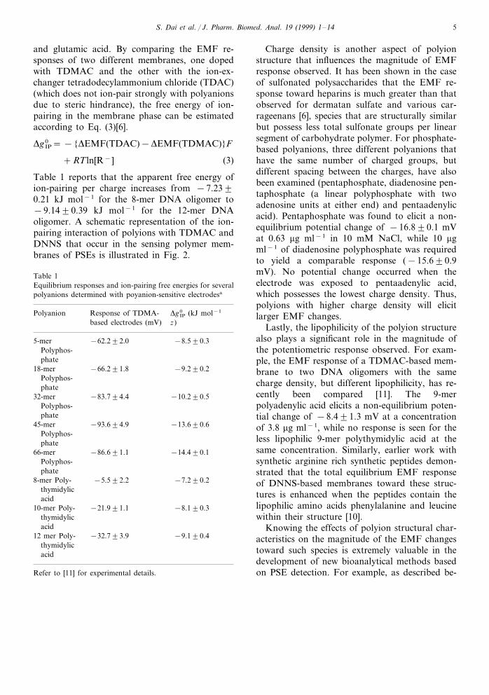
Table 1Equilibrium responses and ion-pairing free energies for severalpolyanions determined with poyanion-sensitive electrodesa
Polyanion Response of TDMA- Dg IP0 (kJ mol−1
based electrodes (mV) z)
−8.590.35-mer −62.292.0Polyphos-phate
18-mer −66.291.8 −9.290.2Polyphos-phate
−10.290.532-mer −83.794.4Polyphos-phate
−93.694.945-mer −13.690.6Polyphos-phate
66-mer −86.691.1 −14.490.1Polyphos-phate
8-mer Poly- −5.592.2 −7.290.2thymidylicacid
10-mer Poly- −8.190.3−21.991.1thymidylicacid
−32.793.912 mer Poly- −9.190.4thymidylicacid
Refer to [11] for experimental details.
S. Dai et al. / J. Pharm. Biomed. Anal. 19 (1999) 1–146
low (Section 3.2), the design of synthetic polypep-tide substrates for new protease assays based onelectrochemical polyion detection has been guidedto a great extent by understanding the role ofcharge density, molecular weight, and lipophilicityon the response of PSEs toward targeted polyionstructures.
3. Biomedical applications
There are two unique response characteristicsthat have contributed to the use of PSEs inbiomedical applications. First, since PSEs respondto only free concentrations of polyion (duringshort measurement periods when the polyion par-ticipates in relatively strong binding reactions(Keq\106 M−1); i.e. slow off rate constants), thismakes them very useful detectors in potentiomet-ric titrations to determine concentrations ofpolyionic drugs, including heparin and protamine(Section 3.1), and in the development of novelnon-separation immunoassays (Section 3.3). Sec-ond, the molecular weight dependence of EMFresponse enables the use of PSEs as detectors forenzyme activity determinations, as well as formonitoring levels of specific enzyme inhibitors(drugs). Both of these bioanalytical applicationswill be described in Section 3.2.
3.1. Heparin determinations
One exciting application of PSEs is for thedetermination of heparin levels in whole bloodsamples from patients undergoing open heartsurgery. Heparin is the most widely used antico-agulant in major surgical and extracorporeal pro-cedures, such as coronary artery bypass surgeryand hemodialysis. Heparin doses given to patientsduring bypass surgery are typically in the range of2–8 U ml−1 (equivalent to 0.8–3.2 mM) [13].However, it is essential to monitor the anticoagu-lant effect and, ideally, also the concentration ofheparin during surgery, since higher doses of hep-arin are often associated with the risk of hemor-rhage [14]. Currently, the most widely usedclinical procedure to monitor anticoagulant activ-ity is the activated clotting time (ACT), which is
the time required for clot formation in wholeblood after contact activation with agents such askaolin. However, the ACT value is not necessarilya very accurate indicator of blood heparin levelssince clotting time can also be affected by varia-tions in temperature and hemodilution, which arecommonly encountered during surgery [15]. Meth-ods used to specifically determine the concentra-tion of heparin in blood include a colorimetricanti-Xa assay (which measures heparin by itsability to inhibit Factor-Xa activity) [16] and aprotamine titration-based clotting assay (HepconHMS, Medtronic Blood Management, Parker,CO). These assays are based on the detection ofclot formation (Hepcon HMS) or a color changein the corresponding plasma sample (anti-Xa as-say using chromogenic substrates) and, hence, canfunction poorly in samples that are highly colored(i.e. whole blood) or devoid of clotting factors(i.e. serum).
PSEs provide a relatively simple means to de-termine heparin levels accurately in whole blood.Such heparin determinations are readily per-formed via classical potentiometric titrations us-ing protamine, a polycationic protein rich inarginine, as the titrant. Protamine binds stoichio-metrically, via electrostatic interactions, with hep-arin (with affinity constant \107 M−1) [3]. SincePSEs respond only to free polyion and not thecomplexed form, they can serve as end-point de-tectors for such titrations.
In our earliest work, TDMAC-doped PSEswere used to determine heparin levels in wholeblood samples via pseudo-titrations with pro-tamine [8]. A direct titration could not be per-formed in this case due to the irreversibility of themembrane electrode response after exposure to asample containing heparin. Such pseudo-titrationswere carried out by adding small volumes (250 ml)of blood to a series of tubes containing known,increasing amounts of protamine. The potentio-metric response of the polyanion-sensitive elec-trode was noted upon sequentially switching theelectrode (and appropriate reference electrode)from the tube containing the highest to the leastamount of protamine. A decrease in potential wasobserved in those tubes where the heparin presentin the blood was in excess of the protamine in
S. Dai et al. / J. Pharm. Biomed. Anal. 19 (1999) 1–14 7
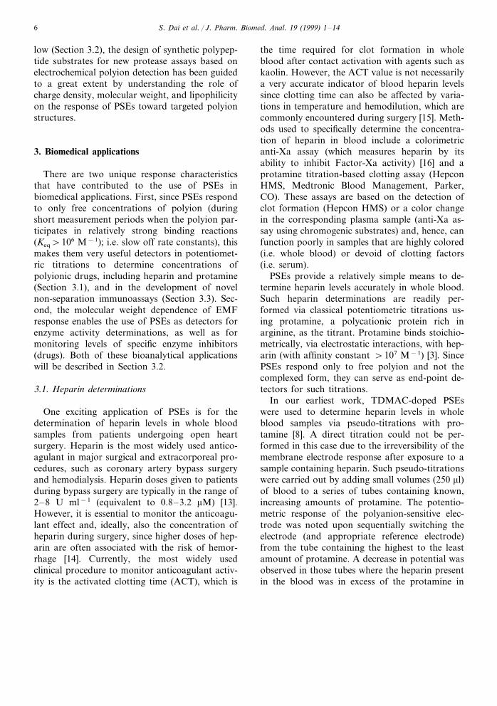
Fig. 3. Typical potentiometric titration of 0.0 ( ), 0.5 (�), 1.0(�) and 2.0 (�) U ml−1 of porcine heparin with protamine asmonitored by polycation-sensitive membrane electrodes con-taining 1 wt.% DNNS, 49.5 wt.% NPOE and 49.5 wt.%polyurethane-M48 in undiluted human plasma. The averageresponse of three identical electrodes is shown.
lations with the Hepcon HMS assay (r=0.934)and the previously mentioned polyanion-sensitiveelectrode method (r=0.973). In addition, reason-ably good correlation (r=0.895) was also foundto a colorimetric anti-Xa assay (Coatest; Chro-mogenix) after correction for blood hematocritlevels (since the anti-Xa assay functions only inplasma samples), thus demonstrating the analyti-cal utility of these electrodes for clinical heparindeterminations.
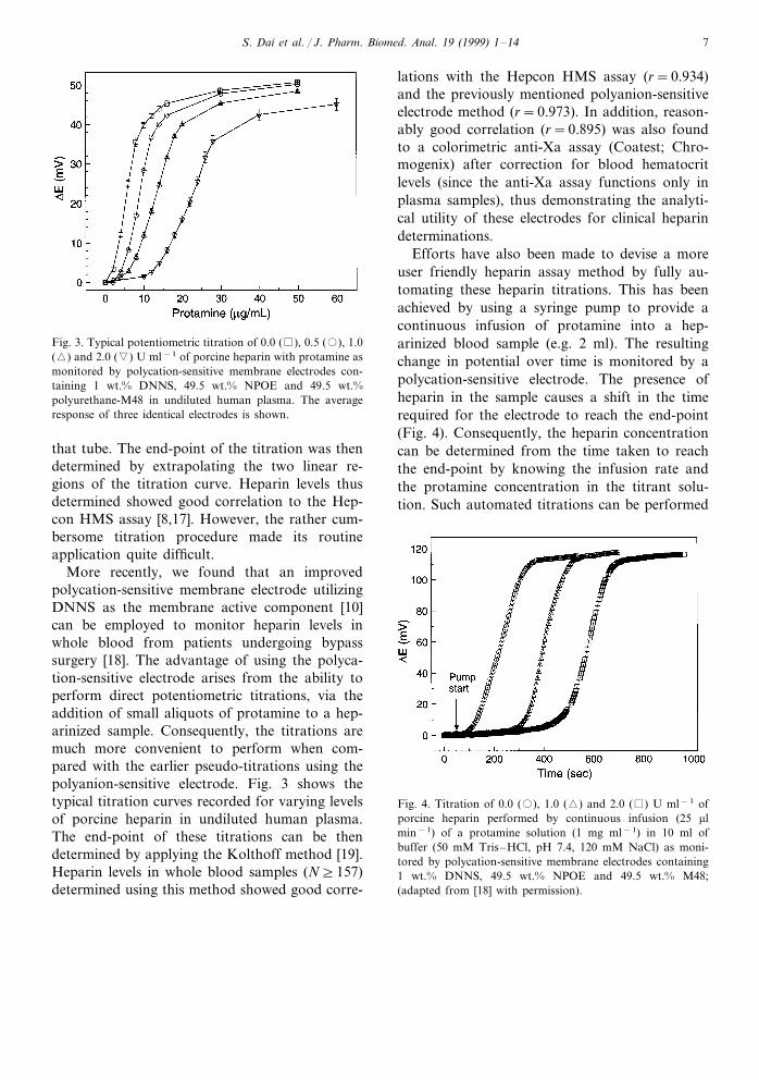
Efforts have also been made to devise a moreuser friendly heparin assay method by fully au-tomating these heparin titrations. This has beenachieved by using a syringe pump to provide acontinuous infusion of protamine into a hep-arinized blood sample (e.g. 2 ml). The resultingchange in potential over time is monitored by apolycation-sensitive electrode. The presence ofheparin in the sample causes a shift in the timerequired for the electrode to reach the end-point(Fig. 4). Consequently, the heparin concentrationcan be determined from the time taken to reachthe end-point by knowing the infusion rate andthe protamine concentration in the titrant solu-tion. Such automated titrations can be performed
that tube. The end-point of the titration was thendetermined by extrapolating the two linear re-gions of the titration curve. Heparin levels thusdetermined showed good correlation to the Hep-con HMS assay [8,17]. However, the rather cum-bersome titration procedure made its routineapplication quite difficult.
More recently, we found that an improvedpolycation-sensitive membrane electrode utilizingDNNS as the membrane active component [10]can be employed to monitor heparin levels inwhole blood from patients undergoing bypasssurgery [18]. The advantage of using the polyca-tion-sensitive electrode arises from the ability toperform direct potentiometric titrations, via theaddition of small aliquots of protamine to a hep-arinized sample. Consequently, the titrations aremuch more convenient to perform when com-pared with the earlier pseudo-titrations using thepolyanion-sensitive electrode. Fig. 3 shows thetypical titration curves recorded for varying levelsof porcine heparin in undiluted human plasma.The end-point of these titrations can be thendetermined by applying the Kolthoff method [19].Heparin levels in whole blood samples (N]157)determined using this method showed good corre-
Fig. 4. Titration of 0.0 (�), 1.0 (�) and 2.0 ( ) U ml−1 ofporcine heparin performed by continuous infusion (25 mlmin−1) of a protamine solution (1 mg ml−1) in 10 ml ofbuffer (50 mM Tris–HCl, pH 7.4, 120 mM NaCl) as moni-tored by polycation-sensitive membrane electrodes containing1 wt.% DNNS, 49.5 wt.% NPOE and 49.5 wt.% M48;(adapted from [18] with permission).
S. Dai et al. / J. Pharm. Biomed. Anal. 19 (1999) 1–148
in a relatively short period of time (B10 min).Multiple automated titrations performed on sam-ples spiked with varying levels of heparin (1.0 and2.0 U ml−1) show good accuracy and precision(1.0090.17 and 1.9790.25 U ml−1), thusproviding an even more rapid means to determineheparin in whole blood samples using PSEs.
Low molecular weight heparins (LMWHs) haveacquired increasing significance for the prophy-laxis of deep venous thromboembolisms (DVT) inpatients undergoing elective surgical proceduressuch as hip-replacement surgery. Almost 50% ofthe patients undergoing such surgical proceduresmay develop DVT and, hence, heparin therapy isnecessary. LMWHs are preferred to unfraction-ated heparin (UFH) in these procedures since theyhave much higher bio-availability (about 95%)and longer elimination half-lives. This makes theiradministration much simpler than UFH, whichhas rather long onset times (:1 h) when adminis-tered subcutaneously. Further, there is a low inci-dence of hemorrhage for patients on LMWHtherapy and this makes the use of this producteven more attractive. However, monitoring levelsof LMWH in blood can be quite difficult becausebroadly used clotting type-based assays, such asthe ACT or aPTT, are not affected significantlyby the presence of LMWHs. Thus, the only usefulapproach to measure the concentration of thesedrugs is to employ colorimetric anti-Xa assayswhich, of course, cannot be performed in wholeblood samples (only on diluted plasma).
The DNNS-based polycation-sensitive elec-trode, however, can also be used to monitor thetitrations of some LMWH preparations usingprotamine as the titrant. Potentiometric titrationsperformed on in vitro blood samples show thatFragmin (Pharmacia) can be determined at con-centrations that are typically encountered in clini-cal practice (0–2 U ml−1), with titration curvesquite similar to that observed for unfractionatedheparins (Fig. 3). However, some other prepara-tions (Lovenox, Rhone-Poulenc) are not as easilytitrated, with titration end-points less pro-nounced. The difference probably arises from thevarying methods adopted in the preparation ofthese LMWHs and the precise molecular weightdistribution of the final products. Indeed, it is
known that Lovenox has a lower average MWcompared to Fragmin, and shorter sulfatedpolysaccharides present in such a preparationlikely bind protamine with much lower affinity,making it more difficult to achieve sharp break-points using the titrimetric approach. However, itmay be possible to synthesize positively chargedpeptides (rich in arginine residues) that may bindmore tightly to these LMWHs than protamine,thus enabling this methodology to be morebroadly applied for measuring a wide range ofcommercial LMWHs in whole blood samples.Efforts in this direction are currently underway inthis laboratory.
3.2. Determination of enzyme acti6ities andenzyme inhibitors
Polyion-sensitive electrodes are also potentiallyuseful detectors in the design of novel electro-chemical enzyme assays. Using PSEs, it is possibleto determine the activity of given enzymes thatcleave polyionic substrate molecules into smallerfragments of less charge and lower molecularweight. Since the EMF response of PSEs is depen-dent on charge and molecular weight, the activityof the enzymes can be directly monitored byobserving the reversal in potentiometric responseof PSEs to specific polyionic substrates.
The first assay based on this novel detectionmethod was designed for trypsin, which cleavesarginine rich peptides, such as protamine, intosmaller fragments [7]. A reversal of the EMFresponse was recorded in real time after the en-zyme was added to a protamine solution. The rateof potential decrease was shown to be directlyproportional to the amount of trypsin activitypresent in the assay mixture.
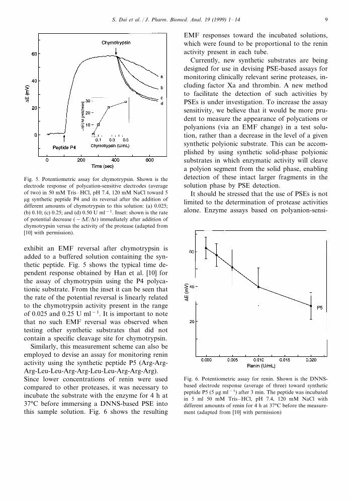
This same potentiometric format has also beensuccessfully employed for the assay of otherprotease activities, including chymotrypsin andrenin [10]. Chymotrypsin and renin cleave only atspecific sites of a polypeptide chain, the carboxylicside of lipophilic amino acids and between se-quential Leu-Leu residues, respectively. Using thesynthetic peptide P4 (Phe-Arg-Arg-Arg-Phe-Val-Arg-Arg-Phe-NH2), which includes the enzymaticcleavage site between Phe-Val, DNNS-based PSEs
S. Dai et al. / J. Pharm. Biomed. Anal. 19 (1999) 1–14 9
Fig. 5. Potentiometric assay for chymotrypsin. Shown is theelectrode response of polycation-sensitive electrodes (averageof two) in 50 mM Tris–HCl, pH 7.4, 120 mM NaCl toward 5mg synthetic peptide P4 and its reversal after the addition ofdifferent amounts of chymotrypsin to this solution: (a) 0.025;(b) 0.10; (c) 0.25; and (d) 0.50 U ml−1. Inset: shown is the rateof potential decrease (−DE/Dt) immediately after addition ofchymotrypsin versus the activity of the protease (adapted from[10] with permission).
EMF responses toward the incubated solutions,which were found to be proportional to the reninactivity present in each tube.
Currently, new synthetic substrates are beingdesigned for use in devising PSE-based assays formonitoring clinically relevant serine proteases, in-cluding factor Xa and thrombin. A new methodto facilitate the detection of such activities byPSEs is under investigation. To increase the assaysensitivity, we believe that it would be more pru-dent to measure the appearance of polycations orpolyanions (via an EMF change) in a test solu-tion, rather than a decrease in the level of a givensynthetic polyionic substrate. This can be accom-plished by using synthetic solid-phase polyionicsubstrates in which enzymatic activity will cleavea polyion segment from the solid phase, enablingdetection of these intact larger fragments in thesolution phase by PSE detection.
It should be stressed that the use of PSEs is notlimited to the determination of protease activitiesalone. Enzyme assays based on polyanion-sensi-
Fig. 6. Potentiometric assay for renin. Shown is the DNNS-based electrode response (average of three) toward syntheticpeptide P5 (5 mg ml−1) after 3 min. The peptide was incubatedin 5 ml 50 mM Tris–HCl, pH 7.4, 120 mM NaCl withdifferent amounts of renin for 4 h at 37°C before the measure-ment (adapted from [10] with permission)
exhibit an EMF reversal after chymotrypsin isadded to a buffered solution containing the syn-thetic peptide. Fig. 5 shows the typical time de-pendent response obtained by Han et al. [10] forthe assay of chymotrypsin using the P4 polyca-tionic substrate. From the inset it can be seen thatthe rate of the potential reversal is linearly relatedto the chymotrypsin activity present in the rangeof 0.025 and 0.25 U ml−1. It is important to notethat no such EMF reversal was observed whentesting other synthetic substrates that did notcontain a specific cleavage site for chymotrypsin.
Similarly, this measurement scheme can also beemployed to devise an assay for monitoring reninactivity using the synthetic peptide P5 (Arg-Arg-Arg-Leu-Leu-Arg-Arg-Leu-Leu-Arg-Arg-Arg).Since lower concentrations of renin were usedcompared to other proteases, it was necessary toincubate the substrate with the enzyme for 4 h at37°C before immersing a DNNS-based PSE intothis sample solution. Fig. 6 shows the resulting
S. Dai et al. / J. Pharm. Biomed. Anal. 19 (1999) 1–1410
tive electrodes have also been developed for thedetection of acid phosphatase and ribonuclease Aactivities [11]. Here the potential reversal aftercleavage of polyanionic substrates (polyphos-phates and pyrimidine polynucleotides) is mea-sured. The linear ranges of the present assays arenot yet optimized; however, sensitivity in the mUml−1 concentration range of these enzymes makessuch PSE-based assays already comparable toconventional spectrophotometric methods interms of sensitivity.
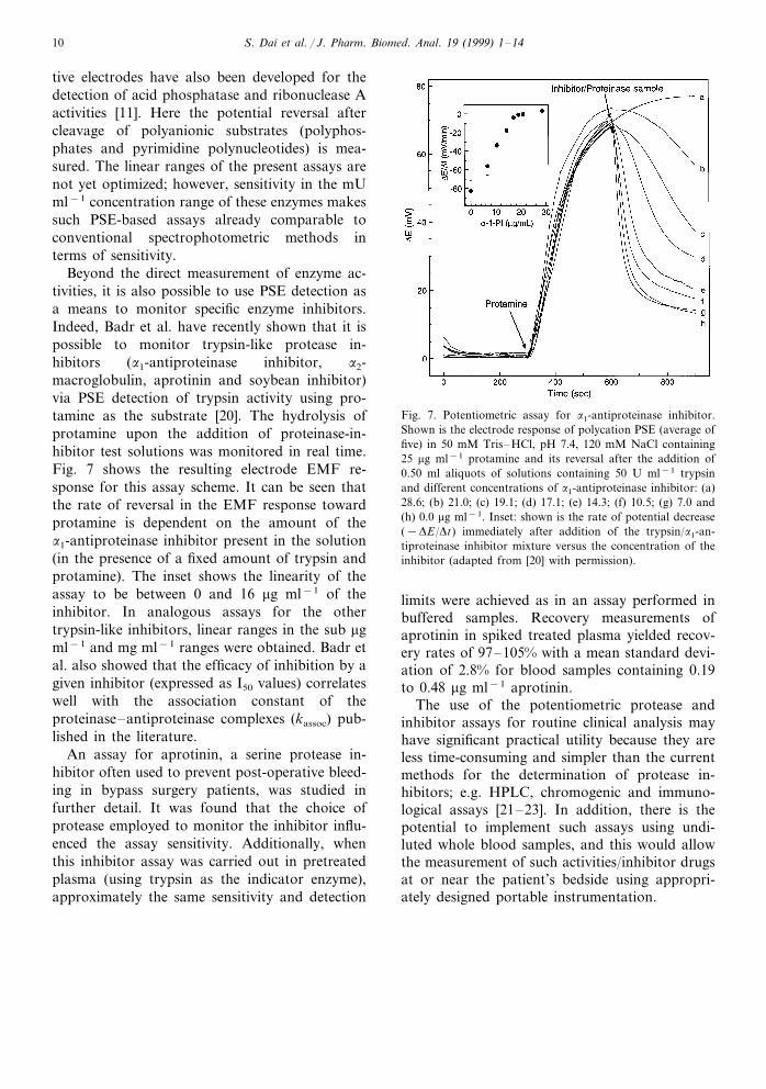
Beyond the direct measurement of enzyme ac-tivities, it is also possible to use PSE detection asa means to monitor specific enzyme inhibitors.Indeed, Badr et al. have recently shown that it ispossible to monitor trypsin-like protease in-hibitors (a1-antiproteinase inhibitor, a2-macroglobulin, aprotinin and soybean inhibitor)via PSE detection of trypsin activity using pro-tamine as the substrate [20]. The hydrolysis ofprotamine upon the addition of proteinase-in-hibitor test solutions was monitored in real time.Fig. 7 shows the resulting electrode EMF re-sponse for this assay scheme. It can be seen thatthe rate of reversal in the EMF response towardprotamine is dependent on the amount of thea1-antiproteinase inhibitor present in the solution(in the presence of a fixed amount of trypsin andprotamine). The inset shows the linearity of theassay to be between 0 and 16 mg ml−1 of theinhibitor. In analogous assays for the othertrypsin-like inhibitors, linear ranges in the sub mgml−1 and mg ml−1 ranges were obtained. Badr etal. also showed that the efficacy of inhibition by agiven inhibitor (expressed as I50 values) correlateswell with the association constant of theproteinase–antiproteinase complexes (kassoc) pub-lished in the literature.
An assay for aprotinin, a serine protease in-hibitor often used to prevent post-operative bleed-ing in bypass surgery patients, was studied infurther detail. It was found that the choice ofprotease employed to monitor the inhibitor influ-enced the assay sensitivity. Additionally, whenthis inhibitor assay was carried out in pretreatedplasma (using trypsin as the indicator enzyme),approximately the same sensitivity and detection
Fig. 7. Potentiometric assay for a1-antiproteinase inhibitor.Shown is the electrode response of polycation PSE (average offive) in 50 mM Tris–HCl, pH 7.4, 120 mM NaCl containing25 mg ml−1 protamine and its reversal after the addition of0.50 ml aliquots of solutions containing 50 U ml−1 trypsinand different concentrations of a1-antiproteinase inhibitor: (a)28.6; (b) 21.0; (c) 19.1; (d) 17.1; (e) 14.3; (f) 10.5; (g) 7.0 and(h) 0.0 mg ml−1. Inset: shown is the rate of potential decrease(−DE/Dt) immediately after addition of the trypsin/a1-an-tiproteinase inhibitor mixture versus the concentration of theinhibitor (adapted from [20] with permission).
limits were achieved as in an assay performed inbuffered samples. Recovery measurements ofaprotinin in spiked treated plasma yielded recov-ery rates of 97–105% with a mean standard devi-ation of 2.8% for blood samples containing 0.19to 0.48 mg ml−1 aprotinin.
The use of the potentiometric protease andinhibitor assays for routine clinical analysis mayhave significant practical utility because they areless time-consuming and simpler than the currentmethods for the determination of protease in-hibitors; e.g. HPLC, chromogenic and immuno-logical assays [21–23]. In addition, there is thepotential to implement such assays using undi-luted whole blood samples, and this would allowthe measurement of such activities/inhibitor drugsat or near the patient’s bedside using appropri-ately designed portable instrumentation.

S. Dai et al. / J. Pharm. Biomed. Anal. 19 (1999) 1–14 11
3.3. Non-separation immunoassays using polyion-sensiti6e membrane electrode detection
In addition to employing PSEs to monitor en-zyme and enzyme inhibitor activities, these elec-trodes may also have applications as detectors innovel non-separation competitive binding im-munoassay designs for monitoring levels of smallorganic molecules (e.g. therapeutic drugs, illicitdrugs, etc.) in undiluted blood samples. Competi-tive immunoassay formats favor detectors thatexhibit good sensitivity toward the labeled reagents(labeled form of analyte), since immunoassay de-tection limits generally decrease as the amount ofrequired reagents decreases. Because PSEs candetect certain polyions at levels as low as 10−8 Min physiological samples, the use of these electrodesin rapid immunoassays is an intriguing concept.There are two non-separation competitive bindingassay formats using PSE detection that are cur-rently under investigation: homogeneous bindingassays and enzyme multiplied immunoassays.
Fig. 8 schematically illustrates how polyion de-tection can be employed to detect hapten typemolecules without discrete separation or washingsteps by using polyions as labels. Since PSEsrespond essentially only to free polyions in solu-tion, this assay scheme involves use of a polyionlabeled-analyte reagent that is capable of beingbound by antibodies/binding proteins directed to-ward the analyte that are either free in solution orimmobilized on a solid support. If the antibodiesare immobilized on a support, such as agarosebeads, polyion-labeled analyte molecules will bindto this surface. The amount of free polyion labeled-analyte in solution will depend inversely on theconcentration of free analyte molecules added tothe sample solution. Free analyte molecules willthen compete with the polycation-labeled analytefor a limited amount of immobilized antibodies andtherefore, a larger EMF change will be observed inthe presence of increasing concentrations of ana-lyte.
To test the feasibility of this assay concept, thebiotin-avidin binding pair has been chosen as amodel system, with polylysine-labeled biotin as thesignal generator. The biotin–avidin system wasselected due to the extremely high binding affinity
of avidin for biotin, while polylysine (MW=9600)was chosen as the label due to the significantresponse of a DNNS-based PSE toward this poly-cationic species and the ease in which conjugationwith N-hydroxysuccinimide biotin can be carriedout with this structure (reaction with primaryamine groups). To demonstrate the assay principle,polylysine was first biotinylated using a 5:1 molarconjugation ratio of biotin to polylysine by reactionwith the N-hydroxysuccinimide derivative of bi-otin. The resulting biotinylated polylysine yieldsalmost an identical EMF response as unmodifiedpolylysine (+65 mV at 10 mg ml−1 biotinylatedpolylysine in Tris buffer, pH 7.4, with 12 mMNaCl).
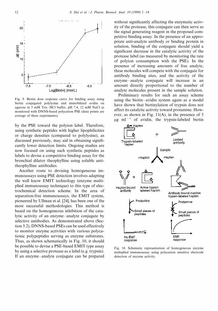
Using immobilized avidin on agarose beads (0.5units), it is possible to achieve an ED50 value of3×10−7 M biotin (1 unit binds 1 mg biotin) using4 mg of polylysine labeled biotin in the assaymixture (Fig. 9). It is important to note that thedetection limit of this novel binding assay is highlydependent on the lower limits of detection exhibited
Fig. 8. Schematic representation of homogeneous binding/im-munoassay system using polycations as labels.
S. Dai et al. / J. Pharm. Biomed. Anal. 19 (1999) 1–1412
Fig. 9. Biotin dose response curve for binding assay usingbiotin conjugated polylysine and immobilized avidin onagarose in 5 mM Tris–HCl buffer, pH 7.4, 12 mM NaCl asmonitored with DNNS-based polycation PSE (data points areaverage of three experiments).
without significantly affecting the enzymatic activ-ity of the protease, this conjugate can then serve asthe signal generating reagent in the proposed com-petitive binding assay. In the presence of an appro-priate anti-analyte antibody or binding protein insolution, binding of the conjugate should yield asignificant decrease in the catalytic activity of theprotease label (as measured by monitoring the rateof polyion consumption with the PSE). In thepresence of increasing amounts of free analyte,these molecules will compete with the conjugate forantibody binding sites, and the activity of theenzyme–analyte conjugate will increase in anamount directly proportional to the number ofanalyte molecules present in the sample solution.
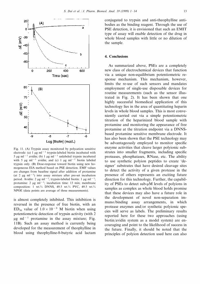
Preliminary results for such an assay schemeusing the biotin–avidin system again as a modelhave shown that biotinylation of trypsin does notaffect its catalytic activity toward protamine. How-ever, as shown in Fig. 11(A), in the presence of 5mg ml−1 of avidin, the trypsin-labeled biotin
by the PSE toward the polyion label. Therefore,using synthetic peptides with higher lipophilicitiesor charge densities (compared to polylysine), asdiscussed previously, may aid in obtaining signifi-cantly lower detection limits. Ongoing studies arenow focused on using such synthetic peptides aslabels to devise a competitive binding assay for thebronchial dilator theophylline using soluble anti-theophylline antibodies.
Another route to devising homogeneous im-munoassays using PSE detection involves adaptingthe well know EMIT technology (enzyme multi-plied immunoassay technique) to this type of elec-trochemical detection scheme. In the area ofseparation-free immunoassays, the EMIT system,pioneered by Ullman et al. [24], has been one of themost successful methodologies. This method isbased on the homogeneous inhibition of the cata-lytic activity of an enzyme–analyte conjugate byselective antibodies. As demonstrated above (Sec-tion 3.2), DNNS-based PSEs can be used effectivelyto monitor enzyme activities with various polyca-tionic polypeptides serving as enzyme substrates.Thus, as shown schematically in Fig. 10, it shouldbe possible to devise a PSE-based EMIT type assayby using a selective protease as a label (e.g. trypsin).If an enzyme–analyte conjugate can be prepared
Fig. 10. Schematic representation of homogeneous enzymemultiplied immunoassay using polycation sensitive electrodedetection of enzyme activity.
S. Dai et al. / J. Pharm. Biomed. Anal. 19 (1999) 1–14 13
Fig. 11. (A) Trypsin assay monitored by polycation sensitiveelectrode: (a) 1 mg ml−1 trypsin-labeled biotin incubated with5 mg ml−1 avidin; (b) 1 mg ml−1 unlabeled trypsin incubatedwith 5 mg ml−1 avidin; and (c) 1 mg ml−1 biotin labeledtrypsin only. (B) Dose-response toward biotin using new ho-mogeneous EIA method based on PSE detection. EMF valuesare changes from baseline signal after addition of protamine(at 2 mg ml−1) into assay mixture after pre-set incubationperiod. Avidin: 2 mg ml−1; trypsin-labeled biotin: 1 mg ml−1;protamine: 2 mg ml−1; incubation time: 13 min; membranecomposition: 1 wt.% DNNS, 49.5 wt.% PVC, 49.5 wt.%NPOE (data points are average of three measurements).
conjugated to trypsin and anti-theophylline anti-bodies as the binding reagent. Through the use ofPSE detection, it is envisioned that such an EMITtype of assay will enable detection of the drug inwhole blood samples with little or no dilution ofthe sample.
4. Conclusions
As summarized above, PSEs are a completelynew class of electrochemical devices that functionvia a unique non-equilibrium potentiometric re-sponse mechanism. This mechanism, however,limits the re-use of such sensors and mandatesemployment of single-use disposable devices forroutine measurements (such as the sensor illus-trated in Fig. 2). It has been shown that onehighly successful biomedical application of thistechnology lies in the area of quantitating heparinlevels in whole blood samples. This is most conve-niently carried out via a simple potentiometrictitration of the heparinized blood sample withprotamine and monitoring the appearance of freeprotamine at the titration endpoint via a DNNS-based protamine sensitive membrane electrode. Ithas also been shown that the PSE technology maybe advantageously employed to monitor specificenzyme activities that cleave larger polyionic sub-strates into smaller fragments, including specificproteases, phosphatases, RNase, etc. The abilityto use synthetic polyion peptides to create ‘de-signer’ substrates that have desired cleavage sitesto detect the activity of a given protease in thepresence of others represents an exciting futuredirection for this technology. Further, the capabil-ity of PSEs to detect sub-mM levels of polyions insamples as complex as whole blood holds promisethat these devices may also have a future role inthe development of novel non-separation im-muno/binding assay arrangements, in whichprotease enzymes and/or synthetic polyionic spe-cies will serve as labels. The preliminary resultsreported here for these two approaches (usingbiotin/avidin system as a model system) are en-couraging and point to the likelihood of success inthe future. Finally, it should be noted that theprinciples of polyion detection used here can also
is almost completely inhibited. This inhibition isreversed in the presence of free biotin, with anED50 value of 1.0×10⁻⁸ M biotin when usingpotentiometric detection of trypsin activity (with 2mg ml−1 protamine in the assay mixture; Fig.11B). Such an assay method is currently beingdeveloped for the measurement of theophylline inblood using theophylline-8-butyric acid lactam
S. Dai et al. / J. Pharm. Biomed. Anal. 19 (1999) 1–1414
be employed to design thin polymeric films thatrespond optically to heparin, protamine, andother polyions [25,26] through the addition of alipophilic proton chromoionophore into the poly-meric films. It is envisioned that such opticalpolyion sensor designs will also find use in hep-arin measurements, enzymatic assays, and im-munoassays, especially when employed in areflectance meter-type spectrophotometric mea-surement configuration.
Acknowledgements
This work was supported in part by grantsfrom the National Institutes of Health(GM28882) and Medtronics Blood Management.O. Lutze also acknowledges a NATO-Fellowshipof the German Academic Exchange Service(DAAD) granted by the Federal Government andthe Federal States of Germany within their HSPIII program.
References
[1] S. Ma, V.C. Yang, M.E. Meyerhoff, Anal. Chem. 64(1992) 694–697.
[2] S. Ma, V.C. Yang, B. Fu, M.E. Meyerhoff, Anal. Chem.65 (1993) 2078–2084.
[3] J.H. Yun, S. Ma, B. Fu, V.C. Yang, M.E. Meyerhoff,Electroanalysis 5 (1993) 719–724.
[4] B. Fu, E. Bakker, J.H. Yun, V.C. Yang, M.E. Meyerhoff,Anal. Chem. 66 (1994) 2250–2259.
[5] B. Fu, J.H. Yun, E. Wang, V.C. Yang, M.E. Meyerhoff,Electroanalysis 7 (1995) 823–829.
[6] B. Fu, E. Bakker, V.C. Yang, M.E. Meyerhoff, Macro-molecules 28 (1995) 5834–5840.
[7] J.H. Yun, V.C. Yang, M.E. Meyerhoff, Anal. Biochem.224 (1995) 212–220.
[8] M.E. Meyerhoff, V.C. Yang, J.A. Wahr, L.M. Lee, J.H.Yun, B. Fu, E. Bakker, Clin. Chem. 41 (1995) 1355–1356.
[9] M.E. Meyerhoff, B. Fu, E. Bakker, J.-H. Yun, V.C.Yang, Anal. Chem. 68 (1996) 168A–175.
[10] I.S. Han, N. Ramamurthy, J.H. Yun, U. Schaller, M.E.Meyerhoff, V.C. Yang, FASEB J. 10 (1996) 1621–1626.
[11] J.M. Esson, M.E. Meyerhoff, Electroanalysis 9 (1997)1325–1330.
[12] T. Hattori, M. Kato, Anal. Sci. 11 (1995) 285–287.[13] L.B. Jaques, Pharmacol. Rev. 31 (1979) 99–166.[14] J. Hirsh, Nouv. Rev. Fr. Hematol. 26 (1984) 261–266.[15] E.J. Cohen, L.J. Camerlengo, J.P.J. Dearing, Extracorpo-
real Tech. 12 (1980) 139–141.[16] A.N. Teien, M. Lie, U. Abildgaard, Thromb. Res. 8
(1976) 413–416.[17] J.A. Wahr, J.-H. Yun, V.C. Yang, L.M. Lee, B. Fu, M.E.
Meyerhoff, J. Cardiothorac. Vasc. Anesth. 10 (1996)447–450.
[18] N. Ramamurthy, N. Baliga, J.A. Wahr, U. Schaller, V.C.Yang, M.E. Meyerhoff, Clin. Chem. 44 (1998) 606–613.
[19] E.P. Sergeant, in: I.M. Kolthoff, P.J. Elwing (Eds.),Chemical Analysis, vol. 69, Wiley, New York, 1985, pp.362–364.
[20] I.H.A. Badr, N. Ramamurthy, V.C. Yang, M.E. Meyer-hoff, Anal. Biochem. 250 (1997) 74–81.
[21] G. Raspi, A.L. Moro, M. Spinetti, J. Chromatogr. 525(1990) 426–432.
[22] M. Jochum, in: H.U. Bergmeyer (Ed.), Methods of Enzy-matic Analysis, vol. 12, 3rd edn, VCH Verlagsgesellschaft,Weinheim, 1986, pp. 257–263.
[23] W. Muller-Esterl, in: H.U. Bergmeyer (Ed.), Methods ofEnzymatic Analysis, vol. 12, 3rd edn, VCH Verlags-gesellschaft, Weinheim, 1986, pp. 246–254.
[24] K.E. Rubenstein, R.S. Schneider, E.F. Ullman, Biochem.Biophys. Res. Commun. 49 (1972) 846–851.
[25] E. Wang, M.E. Meyerhoff, V.C. Yang, Anal. Chem. 67(1995) 522–527.
[26] E. Wang, G. Wang, L. Ma, C.M. Stivanello, S. Lam, H.Petal, Anal. Chim. Acta 334 (1996) 139–147.
.