bio-fabrication of pigment-capped silver nanoparticles
TRANSCRIPT

RSC Advances
PAPER
Ope
n A
cces
s A
rtic
le. P
ublis
hed
on 2
1 M
ay 2
019.
Dow
nloa
ded
on 3
/20/
2022
9:2
5:55
AM
. T
his
artic
le is
lice
nsed
und
er a
Cre
ativ
e C
omm
ons
Attr
ibut
ion-
Non
Com
mer
cial
3.0
Unp
orte
d L
icen
ce.
View Article OnlineView Journal | View Issue
Bio-fabrication o
aLeather Process Technology, Tannery Div
Institute (CLRI), Adyar, Chennai, Ta
[email protected] of Bio & Chemical Engineering,
Technology (Deemed to be University), Ra
600119, IndiacDepartment of Virology, King Institute of Pr
Chennai, Tamilnadu 600 032, IndiadNanotechnology & Catalysis Research Ce
Kuala Lumpur 50603, Malaysia. E-mail: drseDepartment of Physics, Sathyabama Institu
600119, India
Cite this: RSC Adv., 2019, 9, 15874
Received 11th February 2019Accepted 4th May 2019
DOI: 10.1039/c9ra01072f
rsc.li/rsc-advances
15874 | RSC Adv., 2019, 9, 15874–1588
f pigment-capped silvernanoparticles encountering antibiotic-resistantstrains and their cytotoxic effect towards humanepidermoid larynx carcinoma (HEp-2) cells
Lakshmipathy Muthukrishnan,a Muralidharan Chellappa,a Anima Nanda,b
Sudhakar Thukkaram,b Gracyfathima Selvaraj,c Bavanilatha Muthiah,b
Suresh Sagadevan *d and J. Anita Lett e
Bacterial biomolecule-mediated nanoparticle (NP) synthesis constitutes a reliable, eco-friendly approach
that ameliorates green-chemistry principles. In this study, stable silver nanoparticles were synthesized by
exposing aqueous silver ions to an extracellular diffusible pigment produced by Pseudomonas
aeruginosa (PA6) under optimized laboratory conditions. Spectroscopic and microscopic analyses
showed the typical characteristics of silver with an average size of �28.30 nm and spherical shape. The
particles were polydispersed and showed no definite agglomeration with a zeta potential of �32.3 mV,
conferring stability. Antimicrobial studies were carried out using 5, 15, 25 and 50 mg mL�1 concentrations
of pcAgNPs, which showed significant antibacterial activity toward clinically important pathogens at all
concentrations compared to with the control sample. The bactericidal effect induced by pcAgNPs
associated with cell damage was well demonstrated using electron microscopic studies. ROS production
was measured using the DCFH-DA method and the oxidative stress was assessed by measuring the
reduced glutathione (GSH) levels. Cytotoxicity studies on HEp-2 (Human Epidermoid Larynx Carcinoma)
cells exposed to pcAgNPs showed dose-dependent cytotoxic effect with IC50 of 14.8 mg mL�1 compared
to with IC50 of 7.38 mg mL�1 for the Vero cell control. Mechanistically, the pcAgNPs activated p53 that
induced catalase, leading to apoptosis and DNA fragmentation via a p53 transcriptional pathway and
electron transport arrest, which resulted in cell death. This synergistic efficacy of pigment-AgNPs
demonstrated excellent antimicrobial and anti-proliferative activities, providing a potential lead for
developing a broad-spectrum antibacterial agent and improving the therapeutic modalities targeting
carcinoma cells at the gene level.
1. Introduction
Nanotechnology, a major recent technological development,has experienced tremendous growth in the fabrication ofmaterials at a sub-atomic scale for more precise and uniquefunctionalities.1 Such engineered nanomaterials have extended
ision, CSIR – Central Leather Research
milnadu 600 020, India. E-mail:
Sathyabama Institute of Science and
jiv Gandhi Salai, Chennai, Tamilnadu
eventive Medicine and Research, Guindy,
ntre (NANOCAT), University of Malaya,
te of Science and Technology, Chennai-
6
its potential therapeutic and preventive applications in thebiomedical and clinical elds. Specically, the inuence of theirsize extends their ability to surpass physiological barriers andaffinity toward the cellular components of biological origin,allowing them to access the most complicated bio-molecularsystems.2 Moreover, nanoparticles are being used as anti-microbial agents and vehicles for targeted delivery,3 genetherapy,4 and cell labeling.5
Despite several breakthroughs in the formulation ofspecialist drugs from leading pharma on par with theincreasing incidences of infectious diseases, a push fordefending the scope of nanotechnology toward bio-clearanceremains vital. As the use of conventional antibiotics rises,antimicrobial resistance patterns develop,6 demandinga continuous need for novel and more effective therapies.7 Todate, nanotechnology-driven bio-pharmaceuticals have beenthe focus of many investigations and remain one of the highlyspecialized alternative medicines for curing chronic diseases
This journal is © The Royal Society of Chemistry 2019

Paper RSC Advances
Ope
n A
cces
s A
rtic
le. P
ublis
hed
on 2
1 M
ay 2
019.
Dow
nloa
ded
on 3
/20/
2022
9:2
5:55
AM
. T
his
artic
le is
lice
nsed
und
er a
Cre
ativ
e C
omm
ons
Attr
ibut
ion-
Non
Com
mer
cial
3.0
Unp
orte
d L
icen
ce.
View Article Online
such as cancer.8 Although many studies have focused onnanoparticle-induced cytotoxicity and the correspondingmechanisms,9 the decisive factors and accompanying changesinvolved in cytotoxicity remain unclear.10 It is noteworthy thatseveral genes participate in the induction of apoptosis, but therole of the p53 pathway that determines the fate of cancerouscells has rarely been studied. Moreover, in more than half ofhuman cancers, the tumor-suppressing p53 gene remainsdormant.11 Thus, there is a great interest aimed at developingtherapeutic strategies to overcome drug resistance in microbialcommunities and to anticipate the role of p53 for a prolongedexpression through the nanoparticulate system.12
To solve the above-mentioned issues, research studies onsynthesizing nanomaterials that exhibit excellent pharmaco-dynamic and pharmacokinetic properties in an eco-friendlyfashion and following the green chemistry principle havebeen proposed.13 Besides chemical and physical methods, thebioprocess-mediated synthesis of nanomaterials is a fascinatingapproach for the development of a nanoparticle–biomoleculeconjugate.14,15 This is achieved using biocatalysts and/ormetabolites such as pigments, toxins, and siderophores,exhibiting phenomenal characteristics such as virulence,defense mechanism, and stress.16 In recent years, pigments ofmicrobial origin, especially those produced by Pseudomonads,are gaining much importance in the food and agriculture sectoras bio-control agents. The exploitation of such bacteria for theircatabolic versatility and broad-spectrum antibiosis can helpimprove crop production.17,18
In this study, we present an eco-friendly approach forsynthesizing silver nanoparticles harnessing a versatile pigmentproduced by P. aeruginosa in a reliable, stable and cost-effectivemanner. We investigated the biological role of the bacterialpigment-capped AgNPs toward containing antibiotic-resistantstrains and the in vitro antiproliferative effect with respect tothe p53 expression using the human epidermoid larynx carci-noma cell line (HEp-2).
2. Materials and methods2.1. Isolation of Pseudomonas aeruginosa
The bacterium used for the synthesis of silver nanoparticles wasisolated from rhizosphere soil samples collected from the paddyelds (Lat. 12�5404000N and Long. 79�801000E) in Vellore, Tamil-nadu, India. The pseudomonas strains were isolated by per-forming a dilution plate technique19 on Pseudomonas isolationagar (cat no. 17208, Sigma-Aldrich, USA), and the plates wereincubated at 37 � 2 �C for 24 h. Prior to incubation, the plateswere observed for any growth on the selective media. The iso-lated colonies of the Pseudomonas species were further puriedby quadrant streaking on Cetrimide Agar (cat. no. 22470, Sigma-Aldrich, USA) plates and maintained at 4 �C.
2.2. Molecular identication of the potential strain
The growth characteristics of the bacterial isolates, viz., lactosefermentation, odour, and pigmentation were observed alongwith the standard biochemical tests and identied based on
This journal is © The Royal Society of Chemistry 2019
Bergey's manual of determinative bacteriology.20 For identicationat the molecular level, the total DNA from the potential strainwas extracted using a MagAttract Microbial DNA kit (cat. no.27200.4, Qiagen) and the 16S ribosomal RNA gene was ampli-ed using PCR with suitable primers. The PCR product ob-tained was sequenced by an automated sequencer ABI PRISM(Model 3700) and the sequences were compared using theBLAST program (http://www.ncbi.nlm.nih.gov/BLAST/).
2.3. Pigment production
First, 35 pseudomonas strains obtained from various sites wereused for the production, extraction, and characterization ofpigment, and the conditions were optimized as described in ourprevious study.21 In brief, the isolates were inoculated intoErlenmeyer asks containing 100 mL of Pseudomonas broth(cat. no. Z699101, Sigma-Aldrich, USA) and incubated at 37 �C ina rotary shaker for 24 h till the color changed. The pigmentproduced by the potential strain was quantied based on theabsorbance of the pigment in the acidic form at 520 nm and theconcentration was determined using the following formulaaccording to Essar et al. (1990):22
concentration (mg mL�1) ¼ O.D520 � 17.07
The strain that showed the maximum pigment productionprior to incubation was retained for synthesizing AgNPs.
2.4. Bio-synthesis of AgNPs using potential strain
The preliminary screening process was performed by taking10 mL of the diffusible pigment of the strain and adding it to90 mL of 10�3 M AgNO3 (CAS No. 7761-88-8, Sigma-Aldrich,USA) in Erlenmeyer asks till 1 : 1 concentration was ach-ieved. The asks were examined visually for any colour changeprior to the reaction period at 37 �C and 150 rpm, which wasindicative of nanoparticle formation. Aer the formation ofnanoparticles, the asks were removed from the shaker andcentrifuged at 8000 rpm for 20 min, freeze-dried andquantied.23
2.5. Optimization studies on AgNP synthesis
The ideal conditions for nanoparticle synthesis were optimizedby varying the physicochemical parameters such as thetemperature (10, 20, 30, 40 and 50 �C), pH (2–9), the concen-tration of the substrate (0.5–2 mM AgNO3) and the reactionperiod (0–72 h). Sampling was performed by withdrawing 1 mLaliquots at different time intervals; these aliquots were sub-jected to spectrophotometric analysis.24
2.6. Characterization of pcAgNPs
A UV-visible spectrophotometer model UV-1800 (Shimadzu) wasused to record the absorption spectra of the reaction mixture.The reduction of silver ions was monitored by the randomsampling of a 1 mL aliquot in a quartz cuvette and detected inthe scanning range of 800–200 nm at a resolution of 1 nm. ThepcAgNPs were separated and saturated by repeated washing and
RSC Adv., 2019, 9, 15874–15886 | 15875

RSC Advances Paper
Ope
n A
cces
s A
rtic
le. P
ublis
hed
on 2
1 M
ay 2
019.
Dow
nloa
ded
on 3
/20/
2022
9:2
5:55
AM
. T
his
artic
le is
lice
nsed
und
er a
Cre
ativ
e C
omm
ons
Attr
ibut
ion-
Non
Com
mer
cial
3.0
Unp
orte
d L
icen
ce.
View Article Online
centrifugation at 15 000 rpm for 20 min and the nal suspen-sion was freeze-dried. Powder X-ray diffraction patterns weremeasured on a Rigaku multiex diffractometer that operated at30 kV and 100 mA at 3 kW with a Cu target Ka radiation usinga scintillation counter (l ¼ 1.5418 A). For FT-IR analysis, thefreeze-dried pcAgNPs were diluted with potassium bromide ina ratio of 1 : 100 and the spectrum was recorded on a JASCO4400 model in a diffused reectance mode in the region of4000–400 cm�1 and at a resolution of 4 cm�1. HR-TEM (PhilipsCM200) and SAED were performed to identify the size andshape of the nanoparticles. The pcAgNP samples used formicroscopic analyses were prepared on a carbon-coated coppergrid.24,25
The particle size and distribution of the bio-synthesizedsilver nanoparticles were observed using dynamic light scat-tering (DLS) (Malvern Zetasizer, ver. 6.2). For this experiment,the samples were measured at a xed angle of 90� at roomtemperature and the intensity was adjusted in the range of 50–500 kcps by diluting with deionized water. The zeta potential ofthe silver nanoparticles was also measured by following theabove-mentioned protocol and the measurements wereanalyzed in triplicate.
Atomic force microscopy was employed to study the topog-raphy at the nanoscale. The sample was prepared by dissolvingpcAgNPs in ethanol and sonicating (Vibra cell, Sonics) toprevent aggregation. Ten mL of the sample was casted ontoa silicon slide and the solvent was allowed to evaporate. Then,the thin lm of size 1 cm � 1 cm was imaged in the tappingmode with a spring constant of 0.41 N m�1 and resonancefrequency 18 kHz via AFM (Ntegra Prima – NTMDT, Ireland).
2.7. Antibacterial activity of pcAgNPs against clinical isolates
Bacterial strains showing resistance toward two differentgroups of commonly used antibiotics, viz., Methicillin-resistantStaphylococcus aureus (MRSA), Staphylococcus epidermidis(MRSE), Proteus mirabilis, E. coli, Klebsiella pneumoniae, Acine-tobacter baumannii, and the vancomycin-resistant Enterococciwere used for antibacterial studies. The clinical isolates wereprocured fromM s�1. Sharp Laboratories, Chennai, Tamilnadu,and the pure cultures were maintained in Muller Hinton agar(MHA, HiMedia™, India) slants at 4 �C. The antibacterialactivity of pcAgNPs was tested against these drug-resistantbacterial strains using the Kirby Bauer's disk diffusionmethod.26,27 Sterile disks (HiMedia™, India) containing 5, 15,25 and 50 mg mL�1 of pcAgNPs were prepared and placed onMHA plates using sterile forceps along with 1 mM AgNO3 as thecontrol. The zone of inhibition (ZOI) was measured aer incu-bation at 37 �C for 18–24 h and compared with that of thecontrol.
To detect the membrane damage, different volumes of theMH medium, pcAgNPs and the most susceptible strains (105
CFU mL�1) were added to 5 mL broth with a nal concentrationof 25 mg mL�1 AgNPs. Control experiments were conductedwithout AgNPs. The cultures were incubated at 37 � 2 �C withshaking at 150 rpm. One hundred microlitres of cultures weresampled aer being treated for 2 to 4 h. The cultures were
15876 | RSC Adv., 2019, 9, 15874–15886
centrifuged at 8000 rpm, the supernatant was discarded, andthe cells were xed for observation by scanning electronmicroscopy.28
2.8. ROS production and glutathione (GSH) measurements
The intracellular localization of stress-induced reactive oxygenspecies (ROS) was estimated by 20,70-dichlorodihydrouoresceindiacetate (DCFH-DA, cat no. D6883, Sigma-Aldrich, USA) asdescribed.29 The strains were selected based on the antibiogrampattern representing Gram-positive and Gram-negativebacteria. These cells at 105 CFU mL�1 were incubated at 37 �Cwith DCFH-DA (0.5 mM) for 30 min, followed by the addition ofpcAgNPs (5, 15, 25, 50 mg mL�1). The bacterial cells were furtherincubated for 4 h at 37 �C and the uorescence emission wasrecorded at 523 nm with excitation at 503 nm on a Jobin YvonFlurolog-3-11 spectrouorometer. The ROS production wasinterpreted from the emission intensity of the treated cells andcompared against those of the negative (untreated) and positive(1 mM H2O2) controls.
The intracellular GSH level was determined following theprotocol of Park et al. with slight modications.30 In short, thecells (105 CFU mL�1) treated with 5, 15, 25 and 50 mg mL�1
AgNPs were collected and lysed with a 0.6% 5-sulfosalicylic acidsolution for 10 min on ice. Fiy mL of the cell lysate was mixedwith 450 mL of KH2PO4/EDTA buffer (0.1 M potassium phos-phate with 5 mM EDTA, pH 8.0) and 100 mL of 1 mg mL�1 o-phthaldialdehyde solution and incubated for 60 min in dark atroom temperature. The uorescence was measured on a JobinYvon Flurolog-3-11 spectrouorometer with excitation andemission wavelengths of 350 nm and 420 nm, respectively, andcompared against that of controls as described. The concen-tration of GSH was determined from the standard curve andexpressed in mM.
2.9. In vitro antiproliferative activity of pcAgNPs
2.9.1. Culturing of cells. Human epidermoid larynx carci-noma cells (HEp-2) and Vero (African green monkey kidneyepithelial cells) cells were procured from the King Institute ofPreventive Medicine and Research, Guindy, Tamilnadu. Theywere sub-cultured under standard conditions in DMEM (cat. no.5796, Sigma-Aldrich, USA) supplemented with 10% FBS (cat. no.F2442, Sigma-Aldrich, USA) and 100 U mL�1 of penicillin–streptomycin antibiotic (cat. no. P4333, Sigma-Aldrich, USA)solutions in a humidied incubator (MCO-18AIC, Sanyo) at37 �C with 5% CO2.
2.9.2. Cytotoxicity of pcAgNPs-MTT assay. The MTT cellproliferation assay is a colorimetric assay for measuring cellviability through increased metabolic events, leading to thereduction of tetrazolium salt.31 Vero and HEp-2 cells ata concentration of 1 � 105 cells per well in 96-well plates weregrown and incubated with pcAgNPs at various concentrations(0.75, 1.5, 3.1, 6.2, 12.5, 25, 50 and 100 mg mL�1) at 37 �C with5% CO2 for 24 h. MTT (5 mg mL�1) (TC191, HiMedia™, India)was added to the incubated cells and further incubated for 4 h.The purple formazan crystals formed were dissolved in 200 mLof DMSO (cat. no. TC185, HiMedia™, India) and the
This journal is © The Royal Society of Chemistry 2019

Paper RSC Advances
Ope
n A
cces
s A
rtic
le. P
ublis
hed
on 2
1 M
ay 2
019.
Dow
nloa
ded
on 3
/20/
2022
9:2
5:55
AM
. T
his
artic
le is
lice
nsed
und
er a
Cre
ativ
e C
omm
ons
Attr
ibut
ion-
Non
Com
mer
cial
3.0
Unp
orte
d L
icen
ce.
View Article Online
absorbance was measured in an ELISA reader at 570 nm withthe reference lter set at 655 nm. The experiment was per-formed in triplicate and the data were expressed as mean � SD(standard deviation) of the concentration of pcAgNPs.
2.9.3. Apoptotic assays (AO/EB staining). Acridine orange/ethidium bromide (AO/EB) staining was carried out to detectthe morphological evidence of apoptosis on the nanoparticle-treated HEp-2 cells.32 Initially, the cells were xed in methanoland glacial acetic acid (3 : 1) for 1 h at room temperature. Aerincubation, the cells were stained with AO and EB (1 : 1) in PBSand incubated for 5 min; then, they were washed with PBS toremove the excess unbinding dye. The stained cells were visu-alized under UV illumination using the 40� objective (NikonEclipse TS100) and the digitized images were captured.
2.10. Semi-quantitative RT-PCR analysis
Total RNA was extracted from the experimental cells using theTrizol reagent (TRI, Sigma-Aldrich, St. Louis, USA) kit. The RNAisolated from the cells was reverse-transcribed and ampliedusing the one-step RT-PCR System (Fermentas, USA). Theprimer sequences of the forward and reverse p53 were 50-TTGAGGTGCATGTTTGTGCC-30 and 50-AGGAGCTGGTGTTGTTGGAC-30; for b-actin, they were 50-TTCCTCCCTGGAGAAGAGCTA-30 and 50-CACA-GAGTACTTGCGCTCAG-30, respectively.
The PCR conditions were as follows: 94 �C for 5 min; 35cycles of 94 �C for 1 min, 56–62 �C for 1 min, 72 �C for 1 min;and a nal extension step of 72 �C for 10min. The products wereveried by agarose gel electrophoresis and documented usingQuantity One soware (Bio-Rad, USA). The band intensity of theresolved cDNA fragments of p53 was normalized with that of theinternal control b-actin and expressed as OD units relative to b-actin. All of the experiments were performed in two batches andsubjected to the individual Student's ‘t’ test to assess the vari-ation p < 0.05 level.
2.11. DNA fragmentation assay
To investigate the internucleosomal DNA fragmentation causedby pcAgNPs, a DNA fragmentation assay was performedaccording to the standard procedure described by Collins et al.(1997) with slight modications.33 A total of 1 � 105 cells weretreated with pcAgNPs (15 mg mL�1) for 24 h and then collectedby centrifugation. Furthermore, the DNA was isolated usinga commercially available kit (Genei, Bangalore, India) followingthe manufacturer's instructions. The DNA was resolved ona 1.5% agarose gel (0.5 mg mL�1 ethidium bromide in the 1�TAE buffer, pH 8.5) at 100 meV for 90 min and the bands werevisualized using a UV transilluminator.
2.12. Western blotting
First, 1 � 105 cells exposed to pcAgNPs were washed twice withice-cold 1� PBS solution and centrifuged at 5000 rpm for 5 min.The pellet was lysed in a RIPA buffer containing 150 mM NaCl,Triton X-100, 50 mM Tris–HCl (pH 8), 0.5% sodium deoxy-cholate, 0.1% SDS, and protease and phosphatase inhibitors.The protein lysates (20 mg) were subjected to protein
This journal is © The Royal Society of Chemistry 2019
electrophoresis on 10% NuPAGE® Bis-Tris gels (cat. no.NP0301BOX, ThermoFisher Scientic, CA) according to themanufacturer's instructions. Then, the gels were blotted ontoa nitrocellulose membrane (cat. no. 162-0113, Bio-Rad, Italy) for2 h in Tris–glycine buffer. The membranes were blocked ina PBS 0.1% Tween 20 solution containing 3% of BSA, probedwith specic antibodies and developed using the ECL BlottingSubstrate (cat. no. K-12045-C20, Advansta, USA).
In the western blotting analysis, the following primary anti-bodies were used: rabbit polyclonal anti-p21 (1 : 100) (cat. no.TA322940, OriGene, USA), mouse monoclonal anti-p53 (1 : 500)(cat. no. TA327920, OriGene, USA), andmouse monoclonal anti-catalase (1 : 100) (cat. no. TA502496, OriGene, USA). Mousemonoclonal anti-b-actin (1 : 10 000) (cat. no. TA355041,OriGene, USA) was used to control loading and to purify theperoxidase-conjugated affinity goat anti-mouse IgG (cat. no.TA130003, OriGene, USA). The primary and secondary anti-bodies used were diluted in a PBS-0.1% Tween 20 solutioncontaining 3% of BSA (cat. no. P3563, Sigma-Aldrich, USA).
2.13. Densitometric analysis
Protein bands were quantied by densitometric analysis usingthe Image J soware (http://imagej.nih.gov).
2.14. Statistical analysis
Experiments conducted in the present study on the antibiogrampattern, measurement of the zone of inhibition, and bacteri-cidal and anti-proliferative activities were performed in tripli-cate. The results of the experiments were interpreted usingORIGIN 8.0 and expressed as the mean � SD (standard devia-tion) of the concentration of AgNPs. The Student's t-test wasused to demonstrate the statistical signicance. The differencewas considered to be statistically signicant when the p-valuewas at least <0.05.
3. Results and discussion3.1. Identication of potential strain
In this study, NPs were synthesized successfully using a rhizo-sphere-inhabiting environmental isolate Pseudomonas aerugi-nosa isolated from soil samples collected from paddy elds, andit was designated PA6 for laboratory reference. Of the 51 pig-mented strains isolated from six different rhizosphere soils(Table 1), around 68.6% (n ¼ 35) were expected to be Pseudo-monas aeruginosa, which were identied based on thebiochemical characteristics such as non-lactose fermentingproperty, being oxidase-positive, pigment production andgrowth at 40 �C. These strains were then individually screenedfor the production of pyocyanin; sixteen (45%) of themproduced #4 mg mL�1, whereas the remaining 55% showedproduction in the range of 5.3–6.5 mg mL�1. Table 1 reveals thatP. aeruginosapa6 isolated from paddy-cultivated soil showsa maximum pigment production of 6.5 mg mL�1, followed by P.aeruginosaw9 with 5.3 mg mL�1. This variation in the productionof pyocyanin among the Pseudomonas strains can be attributedto regulators.34 The higher pigment producer P. aeruginosapa6
RSC Adv., 2019, 9, 15874–15886 | 15877
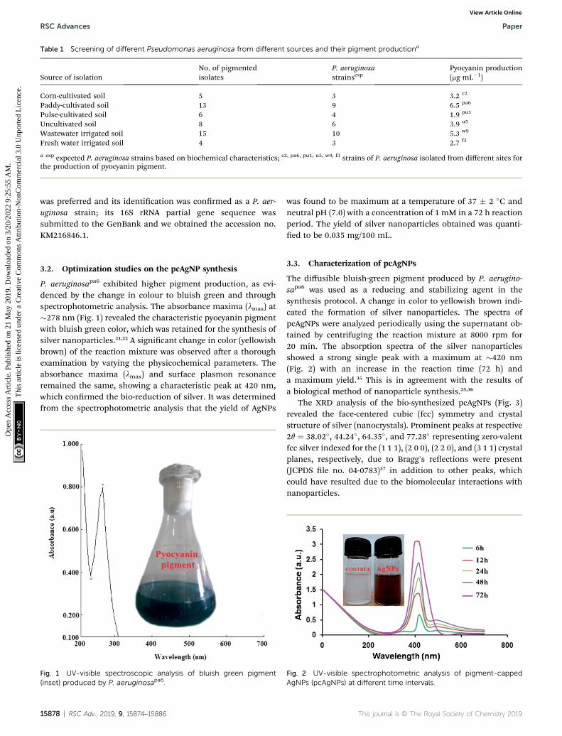
Table 1 Screening of different Pseudomonas aeruginosa from different sources and their pigment productiona
Source of isolationNo. of pigmentedisolates
P. aeruginosastrainsexp
Pyocyanin production(mg mL�1)
Corn-cultivated soil 5 3 3.2 c2
Paddy-cultivated soil 13 9 6.5 pa6
Pulse-cultivated soil 6 4 1.9 pu1
Uncultivated soil 8 6 3.9 u5
Wastewater irrigated soil 15 10 5.3 w9
Fresh water irrigated soil 4 3 2.7 f3
a exp expected P. aeruginosa strains based on biochemical characteristics; c2, pa6, pu1, u5, w9, f3 strains of P. aeruginosa isolated from different sites forthe production of pyocyanin pigment.
RSC Advances Paper
Ope
n A
cces
s A
rtic
le. P
ublis
hed
on 2
1 M
ay 2
019.
Dow
nloa
ded
on 3
/20/
2022
9:2
5:55
AM
. T
his
artic
le is
lice
nsed
und
er a
Cre
ativ
e C
omm
ons
Attr
ibut
ion-
Non
Com
mer
cial
3.0
Unp
orte
d L
icen
ce.
View Article Online
was preferred and its identication was conrmed as a P. aer-uginosa strain; its 16S rRNA partial gene sequence wassubmitted to the GenBank and we obtained the accession no.KM216846.1.
3.2. Optimization studies on the pcAgNP synthesis
P. aeruginosapa6 exhibited higher pigment production, as evi-denced by the change in colour to bluish green and throughspectrophotometric analysis. The absorbance maxima (lmax) at�278 nm (Fig. 1) revealed the characteristic pyocyanin pigmentwith bluish green color, which was retained for the synthesis ofsilver nanoparticles.21,22 A signicant change in color (yellowishbrown) of the reaction mixture was observed aer a thoroughexamination by varying the physicochemical parameters. Theabsorbance maxima (lmax) and surface plasmon resonanceremained the same, showing a characteristic peak at 420 nm,which conrmed the bio-reduction of silver. It was determinedfrom the spectrophotometric analysis that the yield of AgNPs
Fig. 1 UV-visible spectroscopic analysis of bluish green pigment(inset) produced by P. aeruginosapa6.
15878 | RSC Adv., 2019, 9, 15874–15886
was found to be maximum at a temperature of 37 � 2 �C andneutral pH (7.0) with a concentration of 1 mM in a 72 h reactionperiod. The yield of silver nanoparticles obtained was quanti-ed to be 0.035 mg/100 mL.
3.3. Characterization of pcAgNPs
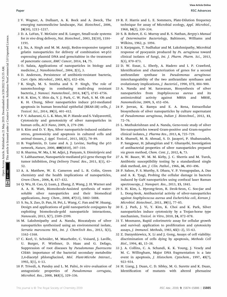
The diffusible bluish-green pigment produced by P. aerugino-sapa6 was used as a reducing and stabilizing agent in thesynthesis protocol. A change in color to yellowish brown indi-cated the formation of silver nanoparticles. The spectra ofpcAgNPs were analyzed periodically using the supernatant ob-tained by centrifuging the reaction mixture at 8000 rpm for20 min. The absorption spectra of the silver nanoparticlesshowed a strong single peak with a maximum at �420 nm(Fig. 2) with an increase in the reaction time (72 h) anda maximum yield.35 This is in agreement with the results ofa biological method of nanoparticle synthesis.25,36
The XRD analysis of the bio-synthesized pcAgNPs (Fig. 3)revealed the face-centered cubic (fcc) symmetry and crystalstructure of silver (nanocrystals). Prominent peaks at respective2q ¼ 38.02�, 44.24�, 64.35�, and 77.28� representing zero-valentfcc silver indexed for the (1 1 1), (2 0 0), (2 2 0), and (3 1 1) crystalplanes, respectively, due to Bragg's reections were present(JCPDS le no. 04-0783)37 in addition to other peaks, whichcould have resulted due to the biomolecular interactions withnanoparticles.
Fig. 2 UV-visible spectrophotometric analysis of pigment-cappedAgNPs (pcAgNPs) at different time intervals.
This journal is © The Royal Society of Chemistry 2019

Fig. 3 X-ray diffraction pattern of the biosynthesized pcAgNPs.
Paper RSC Advances
Ope
n A
cces
s A
rtic
le. P
ublis
hed
on 2
1 M
ay 2
019.
Dow
nloa
ded
on 3
/20/
2022
9:2
5:55
AM
. T
his
artic
le is
lice
nsed
und
er a
Cre
ativ
e C
omm
ons
Attr
ibut
ion-
Non
Com
mer
cial
3.0
Unp
orte
d L
icen
ce.
View Article Online
The topography and size of pcAgNPs were examined byatomic force microscopy (AFM), dynamic light scattering (DLS)and high-resolution transmission electron microscopy(HRTEM). As observed from the atomic microscopy results, thetopography of the silver nanoparticles with no deniteagglomeration conrms the polydispersity of the nanoparticles
Fig. 4 Characterization of pcAgNPs. Atomic Force Microscopic imagediameter in the range of 10–100 nm (B); HR-TEM micrograph at a scalediffraction rings, indicating the purity at a scale bar of 20 nm (D).
This journal is © The Royal Society of Chemistry 2019
(Fig. 4A). Subsequently, the particle size distribution of thesilver nanoparticles (Fig. 4B) unveiled the narrow size distri-bution in the range of 25–55 nm, with the mean diameter of40 nm and zeta potential of �32.3 mV; this indicated a strongelectrostatic attraction between the silver ions and the metab-olites, conferring stability.23,38
Further conrmation of the size of the nanoparticlesanalyzed through HRTEM with SAED patterns (Fig. 4C and D)demonstrated the spherical and nearly spherical shapes of thesilver nanoparticles in the size range 25–50 nm, which werefound to be consistent with the results obtained from dynamiclight scattering. The inset of Fig. 4D shows the selected areadiffraction (SAED) pattern of the silver nanoparticles. Thesmooth surface and ring-like diffraction pattern indicate thatthe particles are crystalline with the characteristic lattice planesof silver.39
FT-IR spectroscopy was performed to analyze the functionalquality of the product and the spectrum was acquired in therange of 4000–400 cm�1 (Fig. 5). The band seen at 3400.9 cm�1
is assigned to the stretching vibrations of O–H and H. Thebending vibration of primary amine can be observed in theregion of 1650–1580 cm�1 and the C–N stretching vibrationscorrespond to aliphatic amines, as represented by a peak in theregion of 1250–1020 cm�1. This indicated the involvement of
of size 10 � 10 mm with height profile at 80 nm (A); hydrodynamicbar of 20 nm (C); and the corresponding SAED pattern showing four
RSC Adv., 2019, 9, 15874–15886 | 15879

Fig. 5 FT-IR spectrum of the biosynthesized pcAgNPs showing thepossible biomolecular interactions of the pigment.
RSC Advances Paper
Ope
n A
cces
s A
rtic
le. P
ublis
hed
on 2
1 M
ay 2
019.
Dow
nloa
ded
on 3
/20/
2022
9:2
5:55
AM
. T
his
artic
le is
lice
nsed
und
er a
Cre
ativ
e C
omm
ons
Attr
ibut
ion-
Non
Com
mer
cial
3.0
Unp
orte
d L
icen
ce.
View Article Online
a polypeptide, as evidenced from the amide linkages betweenthe amino acid residues. In addition, the stretching vibrationsof –C–C– were observed at 1429 cm�1, which accounted for thearomatic compound involved in the reaction; this is probablythe role of pyocyanin (pigment), offering stability to the nano-particles. From the observation, it was conrmed that biomol-ecules such as protein and pigment moieties were involved inthe reduction and stabilization of silver nanoparticles throughintermolecular attractions.40,41
3.4. Antimicrobial activity
The antimicrobial study using nanomaterials was performedbecause of the increase in the multidrug-resistant strains ofbacteria, which could provide insights for an alternative drug ofchoice including AgNPs and silver-based products. pcAgNPsprepared at various concentrations were found to be effectivetoward a series of antibiotic-resistant bacterial strains on parwith those exposed to broad-spectrum antibiotics such asAmikacin, Tetracycline, Vancomycin, Cefepime, Netilmicin,Doxycycline and Chloramphenicol (Table 2) at 25 mg mL�1. Theantibiogram pattern of the clinical isolates showed completeresistance toward a series of antibiotics at 25 mg mL�1 ascompared with the standard antibiotic susceptibility chart.
AgNPs (5, 15, 25 and 50 mg mL�1) exhibited signicantantimicrobial activity toward all antibiotic-resistant Gram-positive and Gram-negative strains in a dose-dependentmanner. Proteus mirabilis showed maximum susceptibilitytoward pcAgNPs with �25 mm ZOI, followed by K. pneumoniae(24 mm) and Acinetobacter baumannii (24 mm). The isolates thatdid not respond to the broad-spectrum antibiotics such asMRSA and MRSE showed susceptibility toward pcAgNPs at thesame concentration as that of the antibiotics. A similar trendwas observed in the case of E. coli and the vancomycin-resistantEnterococcus sp. with a �22 mm inhibition zone.
Furthermore, to manifest the antibacterial effect induced bypcAgNPs, the FESEM technique was adopted, for whichEnterococcus sp. (VRE) and E. coli models were used. RegularFESEM micrographs (Fig. 6) revealed signicant membrane
15880 | RSC Adv., 2019, 9, 15874–15886
damage on both the strains exposed to 25 mg mL�1 of pcAgNPsin 6 h from their exposure (Fig. 6b and d), whereas the controlcells sustained no such damage and retained the coccal androd-shaped morphologies (Fig. 6a and c). It was assumed thatthe dissolved silver ions' interaction with the trans-membraneproteins leads to the release of H+ by the inuence of theproton-motive force,42 resulting in the formation of ‘pits’ thatdamage the cell walls.43 Subsequently, the membrane compo-nents became disorganized from their intact nature, accountingfor the structural changes in the cell walls. This disrupted thepermeability, which led to the death of the cells. The antimi-crobial susceptibility toward the Gram-negative bacterium sus-tained extensive structural damage than the observation for theGram-positive bacterium. It was presumed that the presence ofthe thick peptidoglycan in the Gram-positive bacterium has theability to trap the positively charged silver ions. Thesephenomena suggest a possible antibacterial mechanism exer-ted by pcAgNPs through membrane damage and inhibition ofcellular response.44
3.5. Mechanistic antibacterial effect
It was evident from the electron microscopic study that pcAgNPsadhere and pass through the plasmamembrane, creating pits thatresult in bacterial cell death. Reports on the metal–microbeinteraction induced a signicant rise in ROS, bringing abouta deleterious effect related to oxidative stress.45 Therefore, theoxidative stress in the Gram-positive Enterococci (VRE) and Gram-negative E. coli aer treatment with pcAgNPs was determinedthrough themeasurement of intracellular ROS generated using theDCFH-DA assay. This oxidation-sensitive uorescent probediffuses passively through the cell membrane and gets deacety-lated by esterase to form non-uorescent DCFH.When it comes incontact with ROS, it reacts to form a uorescent product DCtrapped inside the cell. Thus, the ROS level was directly propor-tional with the uorescence intensity at 510 nm and in turn wascorrelated with the antibacterial effect of pcAgNPs on the bacterialcells. Fig. 7c clearly reveals the ROS generation with increasedconcentrations of pcAgNPs. The control cells (untreated withpcAgNPs) showed no difference; however, as the concentration ofpcAgNPs increased, there was increased ROS generation (75–85%)at 50 mg mL�1 as compared with that for the positive control(100%), pertaining to the ROS-mediated cell death.46
Furthermore, to guarantee the state of the organisms thatsuccumbed to cell death, the intracellular glutathione (GSH)concentration wasmeasured. Glutathione, themost potent naturalantioxidant, inuences the physiological and biochemical aspectsof both prokaryotic and eukaryotic cells. Eukaryotic cells possessrelatively higher concentrations of GSH than prokaryotes, but it ischiey found in E. coli, Streptococcus and Enterococcus sp. tomaintain the cellular redox environment and protect the cellsagainst oxidative stress by scavenging ROS. This tripeptide ispresent in the cytosol of cells at a concentration of 1–2 mM GSH.When this reduced form of GSH comes in contact with free radi-cals, it results in the formation of GS* (thiyl radical), which thenreacts with another GS* radical to form glutathione disulphide(GSSG), an important regulator of biological processes.47
This journal is © The Royal Society of Chemistry 2019

Table 2 Antimicrobial susceptibility testing of various concentrations of pcAgNPs and 1mM AgNO3 toward a series of clinical isolates of bacteriaafter 24 h of treatmenta
Test strainsAntibiotics (25 mgmL�1)
Zone of inhibition (in mm) induced by pcAgNPs
AgNO35 mg mL�1 15 mg mL�1 25 mg mL�1 50 mg mL�1
S. aureus (MRSA) AN 7.7 � 0.58 12.8 � 0.79 15.8 � 0.62 19.1 � 0.70 24 � 0.82 10.5 � 1.08TET 9.3 � 0.29VA NZ
S. epidermidis (MRSE) AN 8.7 � 0.26 11.7 � 1.25 15.2 � 0.59 18 � 0.82 23 � 2.16 10.7 � 1.25TET 9.8 � 0.21VA NZ
P. mirabilis AN 12.3 � 0.76 10.8 � 1.03 15.3 � 1.25 18 � 1.41 24.5 � 1.23 13.5 � 0.41FEP 10.9 � 0.36NET 10.1 � 0.51
E. coli AN 12.7 � 0.58 9.8 � 0.85 14 � 2.16 17.3 � 0.94 22.5 � 1.05 11.2 � 0.85TET 9.2 � 0.59NET 10.8 � 0.35
K. pneumoniae AN 11.2 � 0.29 10.3 � 0.47 15.7 � 0.94 18.2 � 0.57 24.2 � 0.85 8.7 � 0.47TET 9.3 � 0.31NET 7.5 � 0.87
A. baumannii AN 11.5 � 0.35 9.6 � 0.42 14.7 � 0.47 18.3 � 0.47 23.7 � 0.94 9.7 � 0.38TET 10.3 � 0.29D 7.6 � 0.23
Enterococcus sp. (VRE) C 10.3 � 0.25 10 � 0.82 16 � 1.41 18.7 � 0.94 22.1 � 0.98 12.3 � 0.47TET 11.8 � 0.75VA 12 � 0.15
a AN – Amikacin; TET – Tetracycline; VA – Vancomycin; FEP – Cefepime; NET – Netilmicin; D – Doxycycline; C – Chloramphenicol; NZ – no zone.Data are representative of at least three independent experiments and expressed as mean � standard deviation (SD) of the concentrations ofAntibiotics and pcAgNPs.
Paper RSC Advances
Ope
n A
cces
s A
rtic
le. P
ublis
hed
on 2
1 M
ay 2
019.
Dow
nloa
ded
on 3
/20/
2022
9:2
5:55
AM
. T
his
artic
le is
lice
nsed
und
er a
Cre
ativ
e C
omm
ons
Attr
ibut
ion-
Non
Com
mer
cial
3.0
Unp
orte
d L
icen
ce.
View Article Online
To decipher the oxidative stress induced by pcAgNPs, theintracellular GSH concentration was measured in pcAgNP-treated cells. As discussed, the initial concentrations of GSH
Fig. 6 FESEM micrographs of antimicrobial susceptibility testing of pcAga scale bar of 1 mm observed after 24 h of treatment with typical membratreated with pcAgNPs showing cell damage (b); E. coli control (c) and E.
This journal is © The Royal Society of Chemistry 2019
were found to be 1.54 � 0.41 and 1.26 � 0.32, respectively, forthe Enterococcus sp. and E. coli (control) cells (Fig. 7a). Besides,the pcAgNP-treated cells showed sequential depletion of GSH
NPs at 25 mg mL�1 toward Gram-positive and Gram-negative strains atne damage. Enterococcus sp. (VRE) control (a); Enterococcus sp. (VRE)coli treated with pcAgNPs (d).
RSC Adv., 2019, 9, 15874–15886 | 15881

Fig. 7 Effect of pcAgNPs on the intracellular GSH concentration (a); DCFH-DA assay (b) for the quantification of ROS generation and the level ofROS production in pcAgNP-treated cells (c).
Fig. 8 Cytotoxicity of Vero and HEp-2 cells exposed to pcAgNPs andtheir corresponding IC50 values. The IC50 value of Vero cells was 7.8 mgmL�1 and that of HEp-2 cells was 14.3 mg mL�1.
RSC Advances Paper
Ope
n A
cces
s A
rtic
le. P
ublis
hed
on 2
1 M
ay 2
019.
Dow
nloa
ded
on 3
/20/
2022
9:2
5:55
AM
. T
his
artic
le is
lice
nsed
und
er a
Cre
ativ
e C
omm
ons
Attr
ibut
ion-
Non
Com
mer
cial
3.0
Unp
orte
d L
icen
ce.
View Article Online
with a subsequent increase in the concentration. At 25 mg mL�1
concentration, there was three-fold reduction in the concen-tration of GSH to 0.54 � 0.14 and 0.41 � 0.06 mM, respectively,for Enterococci sp. and E. coli. Furthermore, the cells treatedwith 50 mg mL�1 pcAgNPs showed steady decline in the GSHconcentration, i.e., #0.2 mM, which was negligible enough tosupport and protect the cells from cessation. The results fromthese studies suggested that the oxidative damage caused byROS associated with reduced glutathione concentration mightbe responsible for the antibacterial activity of pcAgNPs.48
3.6. In vitro antiproliferative effect of pcAgNPs
3.6.1. Cytotoxic effect of pcAgNPs on HEp-2 cells. Cellviability bioassays serve as the basic steps for toxicologicalstudies in explaining the cellular response to a toxicant,providing information on cell death, survival, and metabolicprocesses. The cytotoxicity of the synthesized pcAgNPs in theVero and HEp-2 cells was evaluated using MTT assays. Therewas a signicant cytotoxic effect induced by pcAgNPs in a dose-dependent manner (0.7, 1.5, 3.1, 6.2, 12.5, 25, 50 and 100 mgmL�1). Total cell death, i.e., 100% mortality was observed athigher concentrations and the half maximal inhibitoryconcentration (IC50) values were xed at 7.38 mg mL�1 and 14.8mg mL�1, respectively, for the Vero and HEp-2 cells (Fig. 8). Thiswas further conrmed by the change in morphology associatedwith cell rounding, reduction in cell size and detachment fromthe substratum (Fig. 9b and d). In contrast, the control showeda conuent monolayer (Fig. 9a and c), as evident from the phasecontrast microscopic observation and viable cell staining(Fig. 9i–iv). This difference in cytotoxicities induced by pcAgNPsvaried with the type and source of mammalian cells in vitro,49,50
relying upon the time of exposure and surface properties.3.6.2. Induction of apoptosis by pcAgNPs. AO/EB staining
was performed to study whether the inhibitory effect ofpcAgNPs on HEp-2 cells was due to apoptosis. The stainedadherent cells showed morphological changes associated withthe cell shape and chromatin condensation, which is one of thekey factors in inducing apoptosis. The stained cells weredifferentiated into viable (green) (Fig. 10a), early apoptotic(condensed chromatin and green uorescence) (Fig. 10b), and
15882 | RSC Adv., 2019, 9, 15874–15886
late apoptotic (orange uorescence) (Fig. 10c). The apoptoticfeatures such as cell contraction, nuclear condensation,shredding, membrane blebbing and formation of apoptoticbodies were observed in pcAgNP-treated cells using DAPI,whereas the cells in the control group were conuent, regularand with complete morphological characteristics (Fig. 10d, eand f).51
3.6.3. Interaction of pcAgNPs with DNA. A DNA fragmen-tation assay was performed to evaluate the DNA damage in thepcAgNP-treated HEp-2 cells. As shown in Fig. 11a, the DNAstrand breaks in fragments at 14.8 mg mL; this clearly indicatesthat pcAgNPs can preferentially interact with the nucleotidebases, leading to multi-site cleavage and DNA damage.42 Incontrast, the untreated cells show a clear distinct band. Thestrong affinity between pcAgNPs and DNA is known to act as animportant mediator that can induce apoptosis. The presentstudy is in line with the earlier reports on the mechanism ofAgNP toxicity involving the mitochondrial respiratory chaindisruption, induction of ROS and interruption of ATP synthesis,thereby damaging DNA.52
This journal is © The Royal Society of Chemistry 2019

Fig. 9 Phase contrast microscopic images of Vero and HEp-2 cells exposed to pcAgNPs and their corresponding cells stained with crystal violetobserved with a 10� objective. Vero control (a and i); Vero cells at IC50 concentration (b and ii); HEp-2 control (c and iii); HEp-2 cells at IC50
concentration (d and iv).
Paper RSC Advances
Ope
n A
cces
s A
rtic
le. P
ublis
hed
on 2
1 M
ay 2
019.
Dow
nloa
ded
on 3
/20/
2022
9:2
5:55
AM
. T
his
artic
le is
lice
nsed
und
er a
Cre
ativ
e C
omm
ons
Attr
ibut
ion-
Non
Com
mer
cial
3.0
Unp
orte
d L
icen
ce.
View Article Online
3.6.4. Apoptotic pathway. The possible molecular mecha-nisms of pcAgNP-mediated apoptotic cell death through themRNA expression patterns of the p53 gene were measured byRT-PCR. The target gene expression was normalized with thelevels of the b-actin expression (Fig. 11b and c). The RT-PCRresult revealed signicant upregulation of the p53 geneexpression on HEp-2 cells treated with pcAgNPs, suggesting theinvolvement of the mitochondrial pathway in the induction ofapoptosis.53
The apoptotic regulation mainly depends upon the tran-scriptional regulation of the target gene(s), possibly reectingthe mechanism of the p53 action in different cell types.Although p53 encodes a nuclear phosphoprotein with cancer-inhibiting properties, most of them remain inactive in more
Fig. 10 Fluorescence imaging of AO/EB-stained HEp-2 cells (a–c) and Ddeath. Green coloration denotes viable cells (a); early apoptotic cells witintact nuclei (d); chromatin condensation (e) associated with nuclear fra
This journal is © The Royal Society of Chemistry 2019
than half of human cancers.54 There is compelling evidence ofprotein accumulation in the mitochondria in response toabnormal proliferative signals and oxidative stress includingDNA damage.
We showed in this study that pcAgNPs induced the expres-sion of p53 as HEp-2 cells possess wild-type p53, which are non-functional and normally kept at low levels. The up-regulation ofp53 (Fig. 11c and d) occurred in cells exposed to toxicant(s); inthis case, pcAgNPs determined the tumor suppression functionand apoptosis.55 We found that the pcAgNP treatment in HEp-2cells upregulated the levels of p21 (Fig. 11d), the target of p53,when compared with the observation for control cells. More-over, we also found that pcAgNPs increased the protein levels ofcatalase, one of themost important scavenger enzymes reported
API-stained cells (d–f) exposed to pcAgNPs attributing to apoptotic cellh green fluorescence (b); and late apoptotic cells (c); control cells withgmentation (f).
RSC Adv., 2019, 9, 15874–15886 | 15883

Fig. 11 Apoptotic mechanism of HEp-2 cells treated with pcAgNPs. DNA fragmentation of HEp-2 cells exposed to pcAgNPs (a). RT-PCR analysisof p53 expression, leading to apoptotic cell death (b and c); pcAgNP activation of p53, leading to increased catalase expression. HEp-2 cellstreated with 15 mgmL�1 pcAgNPs for 24 h, showing p53, p21 and catalase expressions assessed usingWestern blotting analysis. b-actin served asa loading control. Numbers were calculated by quantitative densitometric analysis and indicate the ratio of specific proteins versus b-actin. (d)Data are representative of at least three independent experiments.
RSC Advances Paper
Ope
n A
cces
s A
rtic
le. P
ublis
hed
on 2
1 M
ay 2
019.
Dow
nloa
ded
on 3
/20/
2022
9:2
5:55
AM
. T
his
artic
le is
lice
nsed
und
er a
Cre
ativ
e C
omm
ons
Attr
ibut
ion-
Non
Com
mer
cial
3.0
Unp
orte
d L
icen
ce.
View Article Online
to be activated by p53. Altogether, these results indicate thatpcAgNPs activated p53 to induce HEp-2 cell death possiblythrough the reduction of the intracellular ROS production bycatalase.56
Although multiple genes are involved in carcinogenesis, p53is the most widely studied tumor suppressor gene as most of thewild type-carrying p53 cancer cells need re-activation of itsoncosuppressor function. These ndings of the pigment-capped AgNPs from P. aeruginosa could provide an insightinto gene therapeutic approaches applicable to a wide range ofcarcinomas. The growing body of research in the area of cancersmay bring out a number of possible mechanisms by which p53regulates tumours and may provide excellent prospects inclinical applications in optimizing targeting strategies.
4. Conclusion
We demonstrated the synthesis of silver nanoparticles usinga rhizosphere-inhabiting, stress-tolerant P. aeruginosapa6 and itsantimicrobial and anti-proliferative potentials were investi-gated. The pigment-capped AgNPs showed signicant
15884 | RSC Adv., 2019, 9, 15874–15886
antibacterial activity toward drug-resistant bacteria in a dose-dependent fashion on par with standard antibiotics. Thestudy revealed the possible mechanism by which p53 regulatesthe cell cycle through apoptosis, thus providing a guide forfurther optimization of a promising gene therapy system.
Conflicts of interest
There are no conicts to declare.
Acknowledgements
The authors greatly acknowledge CSIR-CLRI and SathyabamaInstitute of Science and Technology (Deemed to be University)for funding and providing the infrastructural facility.
References
1 D. K. Eric, Engines of Creation: The Coming Era ofNanotechnology, Doubleday, 1986.
This journal is © The Royal Society of Chemistry 2019
Paper RSC Advances
Ope
n A
cces
s A
rtic
le. P
ublis
hed
on 2
1 M
ay 2
019.
Dow
nloa
ded
on 3
/20/
2022
9:2
5:55
AM
. T
his
artic
le is
lice
nsed
und
er a
Cre
ativ
e C
omm
ons
Attr
ibut
ion-
Non
Com
mer
cial
3.0
Unp
orte
d L
icen
ce.
View Article Online
2 V. Wagner, A. Dullaart, A. K. Bock and A. Zweck, Theemerging nanomedicine landscape, Nat. Biotechnol., 2006,24(10), 1211–1217.
3 D. A. LaVan, T. McGuire and R. Langer, Small-scale systemsfor in vivo drug delivery, Nat. Biotechnol., 2003, 21(10), 1184–1191.
4 J. Xu, A. Singh and M. M. Amiji, Redox-responsive targetedgelatin nanoparticles for delivery of combination wt-p53expressing plasmid DNA and gemcitabine in the treatmentof pancreatic cancer, BMC Cancer, 2014, 14, 75.
5 O. Salata, Applications of nanoparticles in biology andmedicine, J. Nanobiotechnol., 2004, 2(1), 3.
6 D. Anderson, Persistence of antibiotic-resistant bacteria,Curr. Opin. Microbiol., 2003, 6(5), 452–456.
7 R. Singh, M. S. Smitha and S. P. Singh, The role ofnanotechnology in combating multi-drug resistantbacteria, J. Nanosci. Nanotechnol., 2014, 14(7), 4745–4756.
8 H. R. Kim, Y. Shin da, Y. J. Park, C. W. Park, S. M. Oh andK. H. Chung, Silver nanoparticles induce p53-mediatedapoptosis in human bronchial epithelial (BEAS-2B) cells, J.Toxicol. Sci., 2014, 39(3), 401–412.
9 P. V. Asharani, G. L. K. Mun, M. P. Hande and S. Valiyaveettil,Cytotoxicity and genotoxicity of silver nanoparticles inhuman cells, ACS Nano, 2009, 3, 279–290.
10 S. Kim and D. Y. Ryu, Silver nanoparticle-induced oxidativestress, genotoxicity and apoptosis in cultured cells andanimal tissues, J. Appl. Toxicol., 2013, 33(2), 78–89.
11 B. Vogelstein, D. Lane and A. J. Levine, Surng the p53network, Nature, 2000, 408(6810), 307–310.
12 B. Sharma, W. Ma, I. M. Adjei, J. Panyam, S. Dimitrijevic andV. Labhasetwar, Nanoparticle-mediated p53 gene therapy fortumor inhibition, Drug Delivery Transl. Res., 2011, 1(1), 43–52.
13 A. A. Matthew, W. E. Cameron and L. R. Colin, Greenchemistry and the health implications of nanoparticles,Green Chem., 2006, 8, 417–432.
14 Q. Wu, H. Cao, Q. Luan, J. Zhang, Z. Wang, J. H. Warner andA. A. R. Watt, Biomolecule-Assisted synthesis of water-soluble silver nanoparticles and their biomedicalapplications, Inorg. Chem., 2008, 47(13), 5882–5888.
15 S. Su, X. Zuo, D. Pan, H. Pei, L. Wang, C. Fan and W. Huang,Design and applications of gold nanoparticle conjugates byexploiting biomolecule-gold nanoparticle interactions,Nanoscale, 2013, 5(7), 2589–2599.
16 M. Lakshmipathy and A. Nanda, Biocatalysts of silvernanoparticles synthesized using an environmental isolate,Serratia marcescens S01, Int. J. ChemTech Res., 2013, 5(3),1162–1168.
17 C. Keel, U. Schnider, M. Maurhofer, C. Voisard, J. Laville,U. Burger, P. Wirthner, D. Haas and G. Defago,Suppression of root diseases by Pseudomonas uorescensCHA0: importance of the bacterial secondary metabolite2,4-diacetyl phloroglucinol, Mol. Plant-Microbe Interact.,1992, 5(1), 4–13.
18 P. Trivedi, A. Pandey and L. M. Palni, In vitro evaluation ofantagonistic properties of Pseudomonas corrugata,Microbiol. Res., 2008, 163(3), 329–336.
This journal is © The Royal Society of Chemistry 2019
19 R. F. Harris and L. E. Sommers, Plate-Dilution frequencytechnique for assay of Microbial ecology, Appl. Microbiol.,1968, 16(2), 330–334.
20 S. B. Robert, E. G. Murray and R. S. Nathan, Bergey's Manualof Determinative Bacteriology, Baltimore, Williams andWilkins, 1962, p. 1094.
21 S. Karpagam, T. Sudhakar and M. Lakshmipathy, Microbialresponse of pyocyanin produced by Ps. aeruginosa towardclinical isolates of fungi, Int. J. Pharm. Pharm. Sci., 2013,5(3), 870–873.
22 D. W. Essar, L. Eberly, A. Hadero and I. P. Crawford,Identication and characterization of genes for a secondanthranilate synthase in Pseudomonas aeruginosa:interchangeability of the two anthranilate synthases andevolutionary implications, J. Bacteriol., 1990, 172, 884–900.
23 A. Nanda and M. Saravanan, Biosynthesis of silvernanoparticles from Staphylococcus aureus and itsantimicrobial activity against MRSA and MRSE,Nanomedicine, 2009, 5, 452–456.
24 P. Jeevan, K. Ramya and E. A. Rena, Extracellularbiosynthesis of silver nanoparticles by culture supernatantof Pseudomonas aeruginosa, Indian J. Biotechnol., 2012, 11,72–76.
25 L. Muthukrishnan and A. Nanda, Geno-toxic study of silverbio-nanoparticles toward Gram-positive and Gram-negativeclinical isolates, J. Pharma Res., 2013, 6, 725–729.
26 K. Shameli, M. B. Ahmad, S. D. Jazayeri, P. Shabanzadeh,P. Sangpour, H. Jahangirian and Y. Gharayebi, Investigationof antibacterial properties of silver nanoparticles preparedvia green method, Chem. Cent. J., 2012, 6, 73.
27 A. W. Bauer, W. M. M. Kirby, J. C. Sherris and M. Turck,Antibiotic susceptibility testing by a standardized singledisk method, Am. J. Clin. Pathol., 1966, 36, 493–496.
28 P. Sahoo, P. S. Murthy, S. Dhara, V. P. Venugopalan, A. Dasand A. K. Tyagi, Probing the cellular damage in bacteriainduced by GaN nanoparticles using confocal laser Ramanspectroscopy, J. Nanopart. Res., 2013, 15, 1841.
29 S. H. Kim, L. Hyeong-Seon, R. Deok-Seon, C. Soo-Jae andL. Dong-Seok, Antibacterial activity of silver-nanoparticlesagainst Staphylococcus aureus and Escherichia coli, Korean J.Microbiol. Biotechnol., 2011, 39(1), 77–85.
30 E. J. Park, J. Yi, Y. Kim, K. Choi and K. Park, Silvernanoparticles induce cytotoxicity by a Trojan-horse typemechanism, Toxicol. in Vitro, 2010, 24, 872–878.
31 T. Mosmann, Rapid colorimetric assay for cellular growthand survival: application to proliferation and cytotoxicityassays, J. Immunol. Methods, 1983, 65(1–2), 55–63.
32 Z. Darzynkiewicz, X. Li and J. Gong, Assays of cell viability:discrimination of cells dying by apoptosis, Methods CellBiol., 1994, 41, 15–38.
33 J. A. Collins, C. A. Schandi, K. K. Young, J. Vesely andM. C. Willingham, Major DNA fragmentation is a lateevent in apoptosis, J. Histochem. Cytochem., 1997, 45(7),923–934.
34 H. Liang, J. Duan, C. D. Sibley, M. G. Surette and K. Duan,Identication of mutants with altered phenazine
RSC Adv., 2019, 9, 15874–15886 | 15885

RSC Advances Paper
Ope
n A
cces
s A
rtic
le. P
ublis
hed
on 2
1 M
ay 2
019.
Dow
nloa
ded
on 3
/20/
2022
9:2
5:55
AM
. T
his
artic
le is
lice
nsed
und
er a
Cre
ativ
e C
omm
ons
Attr
ibut
ion-
Non
Com
mer
cial
3.0
Unp
orte
d L
icen
ce.
View Article Online
production in Pseudomonas aeruginosa, J. Med. Microbiol.,2011, 60, 22–34.
35 M. Lakshmipathy and A. Nanda, Bio-processing andprophylactic efficacy of silver nanoparticles, Proc. IndianNatl. Sci. Acad., 2013, 79(3), 473–479.
36 N. Duran, P. D. Marcato, O. L. Alves, G. I. H. De Souza andE. Esposito, Mechanistic aspects of biosynthesis of silvernanoparticles by several Fusarium oxysporum strains, J.Nanobiotechnol., 2005, 3, 8.
37 V. Gopinath, D. MubarakAli, S. Priyadarshini,N. M. Priyadharsshini, N. Thajuddin and P. Velusamy,Biosynthesis of silver nanoparticles from Tribulus terrestrisand its antimicrobial activity: a novel biological approach,Colloids Surf., B, 2012, 96(1), 69–74.
38 S. Das, J. Das, A. Samadder, S. S. Bhattacharyya, D. Das andA. R. Khuda-Bukhsh, Biosynthesized silver nanoparticles byethanolic extracts of Phytolacca decandra, Gelsemiumsempervirens, Hydrastis Canadensis and Thujaoccidentalis induce differential cytotoxicity through G2/Marrest in A375 cells, Colloids Surf., B, 2013, 101, 325–336.
39 T. Shanmugasundaram, M. Radhakrishnan, V. Gopikrishnan,R. Pazhanimurugan and R. Balagurunathan, A study on thebactericidal, anti-biofouling, cytotoxic and antioxidantproperties of actinobacterial synthesized silver nanoparticles,Colloids Surf., B, 2013, 111, 680–687.
40 A. Gole, C. Dash, V. Ramakrishnan, S. R. Sainkar,A. B. Mandale, et al., Pepsin-gold colloid conjugates:Preparation, characterization and enzyme activity,Langmuir, 2001, 17, 1674–1679.
41 D. Raghunandan, B. D. Mahesh, S. Basavaraja, S. D. Balaji,S. Y. Manjunath and A. Venkataraman, Microwave-assistedrapid extracellular synthesis of stable bio-functionalizedsilver nanoparticles from guava (Psidium guajava) leafextract, J. Nanopart. Res., 2011, 13(5), 2021–2028.
42 Q. L. Feng, J. Wu, G. Q. Chen, F. Z. Cui, T. N. Kim andJ. O. Kim, A mechanistic study of the antibacterial effect ofsilver ions on Escherichia coli and Staphylococcus aureus, J.Biomed. Mater. Res., 2008, 52, 662–668.
43 I. Sondi and B. Salopek-Sondi, Silver nanoparticles asantimicrobial agent: a case study on E. coli as a model forGram-negative bacteria, J. Colloid Interface Sci., 2004, 275,177–182.
44 K. B. Holt and A. J. Bard, Interaction of silver (I) ions with therespiratory chain of Escherichia coli: an electrochemical andscanning electrochemical microscopy study of theantimicrobial mechanism of micromolar Ag+, Biochemistry,2005, 44, 13214–13223.
15886 | RSC Adv., 2019, 9, 15874–15886
45 P. V. Vignais, The superoxide-generating NADPH oxidase:structural aspects and activation mechanism, Cell. Mol.Life Sci., 2002, 59(9), 1428–1459.
46 S. Ninganagouda, V. Rathod, D. Singh, J. Hiremath,A. K. Singh, J. Mathew and M. ul-Haq, Growth kinetics andmechanistic action of reactive oxygen species released bysilver nanoparticles from Aspergillus niger on Escherichiacoli, BioMed Res. Int., 2014, 1–9, DOI: 10.1155/2014/753419.
47 G. V. Smirnova and O. N. Oktyabrsky, Glutathione inBacteria, Biochemistry, 2005, 70(11), 1199–1211.
48 H. Xu, F. Qu, H. Xu, W. Lai, Y. Andrew Wang, Z. P. Aguilarand H. Wei, Role of reactive oxygen species in theantibacterial mechanism of silver nanoparticles onEscherichia coli O157:H7, BioMetals, 2012, 25(1), 45–53,DOI: 10.1007/s10534-011-9482-x.
49 S. M. Hussain, K. L. Hess, J. M. Gearhart, K. T. Geiss andJ. J. Schlager, In vitro toxicity of nanoparticles in BRL 3Arat liver cells, Toxicol. In Vitro, 2005, 19(7), 975–983.
50 L. Braydich-Stolle, S. Hussain, J. J. Schlager andM. C. Hofmann, In vitro cytotoxicity of nanoparticles inmammalian germline stem cells, Toxicol. Sci., 2005, 88,412–419.
51 ATSDR (Agency for toxic substances and Disease Registry),Toxicological prole for Silver, Prepared by Clementinternational corporation, under Contract 205-88-0608,U.S. Public Health Service, 1990, ATSDR/TP-90-24.
52 C. Carlson, S. M. Hussain, A. M. Schrand, K. Braydich-Stolle,K. L. Hess, R. L. Jones and J. J. Schlager, Unique cellularinteraction of silver nanoparticles: size-dependentgeneration of reactive oxygen species, J. Phys. Chem. B,2008, 112(43), 13608–13619.
53 M. Schuler, E. Bossy-Wetzel, J. C. Goldstein, P. Fitzgeraldand D. R. Green, p53 induces apoptosis by caspaseactivation through mitochondrial cytochrome c release, J.Biol. Chem., 2000, 275(10), 7337–7342.
54 M. S. Greenblatt, W. P. Bennet, M. Hollstein and C. C. Harris,Mutations in the p53 tumor suppressor gene: clues to canceretiology and molecular pathogenesis, Cancer Res., 1994, 54,4855.
55 K. Oda, H. Arakawa, T. Tanaka, K. Matsuda, C. Tanikawa,T. Mori, H. Nishimori, K. Tamai, T. Tokino, Y. Nakamuraand Y. Taya, p53AIP1, a potential mediator of p53-dependent apoptosis, and its regulation by Ser-46-phosphorylated p53, Cell, 2000, 102(6), 849–862.
56 G. Y. Liou and P. Storz, Reactive oxygen species in cancer,Free Radic. Res., 2010, 44, 479–496.
This journal is © The Royal Society of Chemistry 2019