binding of recombinant t cell receptor ligands (rtl) to antigen presenting cells prevents...
TRANSCRIPT

Journal of Neuroimmunology 225 (2010) 52–61
Contents lists available at ScienceDirect
Journal of Neuroimmunology
j ourna l homepage: www.e lsev ie r.com/ locate / jneuro im
Binding of recombinant T cell receptor ligands (RTL) to antigen presenting cellsprevents upregulation of CD11b and inhibits T cell activation and transfer ofexperimental autoimmune encephalomyelitis
Sushmita Sinha a,b, Lisa Miller a, Sandhya Subramanian a, Owen J.T. McCarty c, Thomas Proctor a,g,Roberto Meza-Romero a,b,g, Jianya Huan b,g, Gregory G. Burrows b,d,g,Arthur A. Vandenbark a,b,e,g,1, Halina Offner a,b,f,g,⁎,1
a Neuroimmunology Research R&D-31, Portland V.A. Medical Center, 3710 SW US Veterans Hospital Rd., Portland, OR 97239, United Statesb Department of Neurology, Oregon Health & Science University, Portland, OR 97239, United Statesc Department of Biomedical Engineering, Oregon Health & Science University, Portland, OR 97239, United Statesd Department of Biochemistry and Molecular Biology, Oregon Health & Science University, Portland, OR 97239, United Statese Department of Molecular Microbiology and Immunology, Oregon Health & Science University, Portland, OR 97239, United Statesf Department of Anesthesiology and Perioperative Medicine, Oregon Health & Science University, Portland, OR 97239, United Statesg Tykeson MS Research Laboratory, Oregon Health & Science University, Portland, OR 97239, United States
⁎ Corresponding author. Neuroimmunology ResearchCenter, 3710 SW US Veterans Hospital Rd., Portland, OR503 721 7893; fax: +1 503 721 7975.
E-mail address: [email protected] (H. Offner).1 Contributed equally to this work.
0165-5728/$ – see front matter © 2010 Elsevier B.V. Adoi:10.1016/j.jneuroim.2010.04.013
a b s t r a c t
a r t i c l e i n f oArticle history:Received 11 March 2010Received in revised form 19 April 2010Accepted 21 April 2010
Keywords:EAERTLsToleranceAPCs
Recombinant T cell ligands (RTLs) ameliorate experimental autoimmune encephalomyelitis (EAE) in an antigen-specificmanner.We evaluated effects of RTL401 (I-Asα1β1+PLP-139-151) on splenocytes from SJL/J micewithEAE to study RTL-T cell tolerance-inducing mechanisms. RTLs bound to B, macrophages and DCs, throughRTL-MHC-α1β1 moiety. RTL binding reduced CD11b expression on splenic macrophages/DC, and RTL401-conditioned macrophages/DC, not B cells, inhibited T cell activation. Reduced ability of RTL- incubatedsplenocytes to transfer EAEwas likelymediated throughmacrophages/DC, since B cells were unnecessary for RTLtreatment of EAE. These results demonstrate a novel pathway of T cell regulation by RTL-bound APCs.
R&D-31, Portland V.A. Medical97239, United States. Tel.: +1
ll rights reserved.
© 2010 Elsevier B.V. All rights reserved.
1. Introduction
Recombinant T cell receptor (TCR) ligands (RTL) containing themembrane distal α1+β1 domains of class II MHC molecules linkedcovalently to specific myelin peptides can induce T cell tolerance andreverse clinical and histological signs of experimental autoimmuneencephalomyelitis (EAE) when administered i.v. or s.c. after onset ofdisease. RTLs containing the syngeneic MHC moiety in combinationwith the cognate encephalitogenic myelin peptide successfullytreated monophasic EAE in Lewis rats (Burrows et al., 1998; Wanget al., 2003), chronic EAE in HLA-DR2 transgenic mice (Chou et al.,2004; Link et al., 2007; Vandenbark et al., 2003), relapsing EAE in SJL/Jmice (Huan et al., 2004; Offner et al., 2005; Sinha et al., 2009b), andchronic EAE in C57BL/6 mice (Sinha et al., 2007). One such construct,RTL1000, containing the human MOG-35-55 peptide linked to the
HLA-DR2 moiety, was recently tested successfully in a Phase 1 safetytrial in Multiple Sclerosis (MS) subjects (Yadav et al., 2010).Understanding the mechanism of action of RTLs is necessary forexploiting their full therapeutic potential in MS.
RTL treatment is very effective at ameliorating clinical andhistological signs of EAE when administered after disease onset,signifying that RTLs can reverse both inflammatory and neurodegen-erative processes in vivo. The strong reduction of infiltrating immunecells and antigen-induced cytokine responses within the CNS of RTL-treated mice suggested that RTLs may interact directly with immunecells to modulate their function. RTLs are designed to interact withTCRs specific for the idiotope produced by the selected MHC/peptidecombination, and we demonstrated that DR2-derived RTLs couldinduce changes in cytokine secretion patterns without proliferation inhuman T cell clones (Burrows et al., 2001). Moreover, RTL201(comprised of the rat RT1.B MHC moiety linked to Gp-MBP-72-89peptide) could induce partial activation of the cognate A1 T cellhybridoma involving a CD3ζ p23/p21 ratio shift, ZAP-70 phosphor-ylation, calcium mobilization, NFAT activation, and transient IL-2production (Wang et al., 2003). TCR ligation with four-domain MHCclass II/peptide idiotopes (and RTL ligation to a lesser degree)

Table 1A. Sequence of RTL molecules used in this study.
RTL Sequence
400 MGGDS (Leader)ERHFVFQFKGECYFTNGTQRIRSVDRYIYNRE-EYLRFDSDVGEYRAVTELGRPDAEYYNKQYLE-QTRAELDTVCRHNYEGVETHTSLRRLGGQDDI-EADHVGVYGTTVYQSPGDIGQYTHEFDGDEWF-YVDLDKKETIWMLPEFGQLTSFDPQGGLQNIAT-GKYTLGILTKRSNSTPATN
401 MGGDS (Leader)HSLGKWLGHPDKF (PLP-139-151)GGGGSLVPRGSGGGG (linker)ERHFVFQFKGECYFTNGTQRIRSVDRYIYNREEYL-RFDSDVGEYRAVTELGRPDAEYYNKQYLEQTRA-ELDTVCRHNYEGVETHTSLRRLGGQDDIEADHVG-VYGTTVYQSPGDIGQYTHEFDGDEWFYVDLDKKE-TIWMLPEFGQLTSFDPQGGLQNIATGKYTLGILTK-RSNSTPATN
B. Secondary structure content of RTL molecules used in this study
RTLs Alphahelix
Antiparallelbeta sheet
Parallel betasheet
Turn Other Total RMSE
RTL400 0.13 0.27 0.01 0.22 0.37 1.00 0.198RTL401 0.14 0.27 0.03 0.21 0.35 1.00 0.206
53S. Sinha et al. / Journal of Neuroimmunology 225 (2010) 52–61
produces a transient and weak binding affinity that would not likelybe detectable without additional cross-linking. Yet, our preliminarystudies demonstrated RTL binding to splenic B cells through the MHCclass II α1β1 moiety in a manner independent of the bound antigenicpeptide (Link, 2007). These findings raised the possibility that RTLmodulation of T cell function could be mediated in part by an initialbinding event to various antigen presenting cells (APCs) that might inturn present a tolerogenic idiotope to antigen-specific T cells.However, RTL binding to cellular receptors leading to T cellmodulation in the context of ongoing inflammation remains largelyuncharacterized.
In this study, we used fluorescent-labeled RTL401 (containing theI-As moiety linked to the PLP-139-151 peptide) to demonstrate thatthe RTL molecule binds strongly to APCs but not T cells in an antigenicpeptide-independent manner. RTL conditioning of macrophagesblocked upregulation of the CD11b marker and inhibited proliferationand cytokine secretion induced in encephalitogenic T cells by freePLP-139-151 peptide. Moreover, using an adoptive transfer approach,we found that encephalitogenic T cells activated with RTL401-armedAPC had reduced capacity to transfer EAE. Coupled with new datashowing that B cells are not required for successful RTL therapy, ourdata demonstrate for the first time a novel cell–cell interaction bywhich RTL-armed myeloid APC deliver tolerogenic signals to cognateT cells to reverse the EAE disease process.
2. Material and methods
2.1. Animals
SJL/J female mice were obtained from Jackson Laboratories (BarHarbor, ME) at 7–8 wk of age. PLP-139-151 TCR Tg 5B6 mice were akind gift from Dr. Vijay Kuchroo, Harvard University and were bred inhouse. B cell deficient (μMT) mice on C57BL/6 background wereobtained from Jackson Laboratories (Bar Harbor, ME) at 7–8 weeks ofage. All mice were housed in the Animal Resource Facility at thePortland Veterans Affairs Medical Center (Portland, OR) in accordancewith institutional guidelines. The study was conducted in accordancewith the National Institutes of Health guidelines for the use ofexperimental animals, and the protocols were approved by theInstitutional Animal Care and Use Committee.
2.2. RTL401 construction
General methods for the design, cloning and expression of RTL(including RTL401 used in this study) have been described previously(Burrows et al., 1999; Huan et al., 2004; Offner et al., 2005). In brief,mRNAwas isolated from the splenocytes of SJL mice using an OligotexDirect mRNA mini-kit (Qiagen,Valencia, CA). cDNA of the Ag binding/TCR recognition domain of murine I-As MHC class II β1 and α1 chainswas derived from mRNA using two pairs of PCR primers. The twochains were sequentially linked by a 5-aa linker (GGQDD) in a two-step PCR with NcoI and XhoI restriction sites being added to the aminoterminus of the β1 chain and to the carboxyl terminus of theα1 chain,respectively, to create RTL400. The PLP 139-151 peptide with a linker(GGGGSLVPRGSGGGG) was covalently linked to the 5′ end of the β1domain of RTL400 to form RTL401. The murine I-As β1α1 insert wasthen ligated into pET21d(+) vector and transformed into Nova blueEscherichia coli host (Novagen, Madison, WI) for positive colonyselection and sequence verification. RTL400 and RTL401 plasmidconstructs were then transformed into E. coli strain BL21(DE3)expression host (Novagen). The purification of proteins has beendescribed previously (Burrows et al., 1999; Huan et al., 2004). Thefinal yield of purified protein varied between 15 and 30 mg/L bacterialculture. The sequence of RTL400 and RTL401 and their secondarystructure content is shown in Table 1A and B respectively.
2.3. EAE induction and RTL401 treatment
SJL/J and 5B6 mice were inoculated in the flanks with 0.2 mLemulsion containing 150 µg of PLP-139-151 peptide and an equalvolume of CFA containing 2 mg/mL of heat-killed Mycobacteriumtuberculosis. B cell deficient (μMT KO) mice were inoculated in theflanks with 0.2 mL of emulsion containing 200 µg of MOG-35-55peptide and an equal volume of CFA containing 4 mg/mL of heat-killed M. tuberculosis. MOG peptide-immunized mice were alsoinjected with 75 ng and 200 ng pertussis toxin (Ptx) intraperitoneallyon days 0 and 2 relative to immunization. Micewere assessed for signsof EAE according to the following scale: 0, normal; 1, limp tail or mildhind limb weakness; 2, moderate hind limb weakness or mild ataxia;3, moderately severe hind limb weakness; 4, severe hind limbweakness or mild forelimb weakness or moderate ataxia; 5,paraplegia with no more than moderate forelimb weakness; and 6,paraplegia with severe forelimb weakness or severe ataxia ormoribund condition. At the onset of clinical signs of EAE (days 10–11), mice were divided into two groups and treated with vehicle(100 µl 20 mM Tris–HCL in 5% dextrose) or with 100 µL of 1 mg/mLRTL subcutaneously for indicated time points. Mice were monitoredfor changes in disease score until they were euthanized for ex vivoanalyses.
2.4. Passive transfer of EAE
Female 5B6 mice were immunized with PLP-139-151/CFA asdescribed in Section 2.3 and spleens were harvested 7 days later.Transgenic 5B6 mice were chosen for this assay since majority of Tcells in these mice expressed TCR specific for PLP-139-151. Spleno-cytes were cultured in vitro in the presence of 10 µg/mL PLP-139-151peptide, 50 µg/mL RTL401 or 10 µg/mL PLP-139-151+50 µg/mLRTL401 (both RTL and peptide added simultaneously) in stimulationmedium (RPMI containing 1% sodium pyruvate, 0.4% of 1:1000 β-mercaptoethanol, 1% L-glutamine, 2% FBS) for 72 h. This dose ofRTL401 was chosen such that it contained at least half the molarconcentration of PLP-139-151 as was added in cultures stimulatedwith free PLP-139-151 peptide, considering that on amolar basis, PLP-139-151 constituted ∼10% of RTL401. Experiments were also carriedout using 100 µg/mL RTL401 to provide an equimolar concentration of

54 S. Sinha et al. / Journal of Neuroimmunology 225 (2010) 52–61
PLP-139-151 peptide. Neither dose of RTL401 was toxic to the cells.Cells were then washed extensively, and 10 million activated cellswere injected i.p. into naive SJL/J recipient mice. The mice wereassessed daily for signs of EAE according to the scale described above.
2.5. Fluorescent staining and antibodies
Four-color (fluoresceinisothiocyanate, phycoerythrin, propi-dium iodide, and allophycocyanin) fluorescence flow cytometryanalyses were performed to determine the phenotypes of cellsfollowing standard monoclonal antibody staining procedures. Afterstaining, cells were washed with staining medium (1× PBScontaining 0.5% bovine serum albumin, 0.02% sodium azide and0.4% of 0.5 M EDTA) and data were acquired immediately with aFACS Calibur (Becton-Dickinson, Mountain View, CA) and analyzedusing FACS express software. Granulocytes specific antibody (Gr1,clone 1A8) was used to exclude them from all the analyses done onthe FACS plots. All antibodies were purchased from BD PharMingen(San Diego, CA). Cells were stained with combinations of thefollowing anti-mouse antibodies: CD3 PE (145-2C11), CD4 PE orAPC (RM4-5), CD19 APC (1D3), CD11b FITC or APC (M1/70), CD11cPE or APC (HL3).
2.6. RTL binding assay
Splenocytes obtained from SJL mice were either incubated alone orwith Alexa-488 labeled RTL401 for 1 h at 37 °C in 0.5 ml of PBS+1%BSA. Cells were washed in PBS+1% BSA, and stained with fluorescentanti-CD3, -CD19, -CD11b or -CD11c antibodies. Data were acquired ona FACS Calibur and analyzedwith FACS express (DeNovo Software, LosAngeles, CA). RTL binding to APCs was also confirmed with anadherence assay. Glass cover slips were coated with BSA or RTL401 for1 h at RT. RTL401-coated surfaces were blocked for 1 hr with BSA(5 mg/ml) to prevent non-specific adhesion. CD19+ (B cells), CD11b+
(macrophages+dendritic cells, DC), CD11c+ (DC) or CD3+ (T cells),were sorted from SJL/J splenocytes and 3.5×10⁵ cells/well wereincubated with immobilized RTL or BSA for 30 min at 37 °C.
2.7. T cell and APC co-culture
Spleens were collected from 5B6 mice 7 days after immunization(as described in section 2.3) with PLP-139-151. Cells were labeledwith CD3, CD19 and CD11b MACS microbeads followed by sorting onan Automacs. Cell purity was N95%. Carboxy fluorescein succinimidylester (CFSE)-labeled PLP-139-151-specific CD3+ T cells were co-cultured with CD3-depleted splenic APCs, or magnetic bead-sortedCD19+ or CD11b+ cells at a 1:2 ratio (for APCs) and a 1:1 ratio (forCD19+ and CD11b+ cells) in 96well round-bottom plates for 5 days at37 °C in the presence of PLP-139-151, RTL401 or PLP-139-151+RTL401. PLP-139-151 and RTL401 concentrations used were asdescribed earlier. More (sorted) APCs were added to the co-culturesas compared to CD19+ or CD11b+ co-cultures due to the presence offewer CD11b+ cells (and more CD19+) in unfractionated APCs.Following incubation, cells were harvested, washed and stained withfluorescent-labeled anti-CD4 to study T cell proliferation concomitantwith CFSE dilution. Culture supernatants were also collected formeasuring secreted IL-17, TNF-α and IFN-γ cytokines using a Luminexassay.
2.8. In vitro splenocyte culture
Splenocytes were obtained from SJL/J mice 10 days post immuni-zation. Immunizations were performed as mentioned in Section 2.3.Cells were cultured either untreated or with previously mentioneddoses of PLP-139-151, RTL401 or PLP-139-151+RTL401 at 37 °C forindicated time points. Cells were collected by scraping with cell
scraper, washed extensively and subjected to CD11b fluorescentstaining as described earlier.
2.9. CD11b expression on blood monocytes
SJL/J mice were immunized with PLP-139-151/CFA and scored asdescribed in Section 2.3. At disease onset mice were either treateddaily with vehicle or 100 µg of RTL401. Blood was collected at theindicated time points after treatment. Following RBC lysis, cells werestained with fluorescent-labeled anti-CD11b antibody. Data acquisi-tion and analysis were carried out using a FACS Calibur and FACSExpress, respectively. Monocytes were gated to study expression ofCD11b+ cells.
2.10. Histopathology
Intact spinal cords were removed from mice that had received invitro stimulated cells and the tissue was fixed in 10% formalin. Spinalcords were dissected after fixation and embedded in paraffin beforesectioning. The sections were stained with Luxol fast blue/periodicacid Schiff–hematoxylin to assess demyelination and inflammatorylesions, and analyzed by light microscopy.
2.11. Statistical analyses
Differences in percentages of cells expressing various cell surfacemarkers, proliferation responses and cytokine responses wereevaluated using Student's t test. Differences in mean, peak andcumulative EAE scores between RTL-treated and control groups weredetermined by the Mann–Whitney test. p values ≤0.05 wereconsidered significant.
3. Results
3.1. RTL binds to APCs
Our preliminary studies demonstrated RTL binding to splenic Bcells through the MHC class II α1β1 moiety in a manner independentof the bound antigenic peptide. To further evaluate if other APCs bindRTL401, 106 splenocytes were incubated with 10 μg of Alexa488labeled RTL401 in 0.5 ml of PBS+1%BSA and washed in PBS+1%BSA.Binding of fluorochrome-labeled RTL to different cell types wasevaluated by FACS Calibur. As depicted in Fig. 1A, RTL401 adhered toCD19+ B cells, CD11b+ macrophages/DC and CD11c+ DC, but did notbind to CD3+ T cells. Furthermore, the binding was concentrationdependent but was independent of the presence of covalently boundpeptide, since empty RTL400 also bound to APCs (data not shown).The binding of RTL to APCs was further illustrated microscopically. Asis shown in Fig. 1B, immobilized RTL401 bound to B cells,macrophages and DC, but not T cells. Taken together with the FACSanalysis, these data demonstrate selective RTL binding to APC in apeptide non-specific manner to as-yet uncharacterized receptors.
3.2. RTL401 prevented upregulation of CD11b integrin on myeloid cells
We studied expression of various activation markers, includingCD11b integrin, on the macrophage subpopulation of splenocytesobtained from PLP-139-151 immunized mice by FACS staining. Total %CD11b expression on live gated monocytes (illustrated in Fig. 2A) didnot change significantly among the groups after 24 and 48 h ofincubation (Fig. 2B). However, while PLP-139-151 stimulated culturesshowed an increase in CD11b expressing cells over time (0.9%±0.7 at24 hvs. 1.27%±0.05at48 h (pb0.01)vs. 2.7±0.2 at72 h (pb0.01)), thepercentage of CD11b expressing cells in RTL401 or PLP-139-151+RTL401 stimulated cultures was significantly less after 72 h ofincubation (RTL401, 1.6±0.2; PLP-139-151+RTL401, 1.7±0.1,

Fig. 1. RTL401binds CD19+B, CD11b+macrophages andCD11c+dendritic cells but not CD3+T cells. A. TenmicrogramsofAlexa488-labeledRTL401was incubatedwithnaïve SJL/Jmousesplenocytes (black line) at37 °C. Cellswerewashed anddatawere collectedon the FACSCalibur gated to detectCD19+CD3-CD11b- B cells, CD3+CD19-CD11b- T cells, CD11b+CD3-CD19-macrophages and CD11c+ CD3-CD19- dendritic cells (DCs). Three experiments were performedwith similar results. Dashed line represents SJL/L splenocyteswith no added RTL. B. NaïveSJL/J splenocytes were sorted by Automacs for CD19+ B cells, CD11b+macrophages, CD11c+ dendritic cells and CD3+ T cells; 3.5×10⁵ cells/well were incubated for 30 min at 37 °C oversurfaces thathadbeencoatedwith eitherBSAorRTL401 (100 μg/mL). Afterwashing to removenon-adherent cells, bound lymphocyteswere imagedonaZeissAxiovert200 Mmicroscope.
55S. Sinha et al. / Journal of Neuroimmunology 225 (2010) 52–61
Fig. 2B). Moreover, among CD11b-expressing cells within PLP-139-151stimulated cultures, therewas a consistent cluster of CD11bhigh cells thatwas significantly decreased in RTL401 and PLP-139-151+RTL401treated groups at all time points studied (Fig. 2C). Cells were alsomock cultured for this assay. Basal level of CD11b expression wasobserved in the un-stimulated cultures (Fig. 2B) and there was noCD11bhigh cells present in these cultures (Fig. 2C). Granulocytes wereexcluded from all the analysis performed on FACS plots.
3.3. In vivo treatment with RTL401 decreased CD11b expression on bloodmonocytes
Because of the ability of RTL401 to modulate CD11b integrinexpression on macrophages in vitro, we further analyzed CD11bexpression ex vivo on blood monocytes at different times aftertreatment with RTL401 or vehicle. As mentioned earlier, granulocyteswere excluded from all the analysis. In vehicle-treated mice, CD11b+
live gated monocytes were 48%±6 one day after treatment, 38%±9after 3 daily treatments and 41%±29 after 5 treatments (Fig. 2D). Thecorresponding percentages of CD11b+cells were significantly lowerat two earlier time points in RTL401 treated mice, with 24±12%positive cells after 1 treatment, 21%±6 positive cells after 3treatments (Fig. 2D).
3.4. RTL401 inhibits peptide-induced proliferation of activated T cellswhen co-cultured with primed splenic APCs and this is mediated byCD11b+cells
To evaluate possible regulatory effects of RTL401-armed APC, T celldepleted splenocytes or purified APC subpopulations were co-incubated with carboxy fluorescein succinimidyl ester (CFSE)-labeledT cells obtained from PLP-139-151 peptide-immunized mice. 5B6 TCR
transgenic mice specific for PLP-139-151 peptide were used for theseco-culture experiments to enrich for PLP-139-151 specific T cells.APCs and negatively selected CD3 positive (no touch) mouse T cellswere sorted from spleens of immunized 5B6 mice using Automacsbeads. Sorted APCs were co-cultured with CFSE-labeled transgenicT cells in the presence of free PLP-139-151 peptide, RTL401 or PLP-139-151+RTL401 in 96 well round-bottom plates for 5 days. Asexpected, splenic APCs induced strong proliferation of CFSE-labeledT cells (77% positive) in the presence of added PLP-139-151 peptide(Fig. 3A). Incubation of the APC+T cell mixture with RTL401 alsoinduced a proliferation response that was significantly greater thanmedium alone (19% vs. 2%) but far less than peptide alone. Of criticalimportance, no additional proliferation of activated T cells occurredwhen free PLP-139-151 peptide was added to the RTL401-armedsplenic APCs (19% positive, Fig. 3A). The ability of RTL401 to decreaseT cell proliferation responses and to inhibit proliferation induced byfree PLP-139-151 peptide was dependent on the covalently tetheredRTL-bound peptide, since incubation of the cell mixture with RTL400(I-Asα1β1with no tethered peptide) had neither effect (Fig. 3A, B andC). Unlike RTL401, RTL400 by itself was not able to induce T cellproliferation nor was the proliferation inhibited when PLP-139-151was added along with RTL400 to the cultures. In accordance with this,we have shown previously that RTL400 has no clinical effect on PLP-139-151 induced EAE (Huan et al., 2004).
We further studied which APC population (CD19+ B cells vs.CD11b+ macrophages and DC) might be responsible for inhibitingRTL401-mediated T cell proliferation. CD11b+, CD19+ and CD3+
mouse cells were sorted from spleens of immunized 5B6 TCR Tg miceusing Automacs beads. Sorted splenic APCs were co-cultured withCFSE-labeled transgenic T cells in the presence of PLP-139-151peptide, RTL401 or PLP-139-151+RTL401 in 96 well round-bottomplates for 5 days. CD11b+ macrophages/DC and CD19+ B cells both

Fig. 2. RTL401 prevents upregulation of CD11b integrin. A. FACS plot showing CD11b and CD11bhigh subpopulations. Percentages shown are CD11bhigh/CD11b+ cells. B. CD11bpositive cells in splenocytes obtained from immunized SJL/J mice weremock cultured or incubated with PLP-139-151, RTL401 or PLP-139-151+RTL401. At the indicated time points,cells were washed and stained with fluorescent anti-CD11b antibody. Data presented are the mean of results from three individual mice. Significant differences (p≤0.05) betweenPLP-139-151 peptide stimulated cells vs. RTL401 and RTL401+peptide-treated groups were determined using the Student's t test. Experiment was done twice with similar results.C. Among CD11b+ cells, PLP139-151 stimulated cultures had a greater percentage of CD11bhigh expressing cells at all the time points studied as compared to other groups. D. CD11bupregulation on blood monocytes is prevented by RTL401 treatment. SJL/J mice were immunized with PLP-139-151/CFA and treated daily with vehicle or 100 μg RTL401 at diseaseonset for 1, 2 or 5 days. Blood was collected at the indicated time points, RBC were lysed and white blood cells were subjected to fluorescent staining. CD11b expression wasevaluated on live gated monocytes. Data presented are from cells pooled from three mice. Repeat experiment was performed with similar results.
56 S. Sinha et al. / Journal of Neuroimmunology 225 (2010) 52–61

Fig. 3. Primed splenic APCs and CD11b+cells incubatedwith RTL401 inhibit proliferation of PLP-139-151 primed TCR transgenic T cells. A. T cell depleted APCs were isolated from the spleens of PLP-139-151/CFA-immunized 5B6mice bylabeling with specific microbeads and sorting on Automacs. Sorted APCs were cultured with CSFE-labeled primed T cells from the same mice at a 1:2 ratio (T:APC) in the presence of free PLP-139-151 peptide, RTL400, RTL401, PLP-139-151+RTL400 or PLP-139-151+RTL401. After five days of culture, cells were washed and evaluated by FACS Calibur for CD4 expression and CFSE dilution. B and C. Similar experiments from PLP-139-151/CFA-immunizedmice were donewith 5B6 T cells cultured with sorted CD11b+ (B) and CD19+ (C) cells plus antigens, except that the T:CD11b and T:CD19 ratio was 1:1. Data presented are representative of three separate experiments.
57S.Sinha
etal./
Journalof
Neuroim
munology
225(2010)
52–61
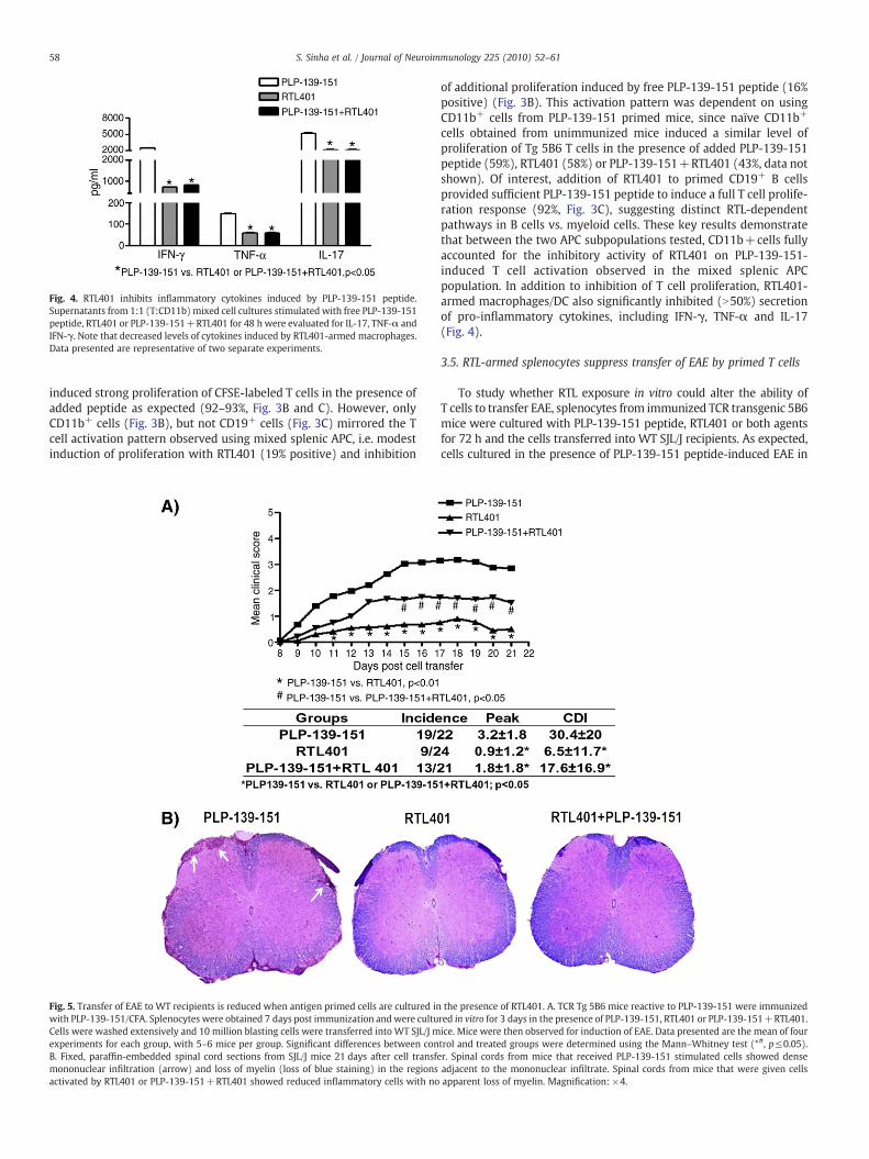
Fig. 4. RTL401 inhibits inflammatory cytokines induced by PLP-139-151 peptide.Supernatants from 1:1 (T:CD11b) mixed cell cultures stimulated with free PLP-139-151peptide, RTL401 or PLP-139-151+RTL401 for 48 h were evaluated for IL-17, TNF-α andIFN-γ. Note that decreased levels of cytokines induced by RTL401-armed macrophages.Data presented are representative of two separate experiments.
58 S. Sinha et al. / Journal of Neuroimmunology 225 (2010) 52–61
induced strong proliferation of CFSE-labeled T cells in the presence ofadded peptide as expected (92–93%, Fig. 3B and C). However, onlyCD11b+ cells (Fig. 3B), but not CD19+ cells (Fig. 3C) mirrored the Tcell activation pattern observed using mixed splenic APC, i.e. modestinduction of proliferation with RTL401 (19% positive) and inhibition
Fig. 5. Transfer of EAE to WT recipients is reduced when antigen primed cells are cultured inwith PLP-139-151/CFA. Splenocytes were obtained 7 days post immunization andwere cultuCells were washed extensively and 10 million blasting cells were transferred into WT SJL/J mexperiments for each group, with 5–6 mice per group. Significant differences between conB. Fixed, paraffin-embedded spinal cord sections from SJL/J mice 21 days after cell transfemononuclear infiltration (arrow) and loss of myelin (loss of blue staining) in the regionsactivated by RTL401 or PLP-139-151+RTL401 showed reduced inflammatory cells with no
of additional proliferation induced by free PLP-139-151 peptide (16%positive) (Fig. 3B). This activation pattern was dependent on usingCD11b+ cells from PLP-139-151 primed mice, since naïve CD11b+
cells obtained from unimmunized mice induced a similar level ofproliferation of Tg 5B6 T cells in the presence of added PLP-139-151peptide (59%), RTL401 (58%) or PLP-139-151+RTL401 (43%, data notshown). Of interest, addition of RTL401 to primed CD19+ B cellsprovided sufficient PLP-139-151 peptide to induce a full T cell prolife-ration response (92%, Fig. 3C), suggesting distinct RTL-dependentpathways in B cells vs. myeloid cells. These key results demonstratethat between the two APC subpopulations tested, CD11b+cells fullyaccounted for the inhibitory activity of RTL401 on PLP-139-151-induced T cell activation observed in the mixed splenic APCpopulation. In addition to inhibition of T cell proliferation, RTL401-armed macrophages/DC also significantly inhibited (N50%) secretionof pro-inflammatory cytokines, including IFN-γ, TNF-α and IL-17(Fig. 4).
3.5. RTL-armed splenocytes suppress transfer of EAE by primed T cells
To study whether RTL exposure in vitro could alter the ability ofT cells to transfer EAE, splenocytes from immunized TCR transgenic 5B6mice were cultured with PLP-139-151 peptide, RTL401 or both agentsfor 72 h and the cells transferred into WT SJL/J recipients. As expected,cells cultured in the presence of PLP-139-151 peptide-induced EAE in
the presence of RTL401. A. TCR Tg 5B6 mice reactive to PLP-139-151 were immunizedred in vitro for 3 days in the presence of PLP-139-151, RTL401 or PLP-139-151+RTL401.ice. Mice were then observed for induction of EAE. Data presented are the mean of fourtrol and treated groups were determined using the Mann–Whitney test (*#, p≤0.05).r. Spinal cords from mice that received PLP-139-151 stimulated cells showed denseadjacent to the mononuclear infiltrate. Spinal cords from mice that were given cellsapparent loss of myelin. Magnification: ×4.

Fig. 6. RTL551 can treat EAE in the absence of B cells. B cell deficient (µMT) mice wereimmunized with MOG-35-55 peptide in CFA plus Ptx and were treated with 5 dailydoses of 100 µg RTL551 or vehicle at onset of clinical signs of EAE. Note significantreduction (p≤0.05) in peak EAE and CDI scores in RTL-treated mice (evaluated by theMann–Whitney test). The experiment was repeated twice with similar results.
59S. Sinha et al. / Journal of Neuroimmunology 225 (2010) 52–61
recipient mice, with peak disease of 3.2±1.8 and cumulative diseaseindex (CDI) of 30.4±20 (Fig. 5A). In contrast, cells cultured in thepresence of RTL401 alone transferredminimal EAE, with peak disease of0.9±1.2 and CDI of 6.5±11.7. Of paramount importance, cells activatedwith PLP-139-151 peptide in the presence of RTL401 had a significantlyreduced capacity to transfer EAE compared to cells stimulatedwith PLP-139-151 peptide alone, with peak disease of 1.8±1.8 and CDI of 17.6±16.9 (Fig. 5A). To rule out the possibility that a lower molarconcentration of RTL-derived PLP-139-151 peptide present in the50 µg/mL dose of RTL401 might lead to suboptimal activation ofT cells in cultures stimulatedwithRTL401alone,we also cultured cells inthe presence of 100 µg/mL of RTL401 that contained an equimolarconcentration of PLP-139-151 compared to 10 µg/mL free peptide. Cellsobtained from this culture did not transfer EAE at all (data not shown),suggesting an RTL401 dose-dependent effect. Spinal cords of micereceiving cells stimulated with RTL401 or PLP-139-151+RTL401showed fewer inflammatory lesions and loss of myelin compared tomice receiving PLP-139-151 stimulated cells (white arrows in spinalcords of mice receiving PLP-139-151 stimulated cells, Fig. 5B). Theseunique results demonstrate that RTL-arming of splenic APC in vitrodown-modulates the disease-causing potential of encephalitogenicPLP-139-151-primed T cells.
3.6. CD19+ B cells are not required for RTL treatment of EAE
Although RTL401 clearly binds to both CD11b/c+ macrophages/DCand CD19+ B cells (Fig. 1), the RTL401-armed B cells directly inducedstrong proliferation and failed to inhibit 5B6 T cell activation inducedby the PLP-139-151 peptide in vitro (Fig. 3), suggesting lack of atolerogenic function. To further evaluate the role of B cells in the RTLtherapeutic mechanism, RTL551 (designed to modulate MOG-35-55specific T cells in C57BL/6 mice) was employed for treatment of B celldeficient µMT knockout mice with EAE. As shown in Fig. 6, RTL551was highly effective for treating EAE induced in B cell deficient mice,reducing the peak disease score from 4.5±0.6 in vehicle-treated miceto 1.6±0.4 in RTL401-treated mice and the cumulative disease index(CDI) from 34.5±6.5 in vehicle-treated mice to 10.2±2.0 in RTL-treatedmice (pb0.01). These data suggest that B cells are not essentialfor successful RTL therapy of EAE.
4. Discussion
Previously we have shown that RTL treatment reduces encepha-litogenicity of T cells in vivo, leading to remission from EAE both inchronic (C57BL/6 and DR2 mice) and relapsing remitting (SJL/J mice)models of EAE (Offner et al., 2008). This study, for the first time,demonstrates RTL-mediated functional effects on pathogenic T cellsafter binding to APCs, including CD19+ B cells, CD11b+ and CD11c+
macrophages and dendritic cells, but not CD3+ T cells. RTL binding toAPC was not dependent on covalently tethered peptide, as emptyRTL400 also bound to these cells with equal intensity. Thus, the robustcell binding event described in the current study involves a putativeRTL receptor that differs from direct ligation of the T cell receptor bysoluble cognate RTLs reported in previous studies using human T cellclones and T cell hybridomas. Our inability to detect direct RTLbinding to CD3+ cells in this study may indicate the absence of theputative RTL receptor on T cells, a low frequency of peptide-specificT cells (typically b1/1000 splenocytes in an unimmunizedmouse) anda low binding avidity of TCR for the RTL idiotope not likely to bedetectable using FACS or immobilization on glass (Davis et al., 1998).Indeed, peptide-specific binding of RTL1000 (containing human DR2covalently bound to MOG-35-55 peptide) to membrane preparationscontaining cognate TCR (from our H2-1 hybridoma) are transient andof low avidity when assessed by Biacore measurements (AAVmanuscript in preparation).
To evaluate the phenotypic effects of RTL401 on CD11b+ cells, wecultured splenocytes obtained from immunized SJL/J mice in thepresence of free PLP-139-151 peptide, RTL401 or both reagents.Initially, we focused on expression of the CD11b (Mac-1, integrin)molecule itself, known to be both a complement receptor (CR3) and acell adhesion molecule present on the surface of macrophages thatundergoes upregulation from preformed cytoplasmic stores uponactivation (Nicholson et al., 2007). CD11b integrin was found toincrease progressively over 72 h in PLP-139-151 stimulated cellcultures (0.9%–2.5%), whereas no increase occurred in RTL-treatedcultures at the 72 h time point (Fig. 2B). Moreover, there weresignificantly lower percentages of the CD11bhigh subset in RTL401-treated cultures at all time points (Fig. 2C). Finally, RTL401 treatmentof EAE in vivo resulted in a profound significant reduction of CD11bupregulation on blood monocytes during the course of EAE (Fig. 2D).
CD11b integrin was found to be critically important for EAEinduction (Bullard et al., 2005), and therefore, down-regulation ofCD11b on monocytes by RTL401 could have a direct consequence onEAE progression. Gordon et al. (1995) found that anti-CD11badministration delayed the onset and reduced the severity of EAE,and it is well established that although T cells initiate EAE, the effectormechanisms provided by infiltrating CD11b+ macrophages andendogenous microglia can lead to inflammation and demyelinationin the CNS (Benveniste, 1997). Mature macrophages and dendriticcells are prominent infiltrates in multiple sclerosis and EAE. CD11b+
macrophages are even more significant for causing damage in EAE asmicroglia are not appropriately activated in the absence of bloodborne macrophages (Bauer et al., 1995). This also suggests thatpossible interaction between infiltrating macrophages and T cellsmight be responsible for the activation of microglia in the CNS.Inhibition of CD11b upregulation on monocytes can block theirantigen presentation, migration and adhesion to vascular endotheli-um leading to reduced accumulation in the CNS. Indeed, wedemonstrated in previous studies that RTL treatment of EAE resultedin significant reduction of inflammatory infiltrates in the spinal cords(Huan et al., 2004; Sinha et al., 2009; Sinha et al., 2010; Sinha et al.,2007; Wang et al., 2006). Taken together, we propose a model whereRTL401 prevents upregulation of CD11b integrin on myeloid cells inthe blood early in the disease and blocks any subsequent entry ofCD11b+ cells into the CNS. This could in turn result in suboptimalactivation of already infiltrated cells and abrogation of the

60 S. Sinha et al. / Journal of Neuroimmunology 225 (2010) 52–61
inflammatory cascade in the CNS, eventually leading to attenuationfrom EAE. It should be noted that strong binding of RTL was alsoobserved with CD11c+ dendritic cells and it is conceivable that likeCD11b, CD11c integrin might also be down-regulated by RTL401, anissue that will be addressed in future studies.
The in vitro experimental paradigm used to assess RTL effects onencephalitogenic T cells was designed to mimic in vivo RTL treatmentof established EAE. Thus, TCR Tg T cells from PLP-139-151 immunized5B6 mice were co-cultured with primed splenic APC from the samemice in the presence of free PLP-139-151 peptide, RTL401 or freepeptide+RTL401 and evaluated for proliferation and cytokinerelease, and ability to transfer EAE to naïve recipient SJL/J mice. PLP-139-151 was included with RTL401 to see whether the presence offree peptide might influence and/ or reverse RTL effects on T cells. Themajor findings from these studies included: 1) Bothmyeloid and B cellAPCs could induce strong T cell proliferation responses when pulsedwith free PLP-139-151 peptide as expected; 2) B cells could inducecomparably strong T cell proliferation responses when pulsed withRTL401 (but not RTL400 lacking the PLP-139-151 peptide), demon-strating an ability to process and present the covalently tethered PLPpeptide derived from RTL401. In contrast, CD11b+ myeloid cells had agreatly reduced capacity (by ∼80%) to induce T cell proliferationresponses with RTL-derived PLP-139-151 peptide; 3) T cell prolifer-ation and cytokine responses to free PLP-139-151 peptide werestrongly inhibited (50–80%) by RTL401-conditioned, T cell depletedsplenocytes and purified myeloid cells but not purified B cells fromPLP-139-151 peptide-primed mice (no effect by myeloid cells fromnaïve mice); 4) The regulatory effects of RTL401-conditioned myeloidcells fully reflected the strong inhibition observed using unfractio-nated splenic APC (that included B cells), indicating a dominant effectof RTL-conditioned myeloid cells; and 5) The inhibitory effects ofRTL401 on T cell responses to PLP-139-151 peptide in vitro werereflected by a significant reduction in severity of passively transferredEAE and reduced inflammatory lesions in spinal cord tissue.
The in vitro T cell activation studies discussed above suggestedimportant functional differences between B cells and myeloid cells,despite comparable binding of RTL by both APC lineages. Unlikemyeloid cells, B cells could induce maximal T cell proliferation withRTL401-derived PLP-139-151 peptide that was comparable to freepeptide alone. Moreover, RTL-armed B cells failed to inhibit T cellproliferation responses. These differences may well be linked to theability of the RTL401 to prevent upregulation of the CD11b integrinpresent on myeloid cells but not B cells. Unlike PLP-139-151stimulated cultures, failure to detect an increase in CD11b in RTL401stimulated cultures suggests suboptimal activation and reducedeffector functions of the macrophages. Indeed anti CD11b antibodyhas been shown to inhibit phagocytosis by neutrophils (Rubel et al.,1999) indicating its role in antigen uptake and therefore subsequentpresentation of peptides, although evidence of a similar role of CD11bon macrophages is currently lacking. Failure of T cells to respond toPLP-139-151 with coordinate down-regulation of CD11b supportsreduced APC function in the presence of RTL. Considering the antigenspecificity of RTL-mediated treatment of EAE, we believe that onlycognate RTL is able to prevent upregulation of CD11b integrin onprimed cells accompanied with inhibition of T cell proliferation.Nonetheless, this will be tested in future studies by using RTL withdifferent target specificity than the immunizing peptide.
To further evaluate the role of CD19+ B cells in RTL therapy, weinduced EAE in B cell deficient µMT KO mice on the C57BL/6background and treated the mice at disease onset with RTL551. Thedata demonstrated unequivocally that RTL551 was highly effective inreversing clinical signs of EAE, indicating that B cells are not necessaryfor RTL treatment effects on EAE. Given the ability of RTL-armedCD11b+ myeloid APC to inhibit T cell activation and transfer of EAE, itis likely that the RTL551 treatment of µMT mice was mediatedthrough residual macrophages and/or dendritic cells. These results do
not eliminate possible regulatory effects of RTL-armed B cells,however, since it is conceivable that B cells and myeloid APCrepresent distinct but redundant RTL-mediated inhibitory pathways.
In conclusion, our results demonstrate for the first time a novelpathway of T cell regulation that involves binding of RTLs to thesurface of myeloid APC and down-regulation of the CD11b macro-phage marker that may contribute to the reduced ability of themacrophages to induce T cell activation and transfer of EAE. Incontrast, B cells could not inhibit T cell activation after RTL bindingand were found to be unnecessary for successful RTL therapy of EAE.
Acknowledgments
The authors wish to thank Ms. Eva Niehaus for assistance inpreparing the manuscript. Dr. Sinha is a Postdoctoral Fellow of theNational Multiple Sclerosis Society and this work was supported inpart by the National Multiple Sclerosis Society Postdoctoral Fellow-ship FG1749-A-1, grants RG3794-A-4 and RG3794-B-6; NIH GrantsNS47661 and AI43960; and the Biomedical Laboratory R&D Service,Department of Veterans Affairs.
Drs. Offner, Burrows, Vandenbark, and OHSU have a significantfinancial interest in Artielle ImmunoTherapeutics, Inc., a companythat may have a commercial interest in the results of this research andtechnology. This potential conflict of interest has been reviewed andmanaged by the OHSU and VAMC Conflict of Interest in ResearchCommittees.
References
Bauer, J., Huitinga, I., Zhao, W., Lassmann, H., Hickey, W.F., Dijkstra, C.D., 1995. The roleof macrophages, perivascular cells, and microglial cells in the pathogenesis ofexperimental autoimmune encephalomyelitis. Glia 15, 437–446.
Benveniste, E.N., 1997. Role of macrophages/microglia in multiple sclerosis andexperimental allergic encephalomyelitis. J. Mol. Med. 75, 165–173.
Bullard, D.C., Hu, X., Schoeb, T.R., Axtell, R.C., Raman, C., Barnum, S.R., 2005. Criticalrequirement of CD11b (Mac-1) on T cells and accessory cells for development ofexperimental autoimmune encephalomyelitis. J. Immunol. 175, 6327–6333.
Burrows, G.G., Bebo Jr., B.F., Adlard, K.L., Vandenbark, A.A., Offner, H., 1998. Two-domainMHC class II molecules form stable complexes with myelin basic protein 69–89peptide that detect and inhibit rat encephalitogenic T cells and treat experimentalautoimmune encephalomyelitis. J. Immunol. 161, 5987–5996.
Burrows, G.G., Chang, J.W., Bachinger, H.P., Bourdette, D.N., Offner, H., Vandenbark, A.A.,1999. Design, engineering and production of functional single-chain T cell receptorligands. Protein Eng. 12, 771–778.
Burrows, G.G., Chou, Y.K., Wang, C., Chang, J.W., Finn, T.P., Culbertson, N.E., Kim, J.,Bourdette, D.N., Lewinsohn, D.A., Lewinsohn, D.M., Ikeda, M., Yoshioka, T., Allen, C.N.,Offner, H., Vandenbark, A.A., 2001. Rudimentary TCR signaling triggers default IL-10secretion by human Th1 cells. J. Immunol. 167, 4386–4395.
Chou, Y.K., Culbertson, N., Rich, C., LaTocha, D., Buenafe, A.C., Huan, J., Link, J., Wands, J.M.,Born, W.K., Offner, H., Bourdette, D.N., Burrows, G.G., Vandenbark, A.A., 2004. T-cellhybridoma specific for myelin oligodendrocyte glycoprotein-35-55 peptide producedfrom HLA-DRB1*1501-transgenic mice. J. Neurosci. Res. 77, 670–680.
Davis, M.M., Boniface, J.J., Reich, Z., Lyons, D., Hampl, J., Arden, B., Chien, Y., 1998. Ligandrecognition by alpha beta T cell receptors. Annu. Rev. Immunol. 16, 523–544.
Gordon, E.J., Myers, K.J., Dougherty, J.P., Rosen, H., Ron, Y., 1995. Both anti-CD11a (LFA-1)and anti-CD11b (MAC-1) therapy delay the onset and diminish the severity ofexperimental autoimmune encephalomyelitis. J. Neuroimmunol. 62, 153–160.
Huan, J., Subramanian, S., Jones, R., Rich, C., Link, J., Mooney, J., Bourdette, D.N.,Vandenbark, A.A., Burrows, G.G., Offner, H., 2004. Monomeric recombinant TCRligand reduces relapse rate and severity of experimental autoimmune encephalo-myelitis in SJL/J mice through cytokine switch. J. Immunol. 172, 4556–4566.
Link, J., 2007. Partial MHC class II molecules preferentially bind to B cells. J. Immunol. 78.Link, J.M., Rich, C.M., Korat, M., Burrows, G.G., Offner, H., Vandenbark, A.A., 2007.
Monomeric DR2/MOG-35-55 recombinant TCR ligand treats relapses of experi-mental encephalomyelitis in DR2 transgenic mice. Clin. Immunol. 123, 95–104.
Nicholson, G.C., Tennant, R.C., Carpenter, D.C., Sarau, H.M., Kon, O.M., Barnes, P.J.,Salmon, M., Vessey, R.S., Tal-Singer, R., Hansel, T.T., 2007. A novel flow cytometricassay of human whole blood neutrophil and monocyte CD11b levels: upregulationby chemokines is related to receptor expression, comparisonwith neutrophil shapechange, and effects of a chemokine receptor (CXCR2) antagonist. Pulm. Pharmacol.Ther. 20, 52–59.
Offner, H., Subramanian, S., Wang, C., Afentoulis, M., Vandenbark, A.A., Huan, J.,Burrows, G.G., 2005. Treatment of passive experimental autoimmune encephalo-myelitis in SJL mice with a recombinant TCR ligand induces IL-13 and preventsaxonal injury. J. Immunol. 175, 4103–4111.
Offner, H., Sinha, S., Wang, C., Burrows, G.G., Vandenbark, A.A., 2008. Recombinant T cellreceptor ligands: immunomodulatory, neuroprotective and neuroregenerative

61S. Sinha et al. / Journal of Neuroimmunology 225 (2010) 52–61
effects suggest application as therapy for multiple sclerosis. Rev. Neurosci. 19,327–339.
Rubel, C., Miliani De Marval, P., Vermeulen, M., Isturiz, M.A., Palermo, M.S., 1999.Lipopolysaccharide enhances FcgammaR-dependent functions in vivo throughCD11b/CD18 up-regulation. Immunology 97, 429–437.
Sinha, S., Subramanian, S., Proctor, T.M., Kaler, L.J., Grafe, M., Dahan, R., Huan, J.,Vandenbark, A.A., Burrows, G.G., Offner, H., 2007. A promising therapeuticapproach for multiple sclerosis: recombinant T-cell receptor ligands modulateexperimental autoimmune encephalomyelitis by reducing interleukin-17 produc-tion and inhibiting migration of encephalitogenic cells into the CNS. J. Neurosci. 27,12531–12539.
Sinha, S., Subramanian, S., Miller, L., Proctor, T.M., Roberts, C., Burrows, G.G.,Vandenbark, A.A., Offner, H., 2009. Cytokine switch and bystander suppression ofautoimmune responses to multiple antigens in experimental autoimmuneencephalomyelitis by a single recombinant T-cell receptor ligand. J. Neurosci. 29,3816–3823.
Sinha, S., Subramanian, S., Emerson-Webber, A., Lindner, M., Burrows, G.G., Grafe, M.,Linington, C., Vandenbark, A.A., Bernard, C.C., Offner, H., 2010. Recombinant TCR
ligand reverses clinical signs and CNS damage of EAE induced by recombinanthuman MOG. J. Neuroimmune. Pharmacol. 5, 231–240.
Vandenbark, A.A., Rich, C., Mooney, J., Zamora, A., Wang, C., Huan, J., Fugger, L., Offner,H., Jones, R., Burrows, G.G., 2003. Recombinant TCR ligand induces tolerance tomyelin oligodendrocyte glycoprotein 35–55 peptide and reverses clinical andhistological signs of chronic experimental autoimmune encephalomyelitis in HLA-DR2 transgenic mice. J. Immunol. 171, 127–133.
Wang, C., Mooney, J.L., Meza-Romero, R., Chou, Y.K., Huan, J., Vandenbark, A.A., Offner,H., Burrows, G.G., 2003. Recombinant TCR ligand induces early TCR signaling and aunique pattern of downstream activation. J. Immunol. 171, 1934–1940.
Wang, C., Gold, B.G., Kaler, L.J., Yu, X., Afentoulis, M.E., Burrows, G.G., Vandenbark, A.A.,Bourdette, D.N., Offner, H., 2006. Antigen-specific therapy promotes repair ofmyelin and axonal damage in established EAE. J. Neurochem. 98, 1817–1827.
Yadav, V., Bourdette, D., Bowen, J.D., Lynch, S.G., Mattson, D., Preinigerova, J., Rose, C.,Stead, R.B., Ferro, A.J., Goldstein, A.S., Burrows, G., Offner, H., Vandenbark, A., 2010.Recombinant T cell receptor ligand (RTL) for the treatment of multiple sclerosis:report of a phase I clinical trial. Neurology 74 (S2), A293–A294.