binasal occlusion (bno), visual motion sensitivity (vms
TRANSCRIPT

brainsciences
Review
Binasal Occlusion (BNO), Visual Motion Sensitivity(VMS), and the Visually-Evoked Potential (VEP) inmild Traumatic Brain Injury and Traumatic BrainInjury (mTBI/TBI)
Kenneth J. Ciuffreda 1,*, Naveen K. Yadav 2 and Diana P. Ludlam 1
1 Department of Biological and Vision Sciences, State University of New York, College of Optometry,New York, NY 10016, USA; [email protected]
2 Chicago College of Optometry, Midwestern University, Downers Grove, IL 60515, USA;[email protected]
* Correspondence: [email protected]; Tel.: +1-212-938-5765
Received: 14 June 2017; Accepted: 4 August 2017; Published: 9 August 2017
Abstract: The diagnosis and treatment of the possible visual sequelae in those with traumatic braininjury (TBI) represents an important area of health care in this special population. One of their mostprevalent yet elusive visual symptoms is visual motion sensitivity (VMS). In this review, we presentthe basic VMS phenomenon and its related symptoms, clinical studies in the area, clinical researchinvestigations using the visual-evoked potential (VEP) as a cortical probe, and possible mechanismsand related neurophysiology that may underlie VMS. Lastly, therapeutic interventions are brieflydescribed, as well as future directions for clinical research and patient care in those with VMS and TBI.
Keywords: traumatic brain injury (TBI); visually-evoked potential (VEP); binasal occlusion (BNO);visual motion sensitivity (VMS); visuomotor; visual motion perception; magnocellular pathway;dorsal stream
1. Introduction
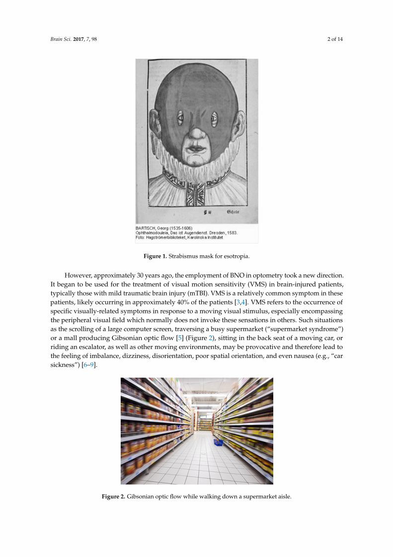
The use of binasal occluders (BNO) and bitemporal occluders (BTO) in medicine, in particularophthalmology, has a long and interesting history for the treatment of strabismus (deviation of aneye from parallelism). Mention of such occlusion dates back to at least 600 AD or so ([1] for a review).Here, BTO was used as a treatment for exotropia (outward deviation on one eye). The concept wasthat, in those with exotropia, temporal occlusion would force them to converge their eyes to themedian plane to attain binocular fixation and fusion. This is basically one of the first mentions ofthe clinical practice of “orthoptics”, the medically-based remediation of abnormal binocular vision,especially fusional problems related to strabismus [1]. Over the next millennium, it evolved furtherfrom small and simple occluders into a more encompassing “strabismus mask”, presumably to havebetter compliance despite its poor cosmesis, primarily for the treatment of esotropia (inward deviationof one eye) ([1] for a review) (Figure 1). Thus, strategic location of the occluders could be used for eitheresotropia or exotropia, and sometimes for hypertropia (vertical deviation of one eye) when placed inthe vertical position. This basic concept continues today both in ophthalmology and optometry forstrabismus, as well as other binocular vision disorders (e.g., spasm of the near reflex) [2], once againusing simple occluders.
Brain Sci. 2017, 7, 98; doi:10.3390/brainsci7080098 www.mdpi.com/journal/brainsci
Brain Sci. 2017, 7, 98 2 of 14Brain Sci. 2017, 7, 98 2 of 14
Figure 1. Strabismus mask for esotropia.
However, approximately 30 years ago, the employment of BNO in optometry took a new
direction. It began to be used for the treatment of visual motion sensitivity (VMS) in brain‐injured
patients, typically those with mild traumatic brain injury (mTBI). VMS is a relatively common
symptom in these patients, likely occurring in approximately 40% of the patients [3,4]. VMS refers to
the occurrence of specific visually‐related symptoms in response to a moving visual stimulus,
especially encompassing the peripheral visual field which normally does not invoke these sensations
in others. Such situations as the scrolling of a large computer screen, traversing a busy supermarket
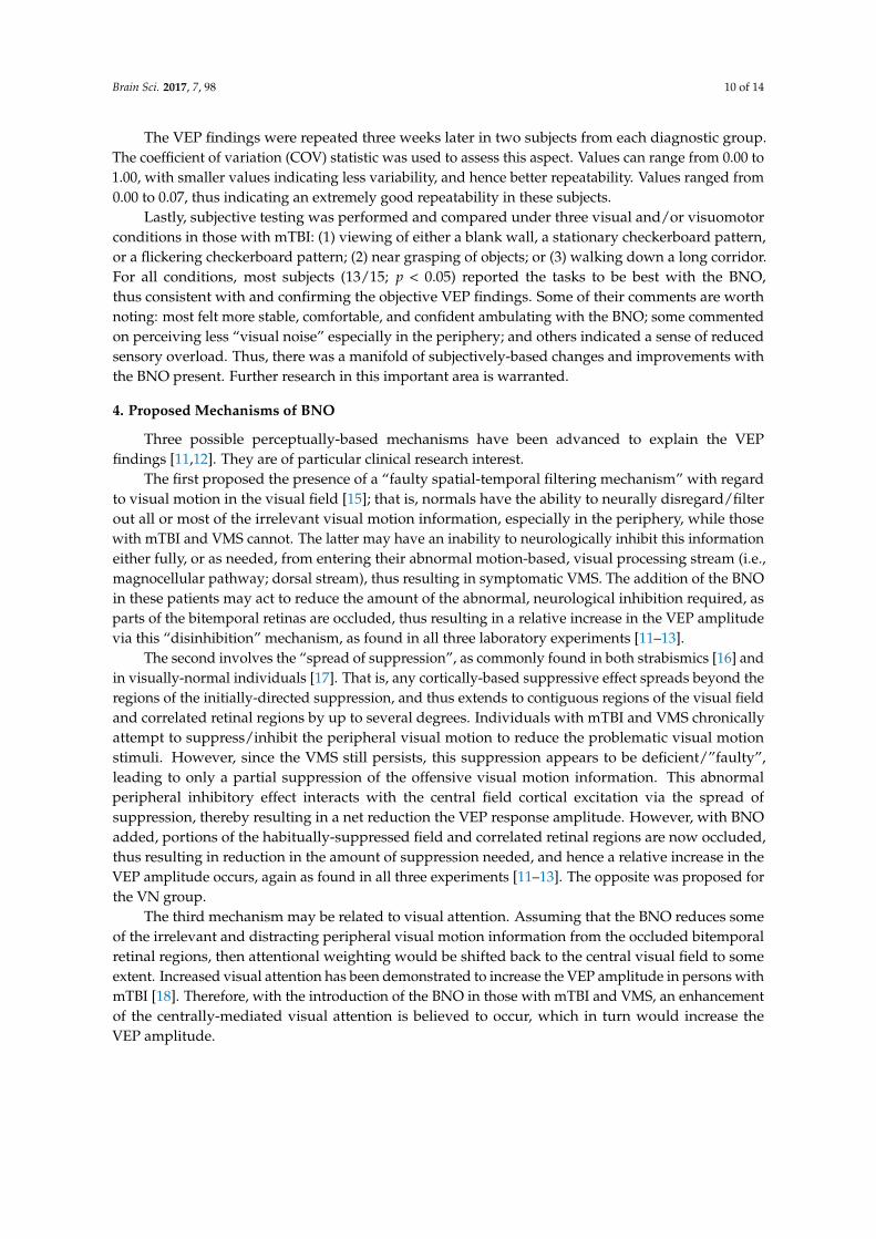
(“supermarket syndrome”) or a mall producing Gibsonian optic flow [5] (Figure 2), sitting in the back
seat of a moving car, or riding an escalator, as well as other moving environments, may be
provocative and therefore lead to the feeling of imbalance, dizziness, disorientation, poor spatial
orientation, and even nausea (e.g., “car sickness”) [6–9].
Figure 2. Gibsonian optic flow while walking down a supermarket aisle.
Figure 1. Strabismus mask for esotropia.
However, approximately 30 years ago, the employment of BNO in optometry took a new direction.It began to be used for the treatment of visual motion sensitivity (VMS) in brain-injured patients,typically those with mild traumatic brain injury (mTBI). VMS is a relatively common symptom in thesepatients, likely occurring in approximately 40% of the patients [3,4]. VMS refers to the occurrence ofspecific visually-related symptoms in response to a moving visual stimulus, especially encompassingthe peripheral visual field which normally does not invoke these sensations in others. Such situationsas the scrolling of a large computer screen, traversing a busy supermarket (“supermarket syndrome”)or a mall producing Gibsonian optic flow [5] (Figure 2), sitting in the back seat of a moving car, orriding an escalator, as well as other moving environments, may be provocative and therefore lead tothe feeling of imbalance, dizziness, disorientation, poor spatial orientation, and even nausea (e.g., “carsickness”) [6–9].
Brain Sci. 2017, 7, 98 2 of 14
Figure 1. Strabismus mask for esotropia.
However, approximately 30 years ago, the employment of BNO in optometry took a new
direction. It began to be used for the treatment of visual motion sensitivity (VMS) in brain‐injured
patients, typically those with mild traumatic brain injury (mTBI). VMS is a relatively common
symptom in these patients, likely occurring in approximately 40% of the patients [3,4]. VMS refers to
the occurrence of specific visually‐related symptoms in response to a moving visual stimulus,
especially encompassing the peripheral visual field which normally does not invoke these sensations
in others. Such situations as the scrolling of a large computer screen, traversing a busy supermarket
(“supermarket syndrome”) or a mall producing Gibsonian optic flow [5] (Figure 2), sitting in the back
seat of a moving car, or riding an escalator, as well as other moving environments, may be
provocative and therefore lead to the feeling of imbalance, dizziness, disorientation, poor spatial
orientation, and even nausea (e.g., “car sickness”) [6–9].
Figure 2. Gibsonian optic flow while walking down a supermarket aisle. Figure 2. Gibsonian optic flow while walking down a supermarket aisle.

Brain Sci. 2017, 7, 98 3 of 14
In this review, we will cover both the clinical and laboratory-based studies involving BNO andVMS, with an emphasis on the objective, visually-evoked potential (VEP) findings. Both the clinicalutility, as well as the possible underlying perceptual and neurological mechanisms, will be addressed.Future directions for research will also be considered.
Methods
The following resources were used for the present review. They included computer searchesusing PubMed, Yahoo, Google, and Google Scholar. Search terms included “visual motion sensitivity”,“VMS”, “visual motion perception”, “binasal occlusion”, “BNO”, “vision in mTBI/TBI”, “brain injury”,“VEP”, and “magnocellular pathway”. In addition, relevant books, book chapters, and publishedpapers in the area were used other than those found in our formal computer searches.
2. Clinical Case Studies
Surprisingly, given the high prevalence and bothersome nature of VMS, there is a relative paucityof clinical studies and case reports in this area of mTBI/TBI, as well as in other forms of acquired braininjury (ABI) and neurological disorders (e.g., cerebral palsy, multiple sclerosis) [7,8]. Unfortunately,visually-evoked potential (VEP) recordings were not obtained, both with and without the BNO inplace, in these clinical reports. However, the cases and their findings provide important insights intothe VMS phenomenon and related symptomatology.
One of the earliest case reports in this area was that of Proctor (2009) in a 46-year-old malepatient [9]. He had been involved in 13 motor vehicle accidents (MVA) over the prior 10 years.The patient later received a full general medical examination, as well as a neurological evaluationincluding the inner ear, with no clear cause found for his persistent visual symptoms. This examinationwas performed eighteen months prior to his optometric vision examination. His recent MRI wasnormal, as were the earlier ones that had been performed. However, his symptoms at the timeof the initial, comprehensive optometric vision examination (including a visual field), and whichhad persisted over the prior ten year period, included VMS/”supermarket syndrome”, dizziness,balance and gait difficulty, especially in crowded environments, nausea when in a moving vehicle,and poor depth perception, all of which are common visually-based symptoms in mTBI [3,4,6]. He wasprescribed a course of vision therapy (i.e., a sequence of specific remedial procedures and protocolsinvolving the oculomotor system and other vision-related aspects incorporating the principles ofperceptual and motor learning) [10], with an oculomotor emphasis, due to the poor fixation, pursuit,and saccadic abilities uncovered. At the sixth therapy session, BNO was attempted. The improvementswere immediate as he walked down a long hallway and performed concurrent visuomotor tasks.The BNO was added to his home therapy; for example, he was instructed to use them when riding asa passenger in a car. The patient reported reduced symptoms in a motor vehicle, as well as furtherimproved mobility with less symptoms, with the BNO. However, he did not use the BNO for driving,as he felt that the occluders restricted his peripheral visual field.
More recently, Gallop (2014) presented two cases of mTBI in which BNO was found to behelpful [7]. The first patient was a 67-year-old male who was involved in an MVA. His initial visualsymptoms included diplopia, pain in his right eye with ocular movement, and balance problems.An ophthalmological vision examination shortly after the MVA revealed diplopia for which base-outprisms were prescribed, but without any relief. Months later, he was examined by his optometristof many years, and thus all of the MVA-related visual symptoms could be compared against thosepre-injury. The patient reported the same visual symptoms as he did earlier to the ophthalmologist,but he now exhibited several new visual signs: reduced distance visual acuity, reduced stereoacuityat near, vertical phoria at near, and large exophoria at near. None of these symptoms and signswere present prior to the MVA. Due to his visual symptoms, especially related to balance, it wasdecided to try BNO on the patient. Immediately, his symptoms were markedly reduced, especiallyduring ambulation. The BNO, along with the oculomotor-based vision therapy, were prescribed with

Brain Sci. 2017, 7, 98 4 of 14
continued success. The second patient was a young, female administrator who had sustained an mTBIone year earlier. She had hit the top of her head and was temporarily “stunned”. Her symptomsincluded reading difficulties, light sensitivity, discomfort on the computer, variable ocular focusing,spatial disorientation, and balance problems. All busy environments, including her office, triggered herbalance and disorientation problems. The patient’s comprehensive optometric vision examination wasunremarkable, and visual acuity was 20/20. However, she was nauseous for several hours after eachsection of the basic vision test battery, which is not uncommon in these patients. Thus, she elected notto perform any further vision testing and to forgo the tentatively suggested vision therapy. About twoyears later, the patient returned to the same optometric clinic with the same persistent visual problems.In the interim, she was being treated for anxiety, with some degree of intermittent relief of her visualsymptoms. There were now fewer symptoms during the subsequent vision examination, and alltesting could be completed. She was recommended a course of vision therapy. In addition, BNOwas attempted. The effect of the added BNO was immediate, with about 80% reduction in hersymptoms as she ambulated about the doctor’s office. The markedly reduced symptoms of VMS inbusy environments, such as the street and her workplace, continued. The combination of the BNO andthe vision therapy has allowed her to remain much less symptomatic under all naturalistic conditionsover the several months of follow-up.
Based on the above clinical studies, what might be the prevailing mechanism? A most intriguingsuggestion was proposed by Gallop (1998) [8]. He speculated that the BNO, essentially functioning asan “object” in the patient’s environment, provided a stable “visual frame of reference” for these patientswith symptoms such as dizziness and disorientation, as the BNO moved with head movement. That is,the patient now had a stable object in the visual field during all tasks which acted as a spatial-perceptualvisual ‘anchor’. This may be even more true when the BNOs are oriented vertically, rather than angledas frequently done (Figure 3), to provide a more direct gravity-based reference.
Brain Sci. 2017, 7, 98 4 of 14
included reading difficulties, light sensitivity, discomfort on the computer, variable ocular focusing,
spatial disorientation, and balance problems. All busy environments, including her office, triggered
her balance and disorientation problems. The patient’s comprehensive optometric vision
examination was unremarkable, and visual acuity was 20/20. However, she was nauseous for several
hours after each section of the basic vision test battery, which is not uncommon in these patients.
Thus, she elected not to perform any further vision testing and to forgo the tentatively suggested
vision therapy. About two years later, the patient returned to the same optometric clinic with the
same persistent visual problems. In the interim, she was being treated for anxiety, with some degree
of intermittent relief of her visual symptoms. There were now fewer symptoms during the
subsequent vision examination, and all testing could be completed. She was recommended a course
of vision therapy. In addition, BNO was attempted. The effect of the added BNO was immediate,
with about 80% reduction in her symptoms as she ambulated about the doctor’s office. The markedly
reduced symptoms of VMS in busy environments, such as the street and her workplace, continued.
The combination of the BNO and the vision therapy has allowed her to remain much less
symptomatic under all naturalistic conditions over the several months of follow‐up.
Based on the above clinical studies, what might be the prevailing mechanism? A most intriguing
suggestion was proposed by Gallop (1998) [8]. He speculated that the BNO, essentially functioning
as an “object” in the patient’s environment, provided a stable “visual frame of reference” for these
patients with symptoms such as dizziness and disorientation, as the BNO moved with head
movement. That is, the patient now had a stable object in the visual field during all tasks which acted
as a spatial‐perceptual visual ‘anchor’. This may be even more true when the BNOs are oriented
vertically, rather than angled as frequently done (Figure 3), to provide a more direct gravity‐based
reference.
Figure 3. Schematic representation of binasal occluders on a subject [11,12].
3. Laboratory Studies
Once again, and surprisingly, there have been a paucity of studies in clinical laboratories
assessing the efficacy of BNO in mTBI/TBI. The first investigation was published nearly 20 years prior
to the more recent studies. All three investigations included the VEP as the primary outcome measure.
Figure 3. Schematic representation of binasal occluders on a subject [11,12].
3. Laboratory Studies
Once again, and surprisingly, there have been a paucity of studies in clinical laboratories assessingthe efficacy of BNO in mTBI/TBI. The first investigation was published nearly 20 years prior to themore recent studies. All three investigations included the VEP as the primary outcome measure.

Brain Sci. 2017, 7, 98 5 of 14
3.1. Padula Group
The first investigation of BNO in mTBI/TBI was by Padula et al. (1994) [13]. This hospital-basedstudy was conducted in ten adult (ages 22–46 years) patients with medically-documented TBI.The presence of VMS was not explicitly indicated, and thus not a prerequisite. All were ambulatory.They were compared with 10 normal adults (ages 23–46 years) from the hospital staff without anyhistory of either TBI or VMS. None of the tested individuals had strabismus, as the VEP testing wasalways conducted under binocular-viewing conditions, and its presence would have contaminated thefindings and their interpretation.
Following a comprehensive optometric vision examination, the VEP (Nicolet system, San Carlos,CA) testing commenced. Important details of the methodology are as follows. A central/nearretinal periphery field, checkerboard pattern was used as the test stimulus. It had high-contrast witha check size of 30 minutes of arc and a stimulus pattern reversal rate of 1.9 reversals/s. An ABAexperimental design was used: A = habitual distance refraction, B = habitual distance refraction plusthe vertically-oriented, translucent tape BNO and 2 BI prisms before each eye, and then conditionA again to assess for and assure a stable baseline. The primary VEP metric was its peak-to-peakamplitude (N1-P1), a standard VEP parameter. The VEP was completed in all ten normal controls andin nine of the ten individuals with TBI.
The VEP findings were as follows. In the normals, the VEP amplitude with the combinedBNO/prisms increased in 6/10, decreased in 2/10, and remained the same in 2/10, with a rangeof −14% to +11%, and a mean decrease in amplitude of −0.41 microvolts, as compared to the baselinevalues. These small, mixed findings suggested a ‘noise’ phenomenon; that is, a lack of any consistentcortically-based physiological effect, as one might predict in this control population. In contrast,in the TBI group, the amplitude increased in 8/9 and decreased in 1/9, with a range of −5% to+54% (mean = +20%, +1.38 microvolts). There was a statistically significant effect of the BNO/prismcombination in the TBI group versus the control group (t-test, p < 0.01). Some patients reported areduction of symptoms (e.g., reduced visual instability) with the BNO/prism combination incorporatedinto their distance spectacle prescription. No other related testing was performed, such as formallyassessing visuomotor ability or general ambulation with and without the BNO/prism combination.
Two mechanisms were proposed, which were hypothesized to increase the effectiveness of thepatient’s “binocular cortical cells”, with the BNO/BI combination. As described above in the ‘clinicalstudies’ section by Gallop (1998) [8], Padula et al. (1994) [13] also proposed that the BNO provided‘structure’ to the patient’s overall visual field. They also proposed that the base-in prisms provideda degree of ‘field expansion’. Prisms do displace and deviate incident light per Snell’s Law [14],and they do so differentially as a function of its angle of incidence. This results in magnification ofthe visual field in a non-uniform manner, and thus some degree of ‘field expansion’ does take place.These proposed mechanisms remain speculative.
3.2. Ciuffreda Group
The next and most recent experiments in this area were performed in the Ciuffreda laboratoryabout 20 years later [11,12]. These two studies extended the earlier one by Padula et al. (1994) [13],with their primary goal being to disambiguate the effects of the BNO/BI prism combination, as wasused in the Padula et al. (1994) study [13].
In the first investigation by Ciuffreda et al. (2013) [11], the question was simple: “What was theeffect of BNO alone on adults with chronic mTBI and symptomatic VMS?” There were 10 patientswith medically-diagnosed mTBI and VMS (mean age 28.9 years), and there were 10 matched,visually-normal (VN) controls (mean age 26.7 years) without VMS, with both groups having 20/20 orbetter corrected visual acuity in each eye. None had a history of seizures, strabismus, amblyopia, or anyocular, systemic, or degenerative neurological disease. All underwent a comprehensive optometricvision examination prior to laboratory testing.

Brain Sci. 2017, 7, 98 6 of 14
The VEP (DIOPSYS system, Pine Brook, NJ, USA) stimulus and test conditions were asfollows. The test target consisted of a 17 H by 15 V degree, alternating (four reversals per second),black-and-white, checkerboard pattern (64 × 64, 20.6 min arc) placed one meter away along thesubject’s midline. Luminance was 64 cd/m-squared with a contrast of 85%. The test duration was 20 s.Two trials per test condition (spectacles only or spectacles with obliquely-oriented, opaque black BNO,with binocular viewing) were averaged in each subject, and then averaged across each group, for theparameters of amplitude and latency. A schematic representation of a subject with the BNOs in place ispresented in Figure 3. A schematic representation of the stimulus array with the subject and the BNOsin place is presented in Figure 4. The BNOs (5.7 H × 15 V degs) were cross-projected 5.5 degrees toeither side of the stimulus array and occluded portions of the bitemporal retinas.
Brain Sci. 2017, 7, 98 6 of 14
The VEP (DIOPSYS system, Pine Brook, NJ, USA) stimulus and test conditions were as follows.
The test target consisted of a 17 H by 15 V degree, alternating (four reversals per second), black‐and‐
white, checkerboard pattern (64 × 64, 20.6 min arc) placed one meter away along the subject’s midline.
Luminance was 64 cd/m‐squared with a contrast of 85%. The test duration was 20 s. Two trials per
test condition (spectacles only or spectacles with obliquely‐oriented, opaque black BNO, with
binocular viewing) were averaged in each subject, and then averaged across each group, for the
parameters of amplitude and latency. A schematic representation of a subject with the BNOs in place
is presented in Figure 3. A schematic representation of the stimulus array with the subject and the
BNOs in place is presented in Figure 4. The BNOs (5.7 H × 15 V degs) were cross‐projected 5.5 degrees
to either side of the stimulus array and occluded portions of the bitemporal retinas.
Figure 4. Representation of the binocular visual‐field with binasal occluders in place, including the
checkerboard visual stimulus. Not drawn to scale. f = fovea [12].
The mean amplitude (microvolts) findings are presented in Figure 5. In the VN group without
BNO, it was 21.6 with a range from 9.1–45.4; with the BNO, it was 17.4 with a range from 7.4–35.9.
This decrease with BNO was found in all 10 subjects, which was statistically significantly (binomial
test, p < 0.01) (Figure 6). In the mTBI group without BNO, it was 19.2 with a range from 12.2–35.5;
with BNO, it was 21.3, with a range from 13.6–36.9. This increase with BNO was found in all 10 subjects
(Figure 6), which was statistically significant (p < 0.01). These findings were repeatable 2–7 days later
in a subset of subjects in each diagnostic group.
Figure 4. Representation of the binocular visual-field with binasal occluders in place, including thecheckerboard visual stimulus. Not drawn to scale. f = fovea [12].
The mean amplitude (microvolts) findings are presented in Figure 5. In the VN group withoutBNO, it was 21.6 with a range from 9.1–45.4; with the BNO, it was 17.4 with a range from 7.4–35.9.This decrease with BNO was found in all 10 subjects, which was statistically significantly (binomialtest, p < 0.01) (Figure 6). In the mTBI group without BNO, it was 19.2 with a range from 12.2–35.5;with BNO, it was 21.3, with a range from 13.6–36.9. This increase with BNO was found in all 10 subjects(Figure 6), which was statistically significant (p < 0.01). These findings were repeatable 2–7 days laterin a subset of subjects in each diagnostic group.

Brain Sci. 2017, 7, 98 7 of 14Brain Sci. 2017, 7, 98 7 of 14
Figure 5. Group mean amplitude for the full field without and with binasal occluders in normals and
in mTBI [12]. * means statically significant.
Figure 6. Diagnostic group mean amplitude difference for individual subjects (%). Positive and
negative percentage values represent an increase or decrease in mean amplitude difference values,
respectively [11].
Latency was also assessed. It was within normal limits, and was not statistically different under
any of the test conditions and diagnostic groups.
Lastly, the subjective impressions of both groups were queried. All VN subjects disliked the
BNO. The blocked field was annoying and produced a sense of visual discomfort. In contrast, eight
of the ten mTBI subjects with VMS liked the BNO (p < 0.01). They reported reduced symptoms (e.g.,
reduced nausea, reduced disorientation). Furthermore, they reported more accurate and comfortable
Figure 5. Group mean amplitude for the full field without and with binasal occluders in normals andin mTBI [12]. * means statically significant.
Brain Sci. 2017, 7, 98 7 of 14
Figure 5. Group mean amplitude for the full field without and with binasal occluders in normals and
in mTBI [12]. * means statically significant.
Figure 6. Diagnostic group mean amplitude difference for individual subjects (%). Positive and
negative percentage values represent an increase or decrease in mean amplitude difference values,
respectively [11].
Latency was also assessed. It was within normal limits, and was not statistically different under
any of the test conditions and diagnostic groups.
Lastly, the subjective impressions of both groups were queried. All VN subjects disliked the
BNO. The blocked field was annoying and produced a sense of visual discomfort. In contrast, eight
of the ten mTBI subjects with VMS liked the BNO (p < 0.01). They reported reduced symptoms (e.g.,
reduced nausea, reduced disorientation). Furthermore, they reported more accurate and comfortable
Figure 6. Diagnostic group mean amplitude difference for individual subjects (%). Positive andnegative percentage values represent an increase or decrease in mean amplitude difference values,respectively [11].
Latency was also assessed. It was within normal limits, and was not statistically different underany of the test conditions and diagnostic groups.
Lastly, the subjective impressions of both groups were queried. All VN subjects disliked theBNO. The blocked field was annoying and produced a sense of visual discomfort. In contrast, eightof the ten mTBI subjects with VMS liked the BNO (p < 0.01). They reported reduced symptoms (e.g.,reduced nausea, reduced disorientation). Furthermore, they reported more accurate and comfortable

Brain Sci. 2017, 7, 98 8 of 14
ambulation, fixating of objects in the field, and grasping of objects. They also indicated that theVEP stimulus appeared clearer, brighter, and flickered less. In the other two mTBI/VMS subjects,they disliked the blocked field of the BNO, despite their objective cortical and subjective improvementswith the BNO.
In the second investigation by Yadav and Ciuffreda (2014) [12], the question was a little morecomplex: “What were the independent and combined effects of the BNO and/or base-in prisms onadults with chronic mTBI and symptomatic VMS?” There were 15 patients with medically-diagnosedmTBI (mean age 35.2 years) with the symptom of VMS, and there were 20 matched, visually-normal(VN) controls (mean age 25.5 years) without the symptom of VMS, with both groups having 20/20 orbetter visual acuity in each eye. None had a history of seizures, strabismus, amblyopia, or any ocular,systemic, or degenerative neurological disease. All underwent a comprehensive optometric visionexamination prior to laboratory testing.
The same VEP apparatus as described in detail in their earlier experiment above was used [11].However, for each test condition in each subject, four trials were now averaged in each subject in eachdiagnostic group and then averaged across each diagnostic group. There were four test conditions,all with distance spectacle correction in place, performed in a counter-balanced manner to minimizeorder effects: (1) baseline, (2) BNO only before each eye, (3) two base-in prisms only before each eye,and (4) BNO plus two base-in prisms before each eye. The statistical analyses (ANOVA) revealedseveral important, significant findings (all p < 0.05).
VN (mean amplitude, microvolts) (Figure 7a): Firstly, the baseline (20.8) and the prism only (20.6)conditions were not significantly different, which suggested that the prisms did not affect the VEPamplitude. Secondly, the BNO only condition (17.1) was significantly less than found in either thebaseline (20.8) or the prism only (20.6) conditions. Thirdly, the BNO (17.1) and the BNO plus prism(18.1) conditions were not significantly different. Together, these findings suggested that the BNOcondition alone was sufficient to have a significant effect on the VEP amplitude; the incorporation ofthe base-in prisms had no cortical advantage.
Brain Sci. 2017, 7, 98 8 of 14
ambulation, fixating of objects in the field, and grasping of objects. They also indicated that the VEP
stimulus appeared clearer, brighter, and flickered less. In the other two mTBI/VMS subjects, they
disliked the blocked field of the BNO, despite their objective cortical and subjective improvements
with the BNO.
In the second investigation by Yadav and Ciuffreda (2014) [12], the question was a little more
complex: “What were the independent and combined effects of the BNO and/or base‐in prisms on
adults with chronic mTBI and symptomatic VMS?” There were 15 patients with medically‐diagnosed
mTBI (mean age 35.2 years) with the symptom of VMS, and there were 20 matched, visually‐normal
(VN) controls (mean age 25.5 years) without the symptom of VMS, with both groups having 20/20 or
better visual acuity in each eye. None had a history of seizures, strabismus, amblyopia, or any ocular,
systemic, or degenerative neurological disease. All underwent a comprehensive optometric vision
examination prior to laboratory testing.
The same VEP apparatus as described in detail in their earlier experiment above was used [11].
However, for each test condition in each subject, four trials were now averaged in each subject in
each diagnostic group and then averaged across each diagnostic group. There were four test
conditions, all with distance spectacle correction in place, performed in a counter‐balanced manner
to minimize order effects: (1) baseline, (2) BNO only before each eye, (3) two base‐in prisms only
before each eye, and (4) BNO plus two base‐in prisms before each eye. The statistical analyses
(ANOVA) revealed several important, significant findings (all p < 0.05).
VN (mean amplitude, microvolts) (Figure 7a): Firstly, the baseline (20.8) and the prism only (20.6)
conditions were not significantly different, which suggested that the prisms did not affect the VEP
amplitude. Secondly, the BNO only condition (17.1) was significantly less than found in either the
baseline (20.8) or the prism only (20.6) conditions. Thirdly, the BNO (17.1) and the BNO plus prism
(18.1) conditions were not significantly different. Together, these findings suggested that the BNO
condition alone was sufficient to have a significant effect on the VEP amplitude; the incorporation of
the base‐in prisms had no cortical advantage.
mTBI (mean amplitude, microvolts) (Figure 7b): Firstly, the baseline (20.9) and the prism only (21.0)
conditions were not significantly different. Secondly, the BNO only (23.2) condition was significantly
greater than either the baseline (20.9) or prism only (21.0) conditions. Thirdly, the BNO only (23.2)
and the BNO plus prism (22.0) conditions were not significantly different. These findings suggested
that the BNO condition alone was sufficient to have a significant effect on the VEP amplitude; the
incorporation of the base‐in prisms had no cortical advantage.
Figure 7. Group mean VEP amplitude for the four test conditions (baseline, prism, BNO, and BNO
plus prism). Plotted is the mean +1 SEM. (a) visually‐normal, (b) mTBI. Brackets with an asterisk (*)
represent significant differences (p < 0.05) [12].
Figure 7. Group mean VEP amplitude for the four test conditions (baseline, prism, BNO, and BNOplus prism). Plotted is the mean +1 SEM. (a) visually-normal; (b) mTBI. Brackets with an asterisk (*)represent significant differences (p < 0.05) [12].
mTBI (mean amplitude, microvolts) (Figure 7b): Firstly, the baseline (20.9) and the prism only (21.0)conditions were not significantly different. Secondly, the BNO only (23.2) condition was significantlygreater than either the baseline (20.9) or prism only (21.0) conditions. Thirdly, the BNO only (23.2) andthe BNO plus prism (22.0) conditions were not significantly different. These findings suggested that the

Brain Sci. 2017, 7, 98 9 of 14
BNO condition alone was sufficient to have a significant effect on the VEP amplitude; the incorporationof the base-in prisms had no cortical advantage.
mTBI (mean latency, milleseconds) (Figure 8a,b): For both diagnostic groups, the mean latency wassignificantly increased for the BNO and/or prism conditions as compared to the baseline by a fewmilliseconds. This suggests small but significant increases in visual processing time for these opticalconditions. However, all were within their laboratory and literature normative values.
Brain Sci. 2017, 7, 98 9 of 14
mTBI (mean latency, milleseconds) (Figure 8a,b): For both diagnostic groups, the mean latency was
significantly increased for the BNO and/or prism conditions as compared to the baseline by a few
milliseconds. This suggests small but significant increases in visual processing time for these optical
conditions. However, all were within their laboratory and literature normative values.
Figure 8. Group mean VEP latency (P100) for the four test conditions (baseline, prism, BNO, and BNO
plus prism). Plotted is the mean +1 SEM. (a) visually‐normal, (b) mTBI. Brackets with an asterisk (*)
represent significant differences (p < 0.05) [12].
The individual amplitude findings (percentage, %) are presented in Figure 9a,b. For the VN
group, all showed a relative decrease in amplitude with the BNO only (range −49.0 to −3.2), nearly all
decreased (17/20) with the BNO plus prism (range −39.5 to −4.9), and there was a mixed response
with the prism only (10/20 decreased, range −18.2 to +18.4). The last result suggested a chance
occurrence or ‘noise’ phenomenon, whereas the first two findings show the key BNO effect. For the
mTBI group, all but two (13/15; p < 0.05) showed an increase in amplitude with the BNO only (range
−9.7 to +40.6), there was a mixed result (7/15 increased) with the combined BNO plus prism (range
−19.4 to +92.3), and again a mixed result (8/15 increased) with the prism only (range −18.8 to +22.7).
Again, the mTBI findings revealed the uniqueness and consistent advantage of the BNO alone on
cortical activity.
Figure 9. Percentage amplitude differences for three test conditions relative to baseline values for each
subject. Negative values indicate a decrease in amplitude and positive values indicate an increase in
amplitude. (a) Visually‐normal, (b) mTBI [12].
Figure 8. Group mean VEP latency (P100) for the four test conditions (baseline, prism, BNO, and BNOplus prism). Plotted is the mean +1 SEM. (a) visually-normal; (b) mTBI. Brackets with an asterisk (*)represent significant differences (p < 0.05) [12].
The individual amplitude findings (percentage, %) are presented in Figure 9a,b. For the VNgroup, all showed a relative decrease in amplitude with the BNO only (range −49.0 to −3.2), nearly alldecreased (17/20) with the BNO plus prism (range −39.5 to −4.9), and there was a mixed response withthe prism only (10/20 decreased, range −18.2 to +18.4). The last result suggested a chance occurrenceor ‘noise’ phenomenon, whereas the first two findings show the key BNO effect. For the mTBI group,all but two (13/15; p < 0.05) showed an increase in amplitude with the BNO only (range −9.7 to +40.6),there was a mixed result (7/15 increased) with the combined BNO plus prism (range −19.4 to +92.3),and again a mixed result (8/15 increased) with the prism only (range −18.8 to +22.7). Again, the mTBIfindings revealed the uniqueness and consistent advantage of the BNO alone on cortical activity.
Brain Sci. 2017, 7, 98 9 of 14
mTBI (mean latency, milleseconds) (Figure 8a,b): For both diagnostic groups, the mean latency was
significantly increased for the BNO and/or prism conditions as compared to the baseline by a few
milliseconds. This suggests small but significant increases in visual processing time for these optical
conditions. However, all were within their laboratory and literature normative values.
Figure 8. Group mean VEP latency (P100) for the four test conditions (baseline, prism, BNO, and BNO
plus prism). Plotted is the mean +1 SEM. (a) visually‐normal, (b) mTBI. Brackets with an asterisk (*)
represent significant differences (p < 0.05) [12].
The individual amplitude findings (percentage, %) are presented in Figure 9a,b. For the VN
group, all showed a relative decrease in amplitude with the BNO only (range −49.0 to −3.2), nearly all
decreased (17/20) with the BNO plus prism (range −39.5 to −4.9), and there was a mixed response
with the prism only (10/20 decreased, range −18.2 to +18.4). The last result suggested a chance
occurrence or ‘noise’ phenomenon, whereas the first two findings show the key BNO effect. For the
mTBI group, all but two (13/15; p < 0.05) showed an increase in amplitude with the BNO only (range
−9.7 to +40.6), there was a mixed result (7/15 increased) with the combined BNO plus prism (range
−19.4 to +92.3), and again a mixed result (8/15 increased) with the prism only (range −18.8 to +22.7).
Again, the mTBI findings revealed the uniqueness and consistent advantage of the BNO alone on
cortical activity.
Figure 9. Percentage amplitude differences for three test conditions relative to baseline values for each
subject. Negative values indicate a decrease in amplitude and positive values indicate an increase in
amplitude. (a) Visually‐normal, (b) mTBI [12].
Figure 9. Percentage amplitude differences for three test conditions relative to baseline values for eachsubject. Negative values indicate a decrease in amplitude and positive values indicate an increase inamplitude. (a) Visually-normal; (b) mTBI [12].
Brain Sci. 2017, 7, 98 10 of 14
The VEP findings were repeated three weeks later in two subjects from each diagnostic group.The coefficient of variation (COV) statistic was used to assess this aspect. Values can range from 0.00 to1.00, with smaller values indicating less variability, and hence better repeatability. Values ranged from0.00 to 0.07, thus indicating an extremely good repeatability in these subjects.
Lastly, subjective testing was performed and compared under three visual and/or visuomotorconditions in those with mTBI: (1) viewing of either a blank wall, a stationary checkerboard pattern,or a flickering checkerboard pattern; (2) near grasping of objects; or (3) walking down a long corridor.For all conditions, most subjects (13/15; p < 0.05) reported the tasks to be best with the BNO,thus consistent with and confirming the objective VEP findings. Some of their comments are worthnoting: most felt more stable, comfortable, and confident ambulating with the BNO; some commentedon perceiving less “visual noise” especially in the periphery; and others indicated a sense of reducedsensory overload. Thus, there was a manifold of subjectively-based changes and improvements withthe BNO present. Further research in this important area is warranted.
4. Proposed Mechanisms of BNO
Three possible perceptually-based mechanisms have been advanced to explain the VEPfindings [11,12]. They are of particular clinical research interest.
The first proposed the presence of a “faulty spatial-temporal filtering mechanism” with regardto visual motion in the visual field [15]; that is, normals have the ability to neurally disregard/filterout all or most of the irrelevant visual motion information, especially in the periphery, while thosewith mTBI and VMS cannot. The latter may have an inability to neurologically inhibit this informationeither fully, or as needed, from entering their abnormal motion-based, visual processing stream (i.e.,magnocellular pathway; dorsal stream), thus resulting in symptomatic VMS. The addition of the BNOin these patients may act to reduce the amount of the abnormal, neurological inhibition required, asparts of the bitemporal retinas are occluded, thus resulting in a relative increase in the VEP amplitudevia this “disinhibition” mechanism, as found in all three laboratory experiments [11–13].
The second involves the “spread of suppression”, as commonly found in both strabismics [16] andin visually-normal individuals [17]. That is, any cortically-based suppressive effect spreads beyond theregions of the initially-directed suppression, and thus extends to contiguous regions of the visual fieldand correlated retinal regions by up to several degrees. Individuals with mTBI and VMS chronicallyattempt to suppress/inhibit the peripheral visual motion to reduce the problematic visual motionstimuli. However, since the VMS still persists, this suppression appears to be deficient/”faulty”,leading to only a partial suppression of the offensive visual motion information. This abnormalperipheral inhibitory effect interacts with the central field cortical excitation via the spread ofsuppression, thereby resulting in a net reduction the VEP response amplitude. However, with BNOadded, portions of the habitually-suppressed field and correlated retinal regions are now occluded,thus resulting in reduction in the amount of suppression needed, and hence a relative increase in theVEP amplitude occurs, again as found in all three experiments [11–13]. The opposite was proposed forthe VN group.
The third mechanism may be related to visual attention. Assuming that the BNO reduces someof the irrelevant and distracting peripheral visual motion information from the occluded bitemporalretinal regions, then attentional weighting would be shifted back to the central visual field to someextent. Increased visual attention has been demonstrated to increase the VEP amplitude in persons withmTBI [18]. Therefore, with the introduction of the BNO in those with mTBI and VMS, an enhancementof the centrally-mediated visual attention is believed to occur, which in turn would increase theVEP amplitude.
Brain Sci. 2017, 7, 98 11 of 14
5. Neurophysiology Underlying BNO
The neurophysiology related to visual motion sensitivity is continuing to emerge and is rapidlyevolving [19,20]. The parvocellular pathway (P-cell) is believed to be involved in the processingof fine detail visual information, generally associated with the fovea; it can be disregarded for thepresent discussion. In contrast, the magnocellular pathway (M-cell) is believed to be involved in theprocessing of general visual motion information typically associated more with the retinal periphery.The M-cell pathway projects to the visual cortical area V5, the medial temporal area (MT), and themedial superior temporal (MST) cortex, where general aspects of visual motion occurring in the visualfield are integrated. It has been suggested that an mTBI/TBI would cause damage to the M-cellpathway, therefore adversely affecting visual motion sensitivity and perception of visual motion inthe environment, hence leading to VMS [19,20]. The addition of the BNO would effectively reducesome of the abnormal visual motion perceived by the patient with mTBI and VMS, thus reducingtheir symptoms.
However, over the past few years, new and exciting research has suggested a more complexscenario, which may better explain the phenomenon and symptoms of VMS in mTBI/TBI.It involves the newly-discovered V6/V6a cortical areas in both monkey and man [21–23]. Extensiveneurophysiological and brain imaging studies have suggested that these two areas are involved inthe perception and analysis of optic flow and “self-motion/egomotion” across the entire visual field,as well as object motion. Thus, combining the older and newer concepts, the following scenario hasbeen proposed [21,22]. The V5/MT/MST areas are involved in the analysis and early processing ofvisual motion signals with regard to their direction and speed, especially in the central visual field.In contrast, cortical areas V6 and V6a are involved in the analysis and processing of both visual objectmotion and self-motion across the entire visual field. It is these latter neural areas that we believeare particularly relevant to VMS. The authors hypothesized that V6/V6a can subtract, or parse out,signals related to object motion in the visual field from signals related to self-motion in the visualfield. If this difference signal is accurate, and there is a match, then visual stability ensues. However,if this difference signal is not accurate, and there is a mismatch, then visual instability may ensue.We speculate that this is what may happen in the patient with mTBI and VMS following damageto the V6/V6a and related cortical areas involved in motion perception. This idea awaits scientificconfirmation, but until then, it may be a reasonable conceptualization of the problem in these patients.The addition of the BNO, which occludes a region of the retinal periphery in each eye, would in effectact to reduce this signal difference or mismatch by reducing the amount of visual field activated,thus resulting in reduced VMS as found both clinically and in the laboratory studies.
Clearly, this is fertile territory for future investigations. This might include clinical and basicexperiments in visual perception, brain imaging, and visuomotor activities.
6. Treatment
Three types of treatment have been proposed in the mTBI/TBI group with VMS [3,4]. The firstis the application of BNO, as described earlier in the paper. The second is the prescription of tintedspectacle lenses. These can be either achromatic (i.e., neutral gray) or chromatic (e.g., reddish-blue).The idea here is to reduce the luminous intensity of the offensive stimulus, so that it becomes lesseffective and therefore less provocative to the patient’s abnormal visual motion processing system.Thirdly, there is visual motion habituation training (Figure 10a,b). This involves the use of eithera slowly moving optokinetic (OKN) drum or the therapist’s moving hands, positioned to the side ofthe patient’s head, thus filling much of the peripheral visual field to simulate Gibsonian optic flow [5],with the therapist standing behind the patient. One would start by presenting the stimulus at a lowvelocity, and as the patient appears to be less symptomatic by its presence, the stimulus velocity/handmotion can be increased, et cetera [24]. This can also be performed with the OKN drum in the centralfield close to the patient’s face, so that much of the visual field in encompassed. The goal here is notto stimulate an OKN eye movement per se, which may occur at times, but rather to simulate visual

Brain Sci. 2017, 7, 98 12 of 14
motion (e.g., Gibsonian optic flow) in the patient’s visual field similar to what they may experience inreal-life conditions (e.g., a long, crowded hallway). Lastly, Gibsonian optic flow can be presented inthe clinic using computer-generated, wide-field, dynamic stimulation [25].
Brain Sci. 2017, 7, 98 12 of 14
may experience in real‐life conditions (e.g., a long, crowded hallway). Lastly, Gibsonian optic flow
can be presented in the clinic using computer‐generated, wide‐field, dynamic stimulation [25].
Figure 10. Visual motion habituation training: (a) optokinetic drum (OKN), (b) hand motion.
7. Study Limitations and Future Directions
There were three primary study limitations. First, sample sizes were relatively small. Second,
the symptom of VMS was only ascertained from the standard clinical case history, and not by using
a formal symptom rating‐scale questionnaire for the perceptual problem of VMS and related aspects.
Third, there was no long‐term follow‐up to assess the effect of the BNO treatment over time.
There are several possible future directions for this important area of clinical and basic research.
First, the application of BNO should be assessed in the clinic and laboratory environments in a larger
sample of those with mTBI and VMS. Given the potential for helping this subset of patients, and the
paucity of potential treatments, a clinical trial is warranted. It should include those with mTBI, both
with and without the symptom of VMS, for comparative and control purposes, as well as for a better
understanding of the possible mechanisms. Second, any new study should include the use of several
visual and visuomotor tests/tasks to assess quantitatively the effects of BNO on visual performance.
Third, since balance and spatial disorientation are common visually‐based symptoms in these
patients, the use of dynamic posturography would be critical to determine the effect of BNO on this
complex process and the related quantitative metrics. This could be performed with a simple
non‐moving environment, as well as with more complex moving environments producing Gibsonian
optic flow patterns per naturalistic situations (e.g., the supermarket). Fourth, testing should be
extended to the moderate and severe TBI populations using the VEP and simpler vision and
visuomotor testing/tasks. Fifth, in all types of TBI, the impact on quality of life (QOL) needs to be
assessed to determine the positive effects on specific visual demands and tasks, both vocational and
avocational. Sixth, and lastly, brain imaging should be performed to determine the site(s) of activity
unique to the application of BNO in all categories of TBI.
8. Clinical Pearls
BNO is a non‐invasive method and is easy to apply in patients with mTBI and VMS.
BNO seems to be an effective treatment method in patients with mTBI in reducing the symptoms
caused by increased VMS.
BNO provides immediate and sustainable effects in patients with mTBI and VMS.
BNO seems to have a positive visuo‐cortical effect in patients with mTBI and VMS.
Using this simple clinical technique would be beneficial in treating these patients and helping
them in performing their activities of daily living (ADL).
Figure 10. Visual motion habituation training: (a) optokinetic drum (OKN); (b) hand motion.
7. Study Limitations and Future Directions
There were three primary study limitations. First, sample sizes were relatively small. Second,the symptom of VMS was only ascertained from the standard clinical case history, and not by usinga formal symptom rating-scale questionnaire for the perceptual problem of VMS and related aspects.Third, there was no long-term follow-up to assess the effect of the BNO treatment over time.
There are several possible future directions for this important area of clinical and basic research.First, the application of BNO should be assessed in the clinic and laboratory environments in a largersample of those with mTBI and VMS. Given the potential for helping this subset of patients, andthe paucity of potential treatments, a clinical trial is warranted. It should include those with mTBI,both with and without the symptom of VMS, for comparative and control purposes, as well as fora better understanding of the possible mechanisms. Second, any new study should include the useof several visual and visuomotor tests/tasks to assess quantitatively the effects of BNO on visualperformance. Third, since balance and spatial disorientation are common visually-based symptomsin these patients, the use of dynamic posturography would be critical to determine the effect ofBNO on this complex process and the related quantitative metrics. This could be performed witha simple non-moving environment, as well as with more complex moving environments producingGibsonian optic flow patterns per naturalistic situations (e.g., the supermarket). Fourth, testing shouldbe extended to the moderate and severe TBI populations using the VEP and simpler vision andvisuomotor testing/tasks. Fifth, in all types of TBI, the impact on quality of life (QOL) needs to beassessed to determine the positive effects on specific visual demands and tasks, both vocational andavocational. Sixth, and lastly, brain imaging should be performed to determine the site(s) of activityunique to the application of BNO in all categories of TBI.
8. Clinical Pearls
• BNO is a non-invasive method and is easy to apply in patients with mTBI and VMS.• BNO seems to be an effective treatment method in patients with mTBI in reducing the symptoms
caused by increased VMS.• BNO provides immediate and sustainable effects in patients with mTBI and VMS.• BNO seems to have a positive visuo-cortical effect in patients with mTBI and VMS.• Using this simple clinical technique would be beneficial in treating these patients and helping
them in performing their activities of daily living (ADL).

Brain Sci. 2017, 7, 98 13 of 14
9. Conclusions
The use of BNO in patients with mTBI should be fruitful in future investigations. The clinicaland laboratory data thus far show promise in the mTBI population with symptomatic VMS. While thepossible mechanisms remain somewhat unclear and elusive, this should not prevent the clinician fromattempting this approach, and related ones, in these patients. The clinician’s goal should be to providesymptom reduction and a better quality of life, which frequently will occur with the prescriptionof BNO.
Acknowledgments: We would like to thanks W. Padula, L. Press, and B. Tannen for their helpful comments.
Author Contributions: All authors contributed equally to the manuscript.
Conflicts of Interest: The authors declare no conflict of interest.
References
1. Medow, N.B. The evolution of strabismus surgery. In Harley’s Pediatric Ophthalmology, 5th ed.; Nelson, L.B.,Olitsky, S.E., Eds.; Lippincott Williams and Wilkins: New York, NY, USA, 2005; pp. 553–557.
2. Tassinari, J.D. Binasal occlusion. J. Behav. Optom. 1990, 1, 16–21.3. Ciuffreda, K.J.; Ludlam, D.P. Conceptual model of optometric vision care in mild traumatic brain injury.
J. Behav. Optom. 2011, 22, 10–12.4. Ciuffreda, K.J.; Ludlam, D.P.; Yadav, N.K. Conceptual model pyramid of optometric vision care in mild
traumatic brain injury (mTBI). Vis. Dev. Rehabil. 2015, 1, 105–108.5. Gibson, J.J. The Perception of the Visual World; Houghton Miffin: Boston, MA, USA, 1950.6. Ciuffreda, K.J. Visual vertigo syndrome: Clinical demonstration and diagnostic tool. Clin. Eye Vis. Care 1999,
11, 41–42. [CrossRef]7. Gallop, S. Binasal occlusion—Immediate sustainable symptomatic relief. Optom. Vis. Perform. 2014, 2, 74–78.8. Gallop, S. A variation on the use of binasal occlusion: A case study. J. Behav. Optom. 1998, 9, 31–35.9. Proctor, A. Traumatic brain injury and binasal occlusion. Optom. Vis. Dev. 2009, 40, 45–50.10. Ciuffreda, K.J. The scientific efficacy of optometric vision therapy in non-strabismic accommodative and
vergence disorders. Optometry 2002, 73, 735–762. [PubMed]11. Ciuffreda, K.J.; Yadav, N.K.; Ludlam, D.P. Effect of binasal occlusion (BNO) on the visual-evoked potential
(VEP) in mild traumatic brain injury (mTBI). Brain Inj. 2013, 27, 41–47. [CrossRef] [PubMed]12. Yadav, N.K.; Ciuffreda, K.J. Effect of binasal occlusion (BNO) and base-in (BI) prisms on the visual-evoked
potential (VEP) in mild traumatic brain injury (mTBI). Brain Inj. 2014, 28, 1568–1580. [CrossRef] [PubMed]13. Padula, W.V.; Argyris, S.; Ray, J. Visual evoked potential (VEP) evaluating treatment for post-traumatic
vision syndrome (PTVS) in patients with traumatic brain injury (TBI). Brain Inj. 1994, 8, 125–133. [CrossRef][PubMed]
14. Ogle, K.N. Optics: An Introduction for Ophthalmologists; Charles, C., Ed.; Thomas: Springfield, IL, USA, 1961.15. Semrud-Clikeman, M. Traumatic Brain Injury in Children and Adolescents; The Guilford Press: New York, NY,
USA, 2001; p. 28.16. Jampolsky, A. Characteristics of suppression in strabismus. Arch. Ophthalmol. 1955, 54, 683–696. [CrossRef]17. de Graef, T.A.; van Ee, R.; Croonenberg, D.; Klink, P.C.; Sack, A.T. Visual suppression at the offset of binocular
rivalry. J. Vis. 2017, 17. [CrossRef] [PubMed]18. Yadav, N.K.; Thiagarajan, P.; Ciuffreda, K.J. Effect of oculomotor vision rehabilitation on the visual-evoked
potential and visual attention in mild traumatic brain injury. Brain Inj. 2014, 28, 922–929. [CrossRef][PubMed]
19. Patel, R.; Ciuffreda, K.J.; Tannen, B.; Kapoor, N. Elevated coherent motion thresholds in mild traumatic braininjury. Optometry 2011, 82, 284–289. [CrossRef] [PubMed]
20. Chapman, C.; Hoag, R.; Giaschi, D. The effect of disrupting the human magnocellular pathway on globalmotion perception. Vis. Res. 2004, 44, 2551–2557. [CrossRef] [PubMed]
21. Pitzalis, S.; Fattori, P.; Galletti, C. The functional role of the medical area V6. Front. Behav. Neurosci. 2013, 16,91. [CrossRef]

Brain Sci. 2017, 7, 98 14 of 14
22. Pitzalis, S.; Fattori, P.; Galletti, C. The human cortical areas V6 and V6a. Vis. Neurosci. 2015, 32, E007.[CrossRef] [PubMed]
23. Wada, A.; Sakano, Y.; Ando, H. Differential responses to a visual motion signal in human medial cortexregion revealed by wide-view stimulation. Front. Psychol. 2016, 7, 309. [CrossRef] [PubMed]
24. Ciuffreda, K.J. Available online: https://www.youtube.com/watch?v=j4-1_y2BWcA&t=3s (accessed on1 April 2017).
25. Cohen, A.; Tannen, B. College of Optometry, State University of New York, New York, NY, USA.Personal communication, 2017.
© 2017 by the authors. Licensee MDPI, Basel, Switzerland. This article is an open accessarticle distributed under the terms and conditions of the Creative Commons Attribution(CC BY) license (http://creativecommons.org/licenses/by/4.0/).