bidirectional ldv system for absolute measurement of blood speed in retinal vessels
TRANSCRIPT
Bidirectional LDV system for absolute measurement of bloodspeed in retinal vessels
Charles E. Riva, Gilbert T. Feke, Bruno Eberli, and Vili Benary
A laser Doppler technique which provides a means of obtaining absolute measurements of the speed of redblood cells (RBCs) flowing in individual retinal vessels is described. Doppler-shift frequency spectra oflaser light scattered from the RBCs are obtained for two directions of the scattered light. Each spectrumexhibits a cutoff frequency that is directly related to the maximum RBC speed (Vmax). The difference incutoff frequencies is used to obtain an absolute measure of Vmax that is independent of the exact orientationof the vessel and of the relative direction of the incident and scattered beams with respect to the flow direc-tion. Preliminary measurements obtained using a prototype instrument are presented.
1. Introduction
The feasibility of using laser Doppler velocimetry(LDV) to measure blood flow in individual retinal ves-sels was first demonstrated in 1972 by Riva et al., whomeasured the Doppler-shift frequency spectrum(DSFS) of laser light scattered from red blood cells(RBCs) flowing in a retinal artery of an anesthetizedrabbit. The maximum Doppler frequency shift fmaxarising from the light scattered by the RBCs flowing atthe maximum speed Vmax was estimated from thespectrum. Vmax was calculated from fmax and fromestimates of the intraocular scattering geometry usingthe general relation
XfmaxVmax ' (1)
n(coso8' - cosoi)
where X is the wavelength in vacuo of the incident laserlight, n is the refractive index of the flowing medium,Oi is the intraocular angle between the incident beamand the flow direction, and 0s is the intraocular anglebetween the collected scattered light and the flow di-rection. It was assumed that the incident laser beamwas perpendicular to the flow direction. Recently, it
When this work was done, the authors were with Eye ResearchInstitute of Retina Foundation, Department of Retina Research,Boston, Massachusetts 02114. Charles Riva is now with Universityof Pennsylvania, Ophthalmology Department, Philadelphia, Penn-sylvania 19114; Bruno Eberli is now with Augenklinik Inselspital,CH-3010 Bern, Switzerland; and Vili Benary is now with Tel AvivUniversity, Physics Department, Tel Aviv, Israel.
Received 13 October 1978.0003-6935/79/132301-06$00.50/0.
3 1979 Optical Society of America.
has been shown2 that fmax can be determined fromDSFS obtained from human retinal vessels using shortmeasurement times (<1 sec). However, calculation ofVmax was not attempted, because of the difficulty en-countered in determining the intraocular scatteringgeometry with the optical arrangement that wasused.
In this paper we present the principles underlying anew approach for determining Vmax. The procedureinvolves collecting the light scattered by the RBCs intwo distinct directions separated by a known angle.Subsequent analysis yields an absolute measure of Vmaxthat is independent of the exact orientation of the vesseland of the relative angular orientation of the incidentand scattered light beams with respect to the flow di-rection. We then present preliminary measurementsobtained using a prototype instrument.
II. Principles of the Technique
A. Determination of RBC Velocity
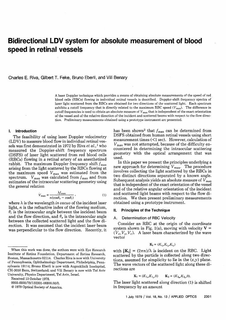
Consider an RBC at the origin of the coordinatesystem shown in Fig. 1(a), moving with velocity V =(Vs, Vy, V). A laser beam characterized by the wavevector
Ki = (KiKiyKi,)
with KjI (2rn)/X is incident on the RBC. Lightscattered by the particle is collected along two direc-tions, assumed for simplicity to lie in the (x,y) plane.The wave vectors of the scattered light along these di-rections are
K1 = (Ki.,K,,O) K2 = (K2XK2O).
The laser light scattered along direction (1) is shiftedin frequency by an amount
1 July 1979 / Vol. 18, No. 13 / APPLIED OPTICS 2301
f = I (K - K) V, (2a)27r
and that scattered along direction (2) is shifted by anamount
f 2 2 (K2 - K;) V. (2b)27r
The difference Af = 2 - f is given by
Ai =-(K2 KI) V2ir
= (K2sVx + K2,V - K,1 VX - K,,V).2ir
From Fig. 1(a), V. = V cos/ cos(7r - y), Vy = V cossin(7r -),
Using elementary rules of trigonometry, Eq. (3a) be-comes
n V cosf3Af = [cos(y + a1 ) - cos(7 + a2)],
A
nV cosf 7snr A ~ sin 2 -Ya, sin 2 - 2,
(3b)
When measurements are performed from vessels in theposterior pole of the eye, y is approximately equal to7r/2; therefore, (7r/2) - y is a small quantity. Further-more, a 1 and a2 are also small, since they are limited bythe size of the pupil. To an accuracy better than 2% for(7r/2) - y - ai (i = 1,2) smaller than 200, sin[(ir/2) - y- ai] = (r/2) - y - ai;so that Eq. (3b) can be writtenas
2irn 2irnK = Ai- cosal, Kly = Ak- sinal,
27rn 27rnK2, = A sina2. K 2 = - cosM2
It follows thatn V cos3
\f = [cos(ir - y) cosa2 + sin(ir - y) sina2
- cos(7r - y) cosal - sin(r - y) sinal].
a
z
Fig. 1. (a) Scattering geometry operative in the general case fdirectional measurements from vessels in the posterior pole of thIncident and scattered beams are indicated as passing throug]pupil of the eye. (b) Separation of the (x,y) plane into two rejdetermined by the direction of zero Doppler shift in the special
of V= V,
,f=n V cos/3b 7r n Vcos#3La/ X2) = 2 C
where Aca = 2 - a,. Thus
V= XAJn/a cosO
(3c)
(4)
In general, the optical frequency shifts f/ and f2 maybe either positive or negative depending upon the or-
(3a) ientations of the incident and scattered beams withrespect to the flow direction. Heterodyne opticalmixing spectroscopy is used to detect the frequencyshifts from RBCs flowing in retinal vessels.2 The lightscattered at a particular angle by the flowing RBCs aswell as the light scattered from the vessel wall is col-lected at the surface of a photodetector. The lightscattered from the wall is unshifted in frequency andthus acts as the reference beam. The resultant photo-current oscillates at the absolute value of the differencefrequency so that only the absolute values Il I and I f21can be measured. Since in practice Af = If21 - If, l, a1and a 2 must be chosen in such a way that both f/ and f2are of the same sign to aid ambiguity in calculatingV.
In the simplified situation shown in Fig. 1(b) whereV is along the y axis, fi and f2 will be of the same sign ifboth vectors K1 and K2 lie either in the shaded or in theunshaded region of the (x,y) plane. The line separatingthe regions is the direction of zero Doppler shift, i.e., thedirection of the vector with components
(-K=,,Kiy,°)-
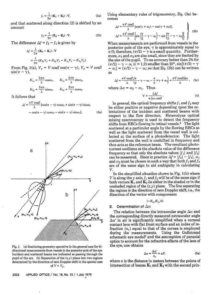
B. Determination of _Aa
The relation between the intraocular angle ŽAa anda) the corresponding directly measured extraocular angle
Aa' in air is significantly simplified when a cornealcontact lens with flat front surface and an index of re-fraction (n,) equal to that of the cornea is employedduring the measurements. Using the Gullstrandschematic eye model3 and the assumption of paraxialoptics to account for the refractive effects of the lens of
)r bi- the eye, one obtainseye.
Oh theIionscase
(5a)lAa =-+ aP,nc
where a is the distance in meters between the points ofintersection of beams K1 and K2 with the second prin-
2302 APPLIED OPTICS / Vol. 18, No. 13 / 1 July 1979
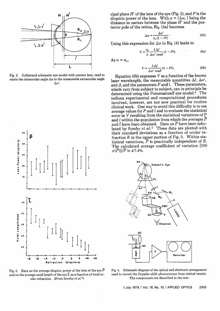
Fig. 2. Gullstrand schematic eye model with contact lens, used to
relate the intraocular angle At to the measurable extraocular angleA'.
26
0
.0
0Q.
4C
-J
24
22
20
18
30
2P
E
- 26
C
_J-
U 24
X4.
22
P
I
f�j.II
i 11 I
cipal plane H' of the lens of the eye (Fig. 2), and P is thedioptric power of the lens. With a = lA a, being thedistance in meters between the plane H' and the pos-terior pole of the retina, Eq. (5a) becomes
n,(1 - PI)
Using this expression for hoe in Eq. (4) leads to
V = n_ Af (1- P).n Aa' cos#
(5b)
(6a)
As n nc,
V = X I (1- P). (6b)A a' cos
Equation (6b) expresses V as a function of the knownlaser wavelength, the measurable quantities zff, Aao',and /3, and the parameters P and 1. These parameters,which vary from subject to subject, can in principle bedetermined using the Pomerantzeff eye model.4 Thetedious experimental and computational proceduresinvolved, however, are not now practical for routineclinical work. One way to avoid this difficulty is to useaverage values for P and I and to evaluate the statisticalerror in V resulting from the statistical variations of Pand I within the population from which the averages Pand I have been obtained. Data on P have been tabu-lated by Sorsby et al. 3 These data are plotted withtheir standard deviations as a function of ocular re-fraction R in the upper portion of Fig. 3. Within sta-tistical variations, P is practically independent of R.The calculated average coefficient of variation [100a(P)]/P is +7.4%.
RVSubject's Eye
Xci
L
I II
I
lII
II I
-8 -6 -4 -2 0 2 4Refraction (diopters)
+6
'p
oc0}000. ~~~~Recorder
+8
Fig. 3. Data on the average dioptric power of the lens of the eye P
and on the average axial length of the eye L as a function of total oc-ular refraction. (From Sorsby et al.
3)
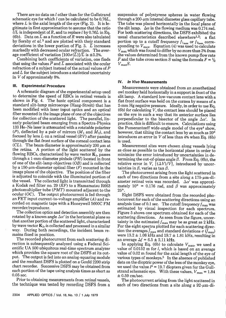
Fig. 4. Schematic diagram of the optical and electronic arrangementused to record the Doppler-shift photocurrent from retinal vessels.
The components are described in the text.
1 July 1979 / Vol. 18, No. 13 / APPLIED OPTICS 2303
-F- l § l l l l l ~~~~~~~~~~~~~~~~~~~~~~~~~~~~~~~~~~~~~i
| § l l l l l l
There are no data on I other than for the Gullstrandschematic eye for which can be calculated to be 0.76L,where L is the axial length of the eye (Fig. 2). It is le-gitimate in first approximation to assume that the ratiol/L is independent of R, and to replace by 0.76L in Eq.(6b). Data on L as a function of R were also tabulatedby Sorsby et al. 3 and are plotted with their standarddeviations in the lower portion of Fig. 3. L increasesmarkedly with decreased ocular refraction. The aver-age coefficient of variation [100of(L)]/L is ±3.2%.
Combining both coefficients of variation, one findsthat using the values P and L associated with the ocularrefraction of a subject instead of the actual values of Pand L for the subject introduces a statistical uncertaintyin V of approximately 8%.
Ill. Experimental Procedure
A schematic diagram of the experimental setup usedto determine the speed of RBCs in retinal vessels isshown in Fig. 4. The basic optical component is astandard slit-lamp microscope (Haag-Streit) that hasbeen modified with laser input optics and an opticalfiber mounted in the image plane of one of the objectivesfor collection of the scattered light. The parallel, lin-early polarized beam emerging from a Spectra-Physics133 He-Ne laser is attenuated with a rotatable polarizer(P), deflected by a pair of mirrors (Ml and M2), andfocused by lens L on a retinal vessel (R V) after passingthrough the flat front surface of the corneal contact lens(CL). The beam diameter is approximately 200 gm atthe retina. A portion of the light scattered by theflowing RBCs, characterized by wave vector K1 , passesthrough a 1-mm-diameter pinhole (PH) located in frontof one of the slit-lamp objectives (OB) and is collectedby a 150-gm-diameter optical fiber (F) mounted in theimage plane of the objective. The position of the fiberis adjusted to coincide with the illuminated portion ofthe vessel. The collected light is transmitted througha Kodak red filter no. 29 (RF) to a Hamamatsu R663photomultiplier tube (PMT) mounted adjacent to theocular (OC). The output photocurrent is fed throughan FET input current-to-voltage amplifier (A) and re-corded on magnetic tape with a Honeywell 5600C FMrecorder/reproducer.
The collection optics and detection assembly are thenrotated by a known angle AXa' in the horizontal plane sothat another portion of the scattered light, characterizedby wave vector K2 ,.is collected and processed in a similarway. During both recordings, the incident beam re-mains fixed in position.
The recorded photocurrent from each scattering di-rection is subsequently analyzed using a Federal Sci-entific UA 500 ubiquitous real-time spectrum analyzerwhich provides the square root of the DSFS at its out-put. The output is fed into an analog-squaring moduleand the resultant DSFS is plotted on a Gould 2200 stripchart recorder. Successive DSFS may be obtained fromeach portion of the tape using analysis times as short as0.05 sec.
Prior to obtaining measurements from retinal vessels,the technique was tested by recording DSFS from a
suspension of polystyrene spheres in water flowingthrough a 200-,gm internal diameter glass capillary tube.The tube was placed horizontally in the focal plane ofthe slit lamp. za in the flowing medium was 0.174 rad.For both scattering directions, the DSFS exhibited theusual characteristics described elsewherel2: a flatportion up to a cutoff frequency fax or f2max corre-sponding to Vmax. Equation (4) was used to calculateVm, which was found to differ by no more than 3% fromthe values determined from the known pump flow rateF and the tube cross section S using the formula F = 1/2VmaxS.
IV. In Vivo Measurements
Measurements were obtained from an anesthetizedowl monkey held horizontally in a support in front of theslit lamp. A standard low-vacuum contact lens with aflat front surface was held on the cornea by means of a2-mm Hg negative pressure. Ideally, in order to use Eq.(6b) for calculating V, the contact lens should be placedon the eye in such a way that its anterior surface liesperpendicular to the bisector of the angle oe'. Inpractice, this is difficult to ensure. Calculations usingthe Pomerantzeff wide-angle model of the eye4 show,however, that tilting the contact lens by as much as 20°introduces an error in V of less than 4% when Eq. (6b)is used.
Measurement sites were chosen along vessels lyingas close as possible to the horizontal plane in order tominimize the error introduced by uncertainties in de-termining the out-of-plane angle . From Eq. (6b), therelative error in V, (V)/VJ, introduced by uncer-tainties in , varies as tan .
The photocurrent arising from the light scattered ineach of two directions from a site along a 175-,m-di-ameter retinal vein was recorded. ZAx' was approxi-mately 100 = 0.176 rad, and /3 was approximately250.
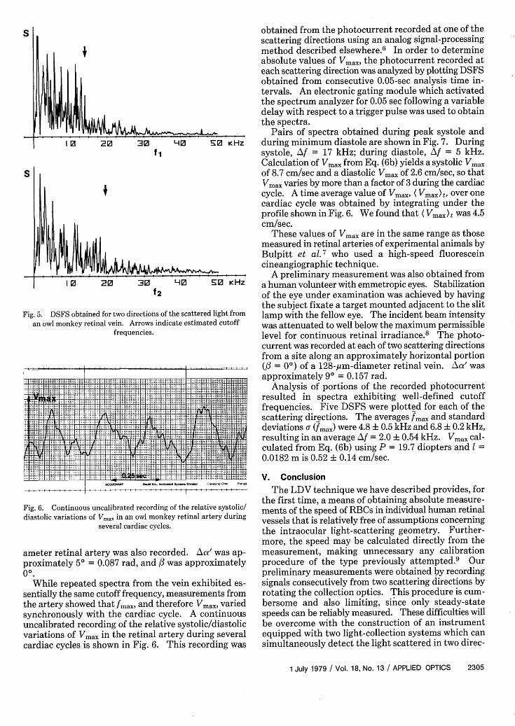
Eight DSFS were obtained from the recorded pho-tocurrent for each of the scattering directions using ananalysis time of 0.1 sec. The cutoff frequency fmax wasestimated by visual inspection for each spectrum.Figure 5 shows one spectrum obtained for each of thescattering directions. As seen from the figure, uncer-tainty in the estimated fmax may be as great as 2 kHz.For the eight spectra plotted for each scattering direc-tion the averages fmax and standard deviations (fmax)were 13.2 +z 1.08 kHz and 19.7 1.81 kHz, resulting inan average Af = 6.5 2.11 kHz.
In applying Eq. (6b) to calculate Vmax, we used avalue of 0.0152 m for 1, which is based on an averagevalue of 0.02 m found for the axial length of the eye ofvarious types of monkeys. 5 In the absence of publisheddata on the dioptric power of the lens of the monkey eye,we used the value P = 19.7 diopters given for the Gull-strand schematic eye. With these values, Vma = 1.84± 0.59 cm/sec.
The photocurrent arising from the light scattered ineach of two directions from a site along a 92-gm-di-
2304 APPLIED OPTICS / Vol. 18, No. 13 / 1 July 1979
S
S
Fig. 5. DSFan owl mc
obtained from the photocurrent recorded at one of thescattering directions using an analog signal-processing
4, method described elsewhere.6 In order to determineabsolute values of Vmax, the photocurrent recorded ateach scattering direction was analyzed by plotting DSFSobtained from consecutive 0.05-sec analysis time in-tervals. An electronic gating module which activatedthe spectrum analyzer for 0.05 sec following a variabledelay with respect to a trigger pulse was used to obtainthe spectra.
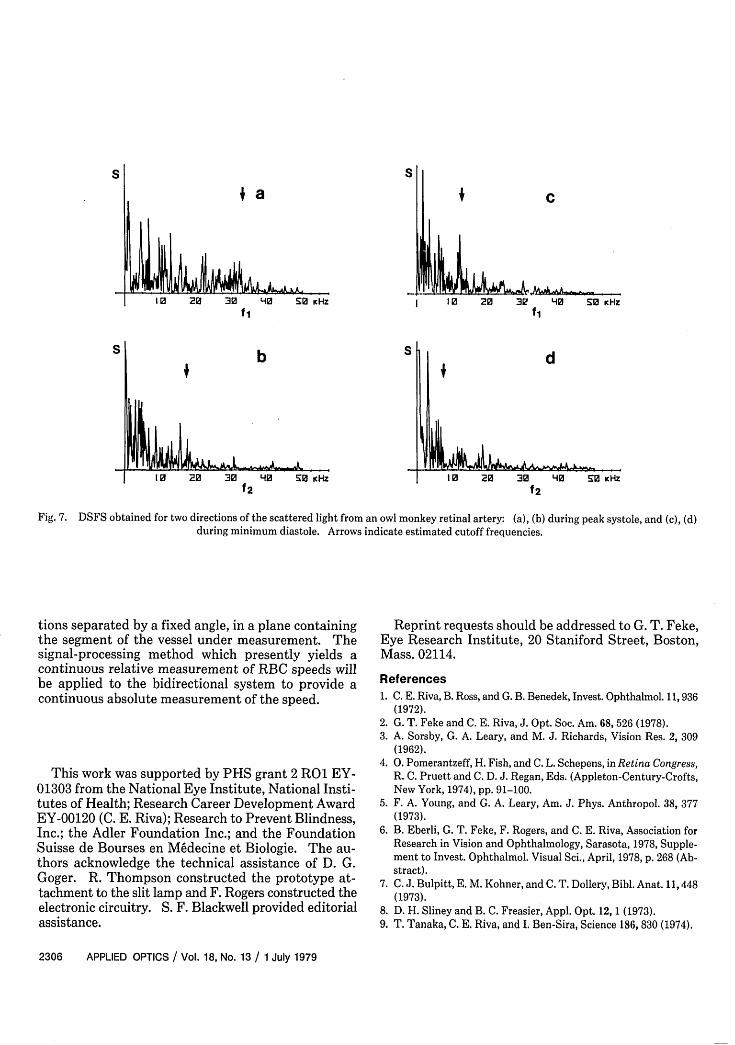
_____________________________________ Pairs of spectra obtained during peak systole and10 20 30 HO E0 KHz during minimum diastole are shown in Fig. 7. During
ft systole, LAf = 17 kHz; during diastole, Af = 5 kHz.Calculation of Vmax from Eq. (6b) yields a systolic Vmaxof 8.7 cm/sec and a diastolic Vmax of 2.6 cm/sec, so thatVmax varies by more than a factor of 3 during the cardiaccycle. A time average value of Vmax, ( Vmax) t, over onecardiac cycle was obtained by integrating under theprofile shown in Fig.6. We found that ( Vmax)t was 4.5cm/sec.
These values of Vmax are in the same range as thosemeasured in retinal arteries of experimental animals by
1^ [h~g~fl I Bulpitt et al. 7 who used a high-speed fluoresceincineangiographic technique.
A preliminary measurement was also obtained fromI 0 20 30 HO E0 KHZ a human volunteer with emmetropic eyes. Stabilization
f2 of the eye under examination was achieved by havingthe subject fixate a target mounted adjacent to the slit
US obtained for two directions of the scattered light from lamp with the fellow eye. The incident beam intensity)nkey retinal vein. Arrows indicate estimated cutoff was attenuated to well below the maximum permissible
frequencies. level for continuous retinal irradiance.8 The photo-
current was recorded at each of two scattering directionsfrom a site along an approximately horizontal portion(/3 = 0') of a 128-,gm-diameter retinal vein. ZAa' wasapproximately 90 = 0.157 rad.
Analysis of portions of the recorded photocurrent... . .... resulted in spectra exhibiting well-defined cutoff. .... . frequencies. Five DSFS were plotted for each of the
. .. .... 'scattering directions. The averages fmax and standarddeviations. a (max) were 4.8 ±0.5 kHz and 6.8 ±0. 2 kHz,resulting in an average zAf =2.0 ±0.54 kHz. Vax cal-
.... .... .. _ X _ _ Mculated from Eq. (6b) using P 19.7 diopters and 1..... .. _ l _ M - _0.0182 m is 0.52 ± 0.14 cm/sec.
r ,.,.,., .S ... , . ..-.. ... . . .. .. .. .. ... . '" 1 'E '' ' '
Fig. 6. Continuous uncalibrated recording of the relative systolic/diastolic variations of Vmax in an owl monkey retinal artery during
several cardiac cycles.
ameter retinal artery was also recorded. Aoe' was ap-proximately 50 = 0.087 rad, and / was approximately00.
While repeated spectra from the vein exhibited es-sentially the same cutoff frequency, measurements fromthe artery showed that fmax, and therefore Vmax, variedsynchronously with the cardiac cycle. A continuousuncalibrated recording of the relative systolic/diastolicvariations of Vmax in the retinal artery during severalcardiac cycles is shown in Fig. 6. This recording was
V. Conclusion
The LDV technique we have described provides, forthe first time, a means of obtaining absolute measure-ments of the speed of RBCs in individual human retinalvessels that is relatively free of assumptions concerningthe intraocular light-scattering geometry. Further-more, the speed may be calculated directly from themeasurement, making unnecessary any calibrationprocedure of the type previously attempted.9 Ourpreliminary measurements were obtained by recordingsignals consecutively from two scattering directions byrotating the collection optics. This procedure is cum-bersome and also limiting, sinice only steady-statespeeds can be reliably measured. These difficulties willbe overcome with the construction of an instrumentequipped with two light-collection systems which cansimultaneously detect the light scattered in two direc-
1 July 1979 / Vol. 18, No. 13 / APPLIED OPTICS 2305
UN
NNIV:M
S
I 0 20 30 H10 SO KHzfl
b
10 20 30 -40f2
+
-o 1w - I * a AAC
I I 0 20 3? L0 S0 KHzfi
3 f
50 KHZ 10 20 30 L40 50 KHz
f2
Fig. 7. DSFS obtained for two directions of the scattered light from an owl monkey retinal artery: (a), (b) during peak systole, and (c), (d)during minimum diastole. Arrows indicate estimated cutoff frequencies.
tions separated by a fixed angle, in a plane containingthe segment of the vessel under measurement. Thesignal-processing method which presently yields acontinuous relative measurement of RBC speeds willbe applied to the bidirectional system to provide acontinuous absolute measurement of the speed.
This work was supported by PHS grant 2 RO EY-01303 from the National Eye Institute, National Insti-tutes of Health; Research Career Development AwardEY-00120 (C. E. Riva); Research to Prevent Blindness,Inc.; the Adler Foundation Inc.; and the FoundationSuisse de Bourses en M6decine et Biologie. The au-thors acknowledge the technical assistance of D. G.Goger. R. Thompson constructed the prototype at-tachment to the slit lamp and F. Rogers constructed theelectronic circuitry. S. F. Blackwell provided editorialassistance.
Reprint requests should be addressed to G. T. Feke,Eye Research Institute, 20 Staniford Street, Boston,Mass. 02114.
References1. C. E. Riva, B. Ross, and G. B. Benedek, Invest. Ophthalmol. 11,936
(1972).2. G. T. Feke and C. E. Riva, J. Opt. Soc. Am. 68, 526 (1978).3. A. Sorsby, G. A. Leary, and M. J. Richards, Vision Res. 2, 309
(1962).4. 0. Pomerantzeff, H. Fish, and C. L. Schepens, in Retina Congress,
R. C. Pruett and C. D. J. Regan, Eds. (Appleton-Century-Crofts,New York, 1974), pp. 91-100.
5. F. A. Young, and G. A. Leary, Am. J. Phys. Anthropol. 38, 377(1973).
6. B. Eberli, G. T. Feke, F. Rogers, and C. E. Riva, Association forResearch in Vision and Ophthalmology, Sarasota, 1978, Supple-ment to Invest. Ophthalmol. Visual Sci., April, 1978, p. 268 (Ab-stract).
7. C. J. Bulpitt, E. M. Kohner, and C. T. Dollery, Bibl. Anat. 11,448(1973).
8. D. H. Sliney and B. C. Freasier, Appl. Opt. 12, 1 (1973).9. T. Tanaka, C. E. Riva, and I. Ben-Sira, Science 186, 830 (1974).
2306 APPLIED OPTICS / Vol. 18, No. 13 / 1 July 1979
S
S
II