best practice guidelines on esc in disasters
DESCRIPTION
World Health Organization Best Practice Guidelines on Emergency Surgical Care on Disaster SituationsTRANSCRIPT
WWWooorrrlllddd HHHeeeaaalll ttthhh OOOrrrgggaaannniiizzzaaattt iiiooonnn
WHO/EHT/CPR 2005 1
BBBeeesssttt PPPrrraaaccctttiiiccceee GGGuuuiiidddeeellliiinnneeesss ooonnn EEEmmmeeerrrgggeeennncccyyy SSSuuurrrgggiiicccaaalll CCCaaarrreee iiinnn DDDiiisssaaasssttteeerrr SSSiiitttuuuaaatttiiiooonnnsss
These guidelines have been extracted from the WHO manual Surgical Care at the District Hospital (SCDH), which is a part of the WHO Integrated Management on Emergency and Essential Surgical Care (IMEESC). The following materials relevant to country's disaster situation should be taken from the IMEESC: 1. Best practice printable protocols (disaster planning, trauma team responsibilities, hand hygiene,
operating room, anaesthesia check list, postoperative management application of cast and splints, cardiac life support, airway management ).
2. Needs assessment 3. Essential Emergency Equipment List 4. Details of anaesthesia, gunshot and landmine injuries in chapters 13, 14, 17 and 18, in SCDH List of Contents
1. Antibiotic Prophylaxis 2. Antibiotic Treatment 3. Tetanus Prophylaxis 4. Failure of Normal Methods of Sterilization 5. Cleaning, Disinfection and Sterilization 6. Waste Disposal 7. Resuscitation 8. Unconsciousness 9. Wound Management 10. Drains 11. Hand Lacerations 12. Specific Lacerations and Wounds 13. Tendon Lacerations 14. Amputations 15. Insertion of Chest Drain and Underwater Seal Drainage 16. Cellulitis and Abscess 17. Open Fractures 18. Upper Extremity injuries 19. Lower Extremity injuries 20. Spine injuries 21. Fractures in children 22. Compartment syndrome 23. Fat embolism

WWWooorrrlllddd HHHeeeaaalll ttthhh OOOrrrgggaaannniiizzzaaattt iiiooonnn
WHO/EHT/CPR 2005 2
AAAnnntttiiibbbiiioootttiiiccc PPPrrroooppphhhyyylllaaaxxxiiisss Antibiotic prophylaxis is different from antibiotic treatment. Prophylaxis is intended to prevent infection or to decrease the potential for infection. It is not intended to prevent infection in situations of gross contamination. Consider using prophylaxis:
1. For traumatic wounds which may not require surgical intervention 2. When surgical intervention will be delayed for more than 6 hours.
Use therapeutic doses if infection is present or likely: • Administer antibiotics prior to surgery, within the 2 hours before the skin is cut, so that tissue levels
are adequate during the surgery • More than one dose may be given if the procedure is long (>6 hours) or if there is significant blood
loss. • The use of topical antibiotics and washing wounds with antibiotic solutions are not recommended. • Use antibiotic prophylaxis in cases where there are:
1. Biomechanical considerations that increase the risk of infection: – Implantation of a foreign body – Known valvular heart disease – Indwelling prosthesis
2. Medical considerations that compromise the healing capacity or increase the infection risk: – Diabetes – Peripheral vascular disease – Possibility of gangrene or tetanus – Immunocompromise
3. High-risk wounds or situations: – Penetrating wounds – Abdominal trauma – Compound fractures – Wounds with devitalized tissue – Lacerations greater than 5 cm or stellate lacerations – Contaminated wounds - High risk anatomical sites such as hand or foot – Biliary and bowel surgery.
• Use intravenous (IV) antibiotics for prophylaxis in clean surgical situations to reduce the risk of postoperative infection, since skin and instruments are never completely sterile.
• For the prophylaxis of endocarditis in patients with known valvular heart disease: - Oral and upper respiratory procedures: give amoxycillin 3 g orally, 1 hour before surgery
and 1.5 g, 6 hours after first dose - Gastrointestinal and genitourinary procedures: give ampicillin 3 g,1 hour before surgery
and gentamicin 1.5 mg/kg intramuscularly (IM) or IV (maximum dose 80 mg), 30 minutes before surgery.

WWWooorrrlllddd HHHeeeaaalll ttthhh OOOrrrgggaaannniiizzzaaattt iiiooonnn
WHO/EHT/CPR 2005 3
AAAnnntttiiibbbiiioootttiiiccc TTTrrreeeaaatttmmmeeennnttt When a wound is extensive and more than 6 hours old, you should consider it to be colonized with
bacteria, and use therapeutic doses and regimens. Penicillin and metronidazole provide good coverage and are widely available. Monitor wound healing and infection regularly. Make use of culture and sensitivity findings if they are available. Continue therapeutic doses of antibiotics for 5–7 days.
TTTeeetttaaannnuuusss PPPrrroooppphhhyyylllaaaxxxiiisss
Active immunization with tetanus toxoid (TT) prevents tetanus and is given together with diphtheria vaccine (TD). Women should be immunized during pregnancy to prevent neonatal tetanus. Childhood immunization regimes include diphtheria, pertussis and tetanus. • Individuals who have not received three doses of tetanus toxoid are not considered immune and
require immunization. • A non-immune person with a minor wound can be immunized if the wound is tetanus prone; give both
TT or TD and tetanus immune globulin (TIG). • A non-immunized person will require repeat immunization at six weeks and at six months to complete
the immunization series. • Examples of tetanus prone wounds include:
- Wounds contaminated with dirt or faeces - Puncture wounds - Burns - Frostbite - High velocity missile injuries.
• Give prophylactic antibiotics in cases of wound contamination • Immunize the non-immune patient against tetanus with tetanus toxoid and give immune globulin if the wound is tetanus prone.
Tetanus prophylaxis regime Clean wounds Moderate risk High risk Immunized and booster within 5 years Nil Nil Nil Immunized and 5–10 years Nil TT or TD TT or TD since booster Immunized and >10 years TT or TD TT or TD TT or TD since booster Incomplete immunization TT or TD TT or TD TT or TD or unknown and TIG and TIG Do not give TIG if the person is known to have had two primary doses of TT or TD
WWWooorrrlllddd HHHeeeaaalll ttthhh OOOrrrgggaaannniiizzzaaattt iiiooonnn
WHO/EHT/CPR 2005 4
FFFaaaiiillluuurrreee ooofff NNNooorrrmmmaaalll MMMeeettthhhooodddsss ooofff SSSttteeerrriiillliiizzzaaatttiiiooonnn Failure of an autoclave or a power supply may suddenly interrupt normal sterilization procedures. If an extra set of sterile equipment and drapes are not available, the following “antiseptic technique” will allow some surgery to continue.
1. Immerse towels and drapes for 1 hour in a reliable antiseptic such as aqueous chlorhexidine, wring them out and lay them moist on the skin of the patient.
2. Treat gauze packs and swabs similarly, but rinse them in diluted (1: 1000) chlorhexidine solution before using them in the wound. From time to time during the operation, rinse gauze in use in this solution.
3. Immerse instruments, needles, and natural suture materials in strong antiseptic for 1 hour and rinse them in weak antiseptic just before use
Cleaning, Disinfection and Sterilization Disinfection
• Disinfectant solutions are used to inactivate any infectious agents that may be present in blood or other body fluids.
• They must always be available for cleaning working surfaces, equipment that cannot be autoclaved and non-disposable items and for dealing with any spillages involving pathological specimens or other known infectious material.
• Needles and instruments should routinely be soaked in a chemical disinfectant for 30 minutes before cleaning.
• Disinfection decreases the viral and bacterial burden of an instrument, but does not clean debris from the instrument or confer sterility.
• The purpose of disinfection is to reduce the risk to those who have to handle the instruments during further cleaning.
• Reusable needles must always be used with great care. After use, they should be placed in a special container of disinfectant before being cleaned and sterilized.
• Thick gloves should be worn when needles and sharp instruments are being cleaned. • There are many disinfectant solutions, with varying degrees of effectiveness. In most countries, the
most widely available disinfectant is sodium hypochlorite solution (commonly known as bleach or chloros), which is a particularly effective antiviral disinfectant solution.
• To ensure effective disinfection, follow the manufacturer’s instructions or any other specific guidelines that have been given and dilute the concentrated solution to the correct working strength.
• It is important to use all disinfectant solutions within their expiry date as some solutions, such as hypochlorite, lose their activity very quickly.
• All disinfectants have what is known as a “contact time”, which means that they must be left in contact with an infectious agent for a certain period of time to ensure that it is completely inactivated. However, some disinfectants are themselves inactivated by the presence of organic material and so higher concentrations of disinfectant and longer contact times must be used in certain situations, such as a large spill of infected blood.
• Linen soiled with blood should be handled with gloves and should be collected and transported in leak-proof bags.
• Wash the linen first in cool water and then disinfect with a dilute chlorine solution. Then wash it with detergent for 25 minutes at a temperature of at least 71°C.
WWWooorrrlllddd HHHeeeaaalll ttthhh OOOrrrgggaaannniiizzzaaattt iiiooonnn
WHO/EHT/CPR 2005 5
Sterilization The methods of sterilization in common use are:
1. Autoclaving or steam sterilization 2. Exposure to dry heat 3. Treatment with chemical antiseptics.
1.Autoclaving • Autoclaving should be the main form of sterilization at the district hospital.
• Before sterilizing medical items, they must first be disinfected and vigorously cleaned to remove
all organic material. Proper disinfection decreases the risk for the person who will be cleaning the instruments.
• Sterilization of all surgical instruments and supplies is crucial in preventing HIV transmission. All viruses, including HIV, are inactivated by steam sterilization (autoclaving) for 20 minutes at 121°C–132°C or for 30 minutes if the instruments are in wrapped packs.
• Appropriate indicators must be used each time to show that sterilization has been accomplished. At the end of the procedure, the outsides of the packs of instruments should not have wet spots, which may indicate that sterilization has not occurred.
2.Dry heat • If items cannot be autoclaved, they can be sterilized by dry heat for1–2 hours at 170°C.
Instruments must be clean and free of grease or oil. • However, sterilizing by hot air is a poor alternative to autoclaving since it is suitable only for
metal instruments and a few natural suture materials. • Boiling instruments is now regarded as an unreliable means of sterilization and is not
recommended as a routine in hospital practice. 3.Antiseptics
• In general, instruments are no longer stored in liquid antiseptic. However, sharp instruments, other delicate equipment and certain catheters and tubes can be sterilized by exposure to formaldehyde, glutaral (glutaraldehyde) or chlorhexidine.
• If you are using formaldehyde, carefully clean the equipment and then expose it to vapour from paraformaldehyde tablets in a closed container for 48 hours. Ensure that this process is carried out correctly.
• Glutaral is a disinfectant that is extremely effective against bacteria, fungi and a wide range of viruses. Always follow the manufacturer’s instructions for use.
WWWaaasssttteee DDDiiissspppooosssaaalll
• All biological waste must be carefully stored and disposed of safely. • Contaminated materials such as blood bags, dirty dressings and disposable needles are also
potentially hazardous and must be treated accordingly. • If biological waste and contaminated materials are not disposed of properly, staff and members of
the community could be exposed to infectious material and become infected. • It is essential for the hospital to have protocols for dealing with biological waste and contaminated
materials. All staff must be familiar with them and follow them. • The disposal of bio hazardous materials is time consuming and expensive, so it is important to
separate non-contaminated material such as waste paper, packaging and non-sterile but not biologically contaminated materials.

WWWooorrrlllddd HHHeeeaaalll ttthhh OOOrrrgggaaannniiizzzaaattt iiiooonnn
WHO/EHT/CPR 2005 6
• Make separate disposal containers available where waste is created so that staff can sort the waste as it is being discarded. Organize things in a way to discourage the need for people to be in contact with contaminated waste.
• All infected waste should then be disposed of by incineration. • Incineration is the ideal method for the final disposal of waste but, if this is not possible, other
suitable methods must be used. • Burying waste is the only option in some areas. If this is the case, you should do as much as
possible before burying it to minimize the risk of infection. • Small amounts of infected waste should be soaked in a hypochlorite solution for at least 12 hours,
put into a pit and then covered. • Larger quantities should be put into a pit with a final concentration of 10% sodium hypochlorite,
before covering immediately. • Do not mix waste chemicals, unless you are certain that a chemical reaction will not take place.
This is essential to prevent any unwanted or even dangerous reactions occurring between the chemicals, which could endanger laboratory staff.
• Always follow local guidelines on the disposal of waste chemicals to ensure that chemical contamination of the surrounding land or water supply does not occur.
• Provide a safe system for getting rid of disposable items such as scalpel blades or needles. The risk of injury with sharp objects increases with the distance they are carried and the amount they are manipulated.
• A container for the safe disposal of sharp objects should be: - Well labelled - Puncture proof - Watertight - Break resistant (a glass container could break and provide a serious hazard to the person
cleaning up the mess) - Opening large enough to pass needles and scalpel blades, but never large enough for
someone to reach into - Secured to a surface, such as a wall or counter, to ensure stability during use - Removable for disposal.
• These containers must then be disposed of safely.
RRReeesssuuusssccciiitttaaatttiiiooonnn Haemorrhage External bleeding can be controlled, usually with pressure. Bleeding into body cavities may be apparent only later; for example, when the circulation has been restored and the rise in blood pressure causes more bleeding and a second fall in blood pressure. Shock Shock is a pathological, life threatening condition in which the oxygen supply to the tissues of the body fails. The cause is usually one of the following: • Hypovolaemia (bleeding)
- the circulating volume is reduced by loss of blood or other fluid (e.g. burn transudate). - Rapid fluid replacement, starting with normal saline or Hartmann’s solution, should restore
the circulation towards normal. • Sepsis
- the circulating volume may be normal, but blood pressure is low and tissue circulation is
WWWooorrrlllddd HHHeeeaaalll ttthhh OOOrrrgggaaannniiizzzaaattt iiiooonnn
WHO/EHT/CPR 2005 7
inadequate. - Support the circulation with volume infusion, but it may not respond as in hypovolaemic
shock. • Acute anaphylaxis: from allergy or drug reaction
- give epinephrine and intravenous fluids. • Neurogenic (after spinal trauma)
- the heart rate is often low and atropine and fluids will be helpful. • Heart failure (left ventricular failure).
Recognize shock by:
- Tachycardia (may be the only sign in a child) - Thready pulse - Narrow pulse pressure: e.g.110/70 becomes 95/75 - Cold hands and feet - Sweating, anxious patient - Breathlessness and hyperventilation - Confusion leading to unconsciousness

WWWooorrrlllddd HHHeeeaaalll ttthhh OOOrrrgggaaannniiizzzaaattt iiiooonnn
WHO/EHT/CPR 2005 8
UUUnnncccooonnnsssccciiiooouuusssnnneeessssss Unconsciousness may have many causes including:
- Head injury - Hypoglycaemia - Ketoacidosis - Cerebrovascular event - Hypoxia - Hypotension - Hypertension and eclampsia - H I V infection - Drug intoxication.
• Assess the response to stimuli, look at the pupils initially and re-examine them later to follow progress. Look for unequal pupils or other localizing signs that may show intracranial haematoma developing.
• In many instances, you may attend to and stabilize other systems first and await the return of consciousness as cerebral perfusion and oxygenation improves.
• After cardiac arrest, a patient who initially had fixed dilated pupils may show smaller pupils after effective CPR. This indicates that a favourable outcome may be possible.
• The supine unconscious patient with a full stomach is at grave risk of regurgitation and aspiration due to the unprotected airway. However, i f a comatose patient has a clear airway and vital signs are normal:
- Avoid intubation as this will involve drug administration and complicate the subsequent diagnosis
- Nurse the patient in the recovery position - Monitor the airway and await progress and diagnosis (Figure 13.4).
• During CPR, ask yourself: is the patient responding? I f not, why not?

WWWooorrrlllddd HHHeeeaaalll ttthhh OOOrrrgggaaannniiizzzaaattt iiiooonnn
WHO/EHT/CPR 2005 9
WWWooouuunnnddd MMMaaannnaaagggeeemmmeeennnttt Surgical wounds can be classified as follows:
• Clean • Clean contaminated: a wound involving normal but colonized tissue • Contaminated: a wound containing foreign or infected material • Infected: a wound with pus present. • Close clean wounds immediately to allow healing by primary intention • Do not close contaminated and infected wounds, but leave them open to heal by secondary
intention • In treating clean contaminated wounds and clean wounds that are more than six hours old, manage
with surgical toilet, leave open and then close 48 hours later. This is delayed primary closure.
Factors that affect wound healing and the potential for infection • Patient:
- Age - Underlying illnesses or disease: consider anaemia, diabetes or immunocompromise - Effect of the injury on healing (e.g. devascularization)
• Wound: - Organ or tissue injured - Extent of injury - Nature of injury (for example, a laceration will be a less complicated wound than a crush
injury) - Contamination or infection - Time between injury and treatment (sooner is better)
• Local factors: - Haemostasis and debridement - Timing of closure
Wound: Primary repair
• Primary closure requires that clean tissue is approximated without tension. • Injudicious closure of a contaminated wound will promote infection and delay healing. • Essential suturing techniques include:
-Interrupted simple -Continuous simple -Vertical mattress -Horizontal mattress -Intradermal. • Staples are an expensive, but rapid, alternative to sutures for skin closure. • The aim with all techniques is to approximate the wound edges without gaps or tension. • The size of the suture “bite” and the interval between bites should be equal in length and proportional to the thickness of tissue being approximated: • As suture is a foreign body, use the minimal size and amount of suture material required to close
the wound • Leave skin sutures in place for 5 days; leave the sutures in longer if healing is expected to be slow
due to the blood supply of a particular location or the patient’s condition • If appearance is important and suture marks unacceptable, as in the face, remove sutures as early
as 3 days. In this case, re-enforce the wound with skin tapes

WWWooorrrlllddd HHHeeeaaalll ttthhh OOOrrrgggaaannniiizzzaaattt iiiooonnn
WHO/EHT/CPR 2005 10
• Close deep wounds in layers, using absorbable sutures for the deep layers. • Place a latex drain in deep oozing wounds to prevent haematoma formation.
Wound: Delayed primary closure
• Irrigate clean contaminated wounds; then pack them open with damp saline gauze. • Close the wounds with sutures at 2 days. • These sutures can be placed at the time of wound irrigation or at the time of wound closure
Wound: Secondary healing
To promote healing by secondary intention, perform wound toilet and surgical debridement. 1. Surgical wound toilet involves: - Cleaning the skin with antiseptics - Irrigation of wounds with saline - Surgical debridement of all dead tissue and foreign matter. Dead tissue does not bleed when cut. 2. Wound debridement involves:
Gentle handling of tissues minimizes bleeding. Control residual bleeding with compression, ligation or cautery. Dead or devitalized muscle is dark in colour, soft, easily damaged and does not contract when
pinched. During debridement, excise only a very thin margin of skin from the wound edge (Figure 5.1).
Figure 5.1 Figure 5.2 Figure 5.3 1 Systematically perform wound toilet and surgical debridement, initially to the superficial layers of tissues and subsequently to the deeper layers (Figures 5.2, 5.3 ).
- After scrubbing the skin with soap and irrigating the wound with saline, prep the skin with antiseptic.
- Do not use antiseptics within the wound. 2 Debride the wound meticulously to remove any loose foreign material such as dirt, grass, wood, glass or clothing.
- With a scalpel or dissecting scissors, remove all adherent foreign material along with a thin margin of underlying tissue and then irrigate the wound again.
- Continue the cycle of surgical debridement and saline irrigation until the wound is completely clean.
3 Leave the wound open after debridement to allow healing by secondary intention.

WWWooorrrlllddd HHHeeeaaalll ttthhh OOOrrrgggaaannniiizzzaaattt iiiooonnn
WHO/EHT/CPR 2005 11
- Pack it lightly with damp saline gauze and cover the packed wound with a dry dressing. - Change the packing and dressing daily or more often if the outer dressing becomes damp with
blood or other body fluids. - Large defects will require closure with flaps or skin grafts but may be initially managed with saline
packing.
Drains Drainage of a wound or body cavity is indicated when there is risk of blood or serous fluid
collection or when there is pus or gross wound contamination. The type of drain used depends on both indication and availability. Drains are classified as open or closed and active or passive:
• Closed drains do not allow the entry of atmospheric air and require either suction or differential pressure to function
• Open drains allow atmospheric air access to the wound or body cavity • Continuous suction drains with air vents are open but active drains.
Drains are not a substitute for good haemostasis or for good surgical technique and should not be left in place too long.
They are usually left in place only until the situation which indicated insertion is resolved, there is no longer any fluid drainage or the drain is not functioning.
Leaving a non-functioning drain in place unnecessarily exposes the patient to an increased risk of infection.
Hand Lacerations
• Treat lacerations promptly with careful evaluation, debridement and lavage • Close wounds only when clean, using suture, spontaneous healing or skin grafts • After injury, elevate the hand to control swelling and begin motion early • Nail bed injuries require special treatment
Evaluation
• Treat open injuries of the hand promptly. • Perform a local examination to check circulation, sensation and motor function. • Gently examine the wound using aseptic technique to determine if it is clean or contaminated. • A contaminated wound contains foreign material and crushed or dead tissue.
Treatment 1. Debride and lavage all wounds in the operating room or emergency area. If a local anaesthetic is needed, use 1% lidocaine without epinephrine. 2. Administer tetanus toxoid and antibiotics. Obtain X-rays to check underlying bones and joints. 3. Stop bleeding by compression with sterile gauze. If necessary, extend the wound, being careful not to cross skin creases in the palm or digits. Remove all foreign material and devitalized tissue, but do not excise any skin unless it is dead. 4. If the wound is clean, repair extensor tendons but not flexor tendons or nerves. 5. Close a clean wound over a drain using interrupted sutures if there is no tension on the skin. 6. If the wound is contaminated, delay closure until after a second debridement. Wounds less than 1 cm square will granulate spontaneously. Use skin grafts for larger wounds, which will not close without skin tension.

WWWooorrrlllddd HHHeeeaaalll ttthhh OOOrrrgggaaannniiizzzaaattt iiiooonnn
WHO/EHT/CPR 2005 12
6.Cover the hand with sterile gauze and a compression dressing. 7. Apply a plaster splint to hold the wrist in 20 degrees of extension, with the metacarpophalangeal joints in 90 degrees of flexion and the interphalangeal joints in full extension. Keep the fingertips exposed unless they are injured. 8. To control oedema, elevate the limb for the first week, either by attachment to an overhead frame or by the use of a triangular sling 9. Begin active exercises as soon as possible and inspect the wound in 2– 3 days to remove drains.
Nail bed injuries • Subungual haematoma causes severe pain resulting from a collection of blood deep under the nail.
This can be seen as a dark red to black collection beneath the nail. • To relieve pain, make one or two small holes in the nail with a hot safety pin or the tip of sterile
number 11 scalpel blade. • I f not repaired, lacerations of the nail bed may result in lasting nail deformity.

WWWooorrrlllddd HHHeeeaaalll ttthhh OOOrrrgggaaannniiizzzaaattt iiiooonnn
WHO/EHT/CPR 2005 13
• Remove the nail and, after debridement and lavage, repair the laceration using fine suture. • I f possible, replace the nail over the sutured laceration until it heals and a new nail has begun to
grow.
Specific Lacerations and Wounds
• Lacerations may be associated with neurovascular or other serious injury; a complete examination is required to identify injuries that are not immediately obvious.
• Minor problems are important because mismanagement can lead to major detrimental consequences.
Blood vessels, nerves and tendons • Assess the function of tendons, nerves and blood vessels distal to the laceration. • Ligate lacerated vessels whether or not they are bleeding, as the vessels which are not bleeding may do
so at a later time. • Large damaged vessels may need to be divided between ligatures. Before dividing these larger vessels
or an end artery, test the effect on the distal circulation by temporary occlusion of the vessel. • Loosely oppose the ends of divided nerves by inserting one or two sutures through the nerve sheath.
Similarly fix tendon ends to prevent retraction. These sutures should be long enough to assist in tendon or nerve identification at a subsequent procedure.
• Formal repair of nerves and flexor tendons is not urgent and is best undertaken later by a qualified surgeon.
Facial Lacerations • It is appropriate to manage most facial wounds in the outpatient department. • Clean the skin with soap and water, while protecting the patient’s eyes. • Irrigate the wound with saline. • Preserve tissue, especially skin, but remove all foreign material and all obviously devitalized tissue. • Close with simple monofilament non-absorbable sutures of 4/0 or 5/0. Reinforce the skin closure with
skin tapes. To avoid skin marking, remove sutures at 3 to 5 days. • If the wound is contaminated, give prophylactic antibiotics to prevent cellulitis. • Large facial wounds or wounds associated with tissue loss require referral for specialized care after
primary management. • Arrest obvious bleeding, clean wounds and remove all foreign material. • Tack the wound edges in place with a few monofilament sutures after the wound is packed with a
sterile saline dressing. Lip Lacerations
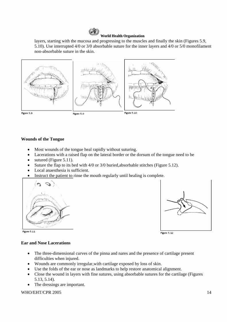
• Small lacerations of the buccal mucosa do not require suturing. • Advise the patient to rinse the mouth frequently, particularly after meals. • Local anaesthesia is adequate for lacerations that do require suturing. • For good cosmesis, proper anatomical alignment of the vermillion border is essential. To achieve
this alignment, place the first stitch at the border (Figure 5.8). This region may be distorted by the swelling caused by local anaesthetic or blanched by adrenaline, so to assure accuracy, premark the vermillion border with a pen. After the initial suture is inserted, repair the rest of the wound in
WWWooorrrlllddd HHHeeeaaalll ttthhh OOOrrrgggaaannniiizzzaaattt iiiooonnn
WHO/EHT/CPR 2005 14
layers, starting with the mucosa and progressing to the muscles and finally the skin (Figures 5.9, 5.10). Use interrupted 4/0 or 3/0 absorbable suture for the inner layers and 4/0 or 5/0 monofilament non-absorbable suture in the skin.
Wounds of the Tongue
• Most wounds of the tongue heal rapidly without suturing. • Lacerations with a raised flap on the lateral border or the dorsum of the tongue need to be • sutured (Figure 5.11). • Suture the flap to its bed with 4/0 or 3/0 buried,absorbable stitches (Figure 5.12). • Local anaesthesia is sufficient. • Instruct the patient to rinse the mouth regularly until healing is complete.
Ear and Nose Lacerations
• The three-dimensional curves of the pinna and nares and the presence of cartilage present difficulties when injured.
• Wounds are commonly irregular,with cartilage exposed by loss of skin. • Use the folds of the ear or nose as landmarks to help restore anatomical alignment. • Close the wound in layers with fine sutures, using absorbable sutures for the cartilage (Figures
5.13, 5.14). • The dressings are important.

WWWooorrrlllddd HHHeeeaaalll ttthhh OOOrrrgggaaannniiizzzaaattt iiiooonnn
WHO/EHT/CPR 2005 15
• Support the pinna on both sides with moist cotton pads and firmly bandage to reduce haematoma formation (Figure 5.15).
• Cover exposed cartilage either by wound closure or split thickness skin grafts. • Wounds of the ear and nose may result in deformities or necrosis of the cartilage.
Nose bleed (epistaxis)
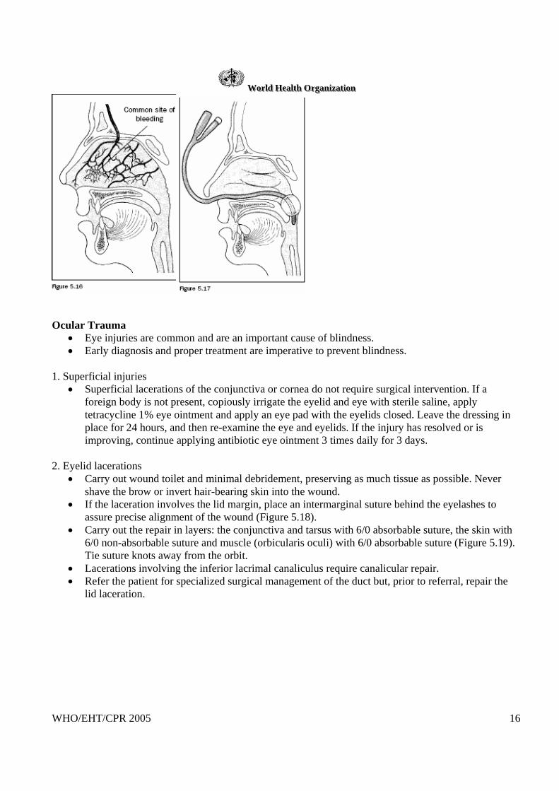
• Epistaxis often occurs from the plexus of veins in the anterior part of the nasal septum (Figure 5.16).
• In children it is often due to nose picking; other causes include trauma, a foreign body, Burkitt’s lymphoma and nasopharyngeal carcinoma.
• Manage epistaxis with the patient in a sitting position. • Remove blood clots from the nose and throat to visualize the site of bleeding and confirm the
diagnosis. • Pinch the nose between your fingers and thumb while applying icepacks to the nose and forehead.
Continue to apply pressure. Bleeding will usually stop within 10 minutes. • If bleeding continues, pack the anterior nares with petroleum impregnated ribbon gauze. • If bleeding continues after packing, the posterior nasopharynx may be the source of bleeding.
Apply pressure using the balloon of a Foley catheter. Lubricate the catheter, and pass it through the nose until the tip reaches the oropharynx. Withdraw it a short distance to bring the balloon into the nasopharynx. Inflate the balloon with water, enough to exert pressure but not to cause discomfort (5–10 ml of water is usually adequate for an adult, but use no more than 5 ml for a child). Gently pull the catheter forward until the balloon is held in the posterior choana (Figure 5.17).Tape the catheter to the forehead or cheek in the same manner as a nasogastric tube. With the catheter in place, pack the anterior nares with petroleum gauze. Deflate the Foley catheter after 48 hours and, if bleeding does not recur, remove it.
WWWooorrrlllddd HHHeeeaaalll ttthhh OOOrrrgggaaannniiizzzaaattt iiiooonnn
WHO/EHT/CPR 2005 16
Ocular Trauma
• Eye injuries are common and are an important cause of blindness. • Early diagnosis and proper treatment are imperative to prevent blindness.
1. Superficial injuries
• Superficial lacerations of the conjunctiva or cornea do not require surgical intervention. If a foreign body is not present, copiously irrigate the eyelid and eye with sterile saline, apply tetracycline 1% eye ointment and apply an eye pad with the eyelids closed. Leave the dressing in place for 24 hours, and then re-examine the eye and eyelids. If the injury has resolved or is improving, continue applying antibiotic eye ointment 3 times daily for 3 days.
2. Eyelid lacerations
• Carry out wound toilet and minimal debridement, preserving as much tissue as possible. Never shave the brow or invert hair-bearing skin into the wound.
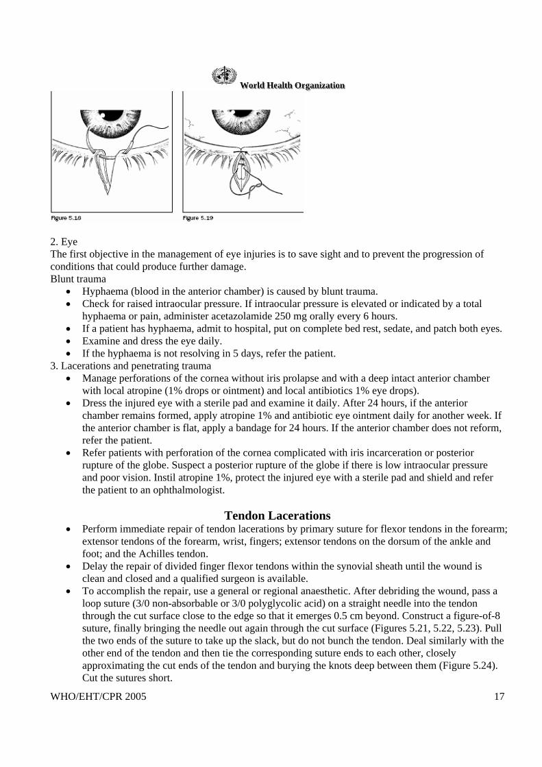
• If the laceration involves the lid margin, place an intermarginal suture behind the eyelashes to assure precise alignment of the wound (Figure 5.18).
• Carry out the repair in layers: the conjunctiva and tarsus with 6/0 absorbable suture, the skin with 6/0 non-absorbable suture and muscle (orbicularis oculi) with 6/0 absorbable suture (Figure 5.19). Tie suture knots away from the orbit.
• Lacerations involving the inferior lacrimal canaliculus require canalicular repair. • Refer the patient for specialized surgical management of the duct but, prior to referral, repair the
lid laceration.
WWWooorrrlllddd HHHeeeaaalll ttthhh OOOrrrgggaaannniiizzzaaattt iiiooonnn
WHO/EHT/CPR 2005 17
2. Eye The first objective in the management of eye injuries is to save sight and to prevent the progression of conditions that could produce further damage. Blunt trauma
• Hyphaema (blood in the anterior chamber) is caused by blunt trauma. • Check for raised intraocular pressure. If intraocular pressure is elevated or indicated by a total
hyphaema or pain, administer acetazolamide 250 mg orally every 6 hours. • If a patient has hyphaema, admit to hospital, put on complete bed rest, sedate, and patch both eyes. • Examine and dress the eye daily. • If the hyphaema is not resolving in 5 days, refer the patient.
3. Lacerations and penetrating trauma • Manage perforations of the cornea without iris prolapse and with a deep intact anterior chamber
with local atropine (1% drops or ointment) and local antibiotics 1% eye drops). • Dress the injured eye with a sterile pad and examine it daily. After 24 hours, if the anterior
chamber remains formed, apply atropine 1% and antibiotic eye ointment daily for another week. If the anterior chamber is flat, apply a bandage for 24 hours. If the anterior chamber does not reform, refer the patient.
• Refer patients with perforation of the cornea complicated with iris incarceration or posterior rupture of the globe. Suspect a posterior rupture of the globe if there is low intraocular pressure and poor vision. Instil atropine 1%, protect the injured eye with a sterile pad and shield and refer the patient to an ophthalmologist.
Tendon Lacerations
• Perform immediate repair of tendon lacerations by primary suture for flexor tendons in the forearm; extensor tendons of the forearm, wrist, fingers; extensor tendons on the dorsum of the ankle and foot; and the Achilles tendon.
• Delay the repair of divided finger flexor tendons within the synovial sheath until the wound is clean and closed and a qualified surgeon is available.
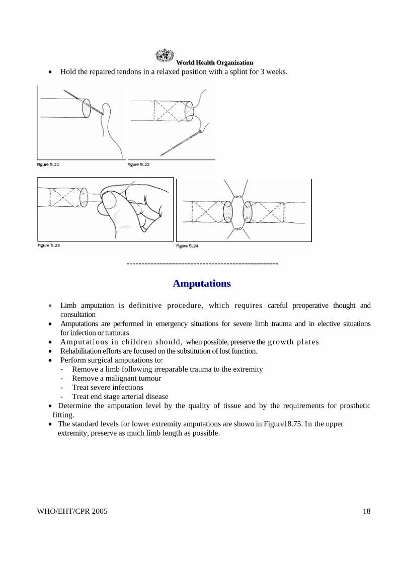
• To accomplish the repair, use a general or regional anaesthetic. After debriding the wound, pass a loop suture (3/0 non-absorbable or 3/0 polyglycolic acid) on a straight needle into the tendon through the cut surface close to the edge so that it emerges 0.5 cm beyond. Construct a figure-of-8 suture, finally bringing the needle out again through the cut surface (Figures 5.21, 5.22, 5.23). Pull the two ends of the suture to take up the slack, but do not bunch the tendon. Deal similarly with the other end of the tendon and then tie the corresponding suture ends to each other, closely approximating the cut ends of the tendon and burying the knots deep between them (Figure 5.24). Cut the sutures short.
WWWooorrrlllddd HHHeeeaaalll ttthhh OOOrrrgggaaannniiizzzaaattt iiiooonnn
WHO/EHT/CPR 2005 18
• Hold the repaired tendons in a relaxed position with a splint for 3 weeks.
--------------------------------------------------
AAAmmmpppuuutttaaatttiiiooonnnsss
• Limb amputation is definitive procedure, which requires careful preoperative thought and consultation
• Amputations are performed in emergency situations for severe limb trauma and in elective situations for infection or tumours
• Amputations in children should, when possible, preserve the growth plates • Rehabilitation efforts are focused on the substitution of lost function. • Perform surgical amputations to:
- Remove a limb following irreparable trauma to the extremity - Remove a malignant tumour - Treat severe infections - Treat end stage arterial disease
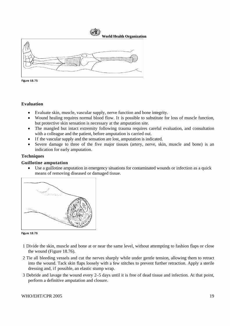
• Determine the amputation level by the quality of tissue and by the requirements for prosthetic fitting.
• The standard levels for lower extremity amputations are shown in Figure18.75. In the upper extremity, preserve as much limb length as possible.
WWWooorrrlllddd HHHeeeaaalll ttthhh OOOrrrgggaaannniiizzzaaattt iiiooonnn
WHO/EHT/CPR 2005 19
Evaluation
• Evaluate skin, muscle, vascular supply, nerve function and bone integrity. • Wound healing requires normal blood flow. It is possible to substitute for loss of muscle function,
but protective skin sensation is necessary at the amputation site. • The mangled but intact extremity following trauma requires careful evaluation, and consultation
with a colleague and the patient, before amputation is carried out. • I f the vascular supply and the sensation are lost, amputation is indicated. • Severe damage to three of the five major tissues (artery, nerve, skin, muscle and bone) is an
indication for early amputation. Techniques Guillotine amputation
• Use a guillotine amputation in emergency situations for contaminated wounds or infection as a quick means of removing diseased or damaged tissue.
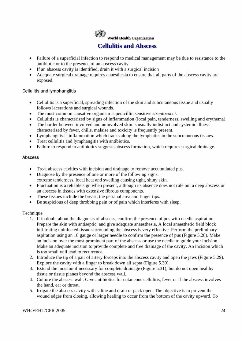
1 Divide the skin, muscle and bone at or near the same level, without attempting to fashion flaps or close the wound (Figure 18.76).
2 Tie all bleeding vessels and cut the nerves sharply while under gentle tension, allowing them to retract into the wound. Tack skin flaps loosely with a few stitches to prevent further retraction. Apply a sterile dressing and, i f possible, an elastic stump wrap.
3 Debride and lavage the wound every 2–5 days until it is free of dead tissue and infection. At that point, perform a definitive amputation and closure.

WWWooorrrlllddd HHHeeeaaalll ttthhh OOOrrrgggaaannniiizzzaaattt iiiooonnn
WHO/EHT/CPR 2005 20
Definitive amputation • Perform a definitive amputation as an elective procedure when the extremity is clean and non-
infected or following a guillotine amputation.
• In the upper limb, preserve as much of the limb as possible.
• The ideal levels for a lower extremity amputation are 12 cm proximal to the knee joint (transfemoral) and 8–14 cm distal to the knee joint (transtibial).
• When possible, save the knee joint to improve function with a prosthesis.
• Amputations through the knee are acceptable in children.
1. Cut the skin flaps 5–6 cm, and the muscles 2–4 cm, distal to the proposed level of bone section (Figure 18.77).
2. Fashion the skin flaps so that the sum of the lengths of the flaps is one and a half times the diameter of the limb. Local conditions may necessitate unequal or irregular flaps.
3. Taper the anterior end of the bone and cut the fibula 3 cm proximal to the tibial cut. 4. Doubly ligate all major vessels (Figure 18.78).
5. Cut the nerves sharply while under gentle tension and allow them to retract into the wound. 6. Stitch opposing muscles over the end of the bone and attach the muscle flaps to the bone through
the periosteum or a drill hole. 7. Release the tourniquet and stop all bleeding before closing further.
8. Suture the skin and fascia loosely in two layers, using interrupted stitches. 9. If skin closure is a problem, use split thick skin grafts on non-weight bearing portions of the stump. 10. Do not close the skin under tension. 11. In most cases, use a drain and plan to remove it in 1–2 days. 12. Apply a firm bandage and place the remaining limb in a plaster splint. 13. Make the stump cylindrical with even muscle distribution. A conical or bulbous stump will be
painful and difficult to fit to the prosthetic socket.
Foot amputations • Perform amputations within the foot at the base of the toes or through the metatarsals, depending on the
level of viable tissue. • Amputations more proximal on the foot (tarsometatarsal joint or midtarsal joint) are acceptable, but may
lead to muscle imbalance. They may require splinting and tendon transfers in order to maintain a plantagrade foot for walking.
Upper extremity amputations • Save as much of the extremity as possible. A prosthesis will often not be available for upper
extremities and any preserved function will be useful. • Split thickness skin grafts work satisfactorily for most stumps. • At the wrist level, preserve carpal joints to allow terminal flexion and
extension movements.

WWWooorrrlllddd HHHeeeaaalll ttthhh OOOrrrgggaaannniiizzzaaattt iiiooonnn
WHO/EHT/CPR 2005 21
• Saving the radial-ulnar joint allows pronation and supination of the forearm. • Patients with bilateral upper extremity amputations may benefit from a Krukenberg operation. This is
an elective procedure that splits the radius and ulna and provides muscle power to each. The resulting forearm has simple grasp and sensation.
Amputations in children Children adapt more easily than adults to amputations and prosthetic use. When possible, preserve the growth plate and the epiphysis to allow normal growth of the extremity. Trans-articular amputations are well tolerated, as is the use of split thickness skin grafts on the weight-bearing surface of the limb.
IIInnnssseeerrrtttiiiooonnn ooofff CCChhheeesssttt DDDrrraaaiiinnn aaannnddd UUUnnndddeeerrrwwwaaattteeerrr SSSeeeaaalll DDDrrraaaiiinnnaaagggeee
IIInnndddiiicccaaatttiiiooonnnsss fffooorrr UUUnnndddeeerrrwwwaaattteeerrr---SSSeeeaaalll DDDrrraaaiiinnnaaagggeee aaarrreee:::
- Pneumothorax - Haemothorax - Haemopneumothorax - Acute empyema.
Technique 1. Prepare the skin with antiseptic and infiltrate the skin, muscle and pleura with 1% lidocaine at the
appropriate intercostal space, usually the fifth or sixth, in the midaxillary line (Figure 16.2). Note the length of needle needed to enter the pleural cavity; this information may be useful later when you are inserting the drain.
2. Aspirate fluid from the chest cavity to confirm your diagnosis (Figure 16.3 ). 3. Make a small transverse incision just above the rib to avoid damaging the vessels under the lower part
of the rib (Figures 16.4, 16.5). In children, it is advisable to keep strictly to the middle of the intercostal space.
4. Using a pair of large, curved artery forceps, penetrate the pleura and enlarge the opening (Figures 16.6, 16.7). Use the same forceps to grasp the tube at its tip and introduce it into the chest (Figures 16.8, 16.9).
5. Close the incision with interrupted skin sutures, using one stitch to anchor the tube. Leave an additional suture untied adjacent to the tube for closing the wound after the tube is removed. Apply a gauze dressing.
6. Connect the tube to the underwater-seal drainage system and mark the initial level of fluid in the drainage bottle (Figure 16.10)

WWWooorrrlllddd HHHeeeaaalll ttthhh OOOrrrgggaaannniiizzzaaattt iiiooonnn
WHO/EHT/CPR 2005 22
Aftercare
• Place a pair of large artery forceps by the bedside for clamping the tube when changing the bottle.

WWWooorrrlllddd HHHeeeaaalll ttthhh OOOrrrgggaaannniiizzzaaattt iiiooonnn
WHO/EHT/CPR 2005 23
• The drainage system is patent i f the fluid level swings freely with changes in the intrapleural pressure. Persistent bubbling over several days suggests a bronchopleural fistula and is an indication for referral.
• Change the connecting tube and the bottle at least once every 48 hours, replacing them with sterile equivalents.
• Wash and disinfect the used equipment to remove all residue before it is resterilized. • If there is no drainage for 12 hours, despite your “milking” the tube, clamp the tube for a further 6
hours and X-ray the chest. I f the lung is satisfactorily expanded, the clamped tube can then be removed.
• To remove the tube, first sedate the patient and then remove the dressing. Clean the skin with antiseptic. Hold the edges of the wound together with fingers and thumb over gauze while cutting the skin stitch that is anchoring the tube. Withdraw the tube rapidly as an assistant ties the previously loose stitch.
WWWooorrrlllddd HHHeeeaaalll ttthhh OOOrrrgggaaannniiizzzaaattt iiiooonnn
WHO/EHT/CPR 2005 24
CCCeeelllllluuullliiitttiiisss aaannnddd AAAbbbsssccceeessssss
• Failure of a superficial infection to respond to medical management may be due to resistance to the antibiotic or to the presence of an abscess cavity
• If an abscess cavity is identified, drain it with a surgical incision • Adequate surgical drainage requires anaesthesia to ensure that all parts of the abscess cavity are
exposed. Cellulitis and lymphangiitis
• Cellulitis is a superficial, spreading infection of the skin and subcutaneous tissue and usually follows lacerations and surgical wounds.
• The most common causative organism is penicillin sensitive streptococci. • Cellulitis is characterized by signs of inflammation (local pain, tenderness, swelling and erythema). • The border between involved and uninvolved skin is usually indistinct and systemic illness
characterized by fever, chills, malaise and toxicity is frequently present. • Lymphangitis is inflammation which tracks along the lymphatics in the subcutaneous tissues. • Treat cellulitis and lymphangitis with antibiotics. • Failure to respond to antibiotics suggests abscess formation, which requires surgical drainage.
Abscess
• Treat abscess cavities with incision and drainage to remove accumulated pus. • Diagnose by the presence of one or more of the following signs: extreme tenderness, local heat and swelling causing tight, shiny skin. • Fluctuation is a reliable sign when present, although its absence does not rule out a deep abscess or
an abscess in tissues with extensive fibrous components. • These tissues include the breast, the perianal area and finger tips. • Be suspicious of deep throbbing pain or of pain which interferes with sleep.
Technique
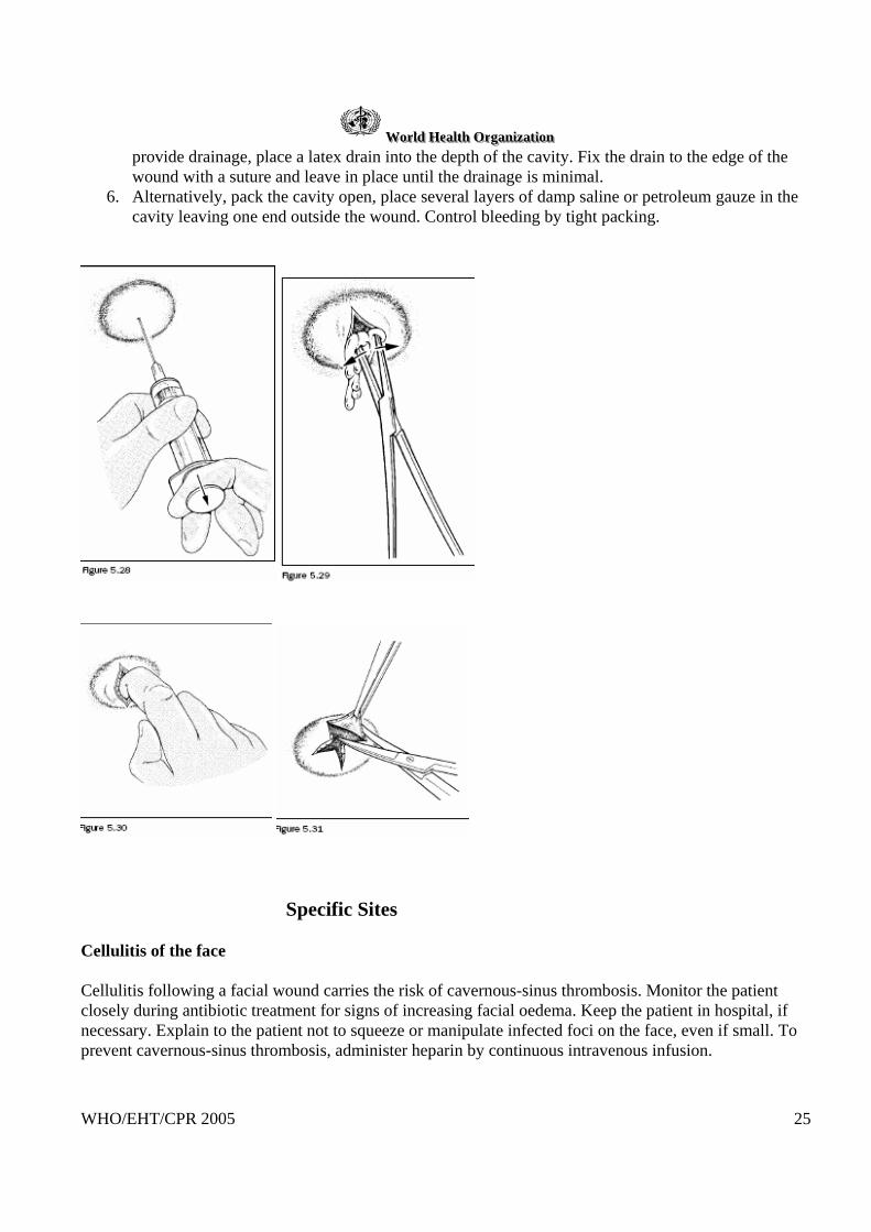
1. If in doubt about the diagnosis of abscess, confirm the presence of pus with needle aspiration. Prepare the skin with antiseptic, and give adequate anaesthesia. A local anaesthetic field block infiltrating uninfected tissue surrounding the abscess is very effective. Perform the preliminary aspiration using an 18 gauge or larger needle to confirm the presence of pus (Figure 5.28). Make an incision over the most prominent part of the abscess or use the needle to guide your incision. Make an adequate incision to provide complete and free drainage of the cavity. An incision which is too small will lead to recurrence.
2. Introduce the tip of a pair of artery forceps into the abscess cavity and open the jaws (Figure 5.29). Explore the cavity with a finger to break down all septa (Figure 5.30).
3. Extend the incision if necessary for complete drainage (Figure 5.31), but do not open healthy tissue or tissue planes beyond the abscess wall.
4. Culture the abscess wall. Give antibiotics for cutaneous cellulitis, fever or if the abscess involves the hand, ear or throat.
5. Irrigate the abscess cavity with saline and drain or pack open. The objective is to prevent the wound edges from closing, allowing healing to occur from the bottom of the cavity upward. To
WWWooorrrlllddd HHHeeeaaalll ttthhh OOOrrrgggaaannniiizzzaaattt iiiooonnn
WHO/EHT/CPR 2005 25
provide drainage, place a latex drain into the depth of the cavity. Fix the drain to the edge of the wound with a suture and leave in place until the drainage is minimal.
6. Alternatively, pack the cavity open, place several layers of damp saline or petroleum gauze in the cavity leaving one end outside the wound. Control bleeding by tight packing.
Specific Sites Cellulitis of the face Cellulitis following a facial wound carries the risk of cavernous-sinus thrombosis. Monitor the patient closely during antibiotic treatment for signs of increasing facial oedema. Keep the patient in hospital, if necessary. Explain to the patient not to squeeze or manipulate infected foci on the face, even if small. To prevent cavernous-sinus thrombosis, administer heparin by continuous intravenous infusion.

WWWooorrrlllddd HHHeeeaaalll ttthhh OOOrrrgggaaannniiizzzaaattt iiiooonnn
WHO/EHT/CPR 2005 26
Ocular infection
• Panophthalmitis is a complication of a neglected penetrating injury of the eye. When efforts to save the eye have failed and the eye is useless, consider evisceration or enucleation. If possible, refer to an ophthalmologist.
• Enucleation of the eye is the surgical removal of the entire globe and requires an ophthalmologist. • Evisceration is the surgical removal of the content of the globe and does not require a specialist.
This procedure involves excision of the anterior globe and curetting of its contents. If necessary, consider evisceration for uncontrolled panophthalmitis. The eviscerated globe is packed open and treated as an abscess cavity. After healing, refer the patient for a prosthesis.
Ear infection
• Middle ear infection presents with chronic drainage of pus from the external meatus. Clean the ear, place a cotton wick and apply a gauze dressing. Continue the administration of antibiotics and give analgesics as needed. Keep the auditory canal dry and change the dressing when necessary.
• Acute mastoiditis is usually a complication of acute otitis media. The patient complains of fever and pain in the affected ear, with disturbed hearing. There may be a discharge from the ear. Characteristically there is a tender swelling in the mastoid area, which pushes the pinna forward and out. Definitive treatment is exposure of the mastoid air cells by a qualified surgeon. When
this is not possible, initial treatment is to relieve immediate pain with an incision and drainage of the abscess down to the periosteum.
Technique 1. Using a general or local anaesthetic, make a curved incision over the most fluctuant part of the
abscess or, if not obvious, at 1.5 cm behind the pinna. Deepen the incision to the periosteum or until pus is found.
2. Take a sample for bacteriological examination and establish free drainage. Apply petroleum gauze or a small latex drain and dress the area with gauze.
3. Continue the administration of antibiotics and analgesics, and change dressings as necessary. 4. Remove the drain after 24– 48 hours.
Infections of the hand
• Staphylococci are the organisms commonly responsible for acute infections of the hand. • An early infection may resolve with antibiotics alone but incision and drainage are usually needed.
Antibiotics should be given until sepsis is controlled. • Patients present with a history of throbbing pain, warm, tender swelling, a flexion deformity of the
finger and pain on movement. • Confirm the abscess with needle aspiration. Obtain an X-ray of the hand to determine if there is • bone involvement and perform a Gram stain on the pus. • Give general or regional anaesthesia and proceed with incision and drainage. • Make an adequate, but not extensive, incision along a skin crease at the site of maximum
tenderness and swelling (Figure 5.42).Aspirate or irrigate away all pus. Open up deeper loculi with artery forceps and insert a latex drain. Obtain a culture. Dress the wound loosely with dry gauze, administer antibiotics and elevate the hand.

WWWooorrrlllddd HHHeeeaaalll ttthhh OOOrrrgggaaannniiizzzaaattt iiiooonnn
WHO/EHT/CPR 2005 27
• Marked swelling on the dorsum of the hand is often due to lymphoedema, which does not require drainage.
• Infection of the nail bed may necessitate excision of a portion of the nail for effective drainage of pus.
• Treat paronychia of the middle finger with an incision over the involved area (Figure 5.43) or excise a portion of the nail (Figure 5.44).
• Treat finger tip abscesses with a “hockey stick” incision (Figure 5.45). • Treat acute septic contracture of an involved digit with antibiotics and prompt surgical drainage of
the flexor tendon sheath through incisions along the lateral or medial borders of the fingers, preferably the junctional area between the palmar and dorsal skin (Figure 5.46).
• Infection of the tendon sheaths of the thumb or little finger may spread to the radial or ulnar bursa, respectively (Figure 5.47), necessitating drainage by short, transverse incisions in the distal palmar crease and/or at the base of the palm.
• Infections of fascial palmar spaces result from extensions of infections of a web-space or a tendon sheath. Drain the affected fascial space through skin incisions directly over the area of maximum swelling and tenderness. Open deeper parts of the abscess with forceps. In general, place incisions for drainage along creases of the palm, along the lateral or medial borders of the fingers, or along the ulnar or radial borders of the forearm (Figure 5.48). Splint the hand in position of function. Encourage active exercises as soon as possible. Give antibiotics and analgesics and remove the drain in 24–48 hours.

WWWooorrrlllddd HHHeeeaaalll ttthhh OOOrrrgggaaannniiizzzaaattt iiiooonnn
WHO/EHT/CPR 2005 28
OOOppp eee nnn FFF rrr aaa ccc tttuuu rrr eee sss • Open fractures, also known as compound fractures, are injuries involving both bone and soft tissue. • The soft tissue injury allows contamination of the fracture site. • All open fractures are contaminated, so primary closure is absolutely contraindicated. • Wound closure predisposes to anaerobic infection and chronic osteomyelitis. • Treat with wound toilet, debridement and fracture immobilization. Prior to debridement, take a
swab for bacteriological examination and administer systemic antibiotics. • When debriding a compound fracture, remove free fragments of bone with no obvious blood supply. • Do not strip muscle and periosteum from the fractured bone. • Leave vessels, nerves and tendons that are intact. • Surgical toilet of these wounds is an emergency. • Perform the debridement within 6 hours and do not delay for referral. • Osteomyelitis is a grave complication, which can be avoided with proper and expeditious wound
toilet. • Stabilize the fracture after wound debridement; perform definitive fracture treatment at a later
time. • Achieve stabilization with a well-padded posterior plaster slab, a complete plaster cast split to prevent
compartment syndrome, traction or, i f available, an external fixator.
Clavicle Fractures • Diagnose fractures from the history and by physical examination • Treat with a sling and early range of motion • Fracture healing takes 4 weeks in children and 6–8 weeks in adults.
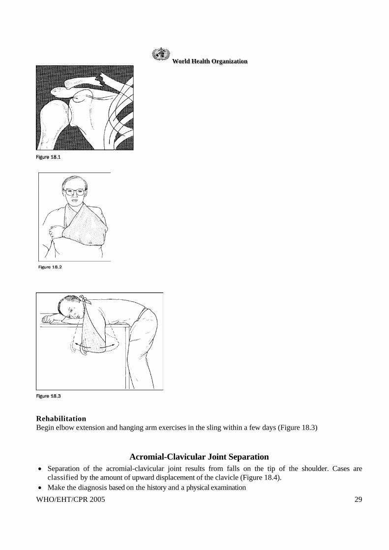
Evaluation A physical examination shows tenderness over the mid or distal clavicle, with swelling, visible and palpable deformity and, often, crepitus. X-rays confirm diagnosis, but are not essential (Figure 18.1).
Treatment
• Apply a sling as shown in Figure 18.2; a figure-of-8 splint provides some comfort, but is not essential for fracture healing
• Fractures are rarely positioned anatomically, but will heal satisfactorily with little or no functional loss. The patient should continue with the sling until pain free. This will take 4–6 weeks in adults
and 3–4 weeks in children.
WWWooorrrlllddd HHHeeeaaalll ttthhh OOOrrrgggaaannniiizzzaaattt iiiooonnn
WHO/EHT/CPR 2005 29
Rehabilitation Begin elbow extension and hanging arm exercises in the sling within a few days (Figure 18.3)
Acromial-Clavicular Joint Separation • Separation of the acromial-clavicular joint results from falls on the tip of the shoulder. Cases are
classified by the amount of upward displacement of the clavicle (Figure 18.4). • Make the diagnosis based on the history and a physical examination

WWWooorrrlllddd HHHeeeaaalll ttthhh OOOrrrgggaaannniiizzzaaattt iiiooonnn
WHO/EHT/CPR 2005 30
• Treat with an arm sling • When comfortable, begin a range of motion and active muscle strengthening in the shoulder.
Evaluation The acromial-clavicular joint is tender to palpation and the end of the clavicle is prominent. X-rays are not essential but confirm the diagnosis and determine i f there is a fracture. Treatment
• Apply an arm sling to support the weight of the arm and to remove the deforming force from the joint. This will not keep the joint in the anatomical position, but the remaining deformity will cause little functional loss.
• Release the sling daily to allow elbow extension. • Begin hanging arm exercises when comfortable and begin active muscle strengthening by the second
week.
Shoulder Dislocation • Anterior dislocations result from injuries that place the arm in abduction, external rotation and
extension. • Posterior dislocations are less common; the arm position is not important as they follow seizures
or electric shocks. • Make the diagnosis by physical examination • Treat with closed manipulation • X-rays help to evaluate the reduction and the presence of fractures • Recurrent dislocations are common, especially in younger patients.
Evaluation
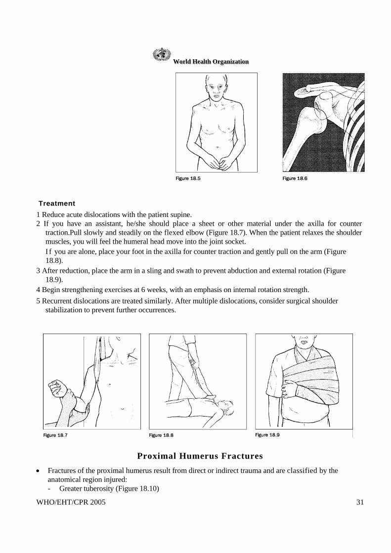
• The contour of the shoulder is changed from the usual curved appearance to one that is much more angular, with a hollow area in place of the humeral head (Figures 18.5 and 18.6)
• Any shoulder movement is painful; X-rays help to determine i f there is a fracture • Perform a careful neurological examination to evaluate for peripheral nerve or brachial plexus injury.
WWWooorrrlllddd HHHeeeaaalll ttthhh OOOrrrgggaaannniiizzzaaattt iiiooonnn
WHO/EHT/CPR 2005 31
Treatment 1 Reduce acute dislocations with the patient supine. 2 If you have an assistant, he/she should place a sheet or other material under the axilla for counter
traction.Pull slowly and steadily on the flexed elbow (Figure 18.7). When the patient relaxes the shoulder muscles, you will feel the humeral head move into the joint socket. I f you are alone, place your foot in the axilla for counter traction and gently pull on the arm (Figure 18.8).
3 After reduction, place the arm in a sling and swath to prevent abduction and external rotation (Figure 18.9).
4 Begin strengthening exercises at 6 weeks, with an emphasis on internal rotation strength. 5 Recurrent dislocations are treated similarly. After multiple dislocations, consider surgical shoulder
stabilization to prevent further occurrences.
Proximal Humerus Fractures • Fractures of the proximal humerus result from direct or indirect trauma and are classified by the
anatomical region injured: - Greater tuberosity (Figure 18.10)

WWWooorrrlllddd HHHeeeaaalll ttthhh OOOrrrgggaaannniiizzzaaattt iiiooonnn
WHO/EHT/CPR 2005 32
- Surgical neck (Figure 18.11) - Anatomic neck - Humeral head.
• The anatomical location of the fracture defines the treatment • X-rays are needed to evaluate the injury • Treat displaced fractures with closed manipulation • The major complication is shoulder stiffness.
Evaluation Suspect the diagnosis from the history and the physical findings of pain, swelling and loss of motion of the shoulder joint. You will need X-rays to confirm the type of fracture and to direct treatment. Treatment • Immobilize non-displaced fractures in a sling and swath. Begin mobilization of the shoulder joint
within a few days. • Treat displaced fractures and fracture dislocations by closed manipulation under anaesthesia. I f the
reduction is not acceptable, consider surgical treatment. • Begin motion as soon as the patient can tolerate hanging arm exercises (Figure 18.3). Begin active motion
against gravity or with weights when the fracture has healed. This is usually at 6–8 weeks.
Humeral Shaft Fractures
Humeral shaft fractures result from direct trauma or rotation of the arm • Treat by closed means in a coaptation splint • The most significant complications are radial nerve injury and non-union.
WWWooorrrlllddd HHHeeeaaalll ttthhh OOOrrrgggaaannniiizzzaaattt iiiooonnn
WHO/EHT/CPR 2005 33
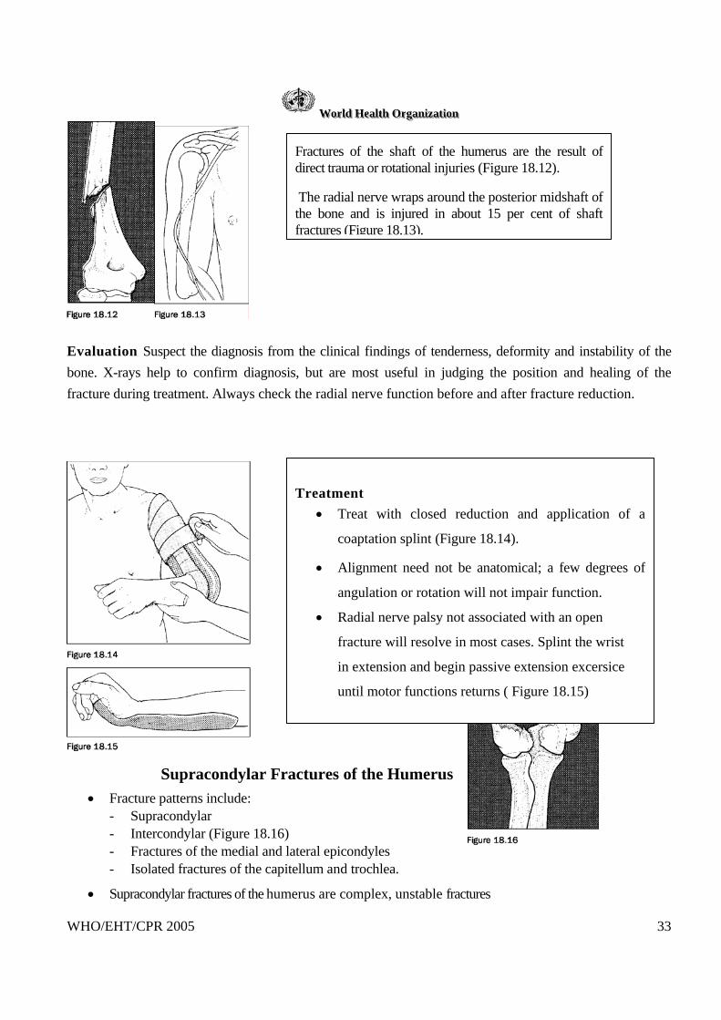
Evaluation Suspect the diagnosis from the clinical findings of tenderness, deformity and instability of the bone. X-rays help to confirm diagnosis, but are most useful in judging the position and healing of the fracture during treatment. Always check the radial nerve function before and after fracture reduction.
Supracondylar Fractures of the Humerus • Fracture patterns include:
- Supracondylar - Intercondylar (Figure 18.16) - Fractures of the medial and lateral epicondyles - Isolated fractures of the capitellum and trochlea.
• Supracondylar fractures of the humerus are complex, unstable fractures
Fractures of the shaft of the humerus are the result of direct trauma or rotational injuries (Figure 18.12).
The radial nerve wraps around the posterior midshaft of the bone and is injured in about 15 per cent of shaft fractures (Figure 18.13).
Treatment • Treat with closed reduction and application of a
coaptation splint (Figure 18.14).
• Alignment need not be anatomical; a few degrees of
angulation or rotation will not impair function.
• Radial nerve palsy not associated with an open
fracture will resolve in most cases. Splint the wrist
in extension and begin passive extension excersice
until motor functions returns ( Figure 18.15)
WWWooorrrlllddd HHHeeeaaalll ttthhh OOOrrrgggaaannniiizzzaaattt iiiooonnn
WHO/EHT/CPR 2005 34
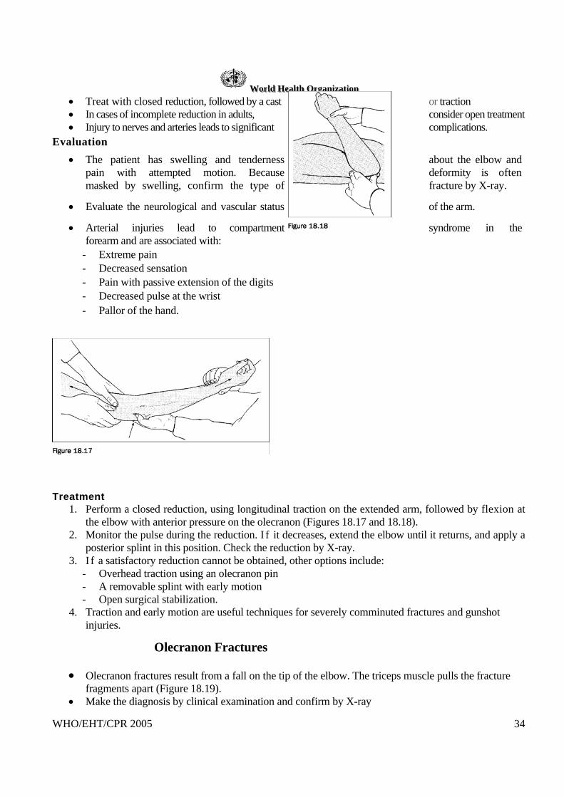
• Treat with closed reduction, followed by a cast or traction • In cases of incomplete reduction in adults, consider open treatment • Injury to nerves and arteries leads to significant complications.
Evaluation • The patient has swelling and tenderness about the elbow and
pain with attempted motion. Because deformity is often masked by swelling, confirm the type of fracture by X-ray.
• Evaluate the neurological and vascular status of the arm.
• Arterial injuries lead to compartment syndrome in the forearm and are associated with:
- Extreme pain - Decreased sensation - Pain with passive extension of the digits - Decreased pulse at the wrist - Pallor of the hand.
Treatment 1. Perform a closed reduction, using longitudinal traction on the extended arm, followed by flexion at
the elbow with anterior pressure on the olecranon (Figures 18.17 and 18.18). 2. Monitor the pulse during the reduction. I f it decreases, extend the elbow until it returns, and apply a
posterior splint in this position. Check the reduction by X-ray. 3. I f a satisfactory reduction cannot be obtained, other options include:
- Overhead traction using an olecranon pin - A removable splint with early motion - Open surgical stabilization.
4. Traction and early motion are useful techniques for severely comminuted fractures and gunshot injuries.
Olecranon Fractures
• Olecranon fractures result from a fall on the tip of the elbow. The triceps muscle pulls the fracture fragments apart (Figure 18.19).
• Make the diagnosis by clinical examination and confirm by X-ray

WWWooorrrlllddd HHHeeeaaalll ttthhh OOOrrrgggaaannniiizzzaaattt iiiooonnn
WHO/EHT/CPR 2005 35
• Treat non displaced fractures with a long arm splint at 90 degrees • Splint displaced fractures with the elbow extended or consider surgical
stabilization. Evaluation
Physical
examination shows swelling about the olecranon and a palpable gap at the fracture site. Examine the ulnar nerve function. X-rays confirm the fracture and associated injuries. Treatment • Treat non-displaced fractures in a splint with the elbow at 90 degrees • Treat displaced fractures with the elbow in full extension; displaced fractures may a have better outcome
i f treated surgically • Simple methods include:
- Suture of the torn triceps tendon (Figure 18.20) - Placement of percutaneous pins with rubber bands (Figures 18.21 and 18.22).
Fractures of the Radial Head and Neck • The radial head is important for pronation and supination of the forearm as well as for flexion and
extension motions at the elbow. • In fractures , with minimal displacement, treat with closed reduction and a posterior splint and begin
motion as soon as comfortable • Treat displaced intra-articular fractures with early motion and consider surgical treatment, if available. • Fractures are classified by the articular involvement (Figure 18.23).

WWWooorrrlllddd HHHeeeaaalll ttthhh OOOrrrgggaaannniiizzzaaattt iiiooonnn
WHO/EHT/CPR 2005 36
Evaluation Patients have pain and swelling over the lateral aspect of the elbow. Some motion remains in minimally displaced fractures. X-rays confirm the diagnosis. Treatment 1 Treat fractures with minimal displacement in an arm sling and begin motion when comfortable. 2 To reduce displaced fractures of the radial neck:
- Place your thumb over the radial head and apply longitudinal traction with a varus stress to the arm
- Gently rotate the forearm while applying medial pressure with your thumb to the radial head - Place the arm in a long arm splint - Begin motion out of the splint at 3 weeks.
3 Treat comminuted or displaced intra-articular fractures with early motion. I f available, alternatives are surgical stabilization or radial head excision.
Elbow Dislocation
.
• Treat with immediate closed reduction • In children, the medial epicondyle may become entrapped in the joint and may require surgical
removal.
Injury occurs with a fall on the outstretched arm . They may be in the posterior or posterior lateral direction (Figure 18.24)

WWWooorrrlllddd HHHeeeaaalll ttthhh OOOrrrgggaaannniiizzzaaattt iiiooonnn
WHO/EHT/CPR 2005 37
1. In children, the medial epicondyle of the humerus is often pulled off as the radius and ulna move posteriorly and laterally. With reduction, this fragment may become lodged in the joint and require surgical removal.
Evaluation
Clinically examine the triangular relationship of the ulna and the two epicondyles to ascertain i f it is disturbed. The olecranon is felt protruding in a posterior direction and any elbow motion is painful. Assess and record ulnar nerve function.
Treatment
1 Treat with immediate closed reduction: apply traction to the arm with the elbow in slight flexion and direct pressure on the tip of the olecranon to push it distally and anteriorly.
2 When reduced, the elbow will have a free range of motion. After reduction, confirm the position of the epicondyle by X-ray.
3 Place the arm in a posterior splint at 90 degrees of flexion.
4 Begin a range of motion at the elbow after 10 days, or as soon as the pain and swelling permit, removing the splint for short periods. Discontinue the splint at 4–6 weeks.
Forearm Fractures Forearm fractures are caused by direct trauma or by a fall on the outstretched arm with an accompanying rotatory or twisting force.
• Forearm fractures are complex fractures which, in adults, usually require surgical stabilization • They occur as three major types:
- Midshaft fractures - Proximal (Monteggia) dislocations - Distal (Galeazzi) fracture dislocations
• The most common complication is loss of forearm rotation. Evaluation 2. The forearm is swollen and tender, with limited motion. 3. Evaluate vascular function by checking pulse, capillary refill and skin temperature of the hand. 4. Check sensory and motor function of the radial, median and ulnar nerves. X-rays confirm the nature of
the fracture.
5. Monteggia fractures involve the proximal ulna with dislocation of the radial head, usually in the anterior direction (Figure 18.25).
6. Galeazzi fractures are the reverse of the above, with a fracture of the distal radius and a dislocation of the radial-ulnar joint at the wrist. The radius fracture is usually oblique, causing the bone to shorten (Figure 18.26)

WWWooorrrlllddd HHHeeeaaalll ttthhh OOOrrrgggaaannniiizzzaaattt iiiooonnn
WHO/EHT/CPR 2005 38
Treatment • Midshaft fractures may involve one or both bones; treat single bone fractures with minimal displacement
in a long arm cast, with the elbow at 90 degrees and the forearm in neutral rotation • Treat displaced fractures by closed reduction and application of a long arm splint; perform the reduction
by applying traction to the fingers and manipulating the forearm with the elbow bent to 90 degrees. Apply counter-traction above the bent elbow (Figure 18.27)
• Reduce Monteggia fractures as described for displaced fractures (Figure 18.28). Apply a long arm cast in supination. It is possible to obtain a satisfactory reduction in children, but adults often require surgical treatment.
• Treat Galeazzi fractures as described for midshaft fractures. They are unstable and often need surgical stabilization.
Rehabilitation Begin motion out of the cast at 6–8 weeks.
Distal Radius Fractures Fractures of the distal radius occur with a fall on the outstretched hand. The direction of the deformity depends on the position of the wrist at the time of impact (Figure 18.28). • The distal radius is one of the most common upper extremity fractures • Treatment is usually by closed reduction and application of a U-shaped splint coaptation • The adequacy of the reduction can be judged by specific parameters visible on the post-reduction X-ray • The most common complication is malposition and loss of motion.

WWWooorrrlllddd HHHeeeaaalll ttthhh OOOrrrgggaaannniiizzzaaattt iiiooonnn
WHO/EHT/CPR 2005 39
• The goal of fracture treatment is to restore the normal anatomy of the following deformities: - Shortening of the radius relative to the ulna (Figure 18.29) - Loss of the volar tilt of the radial articular surface, seen in the lateral X-ray (Figure 18.30) - Disruption of the articular surface.
1. Make the diagnosis based on the history of a fall on the outstretched hand, swelling and tenderness about the wrist and the presence of deformity
2. Evaluate tendon function, vascular supply and sensation in the hand 3. X-rays distinguish radius fractures from carpal injuries and determine i f the fracture is adequately
reduced.
Treatment 1. Anaesthetize for closed reduction, using general anaesthesia (ketamine), an intravenous lidocaine block
or a haematoma block. A haematoma block involves placing 5–10 ml of 2% lidocaine directly into the fracture haematoma, using a strict aseptic technique (Figure 18.31).
2. Reduce the fracture by placing longitudinal traction across the wrist and applying pressure to the distal radial fragment to correct the angular deformity (Figure 18.32). For fractures that are dorsally angulated (Colle’s fractures), this is accomplished by wrist flexion and slight ulnar deviation.
3. Next, apply a sugar tong splint, moulded to maintain the fracture position. Three point moulding involves application of pressure above and below the fracture and counter pressure on the opposite side of the bone near the fracture apex.
4. Between 10 days and 2 weeks, change the sugar tong splint to a short arm cast and check the fracture position by X-ray. Healing takes about 6 weeks.
5. If a satisfactory position of the fracture fragments cannot be obtained or maintained, consider open reduction and internal fixation, placement of an external fixator or closed reduction with percutaneous pin fixation

WWWooorrrlllddd HHHeeeaaalll ttthhh OOOrrrgggaaannniiizzzaaattt iiiooonnn
WHO/EHT/CPR 2005 40
Carpal Fractures and Fracture Dislocations The injury results from a fall on the outstretched hand in hyperextension
• Diagnosis is difficult and is often overlooked • Adequate X-rays are necessary for accurate diagnosis • Closed reduction is the initial treatment, but surgical stabilization may be necessary. • Injuries to the carpal bones fall into three major categories:
- Scaphoid fractures - Trans-scaphoid perilunate fracture/dislocations - Perilunate dislocations.
• The scaphoid bone (S) bridges the proximal and distal rows of carpal bones, making it especially vulnerable to injury. Most commonly, fractures occur at the waist but may also involve the proximal or distal pole (Figure 18.33).
• Perilunate dislocations occur with or without an accompanying scaphoid fracture. • The lunate (L) stays in a volar position while the remaining carpal bones dislocate
posteriorly(Figure18.34).
Evaluation • The wrist appears swollen and painful to move. • Scaphoid fractures are tender in the anatomic snuff box and over the scaphoid tubercle on the volar
aspect of the wrist. I f a perilunate dislocation has occurred, these findings are diffuse about the wrist. X-rays are necessary to make a definitive diagnosis.
• In perilunate dislocations, the lateral X-ray shows an anteriorly displaced lunate bone, with its concavity
WWWooorrrlllddd HHHeeeaaalll ttthhh OOOrrrgggaaannniiizzzaaattt iiiooonnn
WHO/EHT/CPR 2005 41
facing forward (Figure 18.34). The carpus is shortened and the proximal margin of the capitate does not articulate with the concavity of the lunate.
Treatment
• Treat scaphoid fractures with minimal displacement in a thumb spica splint or cast. Healing time is between 6 and 20 weeks.
• Perilunate dislocations require reduction followed by placement in a long arm thumb spica splint. The reduction is usually unstable over time and most patients will need surgical stabilization.
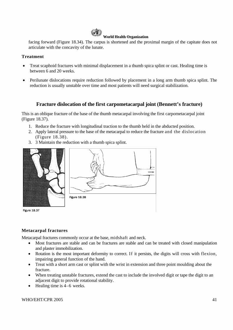
Fracture dislocation of the first carpometacarpal joint (Bennett’s fracture)
This is an oblique fracture of the base of the thumb metacarpal involving the first carpometacarpal joint (Figure 18.37).
1. Reduce the fracture with longitudinal traction to the thumb held in the abducted position. 2. Apply lateral pressure to the base of the metacarpal to reduce the fracture and the dislocation
(Figure 18.38). 3. 3 Maintain the reduction with a thumb spica splint.
Metacarpal fractures Metacarpal fractures commonly occur at the base, midshaft and neck.
• Most fractures are stable and can be fractures are stable and can be treated with closed manipulation and plaster immobilization.
• Rotation is the most important deformity to correct. If it persists, the digits will cross with flexion, impairing general function of the hand.
• Treat with a short arm cast or splint with the wrist in extension and three point moulding about the fracture.
• When treating unstable fractures, extend the cast to include the involved digit or tape the digit to an adjacent digit to provide rotational stability.
• Healing time is 4–6 weeks.
WWWooorrrlllddd HHHeeeaaalll ttthhh OOOrrrgggaaannniiizzzaaattt iiiooonnn
WHO/EHT/CPR 2005 42
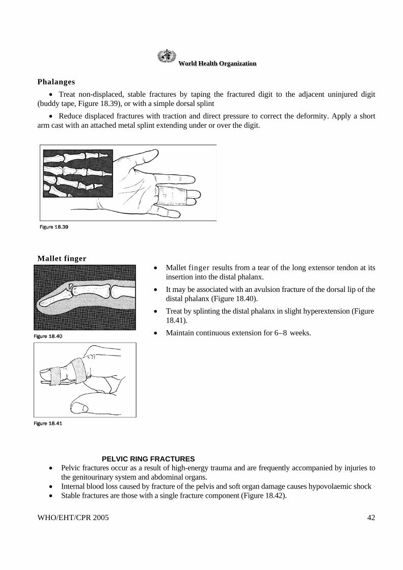
Phalanges
• Treat non-displaced, stable fractures by taping the fractured digit to the adjacent uninjured digit (buddy tape, Figure 18.39), or with a simple dorsal splint
• Reduce displaced fractures with traction and direct pressure to correct the deformity. Apply a short arm cast with an attached metal splint extending under or over the digit.
Mallet finger • Mallet finger results from a tear of the long extensor tendon at its
insertion into the distal phalanx. • It may be associated with an avulsion fracture of the dorsal lip of the
distal phalanx (Figure 18.40). • Treat by splinting the distal phalanx in slight hyperextension (Figure
18.41). • Maintain continuous extension for 6–8 weeks.
PELVIC RING FRACTURES • Pelvic fractures occur as a result of high-energy trauma and are frequently accompanied by injuries to
the genitourinary system and abdominal organs. • Internal blood loss caused by fracture of the pelvis and soft organ damage causes hypovolaemic shock • Stable fractures are those with a single fracture component (Figure 18.42).

WWWooorrrlllddd HHHeeeaaalll ttthhh OOOrrrgggaaannniiizzzaaattt iiiooonnn
WHO/EHT/CPR 2005 43
• Unstable patterns result from fractures at two or more sites, or those associated with disruption of the symphysis pubis or sacroiliac articulation (Figure 18.43).
• Unstable fractures are associated with significant blood loss and multiple system injury • Treat initially with systemic resuscitation and temporary pelvic compression • Complications include deep vein thrombosis, sciatic nerve injury and death from bleeding or internal organ
damage.
Evaluation • Physical examination findings include:
- Flank ecchymosis - Labial or scrotal swelling - Abnormal position of the lower extremities - Pain with pelvic rim compression.
• I f the fracture is unstable, you will feel differential motion of the pelvic components when gently manipulating them. Place your hands on the iliac wings and gently rock the pelvis. Confirm the diagnosis with an anterior-posterior X-ray of the pelvis. Additional inlet and outlet views help determine the extent of the fractures.
• Remember to focus on a systematic examination of the whole patient (see page 16–2). Treatment • Focus the initial management on general resuscitation efforts Manage stable pelvic fractures with bed rest
and analgesics. • Stable fractures are rarely associated with significant blood loss. Unstable fractures Unstable fractures are associated with visceral damage and there is often significant bleeding. As an emergency procedure:
1. Place compression on the iliac wings, using a sheet or sling to close the pelvic space and tamponade active bleeding (Figure 18.44).
2. Treat with a pelvic sling and/or traction on the leg to reduce the vertical shear component of the fracture (Figure 18.45).
3. Maintain the traction until the fracture has consolidated. This usually takes 8–12 weeks.

WWWooorrrlllddd HHHeeeaaalll ttthhh OOOrrrgggaaannniiizzzaaattt iiiooonnn
WHO/EHT/CPR 2005 44
Acetabular Fractures
The fracture disrupts the congruence of the femoral head with the acetabulum and causes damage to the articular surface. A small number of fractures will be combined acetabular and pelvic ring injuries.
• Acetabular fractures result from high-energy pelvic injuries • Treatment a ims to res tore the congruence of the femoral head with the acetabulum by
traction or by surgery if available • Complications include deep venous thrombosis, sciatic nerve injury and late degenerative arthritis of
the hip. Evaluation
• History and physical findings are similar to those in pelvic ring fractures. • Evaluate and treat hypovolaemic shock and visceral organ damage as an emergency (see page 13–8). • Evaluate sciatic nerve function and look for an associated femoral shaft fracture. • Obtain an initial anterior-posterior pelvic X-ray. I f a fracture is evident, oblique views show the
articular surfaces more clearly. X-ray the femoral shaft.
Treatment 1. Minimally displaced fractures Treat minimally displaced fractures with bed rest and gradual mobilization. When comfortable, begin partial weight bearing until fracture healing has occurred. This usually takes about 12 weeks. 2. Displaced and unstable fractures

WWWooorrrlllddd HHHeeeaaalll ttthhh OOOrrrgggaaannniiizzzaaattt iiiooonnn
WHO/EHT/CPR 2005 45
• Treat displaced and unstable fractures with traction to maintain the congruence of the femoral head with the weight-bearing portion of the acetabulum. I f a satisfactory position cannot be maintained, or if there are bone chips within the hip joint, surgical stabilization is indicated.
• Do not send the patient to another hospital unless you are certain that this complicated surgery is available there.
Fractures of the Proximal Femur (hip fractures) Hip fractures in elderly people with osteoporotic (weak) bone frequently occur following simple falls. In younger people, a moderately severe trauma is required to produce a fracture in this region.
Classify fractures by their anatomic location (Figure 18.46): - Intra-capsular (femoral neck fractures) - Extra-capsular: intertrochanteric - Extra-capsular: subtrochanteric.
In intra-capsular fractures, the blood supply to the femoral head is disrupted. This may lead to the secondary complication of avascular necrosis of the femoral head.
Evaluation Make the diagnosis from a history of a fall, pain about the hip and inability to bear weight on the extremity. The physical examination reveals a leg that is shortened and externally rotated. The pain is made worse by attempted motion of the hip, especially with rotation. Confirm diagnosis by X-ray. Treatment Intra-capsular fractures Treat with internal fixation or prosthetic replacement of the femoral head. I f this cannot be done:
• Treat non-displaced or impacted fractures with light skin traction and a gentle range of motion until the fracture has healed; this will be in about 8–12 weeks
• Treat displaced fractures initially in light traction for a few weeks to control pain, then begin sitting and walking with crutches.
Extra-capsular fractures
Treat with Perkin’s traction or surgical fixation. Perkin’s traction will maintain the fracture position while permitting the patient to sit up to move the knee and hip joint, preventing pressure sores and pneumonia.

WWWooorrrlllddd HHHeeeaaalll ttthhh OOOrrrgggaaannniiizzzaaattt iiiooonnn
WHO/EHT/CPR 2005 46
Hip Dislocations

WWWooorrrlllddd HHHeeeaaalll ttthhh OOOrrrgggaaannniiizzzaaattt iiiooonnn
WHO/EHT/CPR 2005 47
Dislocations of the hip joint result from high-energy trauma and are associated with injuries of the acetabulum, femoral shaft and patella. Posterior dislocations are most common. Make the diagnosis from the history and from clinical findings; use X-rays to confirm associated fractures • To avoid the complications of vascular necrosis and loss of joint motion, reduce the dislocation as soon as
possible • Closed reduction is usually successful if carried out promptly.
Evaluation
• Make the diagnosis from the history of the injury and the physical findings of a flexed, adducted, internally rotated hip that is painful to move. The clinical examination is sufficient to make the diagnosis, but X-rays are necessary to identify associated fractures.
• Examine the sciatic nerve function by testing foot and ankle strength and sensation.
Treatment
1 Reduce the dislocation as soon as possible: - With the patient supine, apply traction to the flexed hip
while an assistant holds the pelvis down for counter traction (Figure 18.47); muscle relaxation is usually necessary
- I f you have no assistant, use an alternative method with the patient prone:
- Apply traction downward with the leg flexed over the edge of the table
- Gently rotate the hip while applying pressure on the femoral head in the gluteal region (Figure 18.48).
2 Place the patient in post-reduction skin traction for a few days and then begin non-weight bearing ambulation with crutches. Allow weight bearing after 12 weeks. I f there is a large posterior rim fracture, treat the patient in traction for 8-12 weeks while the fracture unites
Femoral Shaft Fractures
Evaluation • Make the diagnosis based on a history of major trauma and the clinical findings of swelling, pain,
angular or rotational deformity or abnormal motion at the fracture site. Examine the skin and soft tissue on all sides of the limb to check for possible open fractures.
• Evaluate the neurological and vascular status for injury to the sciatic nerve and the femoral artery. Confirm the diagnosis with X-rays of the entire femur, including the femoral neck.
• Femoral shaft fractures result from high-energy trauma and are often associated with other significant injuries
• Debride and lavage open fractures under sterile conditions as soon as possible • Treat in traction and monitor the fracture position with or without X-rays
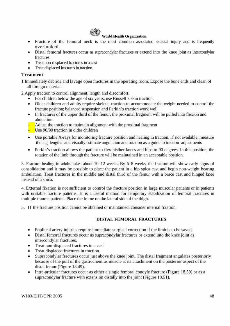
WWWooorrrlllddd HHHeeeaaalll ttthhh OOOrrrgggaaannniiizzzaaattt iiiooonnn
WHO/EHT/CPR 2005 48
• Fracture of the femoral neck is the most common associated skeletal injury and is frequently overlooked.
• Distal femoral fractures occur as supracondylar fractures or extend into the knee joint as intercondylar fractures
• Treat non-displaced fractures in a cast • Treat displaced fractures in traction.
Treatment 1 Immediately debride and lavage open fractures in the operating room. Expose the bone ends and clean of
all foreign material. 2 Apply traction to control alignment, length and discomfort:
• For children below the age of six years, use Russell’s skin traction. • Older children and adults require skeletal traction to accommodate the weight needed to control the
fracture position; balanced suspension and Perkin’s traction work well • In fractures of the upper third of the femur, the proximal fragment will be pulled into flexion and
abduction - Adjust the traction to maintain alignment with the proximal fragment - Use 90/90 traction in older children • Use portable X-rays for monitoring fracture position and healing in traction; i f not available, measure
the leg lengths and visually estimate angulation and rotation as a guide to traction adjustments • Perkin’s traction allows the patient to flex his/her knees and hips to 90 degrees. In this position, the
rotation of the limb through the fracture will be maintained in an acceptable position.
3. Fracture healing in adults takes about 10–12 weeks. By 6–8 weeks, the fracture will show early signs of consolidation and it may be possible to place the patient in a hip spica cast and begin non-weight bearing ambulation. Treat fractures in the middle and distal third of the femur with a brace cast and hinged knee instead of a spica.
4. External fixation is not sufficient to control the fracture position in large muscular patients or in patients with unstable fracture patterns. It is a useful method for temporary stabilization of femoral fractures in multiple trauma patients. Place the frame on the lateral side of the thigh.
5 . I f the fracture position cannot be obtained or maintained, consider internal fixation.
DISTAL FEMORAL FRACTURES
• Popliteal artery injuries require immediate surgical correction if the limb is to be saved. • Distal femoral fractures occur as supracondylar fractures or extend into the knee joint as
intercondylar fractures. • Treat non-displaced fractures in a cast • Treat displaced fractures in traction. • Supracondylar fractures occur just above the knee joint. The distal fragment angulates posteriorly
because of the pull of the gastrocnemius muscle at its attachment on the posterior aspect of the distal femur (Figure 18.49).
• Intra-articular fractures occur as either a single femoral condyle fracture (Figure 18.50) or as a supracondylar fracture with extension distally into the joint (Figure 18.51).
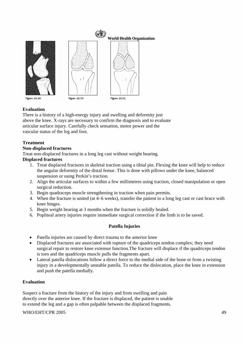
WWWooorrrlllddd HHHeeeaaalll ttthhh OOOrrrgggaaannniiizzzaaattt iiiooonnn
WHO/EHT/CPR 2005 49
Evaluation There is a history of a high-energy injury and swelling and deformity just above the knee. X-rays are necessary to confirm the diagnosis and to evaluate articular surface injury. Carefully check sensation, motor power and the vascular status of the leg and foot. Treatment Non-displaced fractures Treat non-displaced fractures in a long leg cast without weight bearing. Displaced fractures
1. Treat displaced fractures in skeletal traction using a tibial pin. Flexing the knee will help to reduce the angular deformity of the distal femur. This is done with pillows under the knee, balanced suspension or using Perkin’s traction.
2. Align the articular surfaces to within a few millimetres using traction, closed manipulation or open surgical reduction.
3. Begin quadraceps muscle strengthening in traction when pain permits. 4. When the fracture is united (at 4–6 weeks), transfer the patient to a long leg cast or cast brace with
knee hinges. 5. Begin weight bearing at 3 months when the fracture is solidly healed. 6. Popliteal artery injuries require immediate surgical correction if the limb is to be saved.
Patella Injuries
• Patella injuries are caused by direct trauma to the anterior knee • Displaced fractures are associated with rupture of the quadriceps tendon complex; they need
surgical repair to restore knee extensor function.The fracture will displace if the quadriceps tendon is torn and the quadriceps muscle pulls the fragments apart.
• Lateral patella dislocations follow a direct force to the medial side of the bone or from a twisting injury in a developmentally unstable patella. To reduce the dislocation, place the knee in extension and push the patella medially.
Evaluation Suspect a fracture from the history of the injury and from swelling and pain directly over the anterior knee. If the fracture is displaced, the patient is unable to extend the leg and a gap is often palpable between the displaced fragments.

WWWooorrrlllddd HHHeeeaaalll ttthhh OOOrrrgggaaannniiizzzaaattt iiiooonnn
WHO/EHT/CPR 2005 50
A rupture of the quadriceps tendon proximal to the patella, or to the patella tendon distal to it, has similar physical findings. X-rays confirm diagnosis. Treatment Non-displaced fractures Treat non-displaced fractures in a splint or cylinder cast for 4–6 weeks. Permit full weight bearing in the cast. Displaced fractures Treat displaced fractures by surgical repair of the fracture, or by suture of the quadriceps tendon mechanism (Figure 18.52). Remove comminuted fragments and, if necessary, remove a portion of the patella. Place in a splint or cast as for non-displaced fractures.
Tibial Plateau Fractures Tibial plateau fractures are intra-articular injuries of the weight-bearing portion of the knee joint
• Treat non-displaced fractures with a splint or cast • Treat displaced or unstable fractures with traction or surgical stabilization • Evaluate for injury to the popliteal vessels. • Tibial plateau fractures result from a vertical or lateral force driving the femoral condyles into the
tibial articular surface of the knee. • The most unstable fractures involve both plateaux and cross the tibial shaft (Figure 18.53).
Evaluation The knee is swollen, painful and shows deformity at the location of the injury. X-rays determine the location of the fracture and indicate the treatment. Perform a careful examination of the neurological and vascular functions at the foot and ankle. Injury to the popliteal artery requires immediate repair if the leg is to be saved.

WWWooorrrlllddd HHHeeeaaalll ttthhh OOOrrrgggaaannniiizzzaaattt iiiooonnn
WHO/EHT/CPR 2005 51
Treatment Non-displaced fractures Treat non-displaced fractures, and fractures with less than 5 mm of articular surface depression, in a splint initially. In 1–2 weeks, begin a range of motion out of the splint. Keep the patient non-weight bearing for 6 weeks and partial weight bearing with crutches or a stick for an additional 6 weeks. Displaced fractures Treat displaced or unstable fractures by closed reduction followed by a cast, calcaneal traction or surgical reduction and internal fixation. TIBIAL SHAFT FRACTURES
• Healing response and complication rate are related to the extent of soft tissue injury • Open fractures are common and require immediate debridement • Closed reduction and cast application is appropriate for most fractures • External fixation is useful for fractures associated with open wounds or severe comminution and
instability • Complications include compartment syndrome, nonunion and infection. • Fractures in this region are often open because of the proximity of the anterior tibia to the skin
surface. • The amount of soft tissue (skin, muscle, nerve, artery) damage influences the rate of healing and
the chance of subsequent infection. • Fracture patterns include: (Figure 18.54)
- Spiral fractures, from low energy injuries (A) - Short oblique fractures (B) - Transverse fractures (C).
Evaluation • Inspect the skin closely for any wounds. Full thickness breaks in the skin indicate an open fracture and
you should prepare for debridement and lavage of the fracture. • During the initial examination, check the neurological and vascular function • to the foot.

WWWooorrrlllddd HHHeeeaaalll ttthhh OOOrrrgggaaannniiizzzaaattt iiiooonnn
WHO/EHT/CPR 2005 52
• Signs of a developing compartment syndrome include: - Increasing pain - Coolness and pallor of the foot and toes - Pain with passive extension or flexion of the toes or ankle - Increasing tight feeling in the compartments in the calf.
• Treat with surgical release of the four leg compartments as soon as possible Treatment 1 Immediately debride open fractures. 2 Reduce tibial fractures by hanging the leg over the end of the examination table and apply longitudinal traction. 3 Place the limb in a long leg three way splint with the knee in 10–20 degrees of flexion. 4 In 2 – 3 weeks, remove the splint and apply a long leg cast. 5 Recheck the patient about every three weeks. X-rays are useful to check the position of the fracture and the extent of healing. 6 When the fracture position feels stable, place the patient in a patella tendon bearing cast and begin knee motion and weight bearing.The healing time for uncomplicated tibial fractures is about six months. 7. Open fractures that require dressing changes or skin grafts and unstable comminuted fractures are best managed using an external fixation frame. 8. Use either a unilateral or a bilateral frame. When the skin has been closed and the fracture is stable, remove the frame and apply a cast for the remainder of the treatment period.
ANKLE FRACTURES • Ankle fractures result from inversion, eversion/external rotation and vertical forces • The anatomic structures involved include the tibia, fibula and talus and three sets of ligaments • Isolated fibula fractures are stable. Most other injuries involve two or more of the above structures
and require closed reduction or surgical stabilization. External fixation may be used in vertical load fractures.
• Isolated fractures of the distal fibula are caused by an eversion/external rotation force through the ankle. With only one component of the articular ring disrupted, these are stable injuries (Figure 18.55).
• A similar injury combined with a fracture of the medial malleolus or tear of the deltoid ligament (Figure 18.56) is not stable and causes subluxation of the ankle joint.
• Inversion injuries result in medial subluxation of the joint and fractures of both malleoli (Figure 18.57).
• A vertical load causes the distal tibial articular surface to fracture (Figure 18.58), resulting in a compression injury to the cancellous bone and significant disruption of the articular cartilage of the ankle.

WWWooorrrlllddd HHHeeeaaalll ttthhh OOOrrrgggaaannniiizzzaaattt iiiooonnn
WHO/EHT/CPR 2005 53
Evaluation
• Ankle fractures result from low-energy injuries such as a fall from a low step. • Inspection for deformity and palpation of the area of maximum tenderness will enable you to make
an accurate diagnosis. X-rays are most useful to evaluate the position of the ankle joint after closed reduction.
• The reduction is satisfactory if X-rays show the cartilage clear space has auniform thickness on all three sides of the joint when viewed in the mortise view anterior-posterior view with the ankle in 15 degrees of internal rotation) and there is a normal relationship of the distal tibial surface to the talus.
Treatment • Treat isolated fibula fractures in a 3-way splint , followed after 7–10 days by a weight bearing
short leg cast. Unstable fractures
• Reduce unstable fractures with gentle longitudinal traction followed by manipulation in the opposite direction to the deformity:
- Position eversion/external rotation fractures with the heel in inversion, the foot internally rotated and the ankle at 90 degrees of flexion; maintain this position by holding the big toe to support the weight of the leg, while an assistant applies the splint
- Position inversion type fractures with the heel everted slightly, the foot in neutral and the ankle at 90 degrees of flexion.
• Vertical load fractures (Figure 18.58), are difficult to treat by closed reduction. If gentle traction and manipulation of the fragments does not result in a satisfactory reduction, consider calcaneal traction or an external fixation frame.
Foot Injuries
Talus fractures • Clinical examination suggests this fracture, but X-rays are needed to confirm the diagnosis and to
guide treatment • Treat with closed reduction and immobilization • Fracture dislocations may require open reduction. • Talar neck fractures result from an axial load which forces the foot into dorsiflexion. The neck of
the talus is pushed against the anterior tibia, fracturing the neck (Figure 18.59). Continuation of this force produces a dislocation of the subtalar joint as the body of the talus extrudes posterior medially from the ankle joint.

WWWooorrrlllddd HHHeeeaaalll ttthhh OOOrrrgggaaannniiizzzaaattt iiiooonnn
WHO/EHT/CPR 2005 54
Evaluation Diagnosis is based on a history of a dorsiflexion injury, with swelling and pain about the ankle and hind foot. Obtain ankle and foot X-rays to confirm the location and extent of the fracture. Treatment
• Treat minimally displaced fractures in a splint followed by a short leg nonweight bearing cast for 6– 8 weeks.
• Reduce displaced fractures with gentle longitudinal traction, pulling the heel forward and dorsiflexing the foot. Next, evert the foot and bring it into plantar flexion to align the major fragments. Apply a short leg cast. If the talus is islocated, apply direct pressure over the extruded fragment during the reduction manoeuvre.
Calcaneus fractures
• Calcaneal fractures occur either through the body of the calcanous and into the subtalar joint, or as avulsion fractures of the posterior portion of the tuberosity
• The mechanism of the injury is a vertical load which may also cause vertebral body compression fractures
• Treat with compression, elevation, splinting and gradual resumption of weight bearing. • Calcaneous fractures result from a vertical load force driving the talus downward into the subtalar
joint and the body of the calcaneus (Figure 18.60). • Avulsion fractures of the calcaneal tuberosity are produced by the contracting Achilles tendon
(Figure 18.61). These fractures usually do not enter the subtalar joint and have a better prognosis.
Evaluation The physical examination reveals swelling and tenderness about the hind foot. X-rays will confirm diagnosis. Ask about low or mid-back pain and palpate the spine to evaluate for a vertebral fracture.
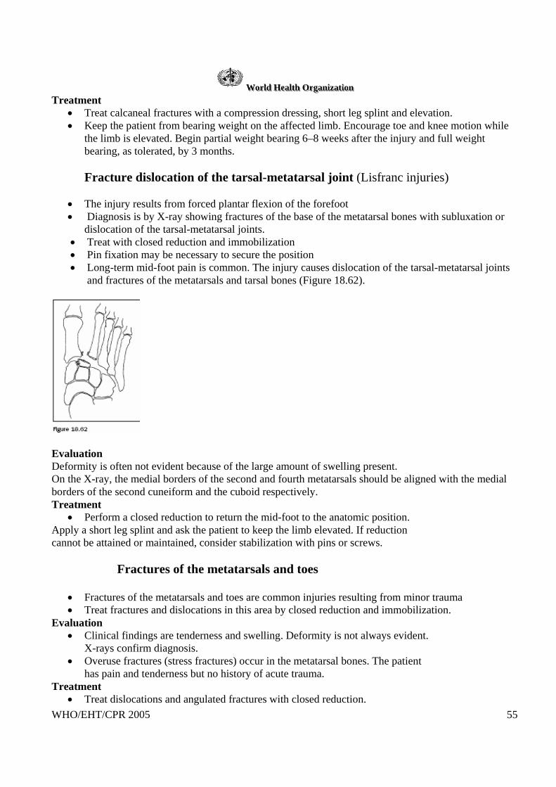
WWWooorrrlllddd HHHeeeaaalll ttthhh OOOrrrgggaaannniiizzzaaattt iiiooonnn
WHO/EHT/CPR 2005 55
Treatment • Treat calcaneal fractures with a compression dressing, short leg splint and elevation. • Keep the patient from bearing weight on the affected limb. Encourage toe and knee motion while
the limb is elevated. Begin partial weight bearing 6–8 weeks after the injury and full weight bearing, as tolerated, by 3 months.
Fracture dislocation of the tarsal-metatarsal joint (Lisfranc injuries)
• The injury results from forced plantar flexion of the forefoot • Diagnosis is by X-ray showing fractures of the base of the metatarsal bones with subluxation or
dislocation of the tarsal-metatarsal joints. • Treat with closed reduction and immobilization • Pin fixation may be necessary to secure the position • Long-term mid-foot pain is common. The injury causes dislocation of the tarsal-metatarsal joints
and fractures of the metatarsals and tarsal bones (Figure 18.62).
Evaluation Deformity is often not evident because of the large amount of swelling present. On the X-ray, the medial borders of the second and fourth metatarsals should be aligned with the medial borders of the second cuneiform and the cuboid respectively. Treatment
• Perform a closed reduction to return the mid-foot to the anatomic position. Apply a short leg splint and ask the patient to keep the limb elevated. If reduction cannot be attained or maintained, consider stabilization with pins or screws.
Fractures of the metatarsals and toes
• Fractures of the metatarsals and toes are common injuries resulting from minor trauma • Treat fractures and dislocations in this area by closed reduction and immobilization.
Evaluation • Clinical findings are tenderness and swelling. Deformity is not always evident.
X-rays confirm diagnosis. • Overuse fractures (stress fractures) occur in the metatarsal bones. The patient
has pain and tenderness but no history of acute trauma. Treatment
• Treat dislocations and angulated fractures with closed reduction.
WWWooorrrlllddd HHHeeeaaalll ttthhh OOOrrrgggaaannniiizzzaaattt iiiooonnn
WHO/EHT/CPR 2005 56
• Immobilize metatarsal fractures in a firm bottom shoe or a short leg cast. • Treat toe fractures and dislocations by taping the toe to a normal adjacent toe (Figure 18.63). • Treat stress fractures by limiting the amount of time the patient spends on his/her feet. If necessary,
use a firm shoe or cast until pain free.
SPINE INJURIESR IES
• Evaluate the spine based on a history of injury, physical examination, a complete neurological examination and X-rays
• Spinal column injuries are stable or unstable, based on bone and ligament damage • Neurological function may be normal, show incomplete injury or complete spinal cord disruption • Base your treatment on the extent of injury. • Fractures are stable if further deformity is unlikely. • They are unstable if a change in the fracture position is expected with mobilization. • Spinal cord injury is complete if there is no nerve function below the level of injury and if
improvement or return of function is unlikely. • Incomplete injuries have some nerve function below the injury level and may show improvement
with treatment.
Evaluation 1 Ask the patient if he/she has neck or back pain or has lost feeling in the arms or legs. Assume that an unconscious patient has a spine injury until he/she wakes up enough to answer these questions or until adequate Xrays show the spine to be normal. 2 Inspect the entire spine by log rolling the patient gently on to his/her side.Look for swelling and bruising. Palpate the spine for areas of tenderness and check for gaps or changes in the alignment of the spinous processes. 3 Perform a careful and complete neurological examination as outlined in Table 18.1 and record your findings. If there is a neurological deficit, determine the level from a motor and sensory examination.The injury is complete if there is no neurological function below that spinal cord level. 3. In incomplete injuries, the sacral nerve roots will often function. During the period of spinal shock (usually the first 48 hours after injury) there may be no spinal cord function. As shock wears off, some neurological recovery may occur with incomplete injuries. The ultimate prognosis cannot be accurately determined during the first several days. Neurological examination in the spinal injury patient
• Sensation - Test sensation to pinprick in the extremities and trunk - Test perianal sensation to evaluate the sacral roots
• Motor function

WWWooorrrlllddd HHHeeeaaalll ttthhh OOOrrrgggaaannniiizzzaaattt iiiooonnn
WHO/EHT/CPR 2005 57
- Evaluate motion and strength of the major muscle groups - Determine if rectal sphincter tone is normal
• Reflexes - Deep tendon reflexes in the upper and lower extremities - Bulbocavernosus reflex: squeeze the glans penis – the bulbocavernosus muscle contracts in a positive test - Anal wink: scratch the skin next to the anus – the anus contracts in a positive test - Babinsky reflex: stroke the bottom of the foot – the toes flex normally - and extend with an upper motor nerve injury
X-ray examination • X-ray the entire spine in patients not mentally responsive enough to cooperate
with the clinical examination. • In patients who are awake:
- X-ray the symptomatic areas of the cervical, thoracic and lumbar spine - X-ray the cervical spine in all patients involved in high-energy multiple trauma.
• Evaluate the cervical spine: - With lateral and AP films - Make sure to include all seven cervical vertebrae - Obtain an open mouth odontoid view. • Take AP and lateral views of the thoracic and lumbar spine.
The most common areas of injury are C2, C5–6 and T12–L1. In patients with pain but normal X-rays, take flexion and extension lateral X-rays of the cervical spine. X-ray interpretation
Injuries are unstable if there is:
- Injury to two or more columns - Rotational mal-alignment - Subluxation or dislocation of one vertebra on another - Fracture of the odontoid - Up to 50% vertebral body compression in the thoracolumbar spine - Increased width between the pedicles on the AP view.
Treatment Cervical spine • C1: The first cervical vertebra has ample room for the spinal cord and neurological injury is unusual:
- Initially, place patients in skull traction to maintain the fracture position and to control discomfort - When stable, change to a Minerva cast or a rigid cervical collar - Healing takes about 3 months
The bony spine is anatomically divided into three sections or columns (Figure18.64).
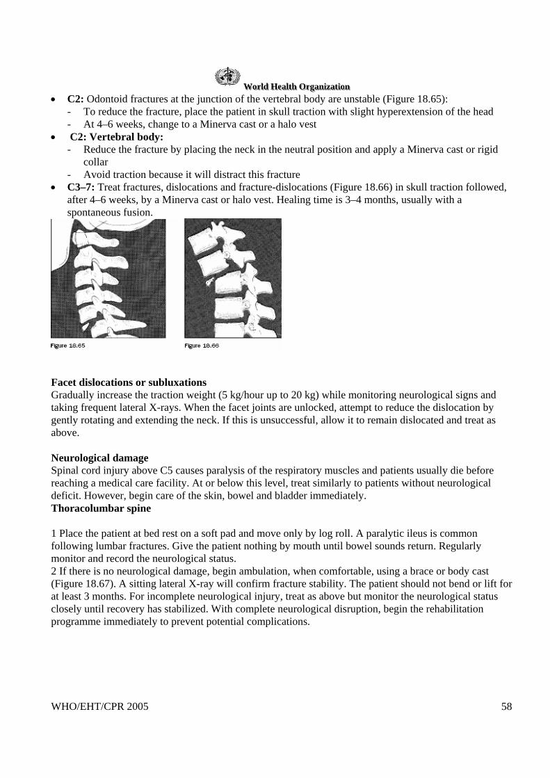
WWWooorrrlllddd HHHeeeaaalll ttthhh OOOrrrgggaaannniiizzzaaattt iiiooonnn
WHO/EHT/CPR 2005 58
• C2: Odontoid fractures at the junction of the vertebral body are unstable (Figure 18.65): - To reduce the fracture, place the patient in skull traction with slight hyperextension of the head - At 4–6 weeks, change to a Minerva cast or a halo vest
• C2: Vertebral body: - Reduce the fracture by placing the neck in the neutral position and apply a Minerva cast or rigid
collar - Avoid traction because it will distract this fracture
• C3–7: Treat fractures, dislocations and fracture-dislocations (Figure 18.66) in skull traction followed, after 4–6 weeks, by a Minerva cast or halo vest. Healing time is 3–4 months, usually with a spontaneous fusion.
Facet dislocations or subluxations Gradually increase the traction weight (5 kg/hour up to 20 kg) while monitoring neurological signs and taking frequent lateral X-rays. When the facet joints are unlocked, attempt to reduce the dislocation by gently rotating and extending the neck. If this is unsuccessful, allow it to remain dislocated and treat as above. Neurological damage Spinal cord injury above C5 causes paralysis of the respiratory muscles and patients usually die before reaching a medical care facility. At or below this level, treat similarly to patients without neurological deficit. However, begin care of the skin, bowel and bladder immediately. Thoracolumbar spine 1 Place the patient at bed rest on a soft pad and move only by log roll. A paralytic ileus is common following lumbar fractures. Give the patient nothing by mouth until bowel sounds return. Regularly monitor and record the neurological status. 2 If there is no neurological damage, begin ambulation, when comfortable, using a brace or body cast (Figure 18.67). A sitting lateral X-ray will confirm fracture stability. The patient should not bend or lift for at least 3 months. For incomplete neurological injury, treat as above but monitor the neurological status closely until recovery has stabilized. With complete neurological disruption, begin the rehabilitation programme immediately to prevent potential complications.
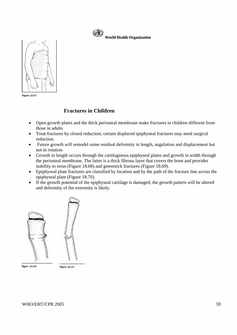
WWWooorrrlllddd HHHeeeaaalll ttthhh OOOrrrgggaaannniiizzzaaattt iiiooonnn
WHO/EHT/CPR 2005 59
Fractures in Children
• Open growth plates and the thick periosteal membrane make fractures in children different from those in adults
• Treat fractures by closed reduction; certain displaced epiphyseal fractures may need surgical reduction
• Future growth will remodel some residual deformity in length, angulation and displacement but not in rotation.
• Growth in length occurs through the cartilaginous epiphyseal plates and growth in width through the periosteal membrane. The latter is a thick fibrous layer that covers the bone and provides stability to torus (Figure 18.68) and greenstick fractures (Figure 18.69).
• Epiphyseal plate fractures are classified by location and by the path of the fracture line across the epiphyseal plate (Figure 18.70).
• If the growth potential of the epiphyseal cartilage is damaged, the growth pattern will be altered and deformity of the extremity is likely.
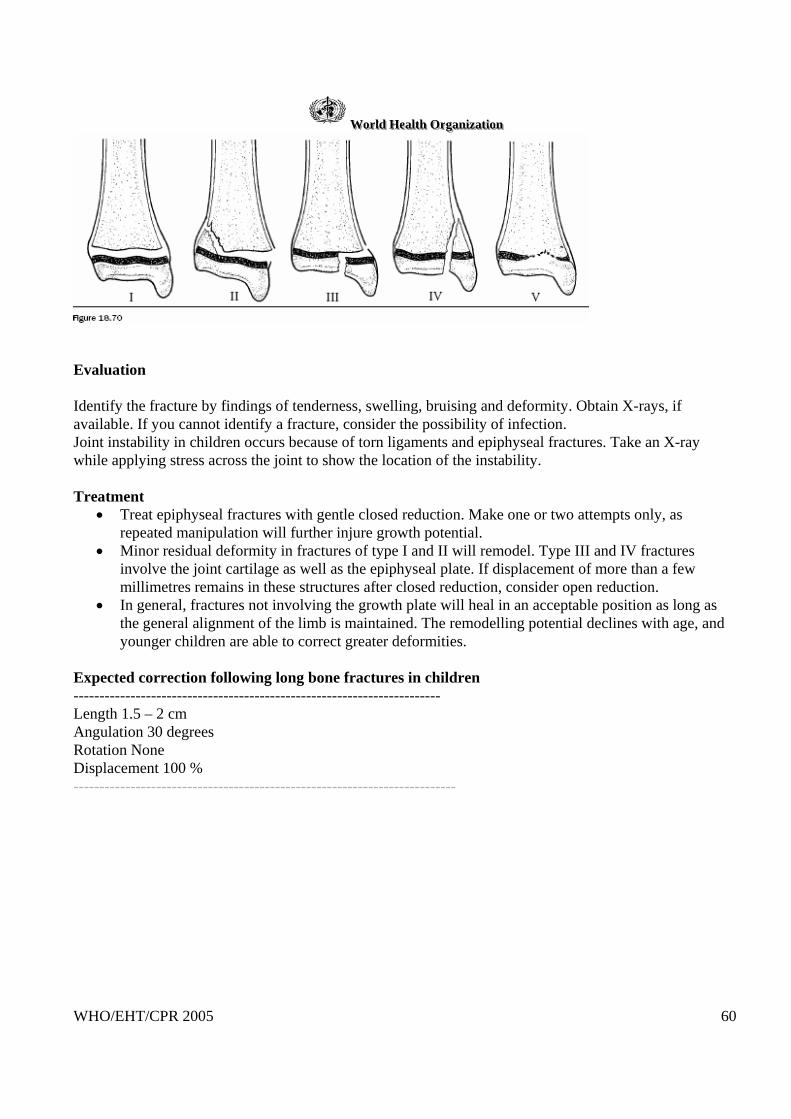
WWWooorrrlllddd HHHeeeaaalll ttthhh OOOrrrgggaaannniiizzzaaattt iiiooonnn
WHO/EHT/CPR 2005 60
Evaluation Identify the fracture by findings of tenderness, swelling, bruising and deformity. Obtain X-rays, if available. If you cannot identify a fracture, consider the possibility of infection. Joint instability in children occurs because of torn ligaments and epiphyseal fractures. Take an X-ray while applying stress across the joint to show the location of the instability. Treatment
• Treat epiphyseal fractures with gentle closed reduction. Make one or two attempts only, as repeated manipulation will further injure growth potential.
• Minor residual deformity in fractures of type I and II will remodel. Type III and IV fractures involve the joint cartilage as well as the epiphyseal plate. If displacement of more than a few millimetres remains in these structures after closed reduction, consider open reduction.
• In general, fractures not involving the growth plate will heal in an acceptable position as long as the general alignment of the limb is maintained. The remodelling potential declines with age, and younger children are able to correct greater deformities.
Expected correction following long bone fractures in children ----------------------------------------------------------------------- Length 1.5 – 2 cm Angulation 30 degrees Rotation None Displacement 100 % --------------------------------------------------------------------------
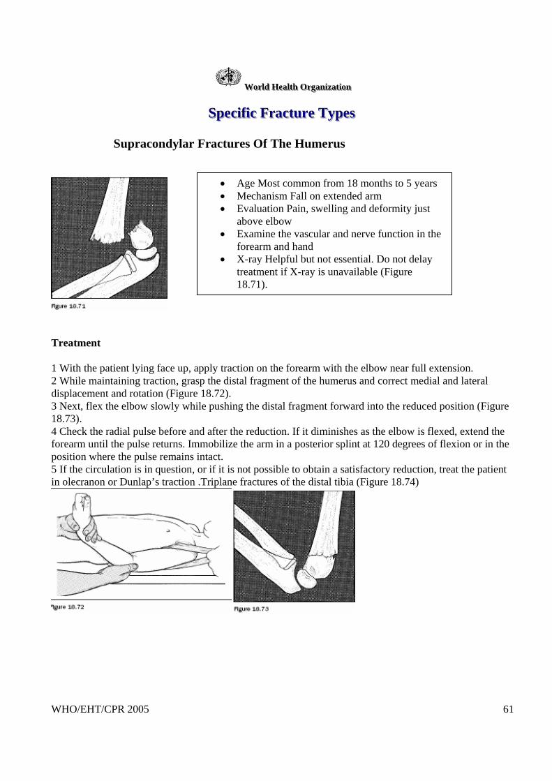
WWWooorrrlllddd HHHeeeaaalll ttthhh OOOrrrgggaaannniiizzzaaattt iiiooonnn
WHO/EHT/CPR 2005 61
SSSpppeeeccciiifffiiiccc FFFrrraaaccctttuuurrreee TTTyyypppeeesss
Supracondylar Fractures Of The Humerus
Treatment 1 With the patient lying face up, apply traction on the forearm with the elbow near full extension. 2 While maintaining traction, grasp the distal fragment of the humerus and correct medial and lateral displacement and rotation (Figure 18.72). 3 Next, flex the elbow slowly while pushing the distal fragment forward into the reduced position (Figure 18.73). 4 Check the radial pulse before and after the reduction. If it diminishes as the elbow is flexed, extend the forearm until the pulse returns. Immobilize the arm in a posterior splint at 120 degrees of flexion or in the position where the pulse remains intact. 5 If the circulation is in question, or if it is not possible to obtain a satisfactory reduction, treat the patient in olecranon or Dunlap’s traction .Triplane fractures of the distal tibia (Figure 18.74)
• Age Most common from 18 months to 5 years • Mechanism Fall on extended arm • Evaluation Pain, swelling and deformity just
above elbow • Examine the vascular and nerve function in the
forearm and hand • X-ray Helpful but not essential. Do not delay
treatment if X-ray is unavailable (Figure 18.71).

WWWooorrrlllddd HHHeeeaaalll ttthhh OOOrrrgggaaannniiizzzaaattt iiiooonnn
WHO/EHT/CPR 2005 62
• Age 12–15 years, at the time of closure of the distal tibial epiphysis • Mechanism Abduction, external rotation force to the ankle joint • Evaluation Painful, swollen ankle with or without other deformity • X-ray is needed to make the diagnosis but, if unavailable, begin treatment
Treatment 1 Apply longitudinal traction to the foot by holding the mid-foot and heel. 2 Invert the heel, bring the ankle from plantar flexion to neutral (90 degrees) and internally rotate the foot. 3 Maintain the reduction by grasping the great toe, allowing the foot to hang, while a three-way splint is applied. 4 Mould the plaster as it dries. 5 Obtain post-reduction X-rays when possible. Compartment Syndrome
• Compartment syndrome is caused by swelling within closed fascial spaces; as the intra-compartmental pressure increases, blood supply to the muscles is lost
• Treat with immediate surgical release of the skin and fascia over the involved compartment. • Increased compartment pressure is commonly caused by:
- Tight casts or dressings - External limb compression - Burn eschar - Fractures - Soft tissue crush injuries - Arterial injury.
• The most common areas involved are the anterior and deep posterior compartment of the leg and the volar forearm compartment. Other areas include the thigh, the dorsal forearm, the foot, the dorsal hand and, rarely, the buttocks.
• Diagnostic physical findings include: - Pain out of proportion to the injury - Tense muscle compartments to palpation - Pain with passive stretch of the involved muscle - Decreased sensation - Weakness of the involved muscle groups - Pallor and decreased capillary refill (late finding) - Elevated compartment pressure (if measurement is possible).

WWWooorrrlllddd HHHeeeaaalll ttthhh OOOrrrgggaaannniiizzzaaattt iiiooonnn
WHO/EHT/CPR 2005 63
Treatment • Split the cast and dressings, if present. • Do not elevate the limb, but observe carefully for improvement. • I f signs and symptoms persist, treat the acute compartment syndrome with immediate surgical
decompression. • Even short delays will increase the extent of irreversible muscle necrosis so, if you suspect a
compartment syndrome, proceed with the decompression immediately. FFFaaattt EEEmmmbbbooo lll iii sssmmm SSSyyynnndddrrrooommmeee
• Fat embolism syndrome follows major long bone trauma. • The etiology remains elusive, but seems to involve a showering of bone marrow contents into the
bloodstream. These lodge in the lungs, brain and other organs. • The syndrome becomes clinically evident on the second or third day post injury. • The lung involvement causes respiratory distress, which is fatal in a small percentage of patients. • Signs include:
- Confusion and anxiety - Increased pulse and respiratory rate - Petechiae located in the axilla, conjunctiva, palate and neck - A chest X-ray showing fluffy infiltrates - Low arterial oxygen content (if test available).
• Administer oxygen and support respiration i f breathing effort becomes great. • The syndrome is self-limited, lasting usually just a few days. Permanent effects are rare, but include
impaired vision, kidney abnormalities and mental changes.