benign tumors of melanocytes
TRANSCRIPT

Benign Tumors of Melanocytes
Part 1. Introduction
Part 2. Pattern recognition
Deba P Sarma, MD
Omaha

Part 1. Introduction

Three germ layers of human embryo are ectoderm, endoderm and mesoderm. From ectoderm comes the neuroectoderm and neural crest. During the early embryonic development, the neural crest cells migrate all the way to the epidermis and settle at the basal layer and hair follicles of the skin starting as melanoblasts and maturing into melanocytes. During this migration, the melanocytes also settle in the eye, ear, mucous membranes, leptomeninges, dermis, and other organs. Melanoblasts are immature cells and do not contain pigment. Melanocytes contain melanosomes containing pigment. Melanocytes are normally located at the basal layer of the epidermis. Melanocyte is a dendritic cell. Remember, along the basal epidermis, about every 10th cell is a melanocyte, others are keratinocytes. One melanocyte is connected to about 40 adjacent keratinocytes by the dendritic processes (together called epidermal melanocyte unit). Melanocyte makes the melanin pigment and through the dendrites, the pigment is transferred to the keratinocytes. As you know, keratinocytes mature and move upwards to the surface keratin layer carrying the pigments. This process continues all our life.

Let’s look at this normal skin. At first glance, all the cells in the epidermis look like keratinocyes. And you are right….most of the epidermal cells are keratinocytes.
Remember, along the basal epidermis, about every 10th cell is a melanocyte, others are keratinocytes. One melanocyte is connected to about 40 adjacent keratinocytes by the dendritic processes (together called epidermal melanocyte unit). Melanocyte makes the melanin pigment and through the dendrites, the pigment is transferred to the keratinocytes. As you know, keratinocytes mature and move upwards to the surface keratin layer carrying the pigments. This process continues all our life.
Look at the cells pointed by the arrows. You see a smaller cell than the adjacent keratinocytes, cell has a clear cytoplasm and a small dark nucleus. This is the cell we are looking for: “MELANOCYTE”.

Yes, it’s true that melanocyes are hard to find in H@E slide. But, if you know where they are located in the skin (basal layer of epidermis) and what they look like, you should be able to find
them . Immunostains, such as S-100 and MITF will readily stain the melanocyte nuclei.
MITF (Microphalmia-Associated Transcription Factor) stains the nuclei of the normal basal melanocytes of the skin.

Melanocytic lesions • Benign- 95%
• Malignant- 4%
• Undetermined- 1%
As you notice, most of the melanocytic lesions that you will diagnose will be benign.
As a pathologist, we always pay a lot of attention not to miss a malignant melanocytic tumor (MELANOMA) because
it is the most feared skin malignancy.
To make the matter worse, textbooks are notorious with extensive, complicated and confusing description of the
melanocytic lesions of the skin. My advice: Use common sense, stick to the basic anatomy and think logically.
Knowing that less than 5% of any pigmented lesion that you will diagnose will be malignant,
CONSIDER EVERY BIOPSY OF A PIGMENTED LESION IS BENIGN UNTIL YOU CAN PROVE OTHERWISE ! INSTANTLY, YOU
ARE IN RIGHT TRACK IN 95% OF SUCH CASES !!
Dr. Sarma’s simple classification of melanocytic tumors Benign: Nevus (Junctional, Compound, Dermal, Congenital, Blue, Spitz, Dysplastic) Lentigo (Simplex, Solar) Potentially malignant: Atypical melanocytic hyperplasia Malignant: Melanoma in-situ Invasive melanoma

Don’t get confused with these few terms: Lentigo, Freckle, Mole, Nevus
• Lentigo: From Latin word for lentil. A dark spot on skin that looks like a lentil bean and does not fade in the winter.
• Freckle: Small brownish spot turning darker or increasing in number upon exposure to the sun and fading in winter. (Freckle=Speckle)
• Mole : Spot, common name for nevus.
MOLE = NEVUS
• Nevus: Latin word meaning birthmark.

Melanocytic nevus is a benign neoplastic proliferation of melanocytes (also called nevocytes or nevus cells) Nevus cell or nevocyte is nothing but a melanocyte except that it does not have any dendrites (except for blue nevus cells that have dendrites). Presence of dendrites enables the melanocytes to move in the tissue whereas mobility of nevus cells is limited.
Mole : Spot, common name for nevus (Pleural: Nevi) MOLE = NEVUS
Melanocytic nevus may be congenital (present at birth or within first few months) or acquired. Most of the melanocytic nevi are acquired (not present at birth but develop during life). Beginning after 2-3 years of age incidence of melanocytic nevi increases up to age 30, reaching a peak incidence in 40-50 years of life, and then gradually involuting and disappearing in each successive decades with a very low incidence in elderly people. Nevi are more common in fair-skinned people. Sun exposure may induce increased number of nevi.
No acquired nevi before 2 yrs Very few nevi in elderly Nevi in 30-50 yr olds
Melanocytes at the basal epidermis are single cells separared by basal keratinocytes. Nevus is formed by proliferation of the melanocytes forming a ‘nevocellular nest’ at the dermoepidermal junction, especially at the tips pf the rete ridges. This is called a ‘junctional nevus’. With further proliferation, the nevocellular nests extend into the dermis. Now, you have nevus nests at the junction as well as in the dermis and we call this nevus a ‘compound nevus’. Over time, the junctional nests disappear leaving only nevus nests in the dermis, and we call this nevus a ‘dermal nevus’. Nevus cells are small round or polygonal cells with small dark nucllei that are smaller than the keratinocyte nuclei.
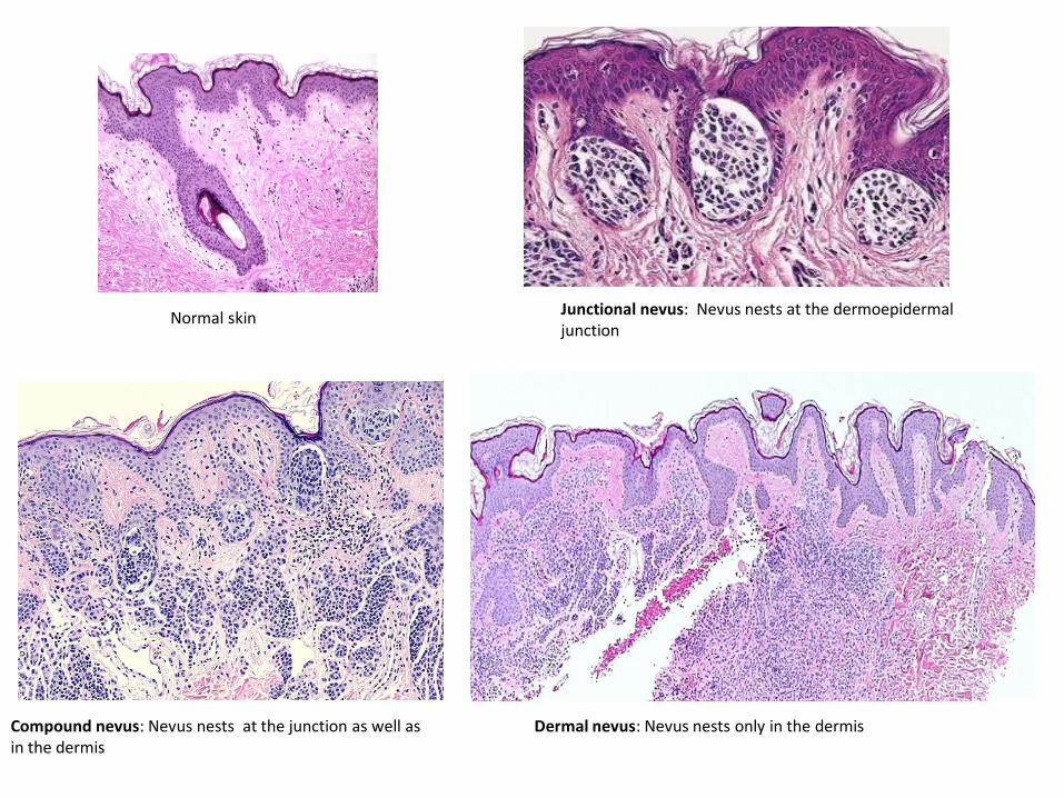
Normal skin Junctional nevus: Nevus nests at the dermoepidermal junction
Compound nevus: Nevus nests at the junction as well as in the dermis
Dermal nevus: Nevus nests only in the dermis

Congenital melanocytic nevus
Congenital melanocytic nevi are present at birth, or arise soon after birth. Nevi that look like congenital nevi, but appear later in life, are called ‘congenital type’ nevi. Congenital nevi may be small (1-5cm), medium (1.5-20cm) or rarely, giant (> 20 cm, bathing trunk variety). Giant nevi have a significantly increased risk of developing melanoma in the lesion (5-10%)
Histologically, congenital nevi may be junctional, compound or dermal. Extension of nevus cells around nerves, vessels and adnexae is typical.

Blue nevus
Flat lesion
No junctional nest
Dermal pigmented spindled melanocytes
Blue nevi are most common in Asian populations, where the prevalence is estimated to be 3-5% in adults. They are found in 1-2% of white adults and are rarely found in blacks. In common blue nevus, dermis shows a vaguely nodular collection of spindled melanocytes and deeply pigmented dendritic melanocytes within thickened collagen bundles. Scattered melanophages are seen. No mitoses are present.

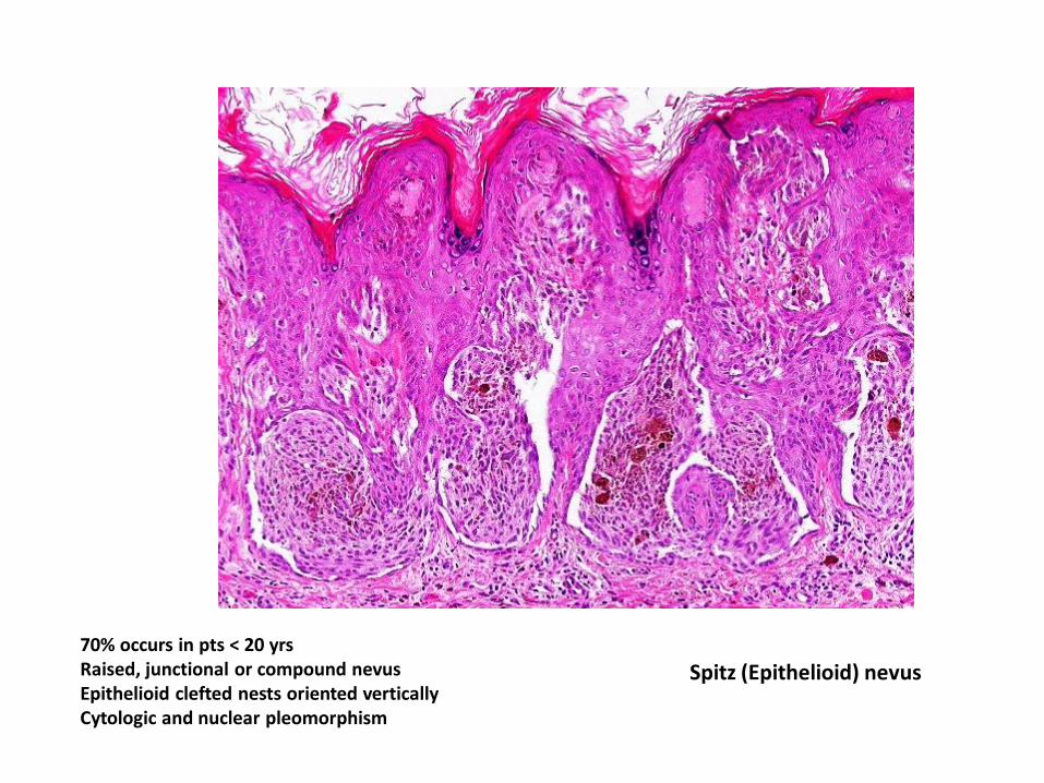
Nevus: Epithelioid (Spitz nevus)
70% occurs in pts < 20 yrs
Raised, junctional or compound nevus
Epithelioid clefted nests oriented vertically
Cytologic and nuclear pleomorphism
Spitz nevi constitute less than 1% of all childhood melanocytic nevi. Single, dome-shaped, red or pigmented papules or nodules typically appear on the face or legs. Misdiagnosis of Spitz nevi as melanomas and misdiagnosis of melanomas as Spitz nevi is a possibility. Histopathologic differentiation from melanomas is equivocal in up to 8% of cases. Criteria in favor of a Spitz nevus in any given case include a young patient, a well-demarcated and symmetrical lesion, maturation of melanocytes at its base, and the presence of epithelial hyperplasia, but no criterion is absolutely reliable.
Most Spitz nevi are compound with large and/or spindle-shaped melanocytes, usually in nests. The nests are composed of an admixture of spindle cells and/or epithelioid cells. Striking symmetry, sharp lateral demarcation, absent (or rare) mitoses, absence of atypical mitoses, presence of eosinophilic and periodic acid-Schiff (PAS)–positive globules (Kamino bodies) in the epidermis are typical for Spitz Maturation of nevus cells in the deeper dermis is an important diagnostic finding.

Atypical nevus (dysplastic nevus) An unusual melanocytic nevus that is often large (> 5 mm), flat and asymmetrical instead of round or oval in shape with indistinct edge. Dysplastic nevus may occur anywhere on the body (scalp, breast, below waist), but it is usually seen in areas exposed to the sun, such as on the back. Most dysplastic nevi are sporadic and do not turn into melanoma . Most remain stable over time. Chance of melanoma is about ten times higher for someone with > 5 dysplastic nevi than for someone who has none, and the more dysplastic nevi a person has, the greater the chance of developing melanoma. Familial dysplastic nevi may be inherited as an autosomal dominant trait. These patients have a very high risk of developing melanoma. People at the highest risk of dysplastic nevi are of northern European background (Celtic) with light-colored hair and freckles. Dysplastic nevi are rare in black, Asian, or Middle Eastern populations.
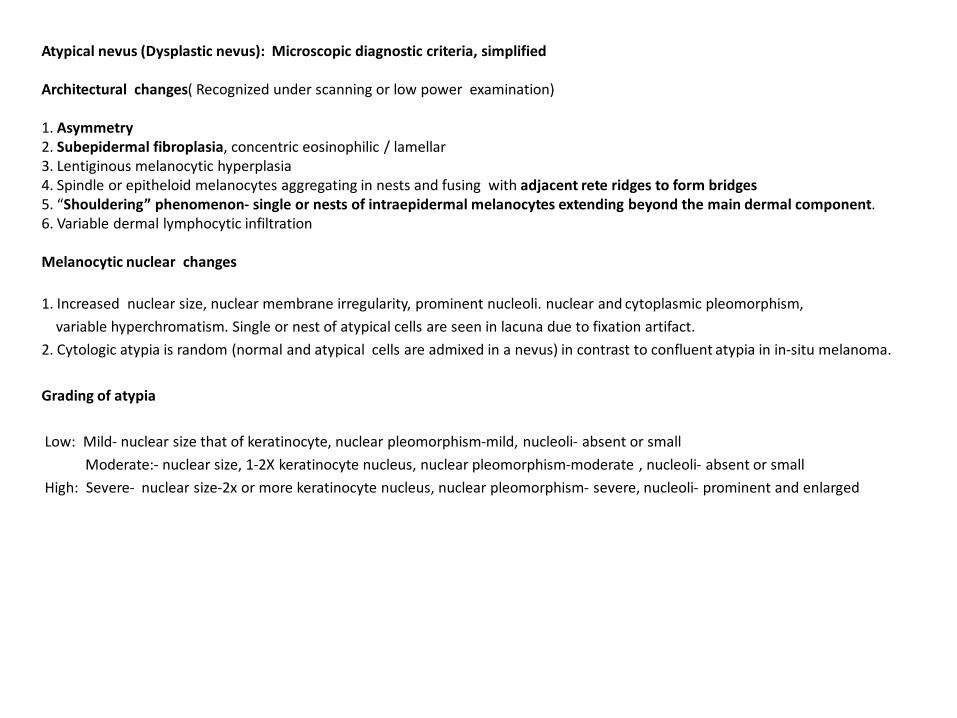
Atypical nevus (Dysplastic nevus): Microscopic diagnostic criteria, simplified Architectural changes( Recognized under scanning or low power examination) 1. Asymmetry 2. Subepidermal fibroplasia, concentric eosinophilic / lamellar 3. Lentiginous melanocytic hyperplasia 4. Spindle or epitheloid melanocytes aggregating in nests and fusing with adjacent rete ridges to form bridges 5. “Shouldering” phenomenon- single or nests of intraepidermal melanocytes extending beyond the main dermal component. 6. Variable dermal lymphocytic infiltration Melanocytic nuclear changes
1. Increased nuclear size, nuclear membrane irregularity, prominent nucleoli. nuclear and cytoplasmic pleomorphism,
variable hyperchromatism. Single or nest of atypical cells are seen in lacuna due to fixation artifact.
2. Cytologic atypia is random (normal and atypical cells are admixed in a nevus) in contrast to confluent atypia in in-situ melanoma.
Grading of atypia
Low: Mild- nuclear size that of keratinocyte, nuclear pleomorphism-mild, nucleoli- absent or small
Moderate:- nuclear size, 1-2X keratinocyte nucleus, nuclear pleomorphism-moderate , nucleoli- absent or small
High: Severe- nuclear size-2x or more keratinocyte nucleus, nuclear pleomorphism- severe, nucleoli- prominent and enlarged

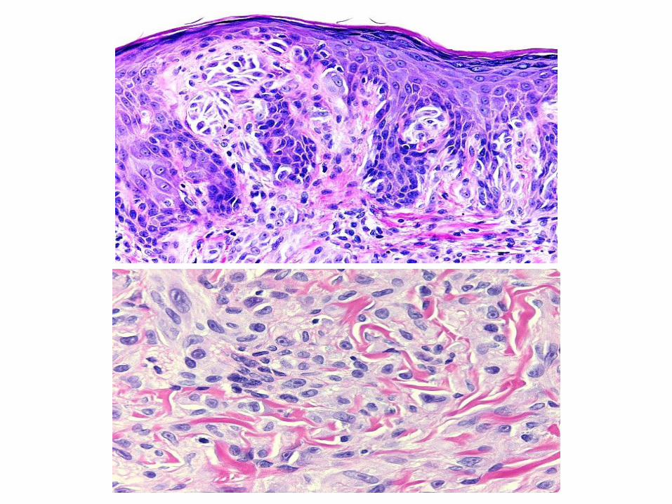
Dysplastic nevus
Dysplastic nevus is a junctional or compound nevus with architectural and cytologic atypia.

Subepidermal fibroplasia
Bridging of the rete ridges by transverse proliferation of the nevocytes
Shouldering by intraepidermal nevus cells lateral to the dermal nevus nests
Dysplastic nevus

Dysplastic nevus
Cytologic and nuclear pleomorphism

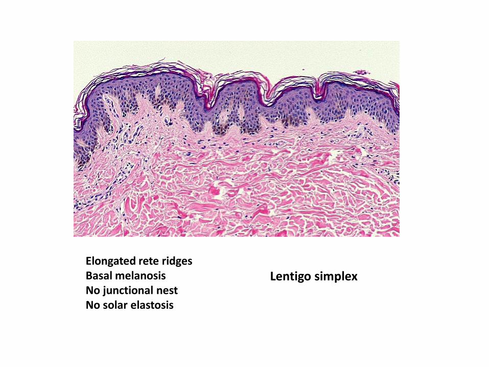
Lentigo simplex
Elongated rete ridges
Basal melanosis
No junctional nest
No solar elastosis
A lentigo is a small, sharply circumscribed, pigmented macule surrounded by normal-appearing skin. Histology: Hyperplasia of the epidermis. Increased pigmentation of the basal layer. Basal melanocytes may be increased in number, but they do not form nests.

Solar lentigo
Elongated rete ridges
Basal melanosis
No junctional nest
Dermal solar elastosis
Flat, dark lesions on the sun-exposed skin in older population, mosly on the hands, face, shoulders, arms and forehead, and the scalp if bald. There is melanocytic proliferation at the basal layer with increased melanin production, but no nesting. Dermis shows actinic change.

Part 2. Pattern recognition
Lentigo simplex Elongated rete ridges Basal melanosis No junctional nest No solar elastosis

Solar lentigo Elongated rete ridges Basal melanosis No junctional nest Dermal solar elastosis
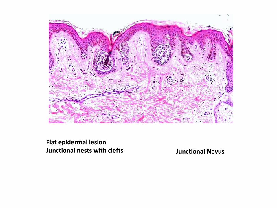
Junctional Nevus
Flat epidermal lesion Junctional nests with clefts
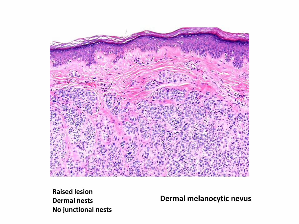
Raised lesion Dermal nests No junctional nests
Dermal melanocytic nevus

Compound melanocytic nevus Raised or flat lesion Junctional clefted nests and dermal nests
70% occurs in pts < 20 yrs Raised, junctional or compound nevus Epithelioid clefted nests oriented vertically Cytologic and nuclear pleomorphism
Spitz (Epithelioid) nevus

Dysplastic nevus All ages Compound nevus (or junctional nevus) Junctional clefted nests with transverse growth pattern (bridging) Cytologic and nuclear pleomorphism

Flat lesion No junctional nest Dermal pigmented spindled melanocytes
Blue nevus

Lentiginous nevus Flat lesion Common melanocytic nevus + lentigo simplex
Now , let me show you examples of several variation of melanocytic nevi

Markedly pigmented compound melanocytic nevus
Pigmented melanocytic nevus
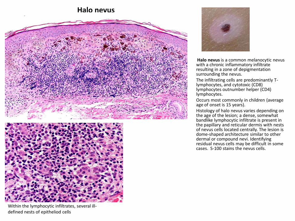
Halo nevus
• Halo nevus is a common melanocytic nevus with a chronic inflammatory infiltrate resulting in a zone of depigmentation surrounding the nevus.
• The infiltrating cells are predominantly T-lymphocytes, and cytotoxic (CD8) lymphocytes outnumber helper (CD4) lymphocytes.
• Occurs most commonly in children (average age of onset is 15 years).
• Histology of halo nevus varies depending on the age of the lesion; a dense, somewhat bandlike lymphocytic infiltrate is present in the papillary and reticular dermis with nests of nevus cells located centrally. The lesion is dome-shaped architecture similar to other dermal or compound nevi. Identifying residual nevus cells may be difficult in some cases. S-100 stains the nevus cells.
Within the lymphocytic infiltrates, several ill-defined nests of epitheliod cells

Neurotized dermal melanocytic nevus (Neuronevus) Old dermal nevi may show degenerative change and the nevus nests may resemble nerves and Meissner corpuscle-like structures as seen in this case. Presence of nevocellular nests is helpful in diagnosis. In the first glance, you may think that it is a neurifibroma.

Let me show you a few more cases of Spitz nevus
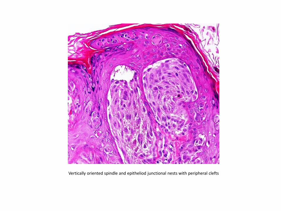
Case 1. M 11 yrs, right upper arm, 8 mm lesion
Symmetrical, raised compound nevus

Vertically oriented spindle and epitheliod junctional nests with peripheral clefts
Kamino body
Kamino bodies are intraepidermal eosinophilic hyaline globules and are seen in 80% of cases of Spitz nevi . Kamino bodies are composed of laminin, type IV collagen and fibronectin.
Vertically oriented spindle and epitheliod junctional nests with peripheral clefts
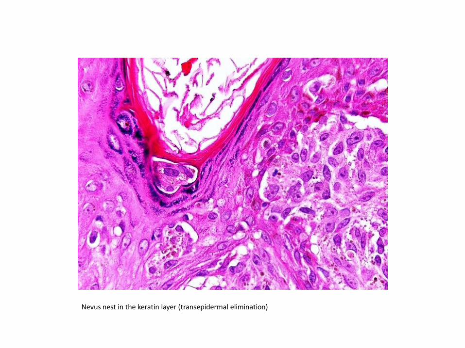
Nevus nest in the keratin layer (transepidermal elimination)
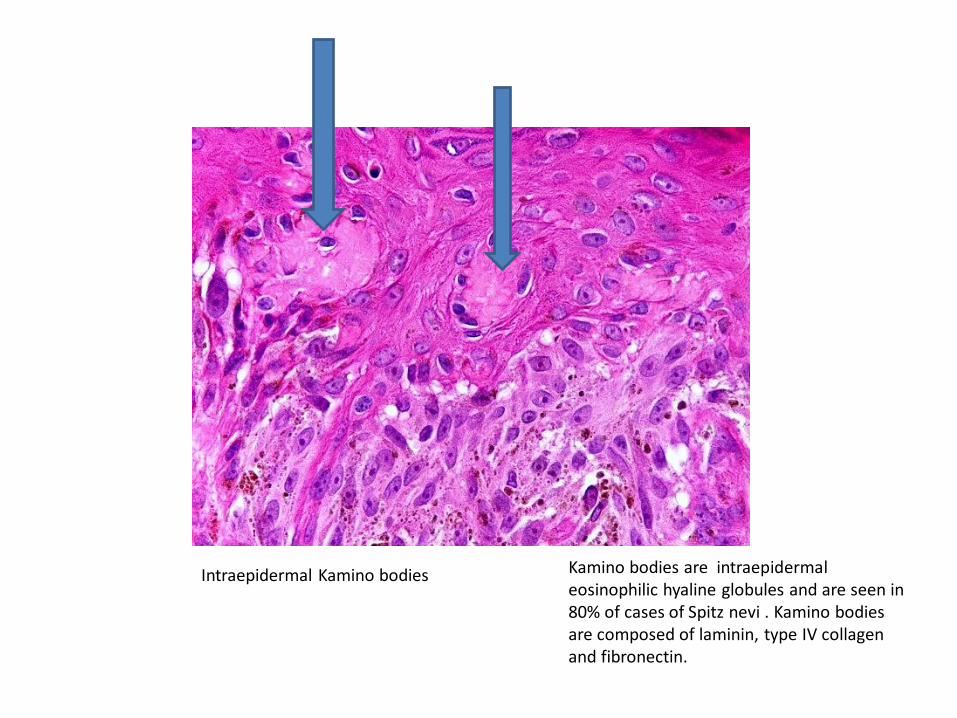
Intraepidermal Kamino bodies
Kamino bodies are intraepidermal eosinophilic hyaline globules and are seen in 80% of cases of Spitz nevi . Kamino bodies are composed of laminin, type IV collagen and fibronectin.
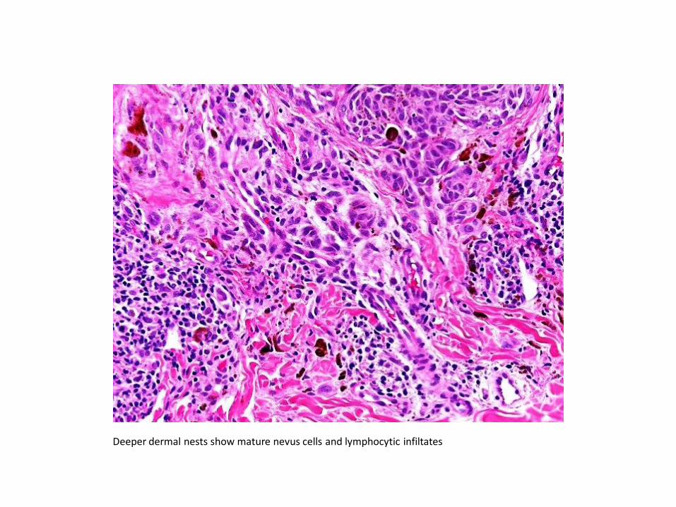
Deeper dermal nests show mature nevus cells and lymphocytic infiltates
Comment
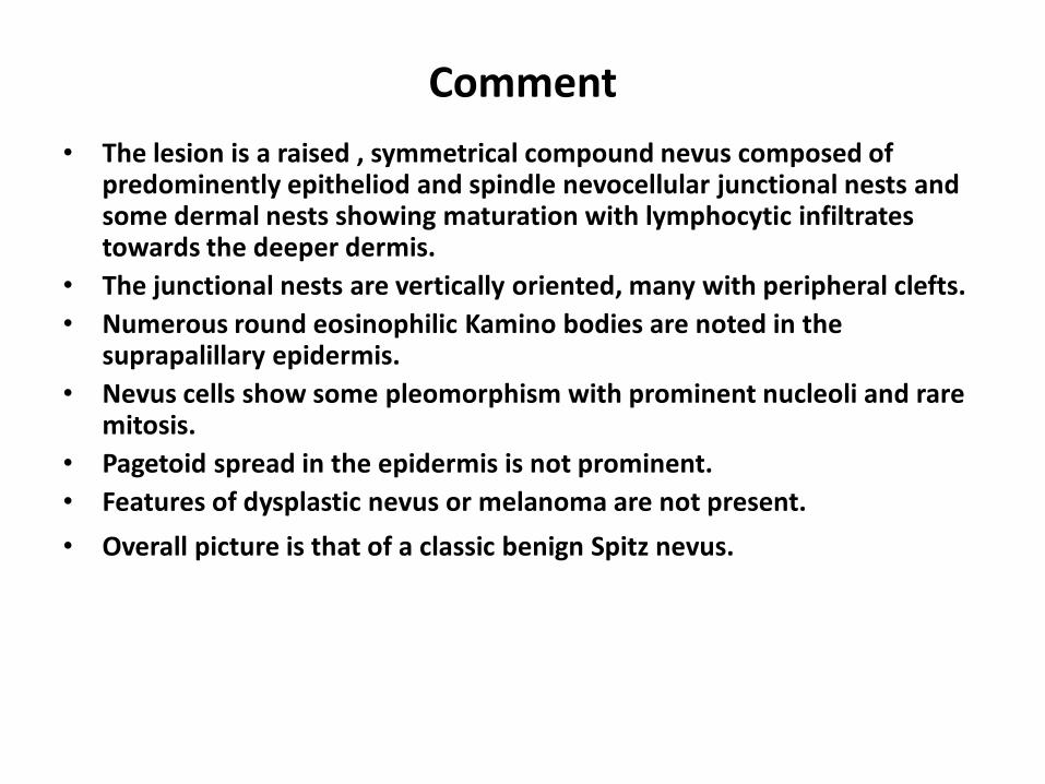
• The lesion is a raised , symmetrical compound nevus composed of predominently epitheliod and spindle nevocellular junctional nests and some dermal nests showing maturation with lymphocytic infiltrates towards the deeper dermis.
• The junctional nests are vertically oriented, many with peripheral clefts.
• Numerous round eosinophilic Kamino bodies are noted in the suprapalillary epidermis.
• Nevus cells show some pleomorphism with prominent nucleoli and rare mitosis.
• Pagetoid spread in the epidermis is not prominent.
• Features of dysplastic nevus or melanoma are not present.
• Overall picture is that of a classic benign Spitz nevus.

Case 2. M 9 yrs, right cheek


Case 3. M 2 yrs, right cheek

You will need to see quite a few cases of Spitz nevus and dysplastic nevus before you feel
confident about your diagnosis. More cases you see, better you will get. Books are helpful,
but real slides from real patients are your best teacher.
Experience cannot be taught, it has to be acquired over time !
Deba P Sarma, MD
Omaha