ben yates,*t msc, fcpod(s), and shaun white,+ bapplsci ... · a bone stress reaction that becomes...
TRANSCRIPT

Ben Yates,*t MSc, FCPod(S), and Shaun White,+ BApplSciFrom the tPodiatry Department, University College Northampton, Northampton, UnitedKingdom, and the tDepartment of Podiatr.x La Trobe Universit.x Melbourne, Australia
Purpose: To identify the incidence of medial tibial stress syndrome (MTSS) in a group of naval recruits undergoing a 10-weekbasic training period and to determine potential risk factors.
Method: One hundred and twenty-four recruits (84 men and 40 women) were followed prospectively during basic training.Anthropometric and lower limb biomechanical data were recorded at the start of the program along with injury history and pre-vious sporting activity for the 3 months prior to enlisting. Recruits were monitored during training for development of medial tib-ial strees syndrome and were asked to complete an exit interview at the end of the program.
Results: Forty recruits (22 men and 18 women) developed medial tibial stress syndrome, giving an incidence of 35%. A signif-icant relationship existed between gender and medial tibial stress syndrome (P = .012), with femare recruits more likely to devel-op medial tibial stress syndrome than male recruits (53% vs 28%). A risk estimate revealed a relative risk of 2.03. The biome-chanical results indicated a more pronated foot type (P = .002) in the medial tibial stress syndrome group when compared to thecontrol group. A risk estimate established that recruits with a more pronated foot type had a relative risk of 1.70.
Conclusion: Identifying a pronated foot type prior to training may help reduce the incidence of medial tibial stress syndrome byearly intervention to control abnormal pronation. Findings of a higher incidence of medial tibial stress syndrome among femalerecruits require further investigation.
Keywords: shin splints; foot pronation; injury rates
Medial tibial stress syndrome (MTSS) is one of manyoveruse lower leg injuries found under the umbrella termof exercise-induced leg pain or shin splints. Stress frac-tures, chronic compartment syndrome, and MTSS are the3 most common forms of exercise-induced leg pain, withMTSS having the highest prevalence. MTSS was firstdefined "as a symptom complex seen in athletes who com-plain of exercise induced pain along the posteriormedialborder of the tibia."36(p209) It is a very common injury expe-rienced by runners and military personnel. It accounts forbetween 13.2% and 17.3% of all running injuries14.18 andup to 22% of injuries seen in aerobic dancers. 51 Among
studies of military personnel, Almeida et al2 followed 176
male and 241 female naval recruits through an II-week(men) and 12-week (women) training period, finding anoverall incidence of 6.417c for MTSS. In the only prospectivecivilian study, an incidence of 13% was identified among125 high school runners. I I
Although various studies have attempted to find theexact pathophysiology for this common condition. it stillremains unresolved. Until recently, inflammation of theperiosteum due to excessive traction was considered themost likely etiology of MTSS.I,23,36,40.4S Detmerl6 opposedthis theory, proposing periostalgia as the likely cause ofMTSS after he found no evidence of inflammatory changesand consistently found adipose tissue interposed betweenthe periosteum and the bone surface. Johnell et al24 firstproposed the bone stress reaction theory after taking biop-sies and finding osseous metabolic changes in 37 limbs\vith MTSS and no evidence of inflammatory changes.
Recent studies have supported this view that ),ITSS isnot an inflammatory process of the periosteum but insteada bone stress reaction that becomes painful.4,s.20 'Vhen aperson begins an exercise program, the bone undergoesmetabolic changes. These changes in the tibia are charac-terized by initial bone porosity due to osteoclastic chan-
'Address correspondence to Ben Yates, Podiatry Department, UniversityCollege Northampton, Boughton Green Road, Northampton, NN27AL, UK.
No author or related institution has received financial benefit fromresearch in this study.
iThe American Journal of Sports Medicine. Vol. 32, No.3001: 10.1177/0095399703258776@ 2004 The American Orthopaedic Society for Sports Medicine
772
f
,]1
fVol. 32, No.3, 2004 Medial Tibial Stress Syndrome Among Naval Recruits 773
ous lower-limb injury risk factors for the development ofMTSS.
METHOD
The study was granted ethics approval from theAustralian Defence Medical Ethics Committee and the LaTrobe University Faculty of Health Sciences HumanEthics Committee. All subjects were recruited from HMASCerberus in Western Port, Victoria, Australia. All recruitsat the commencement of training were offered the oppor-tunity to voluntarily participate in the study. Subjectswere assured that all information obtained would be con-fidential; they would remain anonymous, and a failure toparticipate would have no detrimental effect on theirnaval careers or the management of their MTSS if such acondition was detected.
Prospective data were collected at the start of basictraining and consisted of the measuring of biomechanicalparameters and the administering of a questionnaire. Theexercises undertaken by the recruits during basic trainingtook up an average of 16.2 hours per week; they involved amixture of marching, doubling (running in step), circuittraining, and cross-country running. During the trainingperiod, recruits were monitored for the development ofMTSS. Following the completion of basic training, allrecruits underwent a confidential exit interview to deter-mine the incidence of lower limb injuries incurred duringthe training.
For the purpose of this study, MTSS was defined as"pain along the posteriomedial border of the tibia thatoccurs due to exercise excluding pain from ischaemic ori-gin or signs of stress fracture." The diagnosis of MTSS wasbased on the following criteria.
neling on the compressed concave posteriormedial border.This is followed by the laying down of new bone to resistthese compressive forces and strengthen the bone.3.10 Theresult is that the tibia becomes stronger than the pre-exercise state on the posteriormedial border. However, incases of long-standing MTSS, it has been shown that theaffected part of the tibia is 15% more porous than in con-trol subjects and 23% less than in athletic control sub-. t 30Jec s.
A thorough clinical history and physical examinationcan accurately diagnose MTSS. MTSS commonly presentsas diffuse, palpable pain, localized to the posteriomedial.b. 1 b d 48.1620374055 It h 1 tht1 1a or er. can occur anyw ere a ong e
posteriomedial border but most commonly affects the mid-dle to distal thirds.5.10.11.16.32.36.53.55 The pain is usuallydescribed as a dull ache following exercise, which may lastfor several hours or days. In severe cases, pain may persistduring normal activities of daily living.
Additional investigations such as plain radiographs,bone scans, and magnetic resonance imaging can be usefulto clinically diagnose MTSS as well as rule out other formsof exercise-induced leg pain such as stress fracture. However,all 3 types of investigation have been associated with bothfalse-positive and false-negative results.3.6-8.39.47.57 It hastherefore been suggested that if the clinical picture indi-cates MTSS and there is no diagnostic dilemma, furtherinvestigations are unwarranted.52
Various authors have proposed a wide variety of etiolog-ical factors for MTSS, including training on hard surfacesor uneven terrain, improper training techniques, increas-ing training intensity too quickly, changes in footwear,muscle imbalances or inflexibility, and biomechanicalabnormalities.29.31.35.49.50.55 Although not linked directly toMTSS, a high body mass index (BMI) and a previous his-tory of injury have been linked to the development oflower-limb overuse injuries.25.26.34
Abnormal subtalar joint pronation has been associatedwith MTSS in a number of static and dynamic stud-.31 50 53 Th t d. 11 t '.1es. ..ese s u 1es were a re rospecbve In nature,consisted of small sample groups, and often failed to cor-rectly define the condition as MTSS, using instead thebroader definition of shin splints. The only prospectivestudy that determined foot pronation as a risk factor forMTSS is that of Bennett et al,ll \vho followed high schoolcross-country runners through a training period. Theymeasured the degree of foot pronation by recording theamount the navicular bone lowered between two standingpositions, the neutral calcaneal stance position and therelaxed calcaneal stance position. This distance is termedthe navicular drop, and the procedure measures theamount the medial longitudinal arch (MLA) lowers in thesagittal plane. However, Bennett et al performed this testonly after the runner developed MTSS, so in truth it was aretrospective measurement and may have been an effect ofdeveloping MTSS rather than the cause of the condition.
The purpose of the present study \vas therefore toprospectively measure and monitor naval recruits duringbasic training to determine the incidence and potentialanthropometric, foot posture, exercise history, and previ-
.Pain History. The pain was induced by exercise andlasted for a few hours or days after exercise. Painwas located on the posteriomedial border of thetibia. There was no history of paraesthesia or othersymptoms indicative of other causes of exercise-induced leg pain.
.Location. The recruits identified pain along the pos-teriomedial border of the tibia. The site had to bespread over a minimum of 5 cm. Focal areas of only2 to 3 cm are typical of stress fracture.4,s
.Palpation. Palpation of the posteriomedial border ofthe tibia produced discomfort that was diffuse innature and confined to the posteriomedial border ofthe tibia. In the areas of discomfort, the bone sur-face may feel uneven.
Those subjects who developed MTSS during the studywere placed in the symptomatic MTSS group after meet-ing the following strict criteria:
.positive MTSS diagnosis,
.absence of symptoms indicative of other causes ofexercise-induced leg pain, and
.absence of MTSS at the commencement of thestudy.

Yates and White774 The American Journal of Sports Medicine '..f
:~,~
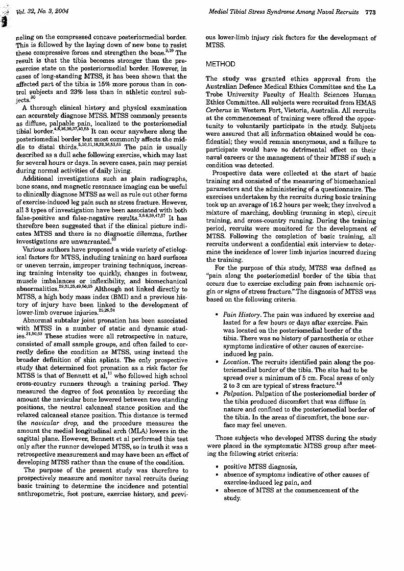
Figure 1. Location of symptoms of exercise-induced legpain. 1, proximal posteromedial tibial border; 2, middle pos-teromedial tibial border; 3, distal posteromedial tibial border;4, anterior compartment; 5, anterior tibial border.
Any subjects with symptoms suggestive of other causes ofexercise-induced leg pain were excluded from the study.Imaging investigations were not undertaken to confirm orexclude MTSS for the reasons previously stated. Any sub-ject with a history of lower limb surgery or fracture likelyto alter their normal lower limb alignment was excludedfrom the study.
Recruits were questioned about anthropometric datasuch as age, sex, height, and weight. A previous history oflower limb injuries, including exercise-induced leg pain,was recorded. A photograph dividing the anterior part ofthe lower leg into 5 distinct areas helped identify the siteof any previous exercise-induced leg pain (Figure 1). Forall previous or current injuries, the duration of pain,treatments implemented, and activity inciting the painwere recorded. All recruits were then questioned abouttheir training schedule or sporting activities in the previ-ous 3 months. The type of exercise, weekly frequency, dura-tion, and type of exercising surface were recorded.Following the questionnaire, all subjects underwent a bio-mechanical assessment including measurements of anklejoint dorsiflexion and foot posture. The person undertakingthese measurements was blinded to the results of thequestionnaire.
The foot posture index (FPU is an observational testthat determines whether a foot is in a pronated, supinat-ed, or neutral position based on 8 parameters.41-43 Theseparameters can also distinguish between frontal, sagittal,or transverse plane positional differences. The FPI hasbeen shown to have good intratester and intertester relia-bility, ranging from 0.73 to 0.87 and 0.66 to 0.78, respec-tively.41.43 This reliability coefficient is much higher thanmost clinical foot biomechanical measurements.
The FPI is based on the observation of 7 visual parame-ters, and the 8th parameter is determined by palpating theposition of the head of the talus. All measurements arerecorded with the patients standing at their natural angleof base of gait. The participants were required to stand ona platform and were instructed to walk up and down onthe spot. After 10 seconds, they were asked to stop, placetheir arms by their side, face for\vard, and distribute theirweight evenly through both legs and feet.
Different observations are seen for both foot pronationand supination. The 8 parameters required to assess over-all foot posture for each foot were the following:
1. Prominence in the region of the talonavicularjoint. Bulging in this area is associated with apronated foot. Indentation is observed in thesupinated foot.
2. Calcaneal frontal plane position. It is everted inthe pronated foot and inverted in the supinatedfoot.
3. Helbing's sign. As the calcaneus everts in thepronated foot, the tendo achilles can "bow," withthe inferior part directed to the lateral side. In asupinated foot, the tendo achilles can bow, with theinferior part directed to the medial side.
4. Supra- and infra-lateral malleolar curvature. In apronated foot, the curve below the malleolus is
more acute than the curve above. In a supinatedfoot, the curve above the malleolus is more acutethan the curve below.
5. Congruence of the lateral border of the foot. In apronated foot, the lateral border develops a con-cave profile as the forefoot abducts on the rear foot.In a supinated foot, the lateral border develops a con-vex profile as the forefoot adducts on the rear foot.
6. Talar head palpation. In a pronated foot, the headof the talus is more prominent on the medial side.and in a supinated foot, it is more prominent onthe lateral side.
7. Congruence of the MLA. The MLA is lo\vered in apronated foot, particularly the proximal part of thearch. In a supinated foot, the proximal part of thearch is high.
8. Abduction/adduction of the forefoot on the rearfoot. In a pronated foot, the forefoot abducts.resulting in more of the toes being visible on thelateral side (the too-many-toes sign). In a supinat-ed foot, the forefoot adducts relative to the rearfoot, resulting in the toes being more visible on themedial side.

8
"; ..
.., ."",.Vol. 32, No.3, 2004 Medial Tibial Stress Syndrome Among Naval Recruits 775
~
Figure 2. Posterior view of a pronated foot as defined by thefoot posture index. 1, talar navicular prominence; 2, cala-caneal frontal plane position; 3, Helbing's sign; 4, inferior andsuperior lateral malleolar curves; 5, congruence of the lateralborder.
symptomatic MTSS group. All subjects not meeting thesestrict criteria were placed in the non-MTSS group.
Statistical analysis included t tests to determine anystatistical significance between variables and bet\veenthose who developed MTSS and those who did not. For anyfactor found to be significant, the clinical degree ofsignifi-cance was determined using relative risks and 95% confi-dence intervals using SPSS software (SPSS Science,Chicago, Ill). Relative risk is the ratio of the t\\"o risks.for example, the risk of developing MTSS with a previ-ous history of the condition compared to risk of devel-oping MTSS with no previous history.
Some of these parameters can be seen in Figure 2. Theparameters for the FPI were measured for both feet. Eachparameter is graded from -2 to +2, whereby +2 is assignedfor marked positive signs of pronation, +1 for moderatesigns of pronation, 0 for neutral, -1 for moderate signs ofsupination, and -2 for marked signs of supination.Therefore, the final score ranges between -16 and +16. Anormal foot has an aggregate score of +1 to +5. A pronatedfoot posture ranges from +6 to +11, with a value of +12 to+ 16 indicating a highly pronated foot. A supinated footposture ranges from 0 to -4, whereas a significantly nega-tive aggregate of -5 to -16 indicates a highly supinatedfoot posture.43 The mean values for groups are categorizedbased on the whole number and are not rounded up ordown.
A decrease in ankle joint dorsiflexion has also been asso-ciated with excessive subtalar joint pronation and hasbeen identified as a risk factor for MTSS.21,50,55 Testinginkle joint dorsiflexion with the knee both fully extendedand flexed at 90° determines any difference in flexibilitybetween the gastrosoleus complex and the soleus musclein isolation. Ankle joint dorsiflexion can be measured clin-ically using a universal goniometer. Most studies of thereliability of goniometric measurement for passive oractive ankle joint dorsiflexion have demonstrated highintratester reliability (0.82-0.90) with comparatively lowintertester reliability (0.20-0.50).17,46,56 Intratester reliabil-ity for ankle joint dorsiflexion, subtalar joint measure-ment, and the FPI were completed during a pilot studyand were 0.83, 0.70, and 0.91, respectively.
The nonweightbearing parameter involved the measure-ment of the degree of ankle joint dorsiflexion with the kneefully extended and then flexed at 90°. Ankle joint dorsi-flexion was measured using a universal goniometer(Zimmer Ltd., United Kingdom) that had numerical divi-sions marked on the goniometer in 1° increments. Toensure greater accuracy, the subtalar joint was held in theneutral position, preventing abnormal dorsiflexion thatoccurs through subtalar and oblique midtarsal joint prona-tion.54 Subtalar joint neutral is found by moving the jointthrough its range of motion to a point where the joint isneither supinated nor pronated. This point is where themedial and lateral aspects of the talus are congruous onboth sides.15
At the completion of the 10-week basic training course,all recruits who participated in the study were required toundergo a brief anonymous and confidential exit inter-view. Recruits were questioned on the development of anylower limb injuries during the training period. For anyinjury, the site, duration, treatment, and activity that pro-duced the discomfort were all recorded. For those with ahistory of exercise-induced shin pain, the criteria estab-lished for the diagnosis of MTSS were applied at thismoment. The subject was questioned about the pain histo-ry and location of the pain using the photograph in Figure1. If the pain was located along the posteriomedial tibialborder, the site was palpated to see if the pain could bereproduced. If all the components of the established crite-ria for MTSS were met, the subject was placed in the
RESULTS
One hundred and twenty-four naval recruits (84 men and40 women) participated in the study. The mean age was21.06 years with a range of 17 to 35 years (SD = 4.26).Prior to naval training, 116 (950/() recruits had undertakensome form of physical exercise. Eight recruits did not per-~

776 Yates and White The American Journal of Sports Medicine
(TABLE 1
Mean, Standard Deviations, and t Tests for the Total Weekly Hours ofExercise by the Medial Tibial Stress Syndrome (MTSS) and Non-MTSS Groups
Non-MTSS Group (n = 72) MTSS Group (n = 40)
SDMean SD Mean t test
t(110) = 0.66, P =.95t(110) = 1.06, P = .29t(110) = -1.33, P = .22
6.124.113.68
4.91
4.825.53
7.454.00
2.41
5.453.257.12
Total weekly hours exercisingTotal weekly weightbearing exercise hoursTotal weekly weightbearing exercise hours excluding walking
The location of symptoms in recruits who experiencedexercise-induced shin pain can be seen in Figure 3. Allrecruits diagnosed with MTSS complained of pain alongthe distal, middle, or proximal posteriormedial border ofthe tibia. The pain occurred mostly in the middle third ofthe posteriomedial tibia, with 61% of all symptoms occur-ring in the middle third.
Twenty-eight recruits (70%) with MTSS did not seekmedical assistance for their injury. Eight (67%) of thosewho sought medical assistance were restricted to lighteractivities (ie, no running or marching). During training,recruits with MTSS missed or "trained lightly on 64 days,compared to a total of 46 days for all other injuries. Fortyof these forty-six days were seen in the non-MTSS groupand six in the MTSS group.
Of the 26 recruits that had a history of MTSS prior toenlisting, 11 (42%) developed the condition during thetraining period. Therefore, 28% of the MTSS group had aprevious history of MTSS. This produced a relative risk of1.52, so recruits with a previous history of MTSS were sta-tistically more likely to develop the condition than weretheir colleagues with no history.
By the beginning of the 4th week of training, 27 recruits(68%) of the MTSS group had developed the condition. Allrecruits diagnosed with MTSS were symptomatic at theend of the training program.
All recruits were questioned about the specific trainingexercises that caused the pain associated with MTSS. Theresults can be seen in Figure 4. Thirty-four of the fortyrecruits identified some form of training as the main causeof the symptoms, with running accounting for 63% ofrecruits' symptoms.
MTSS was bilateral in 35 cases and unilateral in 5.resulting in 75 limbs being examined. The means andstandard deviations for ankle joint dorsiflexion with theknee extended and flexed for both the MTSS and controlgroups can be \iewed in Table 2. These results indicatethat although the MTSS group had slightly less ankle jointdorsiflexion with both the knee extended and flexed, theresults ,vere not statistically or clinically significant (P = .11),
Table 3 reveals the means and standard deviations forthe FPI of the l\ITSS and non-MTSS groups. There was astatistically significantly greater FPI total for the MTSSgroup compared to the non-MTSS group (P = .002). Thisindicates that l\ITSS subjects have a significantly more~
form any weekly physical activity. The mean time spentexercising weekly for all recruits was 6.81 hours (SD =6.06 hours).
During the training period, the recruits were required tocomplete 16.2 hours of intensive physical conditioning perweek. Week 1 and 2 were composed of relatively lighttraining compared to the following weeks. The physicaltraining consisted of marching, doubling (running in step),circuit training, and cross-country running. AustralianDefence force running shoes were worn for circuit trainingand running. In all other weightbearing activities, stan-dard-issue heavy-duty military boots were worn.
At the completion of training, 112 of the initial 124recruits remained in the study, thus representing adropout rate of 10% within the study. Of the 12 recruitsunavailable, 5 were discharged, 3 back classed, 2 hospital-ized, and 2 unavailable due to other commitments. Of the112 recruits followed prospectively through the trainingperiod, 78 (70%) were men and 34 (30%) were women.
Forty naval recruits (22 men and 18 women) developedMTSS during the training period, an incidence of 35%.Female recruits had a high incidence of MTSS, with 18 ofthem (52.9%) developing the condition compared to 22male recruits (28.2%). Using a chi-square analysis, therelationship between group membership and gender wasfound to be P = .012, indicating a significant relationshipbetween the development ofMTSS and gender. A risk esti-mate for gender and the development of MTSS showed arelative risk of 2.03. Therefore, female recruits had twicethe risk of developing MTSS than male recruits.
The mean age of the MTSS group was 20.95 years (SD =3.92) with an age range of 17 to 33 years. The control grouphad a similar mean age of 21.40 (SD = 4.58) with an agerange of 17 to 35 years. The BMI of the MTSS group was23.95 with a range of 18.12 to 30.35 (SD = 2.50). The con-trol group had a mean BMI of 23.90 and a range of 17.91to 32.37. There was no significant difference between the 2groups (P = .917).
Prior to the training regime, 11 (28%.) of the MTSS sub-jects did not perform any weightbearing training com-pared to 7 (10%) of control group subjects. If walking andnonweightbearing training (eg, swimming) are excluded,17 (43%) of the MTSS and 10 (14%) of the control group didnot undertake any exercise. However, as can be seen inTable 1, these differences were not statistically significant.~

j:fI:' Vol. 32, No.3, 2004
I
Medial Tibial Stress Syndrome Among Naval Recruits 777
t/J.DE:J'0..111.DE:JZ
TABLE 2Means and Standard Deviations
for Ankle Joint Dorsiflexion
Group Knee position SDMean (degrees)
MTSSaControlMTSS
Control
ExtendedExtended
FlexedFlexed
10.511.716.8618.5
4.94.55.75.8
Proximal Medial Middle MedialThird Third
Distal MedialThird
Figure 3. Location of symptoms in limbs diagnosed withmedia! tibial stress syndrome.
a MTSS, medial tibial stress syndrome.
TABLE 3Means and Standard Deviations
for the Foot Posture Index-Running
I-Marching
[J Doubling
0 Running/Marching
j ~ Running/Doubling
Ij 0 Marching/Doublin9
I-Basic training
12.
Group Mean SD
MTSS
Control3.173.15
7.455.47
8
~
2
0
Figure 4. Specific training exercises that were associatedwith causing medial tibial stress syndrome.
pronated foot type. A risk estimate was performed, and arelative risk of 1.70 was identified. Recruits with a pronat-ed foot type were almost twice as likely to develop MTSSthan were those with a normal or supinated foot posture.
Figure 5 shows the FPI categories of the MTSS and non-MTSS groups. The MTSS group occupied a higher relativepercentage of the pronated FPI category. Of the MTSS sub-jects, 22.5% (9 of 40) were placed in the normal (+1 to +5)category, compared to 51.4% (37 of 72) of non-MTSS sub-jects. Twenty-nine MTSS subjects (72.5%) occupied thepronated category, compared to thirty-two (44.4%) of non-MTSS subjects. Only 2 subjects fell into the supinated cate-gory, both of which were non-MTSS subjects.
Subjects
Figure 5. The percentage of medial tibial stress syndrome(MTSS) and non-MTSS subjects by foot posture index cate-gory (no subjects were highly supinated).
training for recruits participating in the current study. Theweekly level of exercise in the Andrish et al5 study isunknown.
Recruits' awareness of the condition could have alsoaffected the reported incidence. Prior to the commence-ment of the study, all recruits were briefed and given infor-mation regarding the symptoms of MTSS. They were noti-fied that reporting the condition to the researchers wouldremain confidential and not detrimental to their career. Asa consequence, recruits \vere comfortable in coming for-ward with any type of shin pain to the researchers. It isknown that military trainees often hide injuries from fel-low recruits or officers. In the Almeida et al2 study, 35% ofall injuries were unreported. This may be reflected in thecurrent study as only 30l7c of recruits who developed MTSSsought medical treatment. This hiding of injuries high-lights one of the problems associated with using militarypopulations in injury epidemiological studies.
DISCUSSION
The incidence ofMTSS has varied among civilian and mil-itary populations. In 2 military studies, the incidence hasvaried from 4.0% to 6.4%.2.5 In contrast, the only civilianpopulation study reported a much higher incidence of13%.11 Compared to these reports, the present studyreported a very high incidence ofMTSS. A number offac-tors exist within the study that could explain this higherincidence rate. In the current study, both male and femalerecruits performed an average of 16.2 hours of physicalactivity weekly. In comparison, naval recruits in theAlmeida et al2 study undertook 12.7 hours of physicalactivity weekly. This represented a 33% increase in weekly
70605040302010
0

rhe
American Journal of Sports MedicineYates and White778
It is also possible that the symptoms associated withMTSS were relatively mild in some recruits and thereforedid not require medical treatment. Recruits with minor orvague symptoms were not included in the symptomaticgroup, and all recruits diagnosed with MTSS had com-plained of symptoms for a minimum of 7 days. It is diffi-cult to gauge the severity of MTSS, particularly whenmotivation to continue exercising is high. Some studieshave tried to grade the condition using bone scintigra-phy7.8.13.57 or MRI.6.2o However, the results have beenmixed and are often conflicting. 52 The use of a visual ana-
log pain scale could have helped gauge severity, and this isbeing incorporated in a further study. Confirmation ofthe diagnosis by imaging techniques would havestrengthened the study, and it is recognized that this isa potential limitation.
Two thirds of the MTSS group developed the conditionby the 4th week of training. It is known that the bone-remodeling sequence at the start of an exercise programcommences approximately 5 days after stimulation andthat the bone is relatively weakened for the first 8weeks.9.19 This is one of the main concepts of the bonestress reaction theory in the pathophysiology ofMTSS andstress fractures. The early development of symptoms, mostcommonly in the middle third of the tibia, in relativelyunconditioned recruits as seen in this study would tend tosupport the bone stress reaction theory as the cause ofMTSS.
Until recently, few well-controlled studies reportingwhether gender affected the development of MTSS haveexisted. Yates55 reviewed all MTSS studies, finding acumulative number qfmen and women in previous studiesthat were almost equal (89 men and 80 women). In a largestudy reporting gender differences in musculoskeletalinjury rates, Almeida et al2 followed 176 men and 241women through boot camp army training. During thisperiod, 12 men (6.9%) and 14 women (5.9%) developedMTSS. More recently, Bennett et alII followed 125 highschool long-distance runners through an 8-week trainingperiod. They found that women were at an increased riskof developing MTSS, with 13 of 68 women (19%) and 2 of57 men (3%) developing MTSS.
The increased risk to women was also seen in the pres-ent study, with a risk estimate for gender and MTSSdemonstrating a relative risk of 2.03. Therefore, femalerecruits had twice the risk of developing MTSS than malesin the study. Foot posture, BMI, previous injury history, orprior exercise levels cannot account for the differences inincidence rates between genders as they were comparablebetween the sexes.
Higher female injury rates among military populationshave been well documented.2i.28.38.44 It has been shownthat injury rates are higher when women train alongsidemen and are expected to achieve the same exit fitness lev-I 1222Th ' be s.' 1S may e because women are generally smaller
in stature. Anecdotal evidence in the current study maysupport this, as many female recruits reported that MTSSwas precipitated when they had to "keep up" with malerecruits while marching or doubling (jogging in step). This
may have resulted in overstrenuous gait changes withabnormally long stride lengths, thus increasing the risk ofdeveloping MTSS. This theory is supported by Jones etal,25 who found that the shortest 25% of women had agreater risk of injury than the taller 75% (relative risk =1.7, P = .02). Men and women trained separately in theAlmeida et al2 study, which may have resulted in the non-significant result when comparing gender. Changes havenow been made to the naval-training program with maleand female recruits training separately. The incidence ofMTSS and other lower limb injuries with the new trainingprogram is now the subject of a further study.
There has been some confusion in the literature as tothe exact location of the pain associated with MTSS.Various authors have described MTSS pain as occurring inthe proximal, middle, or distal thirds of the posteriomedi-al tibial border or the anterior border. This study attempt-ed to identify the location of pain associated \vith MTSSclinically, without the use of imaging techniques. This \vasachieved by dividing the tibia into the anterior musclecompartment, anterior tibial crest, and the posteriomedialdistal, middle, and proximal thirds of the tibia. The resultsfrom the current study supported those by Batt et aI,s whofound that the distribution of posteriomedial pain inMTSS subjects occurred mo.,St commonly in the middlethird, followed by the distal third and far less frequently inthe proximal third. Therefore, our results support the com-monly held belief that MTSS can occur within all posteri-omedial tibial thirds but most frequently occurs along theposteriomedial distal t\VO thirds of the tibia.
Although variability existed, 91% of all symptoms ofMTSS from the current study occurred to the posteriome-dial distal and middle tibial thirds. Beck and OsterniglOfound that although large individual differences existedfor inferior attachments of the soleus and flexor digitorumlongus muscles, the mean inferior attachments \vere 48%and 35%, respectively, along the posteriomedial tibia.Therefore, based on our clinical findings, the current studysupports the view held by Batt et al8 that MTSS is morelikely to be related to a bone stress injury rather than aprimary traction periosteal injury because the soleus, tib-ialis posterior, and flexor digitorum longus muscles origi-nate too far proximally to be linked to distal s)mptoms.
Milgrom et al33 found that maximum tibial bendingoccurred at the narro\vest diaphyseal \vidth, the weakestportion of the bone. The middle third of the tibia corre-sponds to the narro\vest diaphyseal width, where thesymptoms of MTSS most commonly occurred. These factswould support the bone stress reaction theory. It is kno\\-nthat bending forces playa significant role in the develop-ment of stress fractures.33 Several authors have hypothe-sized that MTSS and stress fractures are related along acontinuum of bone micro-damage and reparative process-es, whereby MTSS is a relatively mild expression andt fi t .S 1357 T.b.
I fs ress rac ure IS a severe extreme." 1 la stress rac-tures most commonly occur in the middle or distal thirdson the posteriormedial border.
One of the main aims of the study \vas to determine ifthere were biomechanical differences between the 2

Medial Tibial Stress Syndrome Among Naval Recruits 779Vol. 32, No.3, 2004
I
' ! .,.,
Ii, ;,:: ":c
A pre-fitness-training program that incorporates theessential naval-training activities of walking, marching,and running should be designed that gradually increasesin intensity. This program may better prepare recruits forthe demands of physical training, thus decreasing the inci-dence of MTSS.
CONCLUSIONS
MTSS is a common injury among military recruits under-going basic training and frequently results in lost trainingdays. This research has highlighted both gender and apronated foot type as 2 significant risk factors for MTSS.Controlling excessive foot pronation and enabling femaleand male recruits to train separately should be undertak-en to attempt to reduce the incidence ofMTSS among mil-itary recruits.
'-:1REFERENCES
i
groups. The MTSS subjects scored higher on the FPI com-pared to the non-MTSS subjects. The mean score for theMTSS group placed them in the pronated category (+6 to+11), whereas the mean score for the non-MTSS grouppositioned them in the normal category (+1 to +5). Theresults were statistically significant (P = .002). The resultsof the asymptomatic group are comparable to the results ofthe largest study using the FPI, where the mean value was4.9.43 The FPI results of the current study also appear clin-ically significant as the mean score between groups dif-fered by 2 reference points. For a pronated foot type, therelative risk of developing MTSS was 1..70.
The results from the present study support previous ret-rospective studies that have identified a pronated foot typeor excessive subtalar joint pronation as being a cause ofMTSS.11,Sl,50,5S,55 Limitations exist within this studybecause the FPI does not assess dynamic foot function andthe pronated foot posture may not have been reproducedJuring walking, marching, or running gaits. However, ithas many advantages, including its reliability, its validity,the short time it takes to complete the test, and its notrequiring of expensive gait analysis equipment.
Subtalar joint pronation is an integral component inabsorbing ground reaction forces during gait. An excessiveamount or change in timing or velocity of pronation hasbeen proposed to require the smaller intrinsic foot musclesand larger extrinsic anti-pronatory muscles to fire forlonger while contracting eccentrically.5O.55 As a result, mus-cle fatigue occurs earlier, which subsequently increasesthe amount of force absorbed more proximally on thetenoperiosteum and bone. The cause of the increasedpronated foot position seen in the MTSS group was notdue to a lack of ankle joint dorsiflexion.
Excessive eccentric muscle contraction has been linkedto the development of overuse injuries, including MTSS.Richie et al45 studied the difference between eccentric andconcentric muscle activity in 10 runners who ran on 3 dif-ferent surfaces of varying hardness. He found that sub-jects who exercised on hard, noncompliant surfaces suchas concrete produced the greatest eccentric medial shinmuscle activity and muscle pre activation to improveintrinsic shock absorption, subsequently leading tofatigue. Naval training consists of high-impact exercisessuch as running and marching on hard, unyielding sur-faces such as asphalt. Therefore, considerable levels ofintrinsic shock absorption are required, accentuatingeccentric muscle activity, and may contribute to the devel-opment of the condition. Therefore, it is likely that anincreased amount of force is absorbed more proximally onthe tenoperiosteum and bone and this may contribute inthe pathogenesis of MTSS. High-risk groups such aswomen or those with a pronated foot type may requireshoe inserts or orthoses to reduce intrinsic shock absorp-tion, thus reducing the force absorbed by the tenoperios-teum and bone. Possible changes to the running shoe ornavy boot to incorporate more shock-absorbing materialsor anti-pronation structure could be beneficial. Also, train-ing on softer surfaces such as grass will reduce the amountof intrinsic shock absorption required.
.1
:3
1. Abramowitz A, Schepsis A, McArthur C. The medial tibial syndrome:
the role of surgery. Orthop Rev. 1994;24:875-881.2. Almeida S, Trone D, Leone D, Shaffer R, et al. Gender differences in
musculoskeletal injury rates: a function of symptom reporting? Med
SciSports Exerc. 1999;31:1807-1812.3. Anderson MW, Greenspan A. Stress fractures. Radiology. 1996;
199:1-12.4. Anderson M, Ugalde V, Batt M, et al. Shin splints: MR appearance in
a preliminary study. Radiologr 1997;204:177-180.5. Andrish JT, Bergfeld JA, Walheim J. A prospective study on the man-
agement of shin splints. J Bone Joint Surg Am. 1974;56:1697-1700.6. Arendt EA, Griffiths HJ. The use of MR imaging in the assessment
and clinical management of stress reaction in high performance ath-
letes. Clin Sports Med. 1997;16:291-306.7. Bachmann-Nielsen M, Hansen K, Holmer P, et al. Tibial periosteal
reaction in soldiers. Acta Orthop Scand. 1991 ;62:531-534.8. Batt M, Ugalde V, Anderson M, et al. A prospective controlled study
of diagnostic imaging for acute shin splints. Med Sci Sports Exerc.
1998;30:1564-1571.9. Beck B. Tibial stress injuries: an aetiological review for purposes of
guiding management. Sports Med. 1998;26:265-279.10. Beck B, Osternig L. Medial tibial stress syndrome: the location of
muscles in the leg and relation to symptoms. J Bone Joint Surg Am.
1994;76:1057 -1061.11. Bennett JE, Reinking MF, Pluemer B, et al. Factors contributing to the
development of medial tibial stress syndrome in high school runners.
J Orthop Sports Phys Ther: 2001 ;31 :504-51 O.12. Bergman BP, Miller SA. Equal opportunities, equal risks? Overuse
injuries in female military recruits. J Public Health Med. 2001; 23:35-
39.13. Chisin R, Milgrom C, Giladi M, et al. Clinical significance of non-focal
findings in suspected stress fracture. Clin Orthop. 1987;220:200-205.14. Clement D, Taunton J, Smart G. A survey of overuse running injuries.
Phys Sportsmed. 1981;9:47-58.15. Cook A, Gorman I, Morris J: Evaluation of the neutral position of the
subtalar joint. JAm Podiatr Med Assoc. 1988;78:449-451.16. Detmer D. Chronic shin splints: classification and management of
medial tibial stress syndrome. Sports Med. 1986;3:436-446.17. Elveru RA, Rothstein JM, Lamb RL. Goniometric reliability in a clini-
cal setting: subtalar and ankle joint measurements. Phys Ther:
1988;68:672-677.18. Epperly T, Fields K. Epidemiology of running injuries. In: O'Connor F.
Wilder R, eds. Textbook of Running Medicine. New York, NY:
McGraw-Hili; 2001 :1-11.
:]

Yates and White The American Journal of Sports Medicine780
~
L1
E:~r
£:;;.
~
39. Prather J, Nusynowitz M, Snowdy H, et al. Scintigraphic findings instress fractures. J Bone Joint Surg Am. 1977;59:869-874.
40. Puranen J. The medial tibial syndrome: exercise ischaemia in themedial fascial compartment of the leg. J Bone Joint Surg B!:
1974;56:712-715.41. Redmond A, Burns J, Crosbie J, et al. An initial appraisal of the valid-
ity of a criterion based, observational clinical rating system for foot
posture. J Orthop Sport Phys Ther. 2001 ;31 :160.42. Redmond A, Burns J, Crosbie J. A rating system for the quantifica-
tion of foot posture: the foot posture index. Gait Posture. In press.43. Redmond A, Crosbie J, Peat J, et al. A new criterion based compos-
ite clinical rating system for the quantification for foot posture: its val-
idation and use in clinical trials. 19th Australasian Podiatry Conference:May 16-19, 2001; Canberra, Australia. Book of abstracts.
44. Reinker KA, Ozborne S. A comparison of male and femaleorthopaedic pathology in basic training. Mil Med. 1979;144:532-536.
45. Richie DH, DeVries HA, Endo CK. Shin muscle activity and sports
surfaces: an electromyographic study. J Am Podiatr Med Assoc.
1993; 83:181-189.46. Rome K, Cowieson F. Reliability study of the universal goniometer,
fluid goniometer and electrogoniometer for ankle measurement of
ankle dorsiflexion. Foot Ankle Int. 1996;17:28-32.47. Roub LW, Gumerman LW, Hanley EN, et al. Bone stress: a radionu-
clide imaging perspective. Radiology 1979;132:431-438.48. Schon L, Baxter D, Clanton T. Chronic exercise induced leg pain in
active people. Physician Sports Med. 1992;20:100-114.49. Slocum DB. The shin splir]t syndrome. Am J Surg. 1967; 114:875-
881.50. Sommer H, Vallentyne S. Effect of foot posture on the incidence of
medial tibial stress syndrome. Med $ci Sports Exerc. 1995;27:800-
805.51. Taunton JE, McKenzie DC, Clement DB. The role of biomechanics in
the epidemiology of injuries. Sports Med. 1988;6:107-120.52. Ugalde V, Batt M, Chir MBB. Shin splints: current theories and treat-
ment. Crit Rev Phys Rehab Med. 2001 ;13:217-253.53. Viitasalo J, Kvist M. Some biomechanical aspects of the foot and
ankle in athletes with and without shin splints. Am J Sports Med.
1983; 11:125-129.54. Woodburn J. The effect of subtalar joint neutral position on ankle joint
dorsiflexion. Chiropodist. 1991:45:68-72.55. Yates B. Biomechanical Parameters and Surgical Outcome in
Patients With Medial Tibial Stress Syndrome [MSc thesis].
Manchester, UK: Manchester Metropolitan University; 1999.56. Youdas J, Bogard C, Suman V. Reliability of goniometric measure-
ments and visual estimates of ankle joint active range of motionobtained in a clinical setting. Arch Phys MedRehabil. 1993;74:1113-
1118.57. Zwas ST, Elkanovitch R, Frank G. Interpretation and classification of
bone scintigraphic findings in stress fractures. J Nucl Med. 1987;
28:452-457.
19. Eriksen E, Gundersen H, Melson D, et al. Reconstruction of the form-ative site in iliac trabecular bone in 20 normal individuals employinga kinetic model for matrix and mineral apposition. Metab Bone DisReI Res. 1984;5:235-242.
20. Frederickson M, Bergman G, Hoffman K, et al. Tibial stress reactionsin runners: correlation of clinical symptoms and scintigraphy with anew magnetic resonance imaging grading system. Am J Sports Med.
1995;23:472-481.21. Gehlsen G, Segar A. Selected measures of angular displacement,
strength and flexibility in subjects with and without shin splints. Res
Q Exerc Sport. 1980;51:478-485.22. Gemmell 1M. Injuries among female army recruits: a conflict of legis-
lation. J R Soc Med. 2002;95:23-27.23. Holder L, Michael R. The specific scintigraphic pattern of shin splints
in the lower leg. J Nucl Med. 1984;25:865-869.24. Johnell 0, Wendeberg M, Westlin N. Morphological bone changes in
shin splints. Clin Orthop. 1982;167:180-184.25. Jones B, Bovee M, Harris J, et al. Intrinsic risk factors for exercise-
related injuries among male and female army trainees. Am J Sports
Med.1993;21:705-710.26. Jones B, Cowan D, Tomlinson J, et al. Epidemiology of injuries asso-
ciated with physical training among young men in the army. Med SciSports Exerc. 1993;25:197-203.
27. Kimsey CD. The Epidemiology of Lower Extremity Injuries in United
States Marine Corps Recruits [doctoral dissertation]. Columbia:University of South Carolina, School of Public Health; 1993.
28. Kowal OM. Nature and causes of injuries in women resulting from anendurance training program. Am J Sports Med. 1980;8:265-269.
29. Krivickas L. Anatomical factors associated with overuse sportsinjuries. Sports Med. 1997;2:132-146.
30. Magnusson H, Westlin N, Nyqvist F, et al. Abnormally decreasedregional bone density in athletes with medial tibial stress syndrome.Am J Sports Med. 2001 ;29:712-715.
31. Messier SP, Pittala KA. Etiological factors associated with selected
running injuries. Med Sci Sports Exerc. 1988;20:501-505.32. Michael R, Holder L. The soleus syndrome: a cause of medial tibial
stress syndrome (shin splints). Am J Sports Med. 1985;13:87-94.33. Milgrom C, Giladi M, Rand N, et al. The area of moment of inertia of
the tibia: a risk factor for stress fractures. J Biomech. 1989;22:1243-1248.
34. Montgomery L, Nelson F, Norton J, et al. Orthopedic history andexamination in the etiology of overuse injuries. Med Sci Sports Exerc.
1989;21:237-243.35. Moore M. Shin splints: diagnosis, management, prevention. Postgrad
Med.1988;83:199-210.36. Mubarak S, Gould R, Lee Y. The medial tibial stress syndrome. Am J
Sports Med. 1988;10:201-205.37. Nutig M. Orthopaedic injuries in runners. Orthop Rev. 1981;10:97-
100.38. Pester S, Smith PC. Stress fractures in the lower extremities of sol-
diers in basic training. Orthop Rev. 1992;21 :297-303.
'1
1-,: