behavioral/systems/cognitive ligand ... · plexiglas boxes (21 cm 11 cm 17 cm) placed over an...
TRANSCRIPT

Behavioral/Systems/Cognitive
Ligand-Directed Trafficking of the �-Opioid Receptor InVivo: Two Paths Toward Analgesic Tolerance
Amynah A. A. Pradhan,1,2 Wendy Walwyn,2 Chihiro Nozaki,1 Dominique Filliol,1 Eric Erbs,1 Audrey Matifas,1
Christopher Evans,2 and Brigitte L. Kieffer1
1Institut de Genetique et de Biologie Moleculaire et Cellulaire, Centre National de la Recherche Scientifique/Institut National de la Sante et de la RechercheMedicale/Universite de Strasbourg, 67404 Illkirch, France, and 2Semel Institute for Neuropsychiatry and Human Behavior, University of California, LosAngeles, Los Angeles, California 90095
�-Opioid receptors are G-protein-coupled receptors that regulate nociceptive and emotional responses. It has been well established thatdistinct agonists acting at the same G-protein-coupled receptor can engage different signaling or regulatory responses. This concept,known as biased agonism, has important biological and therapeutic implications. Ligand-biased responses are well described in cellularmodels, however, demonstrating the physiological relevance of biased agonism in vivo remains a major challenge. The aim of this studywas to investigate the long-term consequences of ligand-biased trafficking of the �-opioid receptor, at both the cellular and behaviorallevel. We used � agonists with similar binding and analgesic properties, but high [SNC80 ((�)-4-[(�R)-�-((2S,5R)-4-allyl-2,5-dimethyl-1-piperazinyl)-3-methoxybenzyl]-N,N-diethylbenzamide)]- or low [ARM390 (N,N-diethyl-4-(phenyl-piperidin-4-ylidenemethyl)-benzamide)]-internalization potencies. As we found previously, a single SNC80 — but not ARM390 —administration triggered acutedesensitization of the analgesic response in mice. However, daily injections of either compound over 5 d produced full analgesic tolerance.SNC80-tolerant animals showed widespread receptor downregulation, and tolerance to analgesic, locomotor and anxiolytic effects of theagonist. Hence, internalization-dependent tolerance developed, as a result of generalized receptor degradation. In contrast, ARM390-tolerant mice showed intact receptor expression, but �-opioid receptor coupling to Ca 2� channels was abolished in dorsal root ganglia.Concomitantly, tolerance developed for agonist-induced analgesia, but not locomotor or anxiolytic responses. Therefore,internalization-independent tolerance was produced by anatomically restricted adaptations leading to pain-specific tolerance.Hence, ligand-directed receptor trafficking of the �-opioid receptor engages distinct adaptive responses, and this study reveals anovel aspect of biased agonism in vivo.
IntroductionOpioid receptors regulate numerous physiological processes, in-cluding reward, pain, and stress. �-Opioid receptors show a func-tional profile distinct from that of �- or �-opioid receptors(Kieffer and Gaveriaux-Ruff, 2002) and play an important role inchronic pain. � agonists are poor analgesics in acute pain (Gal-lantine and Meert, 2005), but are highly effective followingchronic inflammatory or neuropathic pain (Fraser et al., 2000;Hurley and Hammond, 2000; Cahill et al., 2003). The analysis of� receptor knock-out mice has further established the existence ofan inhibitory � receptor tone, which reduces nociceptive re-
sponses under conditions of persistent pain (Nadal et al., 2006;Gaveriaux-Ruff et al., 2008). �-Opioid receptors also modulateemotional responses. Genetic deletion of either the receptor (Fil-liol et al., 2000) or its endogenous ligand, enkephalin (Konig etal., 1996), results in anxiogenic and depressive-like behaviors.Further, exogenous � agonists produce anxiolytic and antide-pressant effects (Broom et al., 2002; Saitoh et al., 2004; Perrine etal., 2006). Altogether, evidence from both pharmacological andgenetic approaches indicates that the �-opioid receptor is an im-portant player in both nociceptive and emotional processing, andrepresents a promising therapeutic target for pain and mooddisorders.
Opioid receptors are members of the G-protein-coupled re-ceptor (GPCR) superfamily. GPCRs exist under multiple activeconformations, and extensive in vitro pharmacological evidenceindicates that distinct agonists acting at a single receptor mayproduce different active receptor states, which in turn triggerdifferential effects at the receptor or cellular level (Kenakin,2003). These effects include the modulation of specific signalingpathways, or the induction of certain regulatory responses, suchas receptor trafficking and desensitization. These ligand-directedresponses have been defined as “functional selectivity” or “biasedagonism”. The concept of biased agonism has important impli-cations, both in terms of understanding the complexity of GPCR
Received July 19, 2010; revised Sept. 24, 2010; accepted Oct. 7, 2010.This research was supported by the Centre National de la Recherche Scientifique, Institut National de la Sante et
de la Recherche Medicale (INSERM), the Universite de Strasbourg, the French Agence Nationale de la RechercheGrant IMOP, U.S. National Institutes of Health-National Institute on Drug Abuse Grant DA05010, and the Stefan andShirley Hatos Research Foundation. A.A.A.P. was supported by INSERM-Fonds de la Recherche en Sante Quebec. C.N.was supported by the Fondation pour la Recherche Medicale. We thank AstraZeneca R&D Montreal for the gift ofARM390. We thank J. L. Vonesch and the Institut de Genetique et de Biologie Moleculaire et Cellulaire ImagingPlatform. We thank G. Scherrer for development of DOR-eGFP mice. We thank J. Becker, D. Massotte, and C.Gaveriaux-Ruff for advice and helpful discussion.
Correspondence should be addressed to Dr. Amynah A. A. Pradhan, Semel Institute for Neuropsychiatry andHuman Behavior, University of California, Los Angeles, 675 Charles E. Young Drive S, Los Angeles, CA 90095. E-mail:[email protected].
DOI:10.1523/JNEUROSCI.3748-10.2010Copyright © 2010 the authors 0270-6474/10/3016459-10$15.00/0
The Journal of Neuroscience, December 8, 2010 • 30(49):16459 –16468 • 16459

pharmacology and for facilitating drug development (Bosier andHermans, 2007; Galandrin et al., 2007). However, evidence ismostly based on in vitro experiments using recombinant cell sys-tems. A major challenge in GPCR research, today, is to demon-strate the physiological relevance of agonist-biased signaling andregulation in vivo.
The existence of ligand-biased internalization at opioid recep-tors has been demonstrated in vitro, and the in vivo relevance ofthis phenomenon is under intense investigation and debate(Kieffer and Evans, 2002; Kim et al., 2008). In this study, weaddress biased agonism for the � opioid receptor in vivo at thelevel of receptor regulation. The aim of the study was to deter-mine whether agonist-biased receptor trafficking produceddifferential long-term behavioral adaptations. To correlateligand-induced receptor internalization with receptor functionin vivo, we took advantage of a knock-in mouse line expressingfunctional �-opioid receptors with a fluorescent tag (DOR-eGFP) (Scherrer et al., 2006). We examined the pain-relievingeffects of two �-opioid receptor agonists with similar binding andanalgesic properties, but high or low internalization potencies.We found that chronic treatments with the two compounds ledto entirely distinct adaptive responses at both the receptor, cellu-lar, and behavioral levels.
Materials and MethodsDOR-eGFP and wild-type miceAll experiments were performed in accordance with the European Com-munities Council Directive of 24 November 1986 and with AALAC. Inthis study both DOR-eGFP knock-in (male and female) and wild-typeC57BL/6J (male) mice were used. Knock-in mice were produced by ho-mologous recombination. In these mice the eGFP cDNA was introducedinto exon 3 of the �-opioid receptor gene, in frame and 5� from the stopcodon. C57BL/6J mice were purchased from Charles River. Mice aged 12weeks on average, were housed in a temperature- and humidity-controlled animal colony on a 12 h dark-light cycle. Food and water wereavailable ad libitum.
� agonistsSNC80 [(�)-4-[(�R)-�-((2S,5R)-4-allyl-2,5-dimethyl-1-piperazinyl)-3-methoxybenzyl]-N,N-diethylbenzamide] (Calderon et al., 1994) is a non-peptidic � agonist with high selectivity for the �-opioid receptor.AR-M1000390 (ARM390, N,N-diethyl-4-(phenyl-piperidin-4-ylidenem-ethyl)-benzamide) is an SNC80 derivative (Wei et al., 2000), synthesized atAstraZeneca Research and Development Montreal. ARM390 was adminis-tered per os by gavage, as it is an irritant when injected intraperitoneally(AstraZeneca, personal communication).
Ex vivo tissue analysis of DOR-eGFP miceMembrane preparations were performed as described previously (Befortet al., 2001). Whole brain and the lumbar segment of the spinal cord wereremoved, immediately frozen in isopentane or dry ice, and stored at�80°C before use. For the brain, [ 3H]naltrindole and [ 35S]GTP�S assayswere performed on whole brain or spinal cord membranes. Whole brainmembranes were prepared by homogenizing the brain in ice-cold 0.25 M
sucrose solution 10 vol (ml/g wet weight of tissue). Samples were thencentrifuged at 1100 � g for 10 min. Supernatants were collected anddiluted 5 times in buffer containing 50 mM Tris-HCl, pH 7.4, and 1 mM
EDTA, following which they were centrifuged at 35,000 � g for 30 min.The pellets were homogenized in 2 ml of ice-cold sucrose solution (0.32M), aliquoted and kept at �80°C until further use.
For saturation binding studies, 50 �g of membrane proteins wereincubated with variable concentrations of [ 3H]naltrindole (0.1–2 nM)for 1 h at 25°C. Nonspecific binding was determined in the presence of 10�M naloxone. Incubation was terminated by rapid filtration through0.1% polyethyleneimine-presoaked microplate filters (Unifilter GF/B),and washing with ice-cold buffer (50 mM Tris-HCl, pH 7.4). Bound
radioactivity was quantified using a liquid scintillation counter. Bmax andKd values were calculated using Microsoft Excel Software.
For each [ 35S]GTP�S binding assay 5 �g of protein was used per well.Samples were incubated with and without �-opioid receptor agonists(10 �4 to 10 �12
M), for 1 h at 25°C in assay buffer containing 50 mM
Tris-HCl, pH 7.4, 3 mM MgCl2, 100 mM NaCl, 0.2 mM EGTA, 30 �M GDPand 0.1 nM [ 35S]GTP�S. Incubation was terminated by rapid filtrationand washing in ice-cold buffer (50 mM Tris-HCl, 5 mM MgCl2, 50 mM
NaCl, pH 7.4). Bound radioactivity was quantified using a liquid scintil-lation counter. Nonspecific binding was defined as binding in the pres-ence of 10 �M GTP�S, and basal binding indicates binding in the absenceof agonist.
To determine the subcellular distribution of DOR-eGFP after agoniststimulation, mice were anesthetized with ketamine/xylazine (100/10 mg/kg) and intracardially perfused with 10 ml of 9.25% sucrose in ddH2Ofollowed by 30 ml of 4% paraformaldehyde in 0.1 M phosphate buffer(PB; pH 7.4). Brains, spinal cords and dorsal root ganglia were thenpostfixed for 2 h at 4°C in the fixative solution. The tissue was thencryoprotected at 4°C in a 30% sucrose, 0.1 M PB solution until the tissuesank. Tissue was then frozen in isopentane and stored at �80°C until cut.Freely floating sections were cut at 30 �m in a cryostat. Sections weremounted on Superfrost glass slides in 0.01 M PBS, and DOR-eGFP recep-tor distribution was immediately examined in different � receptor-richregions. All samples were observed under Leica confocal microscopes(SP1 or SP2UV; 63� objective and numerical aperture of 1.32), and theLCS (Leica) software was used for image acquisition. Quantification ofcell surface mean fluorescence intensity was determined using ImageJsoftware. Nuclear fluorescence defined the background level and wassubtracted from the cell membrane fluorescence measures (for furtherdetails, see Scherrer et al., 2006). In total, 3– 4 neurons/region/mousewere analyzed, and there were 3–5 mice/group.
Behavioral testingInflammatory pain model. All experiments were performed between8:00 –16:00 h. In all cases mice were habituated to the testing area for 20min daily for 2 d before baseline testing. Two different variations of theComplete Freund’s Adjuvant (CFA) model of inflammatory pain wereused (Pradhan et al., 2009). To assess mechanical pain CFA was injectedinto the paw. To assess thermal pain CFA was injected into the tail.Separate groups of animals were used for each endpoint.
For mechanical responses, the threshold for responses to punctatemechanical stimuli (mechanical allodynia) was tested according to theup-and-down method (Chaplan et al., 1994). In this case, the plantarsurface of the animal hindpaw was stimulated with a series of eight vonFrey filaments (bending force ranging from 0.01 to 2 g). Before the in-jection of CFA baseline mechanical responses (dashed line) were deter-mined. Inflammation was induced by injecting 15 �l of CFA into theplantar surface of the paw, and animals were subsequently tested 48 hlater (Abbadie et al., 2003).
For thermal responses, heat hyperalgesia was assessed by immersingthe tail (5 cm from the tip) into a 46°C water bath. Tail withdrawallatencies were determined, and a cutoff of 40 s was established. Before theinjection of CFA, baseline mechanical responses (dashed line) were de-termined. Inflammation was induced by injecting 20 �l of CFA 3 cmfrom the tip of the tail, and all drug tests occurred at least 48 h later.
To ensure that all animals were treated similarly, each mouse receivedboth intraperitoneal and per os injections. Therefore, animals challengedwith SNC80 (10 mg/kg, i.p.) also received a per os injection of dH2O(SNC80 group), those challenged with ARM390 (10 mg/kg, p.o.) re-ceived an intraperitoneal injection of 0.9% saline (ARM390 group), andcontrol animals were injected with intraperitoneal saline and per osdH2O (control group). Mice were treated chronically with drug or vehi-cle for a maximum of 6 d. Drug was administered every 24 h, unlessotherwise stated, and pain responses were assessed 45 min after drugtreatment.
Quantification of locomotor activity. Before locomotor testing, micewere chronically tested in the CFA tail model to ensure that tolerance to� agonists was established. CFA was applied to the tail to ensure intactlocomotor responses. Locomotor activity was measured using clear
16460 • J. Neurosci., December 8, 2010 • 30(49):16459 –16468 Pradhan et al. • Ligand-Biased Tolerance at �-Opioid Receptors

Plexiglas boxes (21 cm � 11 cm � 17 cm) placed over an infrared plat-form. Locomotor activity was assessed using the ViewPoint videotrack-ing software. Counts were integrated in 5 min bins and added to obtaintotal locomotor activity for a 2 h period. Animals were habituated for 10min to the locomotor test cages 24 h before the test day. On the test day,basal (in the absence of drug) locomotion was assessed for 1 h, mice werethen injected with either vehicle or SNC80 (3 mg/kg, i.p.) followingwhich locomotion was assessed for 2 h.
Elevated plus maze test. Before testing in the elevated plus maze, micewere chronically tested in the CFA tail model to ensure that tolerance to� agonists was established. CFA was applied to the tail to ensure intactlocomotor responses. The elevated plus maze was made of Makrolonplastic sheeting, and consisted of a cross shape, comprising two openarms, and two closed arms (15 cm high walls, Viewpoint, Lyon, France).Each runway was 5 cm wide and 45 cm long. All testing occurred in lowlighting conditions of 25 lux, as determined at the level of the centersquare. Time spent, and entries into each arm were determined using theViewPoint videotracking system. Animals were initially placed in thecentral square of the maze, facing toward a closed arm, and allowed toexplore the maze freely for 5 min. Mice were treated with either SNC80(10 mg/kg, s.c.) or vehicle 60 min before the test. Any animals that fell offthe maze were not included in subsequent data analysis.
Electrophysiological recordingThe whole-cell patch-clamp technique was used to record voltage-dependent Ca 2� channel (VDCC) activity from DRG neurons (Axo-patch 200A amplifier, Molecular Devices). Wild-type C57BL/6J micewere injected with CFA into the paw, and 48 h later were made tolerant tovehicle, SNC80 or ARM390 over 5 d. Mice were killed 6 h after the last
drug injection (day 5), and DRGs were dissected, chemically and physi-cally dissociated, and plated. All recordings were performed within 24 hafter plating. Culture medium was replaced by an external solution thatcontained (in mM): 130 TEA-Cl, 10 CaCl2, 5 HEPES, 25 D-glucose and2.5 � 10 �4 tetrodotoxin at pH 7.2. Recording electrodes contained (inmM): 105 CsCl, 40 HEPES, 5 D-glucose, 2.5 MgCl2, 10 EGTA, 2 Mg 2�-ATP and 0.5 Na �-GTP, pH 7.2. The potential difference between theopen electrode and the bath ground was zeroed before establishing a�1-G61527 resistance seal. No compensation was made for the cancel-lation of liquid junction potential. Ca 2� currents were activated by de-polarizing neurons from �80 to 10 mV for 100 ms at 10 s intervals.Currents were low-pass filtered at 2 kHz and digitized (Digidata, Molec-ular Devices) at 10 kHz. Leak currents were nulled using the P/4 subtrac-tion method. DRG neurons were rapidly and continuously superfused (5ml/min) with external solution. SNC80 (1 �M) was diluted into externalsolution on the day of the experiment and applied through the perfusionsystem. Experiments were performed at room temperature (22–24°C).Mean Ca 2� current amplitudes were measured (pCLAMP 9.0, Molecu-lar Devices) between 5 and 10 ms after initiating the depolarizing step.Mean current amplitudes were then plotted against time. Stable record-ings were fitted by a linear function to compare, by extrapolation, controlcurrent amplitude to the current amplitude recorded in the presence ofopioid receptor agonists.
Statistical analysisData are expressed as mean � SEM. All nonlinear regression analysis wasperformed with GraphPad Prism v4, and all statistical analysis by Sig-maStat software. For pain experiments, two-way repeated-measuresANOVAs were performed followed by Tukey’s post hoc analysis. For
A
B
Injection 1 Injection 2
SN
C80
-S
NC
80
SN
C80
Con
trol
Con
trol
AR
M39
0
AR
M39
0-A
RM
390
0
5
10
15
20ta
il w
ithdr
awal
late
ncy
(s)
12 H
*** ***
Injection 1 Injection 2
SN
C80
-S
NC
80
SN
C80
Con
trol
Con
trol
AR
M39
0
AR
M39
0-A
RM
390
0
5
10
15
20ta
il w
ithdr
awal
late
ncy
(s)
0
5
10
15
20ta
il w
ithdr
awal
late
ncy
(s)
12 H
*** ***
0
5
10
15
20
tail
with
draw
al la
tenc
y (s
)
SN
C80
-SN
C80
SNC
80
Con
trol
Con
trol
AR
M39
0
AR
M39
0-A
RM
390
Injection 1 Injection 224 H
****** ***
***
0
5
10
15
20
tail
with
draw
al la
tenc
y (s
)
0
5
10
15
20
tail
with
draw
al la
tenc
y (s
)
SN
C80
-SN
C80
SNC
80
Con
trol
Con
trol
AR
M39
0
AR
M39
0-A
RM
390
Injection 1 Injection 224 H
****** ***
***
Hip
poca
mpu
sD
RG
ARM390SNC80Control
12 H
25
50
75
***
25
50
75
***
cell surface fluorescence
Hip
poca
mpu
sD
RG
ARM390SNC80Control
12 H
25
50
75
*** 25
50
75
25
50
75
***
25
50
75
*** 25
50
75
***
cell surface fluorescence
8 µM
12 µM
ARM390SNC80Control
24 H
25
50
75
25
50
75
cell surface fluorescence
8 µM
12 µM
ARM390SNC80Control
24 H
8 µM8 µM8 µM
12 µM12 µM12 µM
ARM390SNC80Control
24 H
25
50
75
25
50
75
25
50
75
cell surface fluorescenceDR
GH
ippo
cam
pus
Figure 1. Acute SNC80, but not ARM390, produces transient receptor internalization and behavioral desensitization in DOR-eGFP mice. A, Desensitization (12 H) and recovery (24 H) of theanalgesic response in the CFA tail model of inflammatory pain. Injection 1, Thermal responses in DOR-eGFP mice treated with vehicle (control), SNC80 (10 mg/kg), or ARM390 (10 mg/kg). Injection2, Animals rechallenged 12 (left) or 24 h (right) following Injection 1. Dashed lines represent baseline thermal responses pre-CFA. For drug effects, ***p � 0.001, two-way repeated-measuresANOVA, n � 4 – 6 mice/group. Acute behavioral desensitization occurs for SNC80 only. B, Internalization and recovery of surface receptors 12 and 24 h after drug treatment. Brain and dorsal rootganglia were analyzed by confocal microscopy, and representative images of these regions are shown. Mean cell surface fluorescence was quantified in 3– 4 sections per region per mouse. Whitebars, control group; gray bars, SNC80 group; black bars, ARM390 group; ***p � 0.001, one-way ANOVA, n � 3– 4 mice/group. In vivo internalization is triggered by SNC80 only.
Pradhan et al. • Ligand-Biased Tolerance at �-Opioid Receptors J. Neurosci., December 8, 2010 • 30(49):16459 –16468 • 16461

associative tolerance nonparametric multiplecomparisons (Mann–Whitney U tests) weremade with a Holm-Bonferroni correction. Forlocomotor stimulation and elevated plus mazeanalysis, multiple comparisons were madeusing Bonferroni corrected t tests. For electro-physiological recordings, responses were com-pared using ANOVA followed by a Tukey’s posthoc analysis.
ResultsTwo selective � agonists were used through-out the study: SNC80 (Calderon et al.,1994), and ARM390 (Wei et al., 2000). Bothcompounds show similar receptor bindingand G-protein activation properties (Wei etal., 2000; Marie et al., 2003; Pradhan et al.,2009), and produce comparable pain-relieving effects in models of inflammatorypain (Pradhan et al., 2009). The two com-pounds penetrate the blood brain barrierfollowing systemic injection and producecentrally mediated emotional responses(our unpublished results). However, inboth cell systems and in vivo, SNC80 triggersrobust � receptor internalization, whereasARM390 does not produce significant re-ceptor trafficking (Marie et al., 2003;Pradhan et al., 2009).
Acute SNC80, but not ARM390treatment produces transientdesensitization of theanalgesic responseWe previously showed that SNC80, but notARM390, produced acute behavioral desen-sitization 4 h after acute administration; andthat this response was restored 24 h after theinitial injection (Pradhan et al., 2009). Herewe first examined whether acute behavioraldesensitization could be observed at a latertime point (12 h) and also confirmed therecovery of receptor function after 24 h.CFA was injected into the tail of DOR-eGFPmice, and strong heat hyperalgesia was ob-served 48 h later (Fig. 1A, controls vs dashedlines). An initial injection of either SNC80or ARM390 reversed the CFA-induced hy-peralgesia, and the two compounds wereequally effective (Fig. 1A,B, Injection 1).Twelve hours after the first injection, a sec-ond injection of SNC80 was ineffective,while 24 h after the first injection the antihy-peralgesic effect of the compound was re-stored (Fig. 1A, Injection 2, 12 H and 24 H).ARM390 produced potent antihyperalgesiaat the second injection, both 12 and 24 hafter the first injection (Fig. 1A, Injection 2,12 H and 24 H). A parallel group of DOR-eGFP animals were killed at the time thatthey would have received the second drug injection. We observedrobust DOR-eGFP internalization 12 h after SNC80 (Fig. 1B, 12 H),and cell surface fluorescence was restored at the 24 h time point (Fig.
1B, 24 H). In contrast, ARM390 treatment did not induce receptorinternalization at any time examined (Fig. 1B, 12 H and 24 H).
Thus, as we showed previously (Pradhan et al., 2009), receptorinternalization (SNC80 but not ARM390) leads to acute desensi-
tail
with
dra
wa
l la
tenc
y (s
)
1 2 3 4 50.0
0.5
1.0
1.5
2.0 C ontrol
S NC 80
AR M390
day
me
cha
nica
l thr
es
hold
Mechanical Pain
WT - C57BL/6J
Thermal Pain
WT – C57BL/6J
1 2 3 4 5day
4
8
12
16
tail
with
dra
wa
l la
tenc
y (s
)
1 2 3 4 54
8
12
16
day
Thermal Pain
DOR-eGFP
Figure 2. Chronic SNC80 and ARM390 both produce analgesic tolerance. Development of tolerance in two mouse strains and two painmodels. All animals were tested every 24 h with vehicle (control), SNC80 (10 mg/kg), or ARM390 (10 mg/kg) for 5 d. Left, CFA tail, thermalresponses in DOR-eGFP mice. Middle, CFA tail, thermal responses in wild-type C57BL/6J mice. Right, CFA paw, mechanical responses inwild-type C57BL/6J mice. Dashed lines represent baseline mechanical or thermal responses pre-CFA. n � 5–10 mice/group. Analgesictolerance developed similarly for SNC80 and ARM390, independent of mouse strain or nociceptive endpoint.
Hip
poca
mpu
sD
RG
Control SNC80 ARM390
8 µM
12 µM
-11-10 -9 -8 -7 -6 -5 -4100
150
200
250
300ControlSNC80ARM390
log [SNC80]
Bou
nd/F
ree
0.00
0.05
0.10
0.15
Specific Binding (10-11M)
Control
ARM390SNC80
0.5 1 1.5 2 2.5 3
C Spinal Cord
-11-10 -9 -8 -7 -6 -5 -4100
125
150
175
200
ControlSNC80ARM390
log [SNC80]
% G
TPγS
bind
ing
0
Brain
25
50
75
100
***
25
50
75
100
***
cell surface fluorescence
Hip
poca
mpu
sD
RG
Control SNC80 ARM390
8 µM8 µM
12 µM12 µM
A
-11-10 -9 -8 -7 -6 -5 -4100
150
200
250
300ControlSNC80ARM390
log [SNC80]-11-10 -9 -8 -7 -6 -5 -4
100
150
200
250
300ControlSNC80ARM390
log [SNC80]-11-10 -9 -8 -7 -6 -5 -4
100
150
200
250
300ControlSNC80ARM390
log [SNC80]
B
Bou
nd/F
ree
0.00
0.05
0.10
0.15
Specific Binding (10-11M)
Control
ARM390SNC80
0.5 1 1.5 2 2.5 3
Spinal Cord
-11-10 -9 -8 -7 -6 -5 -4100
125
150
175
200
ControlSNC80ARM390
log [SNC80]
% G
TPγS
bind
ing
Spinal Cord
-11-10 -9 -8 -7 -6 -5 -4100
125
150
175
200
ControlSNC80ARM390
log [SNC80]
Spinal Cord
-11-10 -9 -8 -7 -6 -5 -4100
125
150
175
200
ControlSNC80ARM390
log [SNC80]
% G
TPγS
bind
ing
% G
TPγS
bind
ing
% G
TPγS
bind
ing
% G
TPγS
bind
ing
0
Brain D
25
50
75
100
***25
50
75
100
***
25
50
75
100
25
50
75
100
***
cell surface fluorescence
Figure 3. Chronic SNC80 and ARM390 produce distinct adaptive responses at the receptor level. A, Subcellular DOR-eGFP localization.Brain and dorsal root ganglia were analyzed 24 h after the last drug treatment (day 6) by confocal microscopy, and representative imagesareshown.Meancellsurfacefluorescencewasquantifiedin3– 4sectionsperregionpermouse.Whitebars,controlgroup;graybars,SNC80group; black bars, ARM390 group; ***p�0.001, one-way ANOVA, n�3– 4 mice/group. Both SNC80 and ARM390 animals show surfacefluorescence, but the signal is significantly reduced in the SNC80 group. B, Scatchard plot of [ 3H]naltrindole saturation binding performedon brain membranes. Experiments were performed in triplicate; n � 3–5 mice/group. Kd (nM) and Bmax (pmol/mg) values were compa-rable for control (0.13�0.004 nM, 0.20�0.02 pmol/mg) and ARM390 (0.10�0.006 nM, 0.20�0.03 pmol/mg) groups, while specificbinding was undetectable in the SNC80 group. C, D, SNC80-induced [ 35S]GTP�S binding in brain (C) or spinal cord (D) membrane prepa-rations. The y-axis shows mean�SEM specific [ 35S]GTP�S binding expressed as percentage basal binding (i.e., absence of agonist). n�3 mice/group. For brain, EC50 (nM) and Emax (percentage basal) values are comparable for control (466 � 1.1 nM, 231 � 3.3%) andARM390-tolerant (302�1.1 nM, 245�3.5%) groups, while [ 35S]GTP�S binding was strongly attenuated (239�1.3 nM, 140�1.5%)in the SNC80-tolerant group. Similar results were observed in the spinal cord. EC50 and Emax values were as follows: control, 433�1.2 nM,167.4 � 1.9%), ARM390-tolerant group (455 � 1.2 nM, 176.2 � 2.8%), and SNC80-tolerant group (220 � 1.4 nM, 124.6 � 1.4%).Chronic SNC80 produces DOR-eGFP receptor downregulation, while ARM390 leaves receptors at the cell surface.
16462 • J. Neurosci., December 8, 2010 • 30(49):16459 –16468 Pradhan et al. • Ligand-Biased Tolerance at �-Opioid Receptors

tization of the analgesic response, observable 4H and 12H aftertreatment. Importantly, 24 h following treatment with eitherSNC80 or ARM390, DOR-eGFP was detected on the cell surfaceand these receptors were fully functional, regardless of whetherreceptor internalization had occurred previously or not.
Chronic SNC80 and ARM390 treatments produce fullanalgesic tolerance, independent of receptor internalizationBecause SNC80- and ARM390-exposed animals appeared indis-tinguishable 24 h after the first treatment, we examined the con-sequences of chronic SNC80 or ARM390 treatment every 24 h.We first tested heat hyperalgesia in DOR-eGFP mice (Fig. 2, left).Animals were injected with CFA into the tail, and 48 h later weretreated with vehicle, SNC80 (10 mg/kg) or ARM390 (10 mg/kg)every 24 h for 5 d. Heat hyperalgesia was stably expressed over the5 d, as observed in control animals. As expected, both SNC80 andARM390 efficiently reduced heat hyperalgesia on day 1. The drugeffect gradually decreased over the 5 d, regardless of the internal-ization properties of the compounds. The time course for thedevelopment of tolerance was comparable for the two drugs, andcomplete tolerance was observed at day 5. We repeated the sameexperiment using wild type C57BL/6J mice (Fig. 2, middle). Tol-erance developed similarly for the two � agonists indicating thatthis observation was not specific to the DOR-eGFP strain. Finally,we determined whether tolerance to SNC80 and ARM390 wasendpoint specific. We injected wild type mice with CFA in thepaw, and tested mechanical allodynia by von Frey hair stimula-tion after 48 h (Fig. 2, right). Again, tolerance was observed inboth SNC80- and ARM390-treated animals. As with heat hyper-algesia, tolerance developed to the two drugs at the same rate, andwas complete by day 5. Together, our results show that analgesictolerance develops for both SNC80 and ARM390, independent ofthe high- or low-internalizing properties of the agonist.
SNC80- and ARM390-tolerant animals show distinctreceptor modificationsWe then investigated the status of �-opioid receptors after theestablishment of tolerance to either SNC80 or ARM390. We ex-amined DOR-eGFP receptor subcellular localization, ligandbinding and G-protein coupling. As in Figure 2, DOR-eGFP mice
were subjected to CFA injection in the tail,and treated for 5 d with vehicle, SNC80 orARM390. Full tolerance was observed byday 5 (data not shown), and animals werekilled 24 h after the last injection (day 6)for ex vivo analysis. SNC80-tolerant ani-mals showed cell surface expression ofDOR-eGFP in all areas examined (Fig.3A). Importantly however, the fluores-cent signal was significantly reduced, sug-gesting that receptor downregulation hadoccurred. Further, [ 3H]naltrindole bind-ing was undetectable in brain membranes(Fig. 3B), and [ 35S]GTP�S binding wasseverely attenuated in the brain (Fig. 3C)and spinal cord (Fig. 3D). These resultsindicate that repeated SNC80 treatmentleads to strong receptor downregulationthroughout the nervous system. Thisresult is consistent with our previousobservation of lysosomal targeting ofDOR-eGFP following agonist-inducedreceptor internalization (Pradhan et al.,
2009, see references therein).In sharp contrast, ARM390-tolerant animals showed in-
tense DOR-eGFP surface fluorescence in both DRG and hip-pocampus, comparable to the control group (Fig. 3A).Further, ARM390-tolerant animals showed [ 3H]naltrindole(Fig. 3B) and [ 35S]GTP�S binding in brain (Fig. 3C), andspinal cord (Fig. 3D) membrane preparations that were com-parable to the control group. Hence, �-opioid receptor surfaceexpression, receptor number and receptor coupling toG-proteins are intact in ARM390-tolerant animals. In contrastto SNC80, tolerance to ARM390 does not seem to involveadaptive mechanisms at the receptor level.
SNC80- and ARM390-tolerant animals show cross-toleranceto analgesic responsesAt this stage of the study, the finding that �-opioid receptors wereintact and expressed on the cell surface in ARM390-tolerant an-imals, was most intriguing. This observation led us to examineanalgesic responses of these animals to SNC80, using a cross-overdesign. A first experiment was performed in DOR-eGFP mice,using the CFA paw model (Fig. 4A). As in the previous experi-ments, mice were made tolerant to SNC80 or ARM390 by dailytreatments over 5 d. On the sixth day, SNC80-tolerant animalswere challenged with ARM390, and ARM390-tolerant animalswere challenged with SNC80. An antiallodynic effect was notdetected for either compound, indicating that cross-tolerancehad established in both SNC80- and ARM390-tolerant groups.We performed a second cross-tolerance experiment, using wild-type C57BL/6J mice in the CFA tail model (Fig. 4B). Again, noneof the compounds showed antihyperalgesic activity in tolerantanimals, indicating that cross-tolerance developed independentof mouse strain or analgesic endpoint (mechanical or thermal).
As ARM390 produced tolerance and cross-tolerance in theabsence of changes at the receptor level, we explored the possibil-ity that this tolerance was due to associative tolerance resultingfrom our repeated testing paradigm. Consistent with previousreports (Gamble and Milne, 1989; Williams et al., 2001, see ref-erences therein), repeatedly testing mice for 5 d with vehicle re-sulted in an attenuation of analgesic effects, and this was observedfor both SNC80 and ARM390 (Fig. 4C). However, this associative
A
1 2 3 4 5 60.0
0.5
1.0
1.5
2.0 Control
SNC80
ARM390
day
mec
hani
cal t
hres
hold
Cross-over
Mechanical PainDOR-eGFP
B
1 2 3 4 5 62
6
10
14
18
dayta
il w
ithdr
awal
late
ncy
(s)
Cross-over
Thermal PainC57BL/6J
Control
SNC80ARM390
1 2 3 4 5 62
6
10
14
18
day
1 2 3 4 5 62
6
10
14
18
C
VE H S NC 80 AR M3900.0
0.5
1.0
1.5 novice
tested
mec
hani
cal t
hres
hold
*
####
**
Associative Tolerance
0.0
0.5
1.0
1.5*
####
**
Figure 4. ChronicSNC80andARM390bothproduceanalgesiccross-tolerance.SNC80-tolerantmicewerechallengedwithARM390(10mg/kg),andARM390-tolerantmicewerechallengedwithSNC80(10mg/kg).Graphsshowtoleranceandcross-tolerancedata.A,CFApaw,mechanical response in DOR-eGFP mice. B, CFA tail, thermal response in C57BL/6J wild-type mice. Dashed lines represent baseline me-chanical responses pre-CFA. n�5 mice/group. Cross-tolerance occurs in both treatment groups. C, Repeated nociceptive testing producedassociative tolerance. Inflammatory pain was induced in the paw of C57BL/6J mice, and 48 h later they were either tested daily for 5 d withvehicle (tested) or left in their home cage (novice). On day 6, mice were challenged with vehicle, SNC80 (10 mg/kg), or ARM390 (10 mg/kg).Dashed lines represent baseline mechanical responses pre-CFA. n � 5– 6 mice/group. Antiallodynic effects of SNC80 or ARM390 werereduced in the habituated (tested) groups compared with corresponding novice drug groups (*p � 0.05, **p � 0.01), indicating thatassociative tolerance had developed. Nevertheless, in the habituated/tested animals, the two agonists continued to produce significantantiallodynic effects ( ##p � 0.01 compared with habituated vehicle controls), demonstrating that associative tolerance only partiallyaccounts for the full tolerance and cross-tolerance observed in chronically SNC80- or ARM390-treated animals.
Pradhan et al. • Ligand-Biased Tolerance at �-Opioid Receptors J. Neurosci., December 8, 2010 • 30(49):16459 –16468 • 16463

tolerance only partially contributed to thefull tolerance produced by either � agonistas both still produced a significant antial-lodynic effect in the habituated group.These results indicate that the observedtolerance to either SNC80 or ARM390 re-sulted from both associative and nonasso-ciative (drug) factors, and confirmed thedevelopment of drug-induced tolerancein response to chronic ARM390.
Altogether, these data show that neitherSNC80-tolerant animals, whose receptorsare downregulated, nor ARM390-tolerantanimals, whose receptors are intact at thecellular level, respond to any � agonist in amodel of pain. We conclude that repeatedagonist treatment abolishes �-opioid anal-gesia, either via internalization-dependent orinternalization-independent mechanisms.
ARM390-tolerant mice respond toSNC80 at receptor trafficking andbehavioral levelsWe further investigated �-opioid receptorfunction in ARM390-tolerant animals.We first examined SNC80-induced recep-tor internalization in both SNC80- andARM390-tolerant animals. As in previousexperiments, mice were made tolerant toSNC80 or ARM390 over 5 d. On the sixthday SNC80-tolerant animals were chal-lenged with ARM390, ARM390-tolerantanimals were challenged with SNC80, andtissue from central and peripheral nervoussystems were analyzed by confocal micros-copy. In accordance with our previous ob-servation (Fig. 3A), SNC80-tolerant animalsshowed DOR-eGFP downregulation, as ev-idenced by significantly reduced fluores-cence in the hippocampus, striatum anddorsal root ganglia (Fig. 5A). Interestingly,robust DOR-eGFP internalization was ob-served in ARM390-tolerant animals uponchallenge with SNC80, in both the centraland peripheral nervous system (Fig. 5A). Incombination with receptor binding andG-protein coupling data from tolerant ani-mals (Fig. 2D,E), this result confirms thatchronic treatment with ARM390 leaves � receptors functionally in-tact at the cell surface and readily responsive to agonist-inducedinternalization.
Because SNC80 was able to internalize �-opioid receptorsin the ARM390-tolerant animals, but was unable to produceantiallodynic or antihyperalgesic effects (Fig. 4), we testedwhether SNC80 could elicit other behavioral responses that donot involve pain processing mechanisms. SNC80 is known toproduce locomotor activation (Spina et al., 1998) and anxio-lytic responses (Saitoh et al., 2004) and we therefore testedboth behaviors. We treated three groups of wild-typeC57BL/6J mice with either vehicle (control), SNC80 (SNC80-tolerant) or ARM390 (ARM390-tolerant) and established tol-erance over 5 d, as described previously. On day 6, mice in eachgroup were challenged in a locomotor test with either vehicle or
SNC80 (Fig. 5B). SNC80-tolerant animals were unresponsive tothe locomotor stimulant effect of SNC80, consistent with a pre-vious report (Jutkiewicz et al., 2005). In contrast, robust SNC80-induced locomotor stimulation was observed in both control andARM390-tolerant animals. The anxiolytic effect of SNC80 wasexamined in another experimental group, and similar resultswere obtained (Fig. 5C). Acute SNC80 challenge did not modifythe behavior of SNC80-tolerant animals in the elevated plusmaze. However, control and ARM390-tolerant animals did re-spond to acute SNC80 and spent significantly more time in theopen arms, indicating a reduction in anxiety levels. Therefore,after a 5 d chronic regimen, ARM390 treatment produced toler-ance and cross-tolerance at the level of pain responses, whileagonist-induced locomotor activation and anxiolysis remainedintact.
Control SNC80-ARM390 ARM390-SNC80
Stria
tum
DR
GH
ippo
cam
pus
8 µM
8 µM
12 µM
Control SNC80-ARM390 ARM390-SNC80
Stria
tum
DR
GH
ippo
cam
pus
8 µM8 µM
8 µM8 µM
12 µM12 µM
VVV S SS
Control SNC80 ARM390
0
100
200
300
400
dist
ance
trav
eled
(m)
****
VVV S SS
Control SNC80 ARM390
0
100
200
300
400
dist
ance
trav
eled
(m)
****
C*
*
VVV S SS
Control SNC80 ARM390
0
20
40
60
80
% ti
me
open
arm
s
**
VVV S SS
Control SNC80 ARM390
0
20
40
60
80
0
20
40
60
80
% ti
me
open
arm
s
B
A
25
50
75
25
50
75
25
50
75
25
50
75
25
50
75
25
50
75
cell surface fluorescence
*
****
***
******
Figure 5. Chronic ARM390 induces analgesic tolerance only, leaving other �-opioid receptor responses intact. A, SNC80-induced internalization in SNC80- and ARM390-tolerant animals. Striatum, hippocampus, and dorsal root ganglia were analyzedby confocal microscopy, and representative images are shown. Mean cell surface fluorescence was quantified in 3– 4 sections perregion per mouse. White bars, control group; gray bars, SNC80 group; black bars, ARM390 group; *p � 0.05, ***p � 0.001,one-way ANOVA, n � 3– 4 mice/group. After chronic ARM390, surface � receptors can be internalized by SNC80. B, SNC80-induced locomotor activation in SNC80- and ARM390-tolerant animals. Wild-type C57BL/6J mice were chronically treated withvehicle, SNC80, or ARM390, following which they were challenged with either vehicle or SNC80 (3 mg/kg, i.p.). For drug effects,**p � 0.01, as determined by multiple t tests with Bonferroni corrections, n � 5– 6 mice/group. Control and ARM390-tolerantanimals, but not SNC80-tolerant animals, showed SNC80-induced locomotor activation. C, SNC80-induced anxiolysis in SNC80-and ARM390-tolerant animals. Wild-type C57BL/6J mice were made tolerant to SNC80 or ARM390, and all groups were challengedwith either vehicle or SNC80 (10 mg/kg, s.c.), and tested in the elevated plus maze. Data represent the percentage time spent in theopen arms compared with total time spent in open and closed arms. For drug effects, **p � 0.01, *p � 0.05 as determined bymultiple t tests with Bonferroni corrections, n � 5– 6 mice/group. Control and ARM390-tolerant animals, but not SNC80-tolerantanimals, showed an SNC80-induced anxiolysis. Altogether, chronic SNC80 produces generalized behavioral tolerance, whilechronic ARM390 induces analgesic tolerance only.
16464 • J. Neurosci., December 8, 2010 • 30(49):16459 –16468 Pradhan et al. • Ligand-Biased Tolerance at �-Opioid Receptors
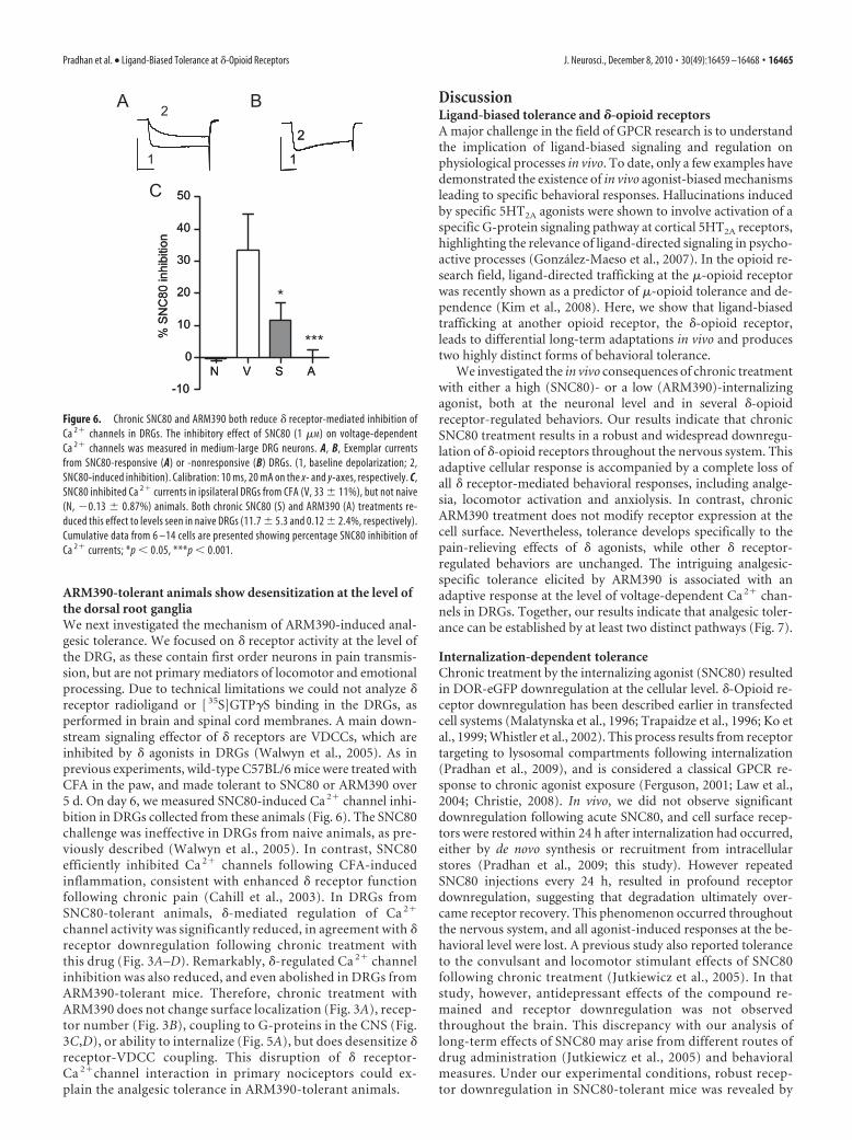
ARM390-tolerant animals show desensitization at the level ofthe dorsal root gangliaWe next investigated the mechanism of ARM390-induced anal-gesic tolerance. We focused on � receptor activity at the level ofthe DRG, as these contain first order neurons in pain transmis-sion, but are not primary mediators of locomotor and emotionalprocessing. Due to technical limitations we could not analyze �receptor radioligand or [ 35S]GTP�S binding in the DRGs, asperformed in brain and spinal cord membranes. A main down-stream signaling effector of � receptors are VDCCs, which areinhibited by � agonists in DRGs (Walwyn et al., 2005). As inprevious experiments, wild-type C57BL/6 mice were treated withCFA in the paw, and made tolerant to SNC80 or ARM390 over5 d. On day 6, we measured SNC80-induced Ca 2� channel inhi-bition in DRGs collected from these animals (Fig. 6). The SNC80challenge was ineffective in DRGs from naive animals, as pre-viously described (Walwyn et al., 2005). In contrast, SNC80efficiently inhibited Ca 2� channels following CFA-inducedinflammation, consistent with enhanced � receptor functionfollowing chronic pain (Cahill et al., 2003). In DRGs fromSNC80-tolerant animals, �-mediated regulation of Ca 2�
channel activity was significantly reduced, in agreement with �receptor downregulation following chronic treatment withthis drug (Fig. 3A–D). Remarkably, �-regulated Ca 2� channelinhibition was also reduced, and even abolished in DRGs fromARM390-tolerant mice. Therefore, chronic treatment withARM390 does not change surface localization (Fig. 3A), recep-tor number (Fig. 3B), coupling to G-proteins in the CNS (Fig.3C,D), or ability to internalize (Fig. 5A), but does desensitize �receptor-VDCC coupling. This disruption of � receptor-Ca 2�channel interaction in primary nociceptors could ex-plain the analgesic tolerance in ARM390-tolerant animals.
DiscussionLigand-biased tolerance and �-opioid receptorsA major challenge in the field of GPCR research is to understandthe implication of ligand-biased signaling and regulation onphysiological processes in vivo. To date, only a few examples havedemonstrated the existence of in vivo agonist-biased mechanismsleading to specific behavioral responses. Hallucinations inducedby specific 5HT2A agonists were shown to involve activation of aspecific G-protein signaling pathway at cortical 5HT2A receptors,highlighting the relevance of ligand-directed signaling in psycho-active processes (Gonzalez-Maeso et al., 2007). In the opioid re-search field, ligand-directed trafficking at the �-opioid receptorwas recently shown as a predictor of �-opioid tolerance and de-pendence (Kim et al., 2008). Here, we show that ligand-biasedtrafficking at another opioid receptor, the �-opioid receptor,leads to differential long-term adaptations in vivo and producestwo highly distinct forms of behavioral tolerance.
We investigated the in vivo consequences of chronic treatmentwith either a high (SNC80)- or a low (ARM390)-internalizingagonist, both at the neuronal level and in several �-opioidreceptor-regulated behaviors. Our results indicate that chronicSNC80 treatment results in a robust and widespread downregu-lation of �-opioid receptors throughout the nervous system. Thisadaptive cellular response is accompanied by a complete loss ofall � receptor-mediated behavioral responses, including analge-sia, locomotor activation and anxiolysis. In contrast, chronicARM390 treatment does not modify receptor expression at thecell surface. Nevertheless, tolerance develops specifically to thepain-relieving effects of � agonists, while other � receptor-regulated behaviors are unchanged. The intriguing analgesic-specific tolerance elicited by ARM390 is associated with anadaptive response at the level of voltage-dependent Ca 2� chan-nels in DRGs. Together, our results indicate that analgesic toler-ance can be established by at least two distinct pathways (Fig. 7).
Internalization-dependent toleranceChronic treatment by the internalizing agonist (SNC80) resultedin DOR-eGFP downregulation at the cellular level. �-Opioid re-ceptor downregulation has been described earlier in transfectedcell systems (Malatynska et al., 1996; Trapaidze et al., 1996; Ko etal., 1999; Whistler et al., 2002). This process results from receptortargeting to lysosomal compartments following internalization(Pradhan et al., 2009), and is considered a classical GPCR re-sponse to chronic agonist exposure (Ferguson, 2001; Law et al.,2004; Christie, 2008). In vivo, we did not observe significantdownregulation following acute SNC80, and cell surface recep-tors were restored within 24 h after internalization had occurred,either by de novo synthesis or recruitment from intracellularstores (Pradhan et al., 2009; this study). However repeatedSNC80 injections every 24 h, resulted in profound receptordownregulation, suggesting that degradation ultimately over-came receptor recovery. This phenomenon occurred throughoutthe nervous system, and all agonist-induced responses at the be-havioral level were lost. A previous study also reported toleranceto the convulsant and locomotor stimulant effects of SNC80following chronic treatment (Jutkiewicz et al., 2005). In thatstudy, however, antidepressant effects of the compound re-mained and receptor downregulation was not observedthroughout the brain. This discrepancy with our analysis oflong-term effects of SNC80 may arise from different routes ofdrug administration (Jutkiewicz et al., 2005) and behavioralmeasures. Under our experimental conditions, robust recep-tor downregulation in SNC80-tolerant mice was revealed by
2
1 1
2
1
2
N V S A-10
0
10
20
30
40
50
% S
NC
80 in
hibi
tion
N V S A-10
0
10
20
30
40
50
% S
NC
80 in
hibi
tion
BA
C
*
***
Figure 6. Chronic SNC80 and ARM390 both reduce � receptor-mediated inhibition ofCa 2� channels in DRGs. The inhibitory effect of SNC80 (1 �M) on voltage-dependentCa 2� channels was measured in medium-large DRG neurons. A, B, Exemplar currentsfrom SNC80-responsive (A) or -nonresponsive (B) DRGs. (1, baseline depolarization; 2,SNC80-induced inhibition). Calibration: 10 ms, 20 mA on the x- and y-axes, respectively. C,SNC80 inhibited Ca 2� currents in ipsilateral DRGs from CFA (V, 33 � 11%), but not naive(N, �0.13 � 0.87%) animals. Both chronic SNC80 (S) and ARM390 (A) treatments re-duced this effect to levels seen in naive DRGs (11.7 � 5.3 and 0.12 � 2.4%, respectively).Cumulative data from 6 –14 cells are presented showing percentage SNC80 inhibition ofCa 2� currents; *p � 0.05, ***p � 0.001.
Pradhan et al. • Ligand-Biased Tolerance at �-Opioid Receptors J. Neurosci., December 8, 2010 • 30(49):16459 –16468 • 16465

four different ex vivo parameters (imag-ing, radioligand and [ 35S]GTP�S bind-ing, and Ca 2� channel conductance)and in three behavioral responses (anal-gesia, locomotion and anxiolysis). To-gether, our comparison of high- andlow-internalizing agonist effects stronglysuggests that �-opioid receptor internaliza-tion in vivo is a major regulatory event thatleads to generalized tolerance.
Internalization-independent toleranceChronic treatment with the low-internalizing agonist (ARM390) pro-duced behavioral tolerance only at thelevel of pain processing, while leaving lo-comotor and emotional responses intact.We investigated the underlying cause forthis particular type of internalization-independent tolerance focusing on adap-tations occurring in DRGs, the first orderneurons of the pain processing pathway.Chronic ARM390 treatment affectedneither the overall receptor number andcellular localization, nor ability of the re-ceptor to internalize in these cells. Be-cause the receptor seemed unaffected,we hypothesized that internalization-independent tolerance resulted from cel-lular adaptations occurring downstreamof the receptor. Many signaling effectorsystems have been described, includingG��-mediated regulation of voltage-dependent Ca 2� channels (Williams etal., 2001; Christie, 2008) in DRGs (Wal-wyn et al., 2005). Our data demonstratethat � receptor inhibition of Ca 2� chan-nel activity, indeed, has desensitized inthese cells, providing a mechanism forthe ARM390 tolerance to analgesic re-sponses. Importantly, this adaptive response seems specific toDRGs, as we did not observe any alteration of G-protein acti-vation in the brain and spinal cord in ARM390-tolerant ani-mals, and centrally mediated behaviors remained intact. Thisanatomically restricted adaptation may have resulted from al-tered � receptor function in DRGs following CFA administra-tion (Gendron et al., 2006) or altered coupling between �receptors and Ca2� channels. Overall our findings demon-strate that chronic receptor activation does not necessarilyinduce universal adaptive responses in vivo, but can recruitspecific processes in restricted neuronal pathways. We alsocannot exclude the possibility that, in addition to DRG-specific mechanisms, other adaptations may occur at the sys-tems level through recruitment of opponent processes withinthe pain neurocircuitry (Ossipov et al., 2005; Simonin et al.,2006).
Cellular or systems level adaptive mechanisms have beenproposed in the context of the �-opioid receptor. The low-internalizing � agonist morphine produces analgesic tolerance. Aproposed hypothesis is that continuous signaling at the receptoris responsible for the development of counter-adaptations thatlead to reduced analgesic effects of the drug (Whistler et al., 1999;He et al., 2002; Kieffer and Evans, 2002). However, the parallel
between the two opioid receptors remains limited to low-internalizing agonists, because �-opioid receptors rapidly recyclefollowing internalization while �-opioid receptors are degraded(Ko et al., 1999; Tanowitz and von Zastrow, 2003; Hanyalogluand von Zastrow, 2008).
Therapeutic implicationsConclusions from this study have therapeutic implications as �agonists are being tested clinically for the treatment of pain, anx-iety and severe depression (Dondio, 2000; Dondio et al., 2001).From our data, it appears that internalizing and noninternalizingagonists would both produce tolerance in pain management.However, the development of noninternalizing � agonists may beof particular interest for the treatment of anxiety and depression,as tolerance is less likely to occur in this context (Jutkiewicz et al.,2005).
In conclusion, this study shows that ligand-directed traf-ficking leads to the development of differential tolerance at�-opioid receptors. The ligand-specific tolerance observed inthis study strengthens the notion that ligand-biased responseshave important implications in GPCR biology in vivo. Ourfindings uncover a novel aspect of biased agonism, which ex-
4H-12H Behavioral desensitization
24H
GG
GG
Restoration of receptor function
Intact receptor function
GG
Receptor degradation
Generalized tolerance
AnxiolysisLocomotion
Analgesia
AcuteTreatment
1 X 24H cycle
ChronicTreatment
5 X 24H cycles
Intact receptor at the cell surface
Analgesic tolerance
Analgesia
Receptor internalization/uncoupling
Analgesia
Intact behavioral response
AnalgesiaLocomotion
Anxiolysis
Intact receptor function
GG
SNC80SNC80SNC80SNC80SNC80SNC80SNC80SNC80 ARM 390ARM 390
ARM 390ARM 390
ARM 390ARM 390
ARM 390
ARM 390
SNC80SNC80 ARM 390
ARM 390
Internalization-dependent Internalization-independent
GGG
SNC80 ARM390
GG
TESTTESTTESTTEST
TESTTESTTESTTEST
DRG
Figure 7. Two distinct paths toward analgesic tolerance. The scheme summarizes receptor analysis at the cellular level (sub-cellular localization, receptor number, G-protein coupling, and Ca 2� channel activity) and behavioral data obtained throughoutthis study. Data highlight the distinct adaptive processes elicited by chronic treatment using either the high-internalizing agonist(SNC80, left) or the low-internalizing agonist (ARM390, right). Top, A single 24 h treatment cycle leads to restored or intactreceptors, with seemingly identical functional properties both at cellular and behavioral levels. Bottom, Five 24 h treatment cyclesproduce highly distinct forms of tolerance. Chronic SNC80 downregulates receptors throughout the nervous system, as classicallydescribed in cellular models, leading to generalized tolerance at the behavioral level. Chronic ARM390 does not modify receptorexpression at the cell surface, but decreases � receptor-mediated Ca 2� channel responses in DRGs. Further, tolerance developsonly for analgesic but not locomotor or emotional responses, indicating that in vivo adaptations occur specifically at the level of painprocessing pathways.
16466 • J. Neurosci., December 8, 2010 • 30(49):16459 –16468 Pradhan et al. • Ligand-Biased Tolerance at �-Opioid Receptors

tends the concept of ligand-specific signaling to ligand-biasedtolerance.
ReferencesAbbadie C, Lindia JA, Cumiskey AM, Peterson LB, Mudgett JS, Bayne EK,
DeMartino JA, MacIntyre DE, Forrest MJ (2003) Impaired neuropathicpain responses in mice lacking the chemokine receptor CCR2. Proc NatlAcad Sci U S A 100:7947–7952.
Befort K, Filliol D, Decaillot FM, Gaveriaux-Ruff C, Hoehe MR, Kieffer BL(2001) A single nucleotide polymorphic mutation in the human mu-opioid receptor severely impairs receptor signaling. J Biol Chem276:3130 –3137.
Bosier B, Hermans E (2007) Versatility of GPCR recognition by drugs: frombiological implications to therapeutic relevance. Trends Pharmacol Sci28:438 – 446.
Broom DC, Jutkiewicz EM, Folk JE, Traynor JR, Rice KC, Woods JH (2002)Convulsant activity of a non-peptidic delta-opioid receptor agonist is notrequired for its antidepressant-like effects in Sprague-Dawley rats. Psy-chopharmacology (Berl) 164:42– 48.
Cahill CM, Morinville A, Hoffert C, O’Donnell D, Beaudet A (2003) Up-regulation and trafficking of delta opioid receptor in a model of chronicinflammation: implications for pain control. Pain 101:199 –208.
Calderon SN, Rothman RB, Porreca F, Flippen-Anderson JL, McNutt RW, Xu H,Smith LE, Bilsky EJ, Davis P, Rice KC (1994) Probes for narcotic receptormediated phenomena. 19. Synthesis of (�)-4-[(alpha R)-alpha-((2S,5R)-4-allyl-2,5-dimethyl-1-piperazinyl)-3-methoxybenzyl]- N, N-diethylbenz-amide (SNC 80): a highly selective, non-peptide delta opioid receptor agonistJ Med Chem 37:2125–2128.
Chaplan SR, Bach FW, Pogrel JW, Chung JM, Yaksh TL (1994) Quantitativeassessment of tactile allodynia in the rat paw. J Neurosci Methods53:55– 63.
Christie MJ (2008) Cellular neuroadaptations to chronic opioids: tolerance,withdrawal and addiction. Br J Pharmacol 154:384 –396.
Dondio G (2000) Development of novel pain relief agents acting throughthe selective activation of the delta-opioid receptor. Farmaco 55:178 –180.
Dondio G, Ronzoni S, Farina C, Graziani D, Parini C, Petrillo P, Giardina GA(2001) Selective delta opioid receptor agonists for inflammatory andneuropathic pain. Farmaco 56:117–119.
Ferguson SS (2001) Evolving concepts in G protein-coupled receptor endo-cytosis: the role in receptor desensitization and signaling. Pharmacol Rev53:1–24.
Filliol D, Ghozland S, Chluba J, Martin M, Matthes HW, Simonin F, Befort K,Gaveriaux-Ruff C, Dierich A, LeMeur M, Valverde O, Maldonado R,Kieffer BL (2000) Mice deficient for delta- and mu-opioid receptors ex-hibit opposing alterations of emotional responses. Nat Genet 25:195–200.
Fraser GL, Gaudreau GA, Clarke PB, Menard DP, Perkins MN (2000) An-tihyperalgesic effects of delta opioid agonists in a rat model of chronicinflammation. Br J Pharmacol 129:1668 –1672.
Galandrin S, Oligny-Longpre G, Bouvier M (2007) The evasive nature ofdrug efficacy: implications for drug discovery. Trends Pharmacol Sci28:423– 430.
Gallantine EL, Meert TF (2005) A comparison of the antinociceptive andadverse effects of the mu-opioid agonist morphine and the delta-opioidagonist SNC80. Basic Clin Pharmacol Toxicol 97:39 –51.
Gamble GD, Milne RJ (1989) Repeated exposure to sham testing proce-dures reduces reflex withdrawal and hot-plate latencies: attenuation oftonic descending inhibition? Neurosci Lett 96:312–317.
Gaveriaux-Ruff C, Karchewski LA, Hever X, Matifas A, Kieffer BL (2008)Inflammatory pain is enhanced in delta opioid receptor-knockout mice.Eur J Neurosci 27:2558 –2567.
Gendron L, Lucido AL, Mennicken F, O’Donnell D, Vincent JP, Stroh T,Beaudet A (2006) Morphine and pain-related stimuli enhance cell sur-face availability of somatic delta-opioid receptors in rat dorsal root gan-glia. J Neurosci 26:953–962.
Gonzalez-Maeso J, Weisstaub NV, Zhou M, Chan P, Ivic L, Ang R, Lira A,Bradley-Moore M, Ge Y, Zhou Q, Sealfon SC, Gingrich JA (2007) Hal-lucinogens recruit specific cortical 5-HT(2A) receptor-mediated signal-ing pathways to affect behavior. Neuron 53:439 – 452.
Hanyaloglu AC, von Zastrow M (2008) Regulation of GPCRs by endocyticmembrane trafficking and its potential implications. Annu Rev Pharma-col Toxicol 48:537–568.
He L, Fong J, von Zastrow M, Whistler JL (2002) Regulation of opioid re-
ceptor trafficking and morphine tolerance by receptor oligomerization.Cell 108:271–282.
Hurley RW, Hammond DL (2000) The analgesic effects of supraspinal muand delta opioid receptor agonists are potentiated during persistent in-flammation. J Neurosci 20:1249 –1259.
Jutkiewicz EM, Kaminsky ST, Rice KC, Traynor JR, Woods JH (2005)Differential behavioral tolerance to the delta-opioid agonist SNC80 ([(�)-4-[(alphaR)-alpha-[(2S,5R)-2,5-dimethyl-4-(2-propenyl)-1-piperazinyl]-(3-methoxyphenyl)methyl]-N,N-diethylbenzamide) in Sprague-Dawley rats.J Pharmacol Exp Ther 315:414–422.
Kenakin T (2003) Ligand-selective receptor conformations revisited: thepromise and the problem. Trends Pharmacol Sci 24:346 –354.
Kieffer BL, Evans CJ (2002) Opioid tolerance—in search of the holy grail.Cell 108:587–590.
Kieffer BL, Gaveriaux-Ruff C (2002) Exploring the opioid system by geneknockout. Prog Neurobiol 66:285–306.
Kim JA, Bartlett S, He L, Nielsen CK, Chang AM, Kharazia V, Waldhoer M,Ou CJ, Taylor S, Ferwerda M, Cado D, Whistler JL (2008) Morphine-induced receptor endocytosis in a novel knockin mouse reduces toleranceand dependence. Curr Biol 18:129 –135.
Ko JL, Arvidsson U, Williams FG, Law PY, Elde R, Loh HH (1999) Visual-ization of time-dependent redistribution of delta-opioid receptors inneuronal cells during prolonged agonist exposure. Brain Res Mol BrainRes 69:171–185.
Konig M, Zimmer AM, Steiner H, Holmes PV, Crawley JN, Brownstein MJ,Zimmer A (1996) Pain responses, anxiety and aggression in mice defi-cient in pre-proenkephalin. Nature 383:535–538.
Law PY, Loh HH, Wei LN (2004) Insights into the receptor transcriptionand signaling: implications in opioid tolerance and dependence. Neuro-pharmacology 47 [Suppl 1]:300 –311.
Malatynska E, Wang Y, Knapp RJ, Waite S, Calderon S, Rice K, Hruby VJ,Yamamura HI, Roeske WR (1996) Human delta opioid receptor: func-tional studies on stably transfected Chinese hamster ovary cells after acuteand chronic treatment with the selective nonpeptidic agonist SNC-80.J Pharmacol Exp Ther 278:1083–1089.
Marie N, Landemore G, Debout C, Jauzac P, Allouche S (2003) Pharmaco-logical characterization of AR-M1000390 at human delta opioid recep-tors. Life Sci 73:1691–1704.
Nadal X, Banos JE, Kieffer BL, Maldonado R (2006) Neuropathic pain isenhanced in delta-opioid receptor knockout mice. Eur J Neurosci23:830 – 834.
Ossipov MH, Lai J, King T, Vanderah TW, Porreca F (2005) Underlyingmechanisms of pronociceptive consequences of prolonged morphine ex-posure. Biopolymers 80:319 –324.
Perrine SA, Hoshaw BA, Unterwald EM (2006) Delta opioid receptor li-gands modulate anxiety-like behaviors in the rat. Br J Pharmacol147:864 – 872.
Pradhan AA, Becker JA, Scherrer G, Tryoen-Toth P, Filliol D, Matifas A,Massotte D, Gaveriaux-Ruff C, Kieffer BL (2009) In vivo delta opioidreceptor internalization controls behavioral effects of agonists. PLoS One4:e5425.
Saitoh A, Kimura Y, Suzuki T, Kawai K, Nagase H, Kamei J (2004) Potentialanxiolytic and antidepressant-like activities of SNC80, a selective delta-opioid agonist, in behavioral models in rodents. J Pharmacol Sci95:374 –380.
Scherrer G, Tryoen-Toth P, Filliol D, Matifas A, Laustriat D, Cao YQ, Bas-baum AI, Dierich A, Vonesh JL, Gaveriaux-Ruff C, Kieffer BL (2006)Knockin mice expressing fluorescent delta-opioid receptors uncover Gprotein-coupled receptor dynamics in vivo. Proc Natl Acad Sci U S A103:9691–9696.
Simonin F, Schmitt M, Laulin JP, Laboureyras E, Jhamandas JH, MacTavishD, Matifas A, Mollereau C, Laurent P, Parmentier M, Kieffer BL, Bour-guignon JJ, Simonnet G (2006) RF9, a potent and selective neuropeptideFF receptor antagonist, prevents opioid-induced tolerance associatedwith hyperalgesia. Proc Natl Acad Sci U S A 103:466 – 471.
Spina L, Longoni R, Mulas A, Chang KJ, Di Chiara G (1998) Dopamine-dependent behavioural stimulation by non-peptide delta opioidsBW373U86 and SNC 80: 1. Locomotion, rearing and stereotypies in intactrats. Behav Pharmacol 9:1– 8.
Tanowitz M, von Zastrow M (2003) A novel endocytic recycling signal that
Pradhan et al. • Ligand-Biased Tolerance at �-Opioid Receptors J. Neurosci., December 8, 2010 • 30(49):16459 –16468 • 16467

distinguishes the membrane trafficking of naturally occurring opioid re-ceptors. J Biol Chem 278:45978 – 45986.
Trapaidze N, Keith DE, Cvejic S, Evans CJ, Devi LA (1996) Sequestration ofthe delta opioid receptor. Role of the C terminus in agonist-mediatedinternalization. J Biol Chem 271:29279 –29285.
Walwyn W, Maidment NT, Sanders M, Evans CJ, Kieffer BL, Hales TG (2005) In-duction of delta opioid receptor function by up-regulation of membranereceptors in mouse primary afferent neurons. Mol Pharmacol 68:1688–1698.
Wei ZY, Brown W, Takasaki B, Plobeck N, Delorme D, Zhou F, Yang H, JonesP, Gawell L, Gagnon H, Schmidt R, Yue SY, Walpole C, Payza K, St-OngeS, Labarre M, Godbout C, Jakob A, Butterworth J, Kamassah A, Morin PE,Projean D, Ducharme J, Roberts E (2000) N, N-Diethyl-4-(phenylpip-
eridin-4-ylidenemethyl)benzamide: a novel, exceptionally selective, po-tent delta opioid receptor agonist with oral bioavailability and itsanalogues. J Med Chem 43:3895–3905.
Whistler JL, Chuang HH, Chu P, Jan LY, von Zastrow M (1999) Func-tional dissociation of mu opioid receptor signaling and endocytosis:implications for the biology of opiate tolerance and addiction. Neuron23:737–746.
Whistler JL, Enquist J, Marley A, Fong J, Gladher F, Tsuruda P, Murray SR,Von Zastrow M (2002) Modulation of postendocytic sorting of Gprotein-coupled receptors. Science 297:615– 620.
Williams JT, Christie MJ, Manzoni O (2001) Cellular and synaptic adap-tations mediating opioid dependence. Physiol Rev 81:299 –343.
16468 • J. Neurosci., December 8, 2010 • 30(49):16459 –16468 Pradhan et al. • Ligand-Biased Tolerance at �-Opioid Receptors