behavioral/cognitive ...the case of linguistically distant languages such as english and japanese...
TRANSCRIPT

Behavioral/Cognitive
Dynamic Neural Network Reorganization Associated withSecond Language Vocabulary Acquisition: A MultimodalImaging Study
Chihiro Hosoda,1,2,3,4 Kanji Tanaka,5,6 Tadashi Nariai,3 Manabu Honda,1,2 and Takashi Hanakawa1,2,7
1Department of Functional Brain Research, National Institute of Neuroscience, National Center of Neurology and Psychiatry, Tokyo 187-8502,Japan, 2Department of Advanced Neuroimaging, Integrative Brain Imaging Center, National Center of Neurology and Psychiatry, Tokyo 187-8551, Japan,3Department of Neurosurgery, Tokyo Medical and Dental University, Tokyo 113-0034, Japan, 4Department of Motor Control and Rehabilitation, ATRComputational Neuroscience Laboratories, Kyoto, 619-0288, Japan, 5Research Center for Advanced Science and Technology, The University of Tokyo,Tokyo, 113-8654, Japan 6Japan Society for the Promotion of Science, Tokyo, 102-0083, Japan, and 7PRESTO, Japan Science and Technology Agency,Kawaguchi, Saitama 332-0012, Japan
It remains unsettled whether human language relies exclusively on innately privileged brain structure in the left hemisphere or is moreflexibly shaped through experiences, which induce neuroplastic changes in potentially relevant neural circuits. Here we show thatlearning of second language (L2) vocabulary and its cessation can induce bidirectional changes in the mirror-reverse of the traditionallanguage areas. A cross-sectional study identified that gray matter volume in the inferior frontal gyrus pars opercularis (IFGop) andconnectivity of the IFGop with the caudate nucleus and the superior temporal gyrus/supramarginal (STG/SMG), predominantly in theright hemisphere, were positively correlated with L2 vocabulary competence. We then implemented a cohort study involving 16 weeks ofL2 training in university students. Brain structure before training did not predict the later gain in L2 ability. However, training interven-tion did increase IFGop volume and reorganization of white matter including the IFGop-caudate and IFGop-STG/SMG pathways in theright hemisphere. These “positive” plastic changes were correlated with the gain in L2 ability in the trained group but were not observedin the control group. We propose that the right hemispheric network can be reorganized into language-related areas through use-dependent plasticity in young adults, reflecting a repertoire of flexible reorganization of the neural substrates responding to linguisticexperiences.
IntroductionLanguage relies on both innate ability and experiences (Kuhl, 2010;Hsu et al., 2011), making understanding of experience-dependentshaping of language systems an essential theme for languageneuroscience. The neural mechanisms of experienced-induced firstlanguage (L1) development are difficult to study since L1 isacquired through infancy-childhood. Understanding of sec-ond language (L2) learning is therefore important because itshould also invoke basic mechanisms of language acquisition.
Vocabulary is important for proficiency in any language (Co-ady and Huckin, 1997; Grogan et al., 2012), but neural mecha-
nisms of L2 vocabulary learning remain unclear. L2 vocabularylearning is achieved through interactions with the already exist-ing L1 lexicon in adults (Ellis et al., 1999). For example, L2 stimulispontaneously activate the L1 lexicon (Thierry and Wu, 2007),and individuals with high L1 vocabulary competence have anadvantage in L2 vocabulary learning (Meschyan, 2002). How-ever, despite these interactions, the L1 and L2 lexicons may bedifferentially structured; it has been proposed that the L2 lexiconis more phonologically organized compared with the L1 lexiconstrongly connected with the semantic system (Laufer, 1989).Hence, L2 vocabulary leaning in adults would induce reorgani-zation predominantly in the phonological system nested with thesemantic system. These features may make L2 vocabulary learn-ing in adults a unique paradigm for exploring repertoires of dy-namic neural reorganization of the language system, especially inthe case of linguistically distant languages such as English andJapanese (Chiswick and Miller, 2005).
L2 learning may accompany macroscopic structural altera-tions of the language system. Recent neuroimaging studies haverevealed plastic changes of gray matter (GM) during learning ofvarious cognitive and motor abilities (Draganski et al., 2004;Mechelli et al., 2004). The analysis of fractional anisotropy (FA)computed from diffusion-weighted magnetic resonance imaging
Received Jan. 29, 2013; revised July 8, 2013; accepted July 12, 2013.Author contributions: C.H. and T.H. designed research; C.H. performed research; C.H. and K.T. contributed un-
published reagents/analytic tools; C.H. analyzed data; C.H., T.N., M.H., and T.H. wrote the paper.This study was supported in part by grants from KAKENHI (20578976), the Neuro Creative Lab (NPO), the Na-
rishige Neuroscience Research Foundation and NEXT to C.H., as well as PRESTO, KAKENHI (24118511), and anintramural research grant from the National Center of Neurology and Psychiatry to T.H. We thank Charles S. DaSallafor his help with English editing.
The authors declare no competing financial interests.Correspondence should be addressed to Dr. Takashi Hanakawa, Department of Advanced Neuroimaging, Inte-
grative Brain Imaging Center, National Center of Neurology and Psychiatry, 4-1-1 Ogawahigashi, Kodaira, Tokyo187-8551, Japan. E-mail: [email protected].
DOI:10.1523/JNEUROSCI.0410-13.2013Copyright © 2013 the authors 0270-6474/13/3313663-10$15.00/0
The Journal of Neuroscience, August 21, 2013 • 33(34):13663–13672 • 13663

(DWI) can index organization of white matter (WM) micro-structure, and allows us to visualize plastic changes of WM(Johansen-Berg, 2012; Zatorre et al., 2012). Very recently, a fewL2 learning studies have shown GM (Mårtensson et al., 2012) andWM (Schlegel et al., 2012) changes in the traditional languagenetwork in the left hemisphere.
Previous neuroimaging studies have, however,demonstratedroles of the right hemisphere in language ability (Vingerhoets etal., 2003; Videsott et al., 2010; Vigneau et al., 2011; Van Ettinger-Veenstra et al., 2012) and in L2 control (Hernandez et al., 2001;Hosoda et al., 2012). One of these found correlations betweenright prefrontal activity and L2 vocabulary competence (Hosodaet al., 2012). To explore neural plasticity associated with L2 vo-cabulary learning, we first explored the neural underpinningscorrelated with L2 vocabulary competence in a cross-sectionalstudy, revealing GM and WM structures correlated with L2 vo-cabulary levels predominantly in the right hemisphere. We thenran a cohort study implementing an L2 training program to testwhether those L2 vocabulary correlates reflected innate predispo-sitions or plastic changes resulting from learning. We furtherexamined the natural course of the induced structural changes.Here we provide evidence that L2 vocabulary learning inducesdynamic reorganization of GM and WM structures outside of thetypical language network in adult brains.
Materials and MethodsSubjects. In the cross-sectional study, 137 native Japanese speakers (71males and 66 females) with a mean age of 24.0 years [SD � 5.3, range18 – 42] were enrolled (Table 1). All subjects were university students orgraduates whose self-reported English (L2) proficiencies varied from lowto very high to the level of Japanese–English bilinguals.
In the cohort study, 67 native Japanese speakers (mean age � 20.1, 31men and 36 women) were initially placed into a training group (TG). Inthe TG, 24 of 47 participants completed the planned 4 month training,whereas the remaining 23 participants opted not to continue during thecourse of training for various reasons. Hence, we studied 24 TG partici-pants (mean age � 20.1, 10 men and 14 women) and an age-matchedcontrol group (CG; n � 20; mean age � 20.1, 10 men and 10 women).The participants in the cohort study were university students and re-cruited from basic level English classes (Table 1). The participants in thecross-sectional study and those in the cohort study did not overlap.
In both studies, the participants had grown up in Japan and started tolearn English as their L2 at a mean age of 11.0 years. None had started tolearn L2 before 7 years old, and thus all participants were regarded as lateL2 learners (Dowens et al., 2010). All the participants were right-handed
as assessed by the Edinburgh Handedness Inventory. All of the partici-pants showed handedness scale of 100 (SD � 0) because we only re-cruited the perfectly right-handed participants, revealing the contents ofhandedness inventory in the recruitment information. All were healthyand neurologically intact, with no history of neuropsychiatric disorders,psychotropic medication use, or head injury. All the participants gavewritten informed consent according to the study protocol approvedby the institutional review board (National Center of Neurology andPsychiatry).
Experimental design. In the cross-sectional study, the participants un-derwent MRI scanning (T1-weighted images and DWI) and assessmentfor English proficiency using the English Vocabulary Test (EVT) and theNational Adult Reading Test (NART). The T1-weighted MRI providedthe data for voxel-based morphometry (VBM) analysis of the GM vol-ume, and the DWI allowed for tract-based spatial statistics (TBSS) andprobabilistic diffusion-based tractography (PDT).
In the cohort experiment, we adopted the Test of English for Interna-tional Communication (TOEIC) as a primary measure to evaluate vari-ous L2 abilities. The TOEIC assesses English proficiency for use inbusiness, providing a fairly accurate measurement of English capabilitiesfor non-native speakers in listening, reading, and grammar. The EVT andNART were used as adjunctive measures of L2 proficiency. Additionally,the Wechsler Adult Intelligence Scale-3 (WAIS-3) and NEO-Five FactorInventory (NEO-FFI) were used to confirm homogeneity of basic intel-ligence and personality traits between the TG and CG.
All participants underwent MRI scanning (T1-weighted MRI andDWI) and an L2 assessment battery before training, hereafter referred toas the “Pre” condition. The L2 training program was developed in-houseusing Visual Basic. Participants undertook 16 weeks of training, and ineach week they learned 60 words or idioms including the meaning, spell-ing, and pronunciation as well as example sentences indicating theirusage. For pronunciation, we referred to the AT&T Natural Voices Text-to-Speech Demo (http://www2.research.att.com/�ttsweb/tts/demo.php). The participants were encouraged to dictate each word, idiom, andsentence 10 times. Each weekend, we distributed a program that includeda review test using the 60 words or idioms of the week. Upon completingthe full program, the participants were expected to master almost 1000words and idioms. Participants in the CG received no particular assign-ment according to previous cohort studies using e-learning (Takeuchi etal., 2010; Mårtensson et al., 2012; Schlegel et al., 2012; Wan et al., 2012;Ghazi Saidi et al., 2013).
The participants in both the TG and CG underwent an L2 assessmentbattery immediately after the training period (“Post-1”). After comple-tion of the training program, the participants were not further engaged inspecific L2 learning programs systemically, and thus the continuation ofL2 learning depended upon each participant’s choice in personal life. Weobtained follow-up behavioral and imaging data from the TG group ayear after the end of the training program (“Post-2”). Statistical tests onthe behavioral parameters were performed using SPSS 17.0 (IBM). Toexplore training-induced changes in L2 abilities, we analyzed totalTOEIC score and its subsections (listening, reading, and grammar) usinga 2-by-2 mixed repeated-measures ANOVA with the time (Pre andPost-1) as a within-subject variable and the group (TG and CG) as abetween-subject variable. Next, a correlation analysis was performed totest if the training-related changes in TOEIC score were correlated withthose of EVT or NART score.
Image data acquisition. MRI data were acquired using a 3 T MRI scan-ner (Siemens Trio) with an 8-channel phased array receiver coil. High-resolution, 3D, T1-weighted anatomical images were obtained with anMPRAGE sequence designed as follows: repetition time (TR) � 2000 ms,echo time (TE) � 4.4 ms, inversion time (TI) � 990 ms, flip angle � 80°,matrix size � 192 � 176, field of view (FOV) � 192 � 176 mm and 1mm 3 isotropic voxels. We also acquired whole-brain DWI as follows:TR � 7900 ms, TE � 80 ms, 65 slices, flip angle � 90°, matrix size � 96 �96, FOV � 192 � 192 mm, 2 � 2 � 2 mm 3 isotropic voxels, 81 volumeswith diffusion weighting (b value � 700 s/mm 2) for different motionprobing gradient directions and 9 volumes without diffusion weighting(b � 0 s/mm 2). Field-map images were acquired in the same scanningspace as the DWI (TE1 � 5.19 ms; TE2 � 7.65 ms). All image data were
Table 1. Means and SDs of subject demographics, L2 proficiency tests, IQ, andpersonality assessments (NEO-FFI)
Cross-sectional study(n � 137)
Cohort study (pretraining data)
TG (n � 24) CG (n � 20)
Age 21.8 � 16.0 20.1 � 1.8 20.5 � 1.7Sex (male: female) 66:71 10:14 10:10TOEIC (200) NA 102.5 � 2.6 100.8 � 2.3EVT (100) 20.6 � 16.0 11.3 � 10.9 12.9 � 12.1NART (100) 41.8 � 6.2 35.6 � 4.5 39.8 � 4.0WAIS-TIQ NA 113.0 � 2.2 116.5 � 1.8WAIS-VIQ NA 101.0 � 2.6 104.0 � 2.1WAIS-PIQ NA 103.5 � 2.5 101.0 � 2.3NEO-FFI neuroticism NA 25.4 � 1.4 26.4 � 1.4NEO-FFI extraversion NA 29.8 � 1.4 30.6 � 1.3NEO-FFI openness NA 32.2 � 1.3 32.3 � 1.3NEO-FFI agreeableness NA 33.9 � 1.0 34.1 � 1.1NEO-FFI conscientiousness NA 31.5 � 1.3 30.7 � 1.2
In the cohort study, no significant differences were found between the TG and CG for all measurement types. NA, notavailable; WAIS-VIQ, Wechsler Adult Intelligence Scale Verbal IQ; WAIS-TIQ, Wechsler Adult Intelligence Scale TotalIQ; WAIS-PIQ, Wechsler Adult Intelligence Scale Performance IQ; NEO-FFI, NEO Five-Factor Inventory.
13664 • J. Neurosci., August 21, 2013 • 33(34):13663–13672 Hosoda et al. • Dynamic Neural Network Reorganization
converted into the Neuroimaging Informatics Technology Initiative for-mat before further processing.
Image data analysis: GM-VBM. The high-resolution 3D T1-weightedimages were subjected to a VBM analysis using VBM8 toolbox(http://dbm.neuro.uni-jena.de/vbm.html) implemented in SPM8 (http://www.fil.ion.ucl.ac.uk/spm). The preprocessing steps were as follows. (1)Each image was segmented into GM, WM, and CSF images in the nativeimage space. (2) The diffeomorphic anatomical registration through expo-nentiated Lie algebra (DARTEL) registration method was used to create astudy-specific mean GM image template by using the aligned images from allthe subjects to improve intersubject registration (Ashburner, 2007). Individ-ual’s GM images were registered to the study-specific mean GM template.(4) The registered GM images were further transformed into the MontrealNeurological Institute (MNI) space. (5) These normalized GM images weresmoothed using a Gaussian kernel of 12 mm full-width at half-maximum.
In the cross-sectional study, we investigated the correlations of EVT orNART scores with regional GM volume. A multiple regression designwas used using EVT or NART score as an independent variable and sex,duration of L2 learning, and age of acquisition as nuisance variables. Weset the voxelwise significance level at p � 0.05 corrected for multiplecomparisons in terms of the familywise error (FWE) rate. After identify-ing the inferior fontal gyrus pars opercularis (IFGop), superior temporalgyrus (STG), and caudate nucleus as the correlates of L2 vocabulary, wefurther investigated which hemisphere was more relevant. We computedthe laterality index (LI) of GM volume correlated with L2 vocabularycompetence (EVT), by using mean �-values computed from 5 mmspherical volumes of interest (VOIs) in IFGop (x, y, z � �40, 9, 24),STG/supramarginal gyrus (SMG; x, y, z � �57, �36, 6), and caudatenucleus as follows (x, y, z � �6, 8, 8). All of these VOIs were defined inthe left hemisphere, and thus mirror-reversed VOIs were created for theright hemisphere.
LI � (mean�LEFT_VOI � mean�RIGHT_VOI)/
(mean�LEFT_VOI � mean�RIGHT_VOI)
The selection of VOI (IFGop, STG, and caudate nucleus) was basedon the results from previous studies (Crinion et al., 2006; Saur et al.,2008). The LI ranges from �1 (completely lateralized to the right) to �1(completely lateralized to the left). Individuals with an LI of ��0.4 or��0.4 were categorized into the left or right hemisphere dominantgroup, respectively, while those with LI between �0.4 and �0.4 wereplaced into the bilateral representation group (Briellmann et al., 2003).
In the cohort study, we first tested the possibility that GM volumebefore L2 training could predict the degree of L2 ability improvementafter training. Positive results from this analysis would support the hy-pothesis that particular brain regions are reserved for L2 vocabularylearning. If such were the case, people with particularly developed struc-tures somewhere in the brain might show better L2 learning ability. Totest this hypothesis, a correlation analysis was performed using the MRIdata of the Pre condition and the training-related improvements in L2ability (differences in TOEIC scores between the Pre and Post-1 condi-tions). Next, to explore training-induced changes in GM, we conducted a2-by-2 mixed repeated-measures ANOVA with time (Pre and Post-1) asa within-subject variable and group (TG and CG) as a between-subjectvariable. The regions showing significant time-by-group interaction( p � 0.05, FWE corrected) were explored. Third, a correlation analysiswas performed to test if the training-related changes in TOEIC score werecorrelated with those of GM volume in right IFGop. This analysis testedif individual differences in brain plastic changes accounted for individualdifferences in L2 performance improvement after training.
Image data analysis: DWI-TBSS. Data preprocessing and analysis ofDWI were performed using Oxford Centre for Functional MRI of theBrain (FMRIB)’s software library (FSL 4.1, UK; http://www.fmrib.ox.ac.uk/fsl/]. All DWI were registered to the b0 images. Nonlinear imagedistortions due to magnetic field (b0) inhomogeneity were correctedbased on the field map images, using FUGUE in the FMRIB softwarelibrary. The registered images were skull stripped using the Brain Extrac-tion Tool. FA maps were calculated using the FMRIB’s Diffusion Tool-
box (FDT) v2.0 (Smith et al., 2004). After calculation of the FA map foreach subject, we implemented a voxelwise statistical analysis of the FAdata using TBSS v1.2 (Smith et al., 2006). In brief, TBSS was performed asfollows: (1) alignment of the FA images from all subjects to a templatethat was arbitrarily selected from those FA images, (2) transformation ofall the aligned FA images into MNI space using affine registration toremove the effect of cross-subject spatial variability (Kerns et al., 2004),(3) creation of a mean FA image and FA skeleton images correspondingto the center of the WM using a threshold of FA � 0.20 (Smith et al.,2006), (4) projection of the individuals’ FAs onto the mean FA skeleton,and (5) voxelwise cross-subject statistical analyses.
As water molecules move faster along the direction of WM fibers thanin the direction perpendicular to them, the direction and size of waterdiffusion in the brain can index structural organization of WM fibers (LeBihan and Johansen-Berg, 2012; Zatorre et al., 2012). DWI can measurewater diffusion noninvasively in vivo. FA is a parameter computed fromDWI according to tensor-based modeling of water diffusion, and repre-sents the degree of directional bias of water diffusion. FA is sensitive tosize and density of axons, degrees of myelination, and the coherence oforganization of fibers within a voxel (Beaulieu, 2002; Alexander et al.,2007; Zatorre et al., 2012) In the cross-sectional study, correlations of L2proficiency (EVT or NART) with FA values as an index of WM organi-zation were tested using Randomize v2.1 in FSL. The statistical thresholdwas set at p � 0.05 (FWE corrected), and the threshold-free cluster en-hancement method was used to define the clusters.
In the cohort study, similar to the VBM analysis, we first tested thepossibility that structural organization of WM before L2 training couldpredict the degree of L2 ability improvement after training. Recent liter-ature indicates that training-induced changes of FA can capture plasticreorganization of WM (Johansen-Berg, 2007, 2012; Floel et al., 2009;Scholz et al., 2009; Johansen-Berg et al., 2012; Zatorre et al., 2012).Activity-dependent myelo-modulation is a potential mechanism bywhich WM is carved by experiences. Such structural WM changes, in-cluding production of myelin basic protein, are correlated with changesin FA (Blumenfeld-Katzir et al., 2011). Changes in myelination may alterconduction velocity and synchronization of signal transmission acrossthe remote areas (Fields, 2005), relating probably to behavioral changes.To capture such learning-induced reorganization of WM, we conducteda 2-by-2 mixed repeated-measures ANOVA with time as a within-subjectvariable and the group as a between-subject variable ( p � 0.05, FWEcorrected), yielding FA changes specific to the L2 training program. Acorrelation analysis was performed using FA images (Pre) and thechanges in TOEIC scores from the Pre to Post-1 conditions.
Image analysis: multifiber PDT. We conducted PDT using FDT(http://fsl.fmrib.ox.ac.uk/fsl/fslwiki/FDT). We performed tractographyby tracing pathways through the estimates of diffusion directions. Amultifiber model was used to handle the issue of fiber crossing. In eachvoxel, two fiber directions were modeled, and a probability distributionfunction (pdf) of diffusion parameters was estimated. We then pro-ceeded with PDT, drawing multiple (5000) streamline samples based onthe pdf and estimated the distribution of connections from each seed totarget voxels. The generated pathways were volumes in which values ateach voxel represented the number of samples passing through thatvoxel. The voxel value corresponded to the probability of the target voxelconnecting to the seed voxel. To remove spurious nonsignificant connec-tions, pathways in individual subjects were thresholded to include onlyvoxels that had at least 50 samples passing through them (out of 5000samples drawn from each seed voxel).
Previous studies have pointed to the role of the caudate nucleus in L2(Crinion et al., 2006; Abutalebi et al., 2008; Grogan et al., 2009; Hosoda etal., 2012; Mårtensson et al., 2012). Moreover, the present TBSS resultsuggested a correlation between L2 ability and WM situated between theIFG and the caudate nucleus and temporoparietal junction (STG/SMG).In the cross-sectional study, therefore, a correlation analysis was per-formed to investigate the correlation between an L2 proficiency index(EVT or NART score) and the WM connectivity parameter computedfrom the bilateral IFGop-caudate and IFGop-STG/SMG correspondingto the dorsal pathway (Rilling et al., 2008; Saur et al., 2008). The wholecaudate nucleus was manually defined for a target mask in each hemi-
Hosoda et al. • Dynamic Neural Network Reorganization J. Neurosci., August 21, 2013 • 33(34):13663–13672 • 13665

sphere according to each subject’s 3D T1-weighted image (Peltier et al.,2011). A spherical VOI with a 5 mm radius was used as a seed mask for theIFGop. The dorsal pathway corresponds to fibers connecting IFGop andSTG, and includes arcuate fasciculus (AF) and parts of superior longitu-dinal fasciculus (SLF; Rilling et al., 2008; Saur et al., 2008). According toa previous study (Saur et al., 2008), we implemented 5 mm radius spher-ical VOIs in the bilateral IFGop and STG/SMG for the dorsal pathway.We performed PDT within each hemisphere: IFGop-caudate nucleusand IFGop-STG/SMG (dorsal pathway) (four tracts in total).
In the cohort study, in addition to the IFGop-caudate and IFGop-STG/SMG pathways, we also assessed correlations between the L2 com-petence and other long frontoparietal/temporal association tracts thatare reported to be relevant to language processing/learning. We consid-ered the ventral language pathways and inferior longitudinal fasciculus(ILF). The ventral pathway connects IFG pars triangularis (IFGtri) andmiddle temporal gyrus (MTG), passing through the extreme capsule. ILFconnects medial temporal lobe with the temporo-occipital junction, andis suggested be relevant to lexical-semantic language processing (Cataniand Mesulam, 2008; Rilling et al., 2008; Saur et al., 2008). We imple-mented 5 mm radius spherical VOIs in the bilateral IFGtri (BA45, x, y, z ��48, 27, 12) and MTG (x, y, z � �48, �60, 18) for the ventral pathway.We placed VOIs in the medial temporal lobe (x, y, z � �40, �20, �7)and temporo-occipital junction (x, y, z � �35 �68, �6) for ILF accord-ing to a previous study (Jou et al., 2011). We performed PDT within eachhemisphere: IFGop-caudate nucleus, IFGop-STG/SMG (dorsal path-way), IFGtri-MTG (ventral pathway), and ILF (eight tracts in total). Thevoxel values indexing probabilistic strength of connectivity across thetwo regions were averaged within each PDT pathway and then subjectedto logarithmic transformation, producing a parameter for statisticalanalyses (hereafter called the connectivity parameter). Statistical tests onthe connectivity parameter computed from PDT were performed usingSPSS 17.0 (IBM).
In the cohort study, we performed a 2-by-2 mixed repeated-measuresANOVA with time as a within-subject variable and the group as abetween-subject variable. In addition, a correlation analysis was per-formed to test if the training-related changes in TOEIC score were cor-related with those of the connectivity parameter from the eight tracts:bilateral IFGop-caudate pathway, dorsal pathway, ventral pathway, andILF. Moreover, to test the possibility that connectivity of the specifictracts before intervention could predict the degree of improvement in L2ability, a correlation analysis was performed between the connectivityparameter of the PDT in the Pre condition and the improvement of L2proficiency.
Analysis of follow-up behavioral and imaging data. Only in the traininggroup, for which follow-up data were available, TOEIC score, GM vol-ume, FA value, and the strength of connectivity of the eight specific tractswere compared across the Pre, Post-1, and Post-2 conditions. To retrievethe GM volume and FA values of interest, we applied spherical VOIs witha 5 mm radius to the right IFGop and WM beneath the right IFG based onthe results from the cross-sectional study. The data were fed into a one-way repeated-measures ANOVA, followed by post hoc comparisons withTukey’s HSD test. Finally, we ran a correlation analysis between the GMchanges from the Pre to Post-1 or Post-1 to Post-2 and FA changes fromthe Pre to Post-1 or Post-1 to Post-2. This analysis suggested the existenceof different subgroups following the completion of training. Indeed,three participants continued L2 learning of their own motivation afterour intervention, while the rest of the participants (n � 21) did not.Hence, a subgroup analysis was conducted to compare the changes ofTOEIC, GM, or FA from Post-1 to Post-2 between the two groups. Be-cause one of the groups consisted of a small number of participants, weperformed this test with a nonparametric, Mann–Whitney U test.
ResultsCross-sectional studyMean EVT and NART scores were 20.6 and 41.8, respectively(Table 1). The large variance of these values supported that L2proficiency of the participants was variable.
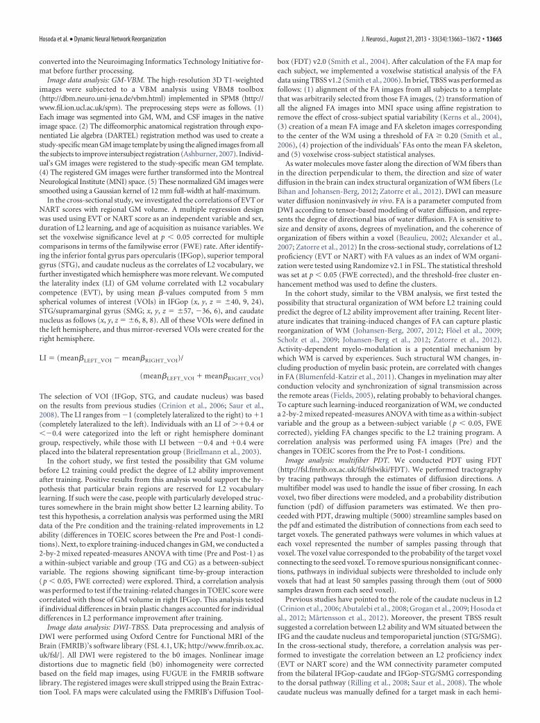
The VBM analysis showed that individuals with more exten-sive L2 vocabulary (EVT) had significantly larger GM volume inthe IFGop corresponding to Brodmann area (BA) 44, caudatenuclei, STG/SMG, and anterior cingulate cortex, all bilaterally(Fig. 1, Table 2). The correlation of the EVT score and GM vol-ume was found more strongly in the right hemisphere for theIFGop (LI � �0.40) and within the range of symmetry for thecaudate nucleus (LI � �0.15) and STG (LI � �0.17). The TBSSanalysis showed that individuals with richer L2 vocabularyshowed higher FA values in the subcortical WM beneath theIFGop (sub-IFGop), ILF, and AF only in the right hemisphere.The PDT identified bilateral IFGop-caudate nucleus and IFGop-STG/SMG (dorsal pathway) in all individuals. A statistical anal-ysis of the connectivity parameters retrieved from the four tractsrevealed correlation of the EV score with connectivity of theIFGop-caudate nucleus (p � 0.001) and the IFGop-STG/SMG(dorsal pathway) (p � 0.001) in the right hemisphere, but not in
Figure 1. Results from the cross-sectional study. a, 3D rendered images showing the correlation between EVT score and GM volume from the VBM analysis. GM volume for the bilateral IFGop,STG/SMG, and caudate nucleus (CN) correlated with EVT score, reflecting differential levels of L2 proficiency across participants ( p � 0.05 FWE corrected). The plots show the correlation betweenEVT and GM volume in the right IFGop (green circle). b, The TBSS analysis displays voxels with a positive correlation between EVT score and fractional anisotropy reflecting integration of WMstructure. Significant correlation was found in the subcortical region beneath the IFGop and the AF. The plots show the correlation between EVT scores and FA values in the WM beneath the rightIFGop (green circle). c, The PDT analysis shows fiber connections between the right IFGop and the CN in all participants. The EVT score was significantly correlated with connectivity of theIFGop-caudate head (plot). R, right; L, left.
13666 • J. Neurosci., August 21, 2013 • 33(34):13663–13672 Hosoda et al. • Dynamic Neural Network Reorganization
the left hemisphere (IFGop-caudate, p � 0.09; dorsal pathway,p � 0.11; Fig. 1).
NART score did not correlate with GM volume (VBM analy-sis) or FA value (TBSS analysis) at the predetermined statisticalthreshold. When a lenient threshold (uncorrected p � 0.001) wasapplied to the VBM analysis, we detected a trend toward correla-tion between NART score and GM volume in the middle tempo-ral and postcentral gyri in the left hemisphere.
Cohort study: behavioral dataBaseline profiles did not differ between the TG and CG partici-pants in terms of age, sex, IQs, personality traits, or L2 ability(Table 1).
The total learning time, recorded on log files, was 45.5 h (SD �3.6, range 25.1 � 64.6) across the whole training period. Just afterL2 training (Post-1), the TG showed a 29 � 21.3% (mean � SD)improvement in total TOEIC score, whereas the CG groupshowed no change (0 � 8.1%; Fig. 2a), revealing the significanteffect of L2 training (F(3,82) � 27.15, p � 0.001 by mixedrepeated-measures ANOVA; Table 3). Specifically, improvementwas found in the listening (F(3,82) � 17.64, p � 0.001) and reading(F(3,82) � 10.15, p � 0.001) sections of TOEIC, but not in thegrammar section (F(3,82) � 0.50, p � 0.42; mixed repeated-measures ANOVA). This finding is reasonable because the train-ing program included items for vocabulary and listening (i.e.,pronunciation), but not for grammar. The changes in TOEICscore paralleled those of the EVT score (r � 0.41, p � 0.03),whereas no correlation was found with those of the NART score(r � 0.15, p � 0.53).
Follow-up L2 proficiency tests were obtained in the TG par-ticipants a year after the training program. One-way repeated-measures ANOVA showed significant differences in TOEIC scoreacross the three time points (F(1.8, 38.6) � 24.96, p � 0.001 byrepeated-measures ANOVA with sphericity correction). Post hoccomparisons (Tukey’s HSD test) demonstrated significant in-creases in score from the Pre to Post-1 (p � 0.001) conditionsand significant decreases from Post-1 to Post-2 (p � 0.02), re-sulting in no difference in score between Pre and Post-2.
Cohort study: imaging dataBefore the training (Pre), no significant differences in brainstructure were detected in the GM-VBM, WM-TBSS, and con-nectivity of the eight specific tracts (bilateral IFGop-caudate, dor-sal pathway, ventral pathway, and ILF) between the TG and theCG, supporting the homogeneity of the two groups. Further, weexamined whether brain architecture at the Pre stage could pre-dict L2 ability improvement gains after the training program inthe TG. However, we failed to find significant correlationsbetween the training-induced improvement of TOEIC scores
and Pre-GM volume (VBM), Pre-FA values (TBSS), or Pre-connectivity of the eight tracts (PDT).
The analyses of the MRI data (Post-1 vs Pre) identifiedtraining-induced increases in both GM and WM of the righthemisphere for the TG compared with the CG (significant time-by-group interaction). In the TG compared with the CG, VBManalysis identified significant increases in GM volume only inthe right IFGop (5.1 � 2.3%), corresponding to the neuraloutcome of plastic changes induced by the L2 vocabularylearning (Fig. 2b). The training-induced increases of GM vol-ume in the right IFGop were correlated with those of TOEICscore (r � 0.52, p � 0.02).
In the TG, FA values increased from Pre to Post-1 in WM inthe right sub-IFGop region (3.6 � 1.6%; Fig. 2c). This increasein FA showed a strong tendency to correlate with the increase inTOEIC score (r � 0.33, p � 0.05). Using the PDT analyses, wefurther discovered learning-related increases in the connectivityparameter of the IFGop-caudate head pathway (4.5%�6.4,F(3,82) � 7.83, p � 0.01) and that of the dorsal pathway (3.7 �6.4%, F(3,82) � 6.72, p � 0.01) in the right hemisphere (2-by-2repeated-measures ANOVA). The homologous tracts in the lefthemisphere did not show the corresponding changes (2.5 �4.4%, F(3,82) � 0.16, p � 0.69 for the left IFGop-caudate connec-tivity; 2.7 � 5.2%, F(3,82) � 0.16, p � 0.65 for the left dorsalpathway connectivity; Fig. 3). There were no significant L2training-induced connectivity changes in the ventral pathway(2.1 � 4.0%, F(3,82) � 0.16, p � 0.69 for the right; 1.9 � 4.5%,F(3,82) � 0.26, p � 0.70 for the left) or in the ILF (1.8 � 5.0%,F(3,82) � 0.22, p � 0.41 for the right; 2.0 � 5.1%, F(3,82) � 0.31,p � 0.49 for the left). The training-induced increases in TOEICscore were correlated with those of the right IFGop-caudate connec-tivity (p � 0.02, r � 0.59), but not of the left IFGop-caudate (p �0.13, r � 0.25), right dorsal pathway (p � 0.07, r � 0.29), left dorsalpathway (p � 0.30, r � 0.14), right ILF (p � 0.14, r � 0.19), or leftILF connectivity (p � 0.25, r � 0.20; Fig. 3a,e,f,j,k,o).
We re-examined L2 proficiency and brain structure of theparticipants in the TG a year after the completion of the trainingprogram. One-way repeated-measures ANOVA showed sig-nificant differences in GM (F(1.8, 38.6) � 477.0, p � 0.001), FA(F(1.8, 38.6) � 233.8, p � 0.001), and right IFGop-caudate connectiv-ity (F(1.8, 38.6) � 331.0, p � 0.001) across the three time points. Con-sistent with the aforementioned group comparison, post hoccomparisons (Tukey’s HSD test) between Pre and Post-1 demon-strated significant increases in GM volume in the right IFG (p �0.001), FA beneath the right IFGop (p � 0.03), and connectivitybetween the right IFGop and caudate head (p � 0.02). Furthermore,the increment of GM volume and FA values from Pre to Post-1 weresignificantly correlated (Fig. 4a), suggesting parallel plastic changesin GM and WM. Nevertheless, post hoc comparisons (Tukey’s HSDtest) between Post-1 and Post-2 showed significant decreases in GMvolume in the right IFGop (�4.3 � 2.5%, p � 0.04) and FA beneaththe right IFG (�3.7 � 4.2%, p � 0.04). In addition, the decreases ofGM volume from Post-1 to Post-2 were significantly correlated withthose of FA values (p � 0.001, r � 0.65; Fig. 4b). That means theparallelism of the “negative” changes in GM and WM. We also no-ticed a non-negligible trend toward decreases in right IFGop- cau-date head connectivity (�2.3 � 8.1%, p � 0.05) in the connectivityof the dorsal pathway (�2.0 � 7.2%, p � 0.06). Resultantly, nodifference was found between the Pre and Post-2 values for any of theimaging parameters. These findings indicate that in most of the par-ticipants, training-induced behavioral values and macro-scopic neural structure returned to the pretraining state, suggestinga phenomenon of neural “elasticity.” Through the assessment of the
Table 2. Significant correlation between EVT score and GM volume ( p < 0.05corrected) in the cross-sectional study
Anatomical location
Coordinates
Z-value P correctedx y z
Right IGFtri (BA44) 36 11 28 7.84 0.00Right caudate nucleus 12 8 19 5.92 0.00Right STG/SMG 56 �31 21 5.42 0.00Right MTG 49 �20 0 4.08 0.00Left IFG (BA44, 45) �38 16 30 6.69 0.00Left superior frontal gyrus �22 35 45 5.45 0.00Left caudate nucleus �4 16 �2 5.26 0.00Left STG/SMG �48 �50 26 4.92 0.00
The coordinates (x, y, z) indicate local maxima in each brain region according to the MNI template.
Hosoda et al. • Dynamic Neural Network Reorganization J. Neurosci., August 21, 2013 • 33(34):13663–13672 • 13667

Post-2 data, however, we noticed that three participants showed fur-ther improvement in L2 ability from Post-1 to Post-2 (8.3, 7.5, and7.8% increases in TOEIC score), despite the overall decreases of theTOEIC score as a group. Two of those exceptional participants con-tinued to study L2 to pass a certification examination, and one par-ticipated in a short-term study abroad program after finishing ourtraining program. Even though these were anecdotal findings in asmall number of participants, we deemed them important and per-formed a subgroup analysis: three participants with increasedTOIEC score from Post-1 to Post-2 (continued subgroup) and therest (n � 21; discontinued subgroup). There were significant differ-ences in changes of TOEIC score from Post-1 to Post-2 be-tween the continued and discontinued (mean �21% � 8.5)subgroups (Mann–Whitney U test, p � 0.001). Intriguingly, thecontinued group showed increases in GM volume in the rightIFGop (3.1, 2.9, and 2.5%, respectively) from Post-1 to Post-2 asopposed to the discontinued group (mean �4.5% � SD 1.2). Thesame held true for the FA value in the right sub-IFGop (2.8, 3.2,and 2.1%, respectively, for the continued group) as opposed tothe discontinued group (mean �3.6% � 1.5; Fig. 4b). Thesedifferences reached significance for the GM change (p � 0.001 byMann–Whitney U test) and for the FA change (p � 0.001 byMann–Whitney U test).
DiscussionCurrently, international communication relies heavily on Eng-lish. This fact imposes educational challenges to non-English na-tive cultures in the rapidly globalizing world, prompting us toadvance our understanding of the mechanisms of L2 acquisition.The human capacity for language relies on neural networks that
orchestrate lexicosemantic, phonological, and syntactic subsys-tems, which should undergo adequate tuning for L2 learning.However, nonsyntactic aspects of L2 learning are scarcely under-stood hitherto. Here we focused on L2 vocabulary learning,which should involve the lexicosemantic and phonological sub-systems. The present cross-sectional study showed that bilateralfront-subcortical-parietotemporal areas, predominantly in theright hemisphere, might underlie superior L2 vocabulary abilityin late L2 learners. The cohort experiment showed that the brainarchitecture before learning could not predict the future gain inL2 ability after learning. This finding argues against the idea thatprivileged persons with developed language network have advan-tages in L2 learning, although it holds true for discriminatingspeech sounds of unfamiliar L2 (Golestani and Pallier, 2007;Golestani et al., 2007). This discrepancy suggests that relativeimportance of predispositions and environmental effects maydiffer, depending on which aspects of L2 should be learned. Thecohort experiment clearly disclosed that the gains and losses of L2ability correlated with increases and decreases, respectively, ofGM structure and WM connectivity. This longitudinal consis-tency of the behavior–structure relationship within individualshighlights the notion that use-dependent plastic changes of neu-ral networks underlie L2 learning. No overall gains of TOEICscore in the Post-2 suggested that the present e-training protocolwas not sufficiently effective in leaving a long-term signature ofL2 vocabulary learning. Note, however, that the loss of L2 abilitymeasured by the behavioral test does not necessarily mean thatthe once-remembered items had completely disappeared fromlong-term memory. It is also possible that the items could notbe retrieved in a contextually timely manner. Such presumablemechanisms agree with reduction of connectivity (i.e., discon-nection) between the frontal executive areas and temporoparietalcortices.
Recent imaging studies have begun to show GM/WM changesrelated to learning. Biological interpretations of those imagingmeasures are still challenging. Many possible mechanisms couldbe involved: neurogenesis, gliogenesis, synaptogenesis, and vas-
Figure 2. Results from the cohort study. Shown are changes in L2 competence (TOEIC), GM, and FA in the cohort study; *p � 0.05. There were no significant differences in behavioral or imagingparameters between the TG and CG at the Pre stage. a, Mean total TOEIC score changes from Pre to Post-1 in CG (left of the dotted line) and from Pre to Post-1 and Post-2 in TG (right of the dottedline). Error bars indicate standard error of the mean. Asterisks show significant differences with p � 0.05. b, Right, Colored voxels represent a cluster in the right IFGop (x, y, z � 36, 11, 28; z value �7.84) showing significant GM increases from Pre to Post-1 in TG compared with those in CG (group-by-time interaction, p � 0.05 FWE-corrected). Mean GM volume changes are shown (upper left)for the right IFGop from Pre to Post-1 in CG and from Pre to Post-1 and Post-2 in TG. Changes in GM volume for the right IFGop significantly correlated with improvement in total TOEIC score acrossindividuals (lower left). c, Right, Colored voxels represent WM clusters showing significant FA increases from Pre to Post-1 in TG than those in CG (group-by-time interaction, p � 0.05 FWE-corrected). Upper left, Shows mean FA changes from Pre to Post-1 in CG and from Pre to Post-1 and Post-2 in TG. The change in FA value beneath the right IFGop was significantly correlated withchange in total TOEIC score ( p � 0.05, lower left). R, right.
Table 3. Means and SDs of subject L2 proficiency tests
TG (n � 24) CG (n � 20)
Pre Post-1 Post-2 Pre Post-1 Post-2
TOEIC (200) 102.5 � 4.0 129.6 � 5.1 114.1 � 4.5 100.8 � 4.3 103.7 � 4.3 —EVT (100) 11.3 � 10.9 16.9 � 8.1 12.4 � 11.0 12.9 � 12.1 12.0 � 10.1 —NART (100) 35.6 � 4.5 37.8 � 4.6 37.0 � 4.0 39.8 � 4.0 38.0 � 4.4 —
13668 • J. Neurosci., August 21, 2013 • 33(34):13663–13672 Hosoda et al. • Dynamic Neural Network Reorganization
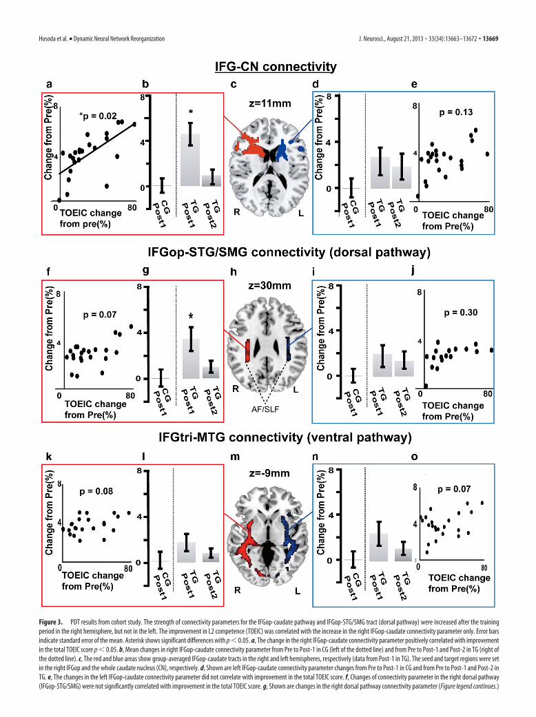
Figure 3. PDT results from cohort study. The strength of connectivity parameters for the IFGop-caudate pathway and IFGop-STG/SMG tract (dorsal pathway) were increased after the trainingperiod in the right hemisphere, but not in the left. The improvement in L2 competence (TOEIC) was correlated with the increase in the right IFGop-caudate connectivity parameter only. Error barsindicate standard error of the mean. Asterisk shows significant differences with p � 0.05. a, The change in the right IFGop-caudate connectivity parameter positively correlated with improvementin the total TOEIC score p � 0.05. b, Mean changes in right IFGop-caudate connectivity parameter from Pre to Post-1 in CG (left of the dotted line) and from Pre to Post-1 and Post-2 in TG (right ofthe dotted line). c, The red and blue areas show group-averaged IFGop-caudate tracts in the right and left hemispheres, respectively (data from Post-1 in TG). The seed and target regions were setin the right IFGop and the whole caudate nucleus (CN), respectively. d, Shown are left IFGop-caudate connectivity parameter changes from Pre to Post-1 in CG and from Pre to Post-1 and Post-2 inTG. e, The changes in the left IFGop-caudate connectivity parameter did not correlate with improvement in the total TOEIC score. f, Changes of connectivity parameter in the right dorsal pathway(IFGop-STG/SMG) were not significantly correlated with improvement in the total TOEIC score. g, Shown are changes in the right dorsal pathway connectivity parameter (Figure legend continues.)
Hosoda et al. • Dynamic Neural Network Reorganization J. Neurosci., August 21, 2013 • 33(34):13663–13672 • 13669

cular changes for GM, and remodeling ofthe myelin sheath and activity-dependentaxonal changes for WM reorganization in-dexed by FA (Scholz et al., 2009; Zatorre etal., 2012). Recently, an animal experimentassociated increases in FA with increasedexpression of a marker of myelination(Blumenfeld-Katzir et al., 2011). An impor-tant finding here was that the bidirectionalchanges of the behavioral, GM, and WMparameters paralleled each other, incontrast to a previous study showingdiscrepancy between training-inducedchanges in GM and WM (Taubert et al.,2010). The coupling of behavioral, GM, andWM changes supports that learning de-pends upon experience-dependent activi-ties correlated across connected regions(Fields, 2005; Zatorre et al., 2012).
The present study has provided the firstcompelling evidence for increased connec-tivity of specific tracts underlying L2 abilityand acquisition. We found that the IFGop constituted networkswith important language-related nodes: the STG/SMG (Price etal., 1999; Price and Crinion, 2005; Saur et al., 2008; Carreiras et al.,2009) and the caudate nucleus (Crinion et al., 2006; Friederici,2006). The IFGop and caudate nucleus probably constitute thecorticobasal ganglia circuits relevant to language processing andlearning. Impairments of the caudate nucleus are identified in peo-ple with mutation of FOXP2, which is an important genetic predis-position for language acquisition (Liegeois et al., 2003; Enard et al.,2009). Additionally, caudate activity underlies lexicosemantic con-trol in bilinguals (Crinion et al., 2006), and reduced striatal dopa-mine releases impair language processing (Tettamanti et al., 2005).The corticobasal ganglia circuits are suggested for reward-based re-inforcement learning since they receive both contextual informationfrom the cortex and reward signals from dopaminergic neurons(Doya, 2008). Reinforcement learning might play a part in enhanc-ing executive control of the IFG over the mechanisms for acquiringL2 vocabulary.
We found increased connectivity of the “dorsal pathway,” corre-sponding mainly to the temporal part of AF involved in phonologi-cal processing (Rilling et al., 2008). AF underlies successfulassociative learning between sounds and words (Wong et al., 2011;Yeatman et al., 2011). AF may thus integrate phonological and se-
mantic aspects of L2 vocabulary. The predominant involvement ofthe phonological subsystem in L2 vocabulary learning agrees withthe present finding showing little connectivity changes in the ventralpathway and ILF, which are involved mainly in semantic processing(Saur et al., 2008). The predominant involvement of the dorsal path-way suggests that phonology learning may have played a pivotal rolein the present L2 training paradigm. A future study, however, will beneeded to dissect the substrates for the lexicosemantic and phono-logical aspects of L2 vocabulary learning.
A rather surprising outcome was that the IFGop-caudate-STG/SMG network correlated with L2 vocabulary competence and itslearning-induced plastic changes were lateralized to the right hemi-sphere. These observations appear to contradict with mounting ev-idence indicating significance of the left hemispheric language areasfor L2 (Tettamanti et al., 2002; Musso et al., 2003; Abutalebi andGreen, 2007; Abutalebi et al., 2008; Sakai et al., 2009; Mårtensson etal., 2012; Schlegel et al., 2012; Ghazi Saidi et al., 2013). Moreover,learning of new languages induces plastic changes in GM and WM inthe left hemisphere (Schlegel et al., 2012). In early learning stages,however, the right frontal cortex is involved in acquisition of artifi-cial grammar and natural language (Fletcher et al., 1999, 2005; Tet-tamanti et al., 2002; Musso et al., 2003). Recent evidence has begunto highlight the importance of the right hemisphere for vocabularyand phonological aspects of L2 processing. The right STG/SMG isinvolved in non-L1 vocabulary learning (Smith et al., 2006; Jeong etal., 2010; Raboyeau et al., 2010; Veroude et al., 2010). We previouslyshowed that right IFGop activity for switching phonology from L1 toL2 was correlated with L2 vocabulary levels (Hosoda et al., 2012).Others showed enhanced activity in the right prefrontal areas duringpicture naming in L2 (Videsott et al., 2010), coupling of right IFGactivity with proficiency of word production in L2 (Calabrese et al.,2001; Vingerhoets et al., 2003; van Ettinger-Veenstra et al., 2010),and involvement of the right hemisphere in the control of verbalinterference in bilinguals (Filippi et al., 2011). Moreover, a case studysuggested an essential role of the right IFG for L2 (April and Tse,1977); a dextral late bilingual patient suffering from a right IFG le-sion had severe difficulty in finding words and reading more in L2than in L1. Hence, evidence certainly supports the roles of the rightIFG and STG in nongrammatical aspects of L2 usage, although theymay not be as well recognized as the correlates of L2 compared withthe left counterparts.
4
(Figure legend continued.) from Pre to Post-1 in CG and from Pre to Post-1 and Post-2 in TG.Error bars indicate SEM. Asterisks show significance differences with p � 0.05. h, The red andblue areas represent the group-averaged dorsal pathway tractography including AF and SLF inthe right and left hemispheres, respectively (data from Post-1 in TG). i, Mean changes in the leftdorsal pathway connectivity parameter from Pre to Post-1 in CG and from Pre to Post-1 andPost-2 in TG. j, Changes in the left dorsal pathway connectivity parameter did not correlate withthe change in the total TOEIC score. k, Changes in the right ventral pathway (IFGtri-MTG) pa-rameter were not significantly correlated with improvement in the total TOEIC score. l, Changesin the right ventral pathway connectivity parameter from Pre to Post-1 in CG and from Pre toPost-1 and Post-2 in TG. m, The red and blue areas represent the group-averaged ventralpathway tractography in the right and left hemispheres, respectively (data from Post-1 in TG).n, Mean changes in the left ventral pathway connectivity parameter from Pre to Post-1 in CGand from Pre to Post-1 and Post-2 in TG. o, Changes in the left ventral pathway connectivityparameter did not correlate with improvement in the total TOEIC score. The connectivity pa-rameters for the inferior longitudinal fascicles did not change in either hemisphere (data notshown). R, right; L, left.
Figure 4. CorrelationbetweenGMandWMchangesinducedbyL2training.a,ChangesinGMvolume(abscissa)andFA(ordinate)fromPre to Post-1 in TG. The plots show significant correlation between GM volume and FA changes ( p�0.03). b, Changes in GM volume andFA from Post-1 to Post-2. The plots show significant correlation between GM volume change and FA change ( p � 0.001, r � 0.65). Thecircles indicate participants with decreased TOEIC scores and the triangles indicate subjects with increased TOEIC scores from Post-1 toPost-2.Thefilledcircles indicateparticipantswhoshoweddecrementofthetotalTOEICscoresfromPost-1toPost-2whilethefilledtrianglesindicate participants who showed increment of the total TOEIC scores for the same period (see main text for the subgroup analysis).
13670 • J. Neurosci., August 21, 2013 • 33(34):13663–13672 Hosoda et al. • Dynamic Neural Network Reorganization

A limitation of the cohort experiment was no behavioral controlover CG during the training period. Although we cannot completelyexclude the effects of nonlanguage e-learning factors on the training-induced changes, we considered them unlikely as the sole explana-tion of the learning-induced plastic changes because of the followingreasons. First, there was no correlation of the total e-training timewith the GM/WM changes. Second, previous long-term computer-learning studies did not report changes of right IFG, caudate nucleus,or STG (Takeuchi et al., 2010; Wan et al., 2012). Consistently, pre-liminary results from our computer-based sequence learning exper-iment for 10 weeks failed to find changes in the right IFGop, caudatenucleus, or STG (C. Hosoda, M. Honda, T. Hanakawa, unpublishedobservation).
Paucity of evidence allows us only to speculate specific functionsof the right IFGop in L2 vocabulary learning. We theorize that theright IFGop might link lexicosemantic-phonological knowledge be-tween L1 and L2. For late L2 vocabulary learning, one should asso-ciate new L2 vocabulary temporarily represented in short-termmemory with existing L1 vocabulary represented in the left hemi-sphere. This agrees with a finding that acquisition of new L2 phonol-ogy recruits the left-lateralized language network (Paulesu et al.,2009). However, more long-term encoding-retrieval processes maymodify bilateral networks since the right prefrontal areas are impor-tant for accessing to long-term memory (Ranganath et al., 2007).Another simple, yet plausible, explanation is a spillover of languagerepresentations/control functions from the left to the right hemi-sphere. This could be particularly important here since Japanese isone of the most linguistically distant languages from English (Chis-wick and Miller, 2005). The spillover concept is consistent with find-ings that mathematics/arithmetic experts show greater righthemispheric activity than control subjects for whom left hemi-spheric activity is more relevant (Hanakawa et al., 2003; Aydin et al.,2007). These findings may characterize a repertoire of use-dependent dynamic reorganization of the brain. A dramatic exam-ple is a child who shifted the originally left hemispheric languagecenters to the mirror-reversed, right hemispheric sites after surgicalremoval of the left hemisphere (Hertz-Pannier et al., 2002). Lan-guage experience-dependent changes may likely occur in the rightIFG because of weaker genetic influences on the right IFG comparedwith the left (Thompson et al., 2001).
In conclusion, the present study indicates that the macro-scopic reorganization of the IFGop-caudate-STG network, espe-cially on the right, can underlie L2 vocabulary learning in adults(1500/1500 words).
ReferencesAbutalebi J, Green DW (2007) Bilingual language production: the neurocogni-
tion of language representation and control. J Neurolinguistics 20:242–275.CrossRef
Abutalebi J, Annoni JM, Zimine I, Pegna AJ, Seghier ML, Lee-Jahnke H, LazeyrasF, Cappa SF, Khateb A (2008) Language control and lexical competition inbilinguals: an event-related FMRI study. Cereb Cortex 18:1496–1505.Medline
Alexander AL, Lee JE, Lazar M, Field AS (2007) Diffusion tensor imaging ofthe brain. Neurotherapeutics 4:316 –329. CrossRef Medline
April RS, Tse PC (1977) Cross aphasia in a Chinese bilingual dextral. ArchNeurol 34:766 –770. CrossRef Medline
Ashburner J (2007) A fast diffeomorphic image registration algorithm.Neuroimage 38:95–113. CrossRef Medline
Aydin K, Ucar A, Oguz KK, Okur OO, Agayev A, Unal Z, Yilmaz S, Ozturk C(2007) Increased gray matter density in the parietal cortex of mathemati-cians: a voxel-based morphometry study. AJNR Am J Neuroradiol 28:1859–1864. CrossRef Medline
Beaulieu C (2002) The basis of anisotropic water diffusion in the nervoussystem–a technical review. NMR Biomed 15:435– 455. CrossRef Medline
Blumenfeld-Katzir T, Pasternak O, Dagan M, Assaf Y (2011) Diffusion MRI
of structural brain plasticity induced by a learning and memory task. PLoSOne 6:e20678. CrossRef Medline
Briellmann RS, Mitchell LA, Waites AB, Abbott DF, Pell GS, Saling MM,Jackson GD (2003) Correlation between language organization and dif-fusion tensor abnormalities in refractory partial epilepsy. Epilepsia 44:1541–1545. CrossRef Medline
Calabrese P, Neufeld H, Falk A, Markowitsch HJ, Muller C, Heuser L, GehlenW, Durwen HF (2001) Wortgenerierung bei Bilingualen-eine fMRT-Studie mit Implikationen fur Sprach-und Gedachtnisprozesse. FortschrNeurol Psychiatr 69:42– 49. CrossRef Medline
Carreiras M, Seghier ML, Baquero S, Estevez A, Lozano A, Devlin JT, Price CJ(2009) An anatomical signature for literacy. Nature 461:983–986.CrossRef Medline
Catani M, Mesulam M (2008) The arcuate fasciculus and the disconnectiontheme in language and aphasia: history and current state. Cortex 44:953–961. CrossRef Medline
Chiswick BR, Miller PW (2005) Linguistic distance: a quantitative measureof the distance between English and other languages. J Multiling MulticultDev 26:1–11. CrossRef
Coady J, Huckin T (1997) Second Language Vocabulary Acquisition: Cam-bridge UP.
Crinion J, Turner R, Grogan A, Hanakawa T, Noppeney U, Devlin JT, Aso T,Urayama S, Fukuyama H, Stockton K, Usui K, Green DW, Price CJ(2006) Language control in the bilingual brain. Science 312:1537–1540.CrossRef Medline
Dowens MG, Vergara M, Barber HA, Carreiras M (2010) Morphosyntacticprocessing in late second-language learners. J Cogn Neurosci 22:1870–1887.CrossRef Medline
Doya K (2008) Modulators of decision making. Nat Neurosci 11:410 – 416.CrossRef Medline
Draganski B, Gaser C, Busch V, Schuierer G, Bogdahn U, May A (2004) Neu-roplasticity: changes in grey matter induced by training. Nature 427:311–312.CrossRef Medline
Ellis R, Heimbach R, Tanaka Y, Yamazaki A (1999) Learning a second lan-guage through interaction. Amsterdam/Philadelphia: John BenjaminsPublishing Company.
Enard W, Gehre S, Hammerschmidt K, Holter SM, Blass T, Somel M, Bruck-ner MK, Schreiweis C, Winter C, Sohr R, Becker L, Wiebe V, Nickel B,Giger T, Muller U, Groszer M, Adler T, Aguilar A, Bolle I, Calzada-WackJ, et al. (2009) A humanized version of Foxp2 affects cortico-basal gan-glia circuits in mice. Cell 137:961–971. CrossRef Medline
Fields RD (2005) Myelination: an overlooked mechanism of synaptic plas-ticity? Neuroscientist 11:528 –531. CrossRef Medline
Filippi R, Richardson FM, Dick F, Leech R, Green DW, Thomas MS, Price CJ(2011) The right posterior paravermis and the control of language inter-ference. J Neurosci 31:10732–10740. CrossRef Medline
Fletcher P, Buchel C, Josephs O, Friston K, Dolan R (1999) Learning-relatedneuronal responses in prefrontal cortex studied with functional neuroim-aging. Cereb Cortex 9:168 –178. CrossRef Medline
Fletcher PC, Zafiris O, Frith CD, Honey RA, Corlett PR, Zilles K, Fink GR(2005) On the benefits of not trying: brain activity and connectivity re-flecting the interactions of explicit and implicit sequence learning. CerebCortex 15:1002–1015. Medline
Floel A, de Vries MH, Scholz J, Breitenstein C, Johansen-Berg H (2009)White matter integrity in the vicinity of Broca’s area predicts grammarlearning success. Neuroimage 47:1974 –1981. CrossRef Medline
Friederici AD (2006) What’s in control of language? Nat Neurosci 9:991–992.CrossRef Medline
Ghazi Saidi L, Perlbarg V, Marrelec G, Pelegrini-Issac M, Benali H, AnsaldoAI (2013) Functional connectivity changes in second language vocabu-lary learning. Brain Lang 124:56 – 65. CrossRef Medline
Golestani N, Pallier C (2007) Anatomical correlates of foreign speech soundproduction. Cereb Cortex 17:929 –934. Medline
Golestani N, Molko N, Dehaene S, LeBihan D, Pallier C (2007) Brain structurepredicts the learning of foreign speech sounds. Cereb Cortex 17:575–582.Medline
Grogan A, Green DW, Ali N, Crinion JT, Price CJ (2009) Structural corre-lates of semantic and phonemic fluency ability in first and second lan-guages. Cereb Cortex 19:2690 –2698. CrossRef Medline
Grogan A, Parker Jones O, Ali N, Crinion J, Orabona S, Mechias ML, Rams-den S, Green DW, Price CJ (2012) Structural correlates for lexical effi-
Hosoda et al. • Dynamic Neural Network Reorganization J. Neurosci., August 21, 2013 • 33(34):13663–13672 • 13671

ciency and number of languages in non-native speakers of English.Neuropsychologia 50:1347–1352. CrossRef Medline
Hanakawa T, Honda M, Okada T, Fukuyama H, Shibasaki H (2003) Neuralcorrelates underlying mental calculation in abacus experts: a functionalmagnetic resonance imaging study. Neuroimage 19:296 –307. CrossRefMedline
Hernandez AE, Dapretto M, Mazziotta J, Bookheimer S (2001) Languageswitching and language representation in Spanish-English bilinguals: anfMRI study. Neuroimage 14:510 –520. CrossRef Medline
Hertz-Pannier L, Chiron C, Jambaque I, Renaux-Kieffer V, Van de MoortelePF, Delalande O, Fohlen M, Brunelle F, Le Bihan D (2002) Late plasticityfor language in a child’s non-dominant hemisphere: a pre- and post-surgery fMRI study. Brain 125:361–372. CrossRef Medline
Hosoda C, Hanakawa T, Nariai T, Ohno K, Honda M (2012) Neural mech-anisms of language switch. J Neurolinguistics 25:44 – 61. CrossRef
Hsu AS, Chater N, Vitanyi PM (2011) The probabilistic analysis of languageacquisition: theoretical, computational, and experimental analysis. Cog-nition 120:380 –390. Medline
Jeong H, Sugiura M, Sassa Y, Wakusawa K, Horie K, Sato S, Kawashima R (2010)Learning second language vocabulary: neural dissociation of situation-basedlearning and text-based learning. Neuroimage 50:802–809. CrossRefMedline
Johansen-Berg H (2007) Structural plasticity: rewiring the brain. Curr Biol17:R141–R144. CrossRef Medline
Johansen-Berg H (2012) The future of functionally-related structuralchange assessment. Neuroimage 62:1293–1298. CrossRef Medline
Johansen-Berg H, Baptista CS, Thomas AG (2012) Human structural plas-ticity at record speed. Neuron 73:1058 –1060. CrossRef Medline
Jou RJ, Jackowski AP, Papademetris X, Rajeevan N, Staib LH, Volkmar FR(2011) Diffusion tensor imaging in autism spectrum disorders: prelimi-nary evidence of abnormal neural connectivity. Aust N Z J Psychiatry45:153–162. CrossRef Medline
Kerns JG, Cohen JD, MacDonald AW 3rd, Cho RY, Stenger VA, Carter CS(2004) Anterior cingulate conflict monitoring and adjustments in con-trol. Science 303:1023–1026. CrossRef Medline
Kuhl PK (2010) Brain mechanisms in early language acquisition. Neuron67:713–727. CrossRef Medline
Laufer B (1989) A factor of difficulty in vocabulary learning: deceptivetransparency. AILA Rev 6:10 –20.
Le Bihan D, Johansen-Berg H (2012) Diffusion MRI at 25: exploring braintissue structure and function. Neuroimage 61:324 –341. Medline
Liegeois F, Baldeweg T, Connelly A, Gadian DG, Mishkin M, Vargha-KhademF (2003) Language fMRI abnormalities associated with FOXP2 genemutation. Nat Neurosci 6:1230 –1237. CrossRef Medline
Mårtensson J, Eriksson J, Bodammer NC, Lindgren M, Johansson M, NybergL, Lovden M (2012) Growth of language-related brain areas after for-eign language learning. Neuroimage 63:240 –244. CrossRef Medline
Mechelli A, Crinion JT, Noppeney U, O’Doherty J, Ashburner J, FrackowiakRS, Price CJ (2004) Neurolinguistics: structural plasticity in the bilin-gual brain. Nature 431:757. CrossRef Medline
Meschyan GaH, A (2002) Is native-language decoding skill related tosecond-language learning? J Educ Psychol 94:14 –22. CrossRef
Musso M, Moro A, Glauche V, Rijntjes M, Reichenbach J, Buchel C, Weiller C(2003) Broca’s area and the language instinct. Nat Neurosci 6:774 –781.CrossRef Medline
Paulesu E, Vallar G, Berlingeri M, Signorini M, Vitali P, Burani C, Perani D,Fazio F (2009) Supercalifragilisticexpialidocious: how the brain learnswords never heard before. Neuroimage 45:1368 –1377. CrossRef Medline
Peltier J, Nicot B, Baroncini M, Zunon-Kipre Y, Haidara A, Havet E, FoulonP, Page C, Lejeune JP, Le Gars D (2011) [Anatomy of the periventricularwhite matter]. Neurochirurgie 57:151–155. CrossRef Medline
Price CJ, Crinion J (2005) The latest on functional imaging studies of apha-sic stroke. Curr Opin Neurol 18:429 – 434. CrossRef Medline
Price CJ, Green DW, von Studnitz R (1999) A functional imaging study oftranslation and language switching. Brain 122:2221–2235. CrossRefMedline
Raboyeau G, Marcotte K, Adrover-Roig D, Ansaldo AI (2010) Brain activa-tion and lexical learning: the impact of learning phase and word type.Neuroimage 49:2850 –2861. CrossRef Medline
Ranganath C, Heller AS, Wilding EL (2007) Dissociable correlates of two classesof retrieval processing in prefrontal cortex. Neuroimage 35:1663–1673.CrossRef Medline
Rilling JK, Glasser MF, Preuss TM, Ma X, Zhao T, Hu X, Behrens TE (2008)The evolution of the arcuate fasciculus revealed with comparative DTI.Nat Neurosci 11:426 – 428. CrossRef Medline
Sakai KL, Nauchi A, Tatsuno Y, Hirano K, Muraishi Y, Kimura M, BostwickM, Yusa N (2009) Distinct roles of left inferior frontal regions that ex-plain individual differences in second language acquisition. Hum BrainMapp 30:2440 –2452. CrossRef Medline
Saur D, Kreher BW, Schnell S, Kummerer D, Kellmeyer P, Vry MS, UmarovaR, Musso M, Glauche V, Abel S, Huber W, Rijntjes M, Hennig J, Weiller C(2008) Ventral and dorsal pathways for language. Proc Natl Acad SciU S A 105:18035–18040. CrossRef Medline
Schlegel A, Rudelson JJ, Tse P (2012) White matter structure changes asadults learn a second language. J Cogn Neurosci 24:1664 –1670. CrossRef
Scholz J, Klein MC, Behrens TE, Johansen-Berg H (2009) Training induceschanges in white-matter architecture. Nat Neurosci 12:1370 –1371.CrossRef Medline
Smith SM, Jenkinson M, Woolrich MW, Beckmann CF, Behrens TE,Johansen-Berg H, Bannister PR, De Luca M, Drobnjak I, Flitney DE,Niazy RK, Saunders J, Vickers J, Zhang Y, De Stefano N, Brady JM, Mat-thews PM (2004) Advances in functional and structural MR image anal-ysis and implementation as FSL. Neuroimage 23 [Suppl 1]:S208 –S219.Medline
Smith SM, Jenkinson M, Johansen-Berg H, Rueckert D, Nichols TE, MackayCE, Watkins KE, Ciccarelli O, Cader MZ, Matthews PM, Behrens TE(2006) Tract-based spatial statistics: voxelwise analysis of multi-subjectdiffusion data. Neuroimage 31:1487–1505. CrossRef Medline
Takeuchi H, Sekiguchi A, Taki Y, Yokoyama S, Yomogida Y, Komuro N,Yamanouchi T, Suzuki S, Kawashima R (2010) Training of workingmemory impacts structural connectivity. J Neurosci 30:3297–3303.CrossRef Medline
Taubert M, Draganski B, Anwander A, Muller K, Horstmann A, Villringer A,Ragert P (2010) Dynamic properties of human brain structure:learning-related changes in cortical areas and associated fiber connec-tions. J Neurosci 30:11670 –11677. CrossRef Medline
Tettamanti M, Alkadhi H, Moro A, Perani D, Kollias S, Weniger D (2002)Neural correlates for the acquisition of natural language syntax. Neuro-image 17:700 –709. CrossRef Medline
Tettamanti M, Moro A, Messa C, Moresco RM, Rizzo G, Carpinelli A, Ma-tarrese M, Fazio F, Perani D (2005) Basal ganglia and language: phonol-ogy modulates dopaminergic release. Neuroreport 16:397– 401. CrossRefMedline
Thierry G, Wu YJ (2007) Brain potentials reveal unconscious translationduring foreign-language comprehension. Proc Natl Acad Sci U S A 104:12530 –12535. CrossRef Medline
Thompson PM, Cannon TD, Narr KL, van Erp T, Poutanen VP, Huttunen M,Lonnqvist J, Standertskjold-Nordenstam CG, Kaprio J, Khaledy M, DailR, Zoumalan CI, Toga AW (2001) Genetic influences on brain struc-ture. Nat Neurosci 4:1253–1258. CrossRef Medline
van Ettinger-Veenstra HM, Ragnehed M, Hallgren M, Karlsson T, LandtblomAM, Lundberg P, Engstrom M (2010) Right-hemispheric brain activa-tion correlates to language performance. Neuroimage 49:3481–3488.CrossRef Medline
Van Ettinger-Veenstra H, Ragnehed M, McAllister A, Lundberg P, EngstromM (2012) Right-hemispheric cortical contributions to language abilityin healthy adults. Brain Lang 120:395– 400. CrossRef Medline
Veroude K, Norris DG, Shumskaya E, Gullberg M, Indefrey P (2010) Func-tional connectivity between brain regions involved in learning words of anew language. Brain Lang 113:21–27. CrossRef Medline
Videsott G, Herrnberger B, Hoenig K, Schilly E, Grothe J, Wiater W, SpitzerM, Kiefer M (2010) Speaking in multiple languages: neural correlates oflanguage proficiency in multilingual word production. Brain Lang 113:103–112. CrossRef Medline
Vigneau M, Beaucousin V, Herve PY, Jobard G, Petit L, Crivello F, Mellet E,Zago L, Mazoyer B, Tzourio-Mazoyer N (2011) What is right-hemisphere contribution to phonological, lexico-semantic, and sentenceprocessing? Insights from a meta-analysis. Neuroimage 54:577–593.CrossRef Medline
Vingerhoets G, Van Borsel J, Tesink C, van den Noort M, Deblaere K,Seurinck R, Vandemaele P, Achten E (2003) Multilingualism: an fMRIstudy. Neuroimage 20:2181–2196. CrossRef Medline
Wan X, Takano D, Asamizuya T, Suzuki C, Ueno K, Cheng K, Ito T, Tanaka
13672 • J. Neurosci., August 21, 2013 • 33(34):13663–13672 Hosoda et al. • Dynamic Neural Network Reorganization

K (2012) Developing intuition: neural correlates of cognitive-skill learn-ing in caudate nucleus. J Neurosci 32:17492–17501. Medline
Wong FC, Chandrasekaran B, Garibaldi K, Wong PC (2011) White matteranisotropy in the ventral language pathway predicts sound-to-wordlearning success. J Neurosci 31:8780 – 8785. CrossRef Medline
Yeatman JD, Dougherty RF, Rykhlevskaia E, Sherbondy AJ, Deutsch GK,
Wandell BA, Ben-Shachar M (2011) Anatomical properties of the arcu-ate fasciculus predict phonological and reading skills in children. J CognNeurosci 23:3304 –3317. CrossRef Medline
Zatorre RJ, Fields RD, Johansen-Berg H (2012) Plasticity in gray and white:neuroimaging changes in brain structure during learning. Nat Neurosci15:528 –536. CrossRef Medline
Hosoda et al. • Dynamic Neural Network Reorganization J. Neurosci., August 21, 2013 • 33(34):13663–13672 • 13672a