basics of treatment planning ii - uthgsbsmedphys.org · 3/16/15 1 basics of treatment planning ii...
TRANSCRIPT

3/16/15
1
Basics of treatment planning II
Sastry Vedam PhD DABR
Introduction to Medical Physics III: Therapy Spring 2015
Dose calculation algorithms
! Correction based
! Model based

3/16/15
2
Dose calculation algorithms
! Representation of patient and dose distribution ! Block of tissue of uniform density ! Contour external surface with solder
wire ! Contours obtained from CT
Dose calculation algorithms
! Modern algorithms ! 3D point by point/voxel by voxel description of
patient (CT) ! Spatial reliability of CT (<2%) ! Dose uncertainty (photon beams) <1% ! Typical CT scan ! 50 – 100 images ! 2.5 – 5 mm slice thickness ! 512x512 pixels per imaging plane ! 2-16 bytes to store HU value data

3/16/15
3
Dose calculation algorithms
! Speed ! Processor power ! Grid spacing ! Non uniform sample spacing within
grid ! Calculation algorithm
Correction based algorithms

3/16/15
4
Dose calculation algorithms
! Correction based ! Semi empirical ! Based on measured data (PDD, Profiles etc.,)
! Reference calibration condition ! Dose/MU @ a defined location in water phantom for a defined field
size
! Corrections for: ! Attenuation
! Contour irregularity ! Beam modifiers ! Tissue inhomogeneities
! Scatter (Scattering volume, field size, shape and radial distance) ! Geometry (Non reference SSD/depth)
MU – Isocentric setup

3/16/15
5
MU – Non Isocentric setup
Correction based algorithms
! Limited accuracy ! 3D heterogeneity corrections at tissue interfaces
! Lack of complete electronic equilibrium
! Secondary check for MUs calculated from more complex model based algorithms

3/16/15
6
Model based algorithms
Model based algorithms
! Compute dose distribution with a physical model that actually simulates radiation transport through a patient
! Radiation transport ! Production of megavoltage X-rays in treatment head
! Interaction and scattering of photons by Compton Effect
! Effects of transport of charged particles near boundaries and tissue heterogeneities

3/16/15
7
Radiation Transport
Electron disequilibrium due to greater lateral range of electrons compared to field size
Radiation Transport
Pencil beam charge particle tracks in phantom

3/16/15
8
Convolution
Convolution
Energy fluence
Energy deposition kernel (Patient density map)
Dose

3/16/15
9
Convolution/Superposition ! Several variations
! Common/essential components ! Energy imparted to medium by
interactions of primary photons (TERMA)
! Energy deposited about a primary interaction site (Kernel)
! Kernel ! Primary (Primary dose) ! First and multiple scatter dose (Can
be calculated together or separately)
! Kernel also referred to as: ! Dose spread array ! Differential pencil beam ! Point spread function ! Energy deposition kernel
TERMA
! Total energy released per unit mass ! Energy imparted to secondary charged particles
! Energy retained by scattered photon
! Sum of the above should equal energy of the primary photon for each interaction
Energy fluence
Mass attenuation coefficient TERMA
3/16/15
10
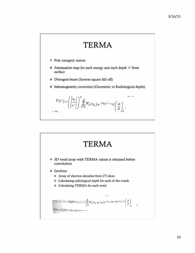
TERMA
! Poly energetic nature
! Attenuation map for each energy and each depth ‘r’ from surface
! Divergent beam (Inverse square fall off)
! Inhomogeneity correction (Geometric vs Radiological depth)
TERMA
! 3D voxel array with TERMA values is obtained before convolution
! Involves: ! Array of electron densities from CT slices
! Calculating radiological depth for each of the voxels
! Calculating TERMA for each voxel
3/16/15
11
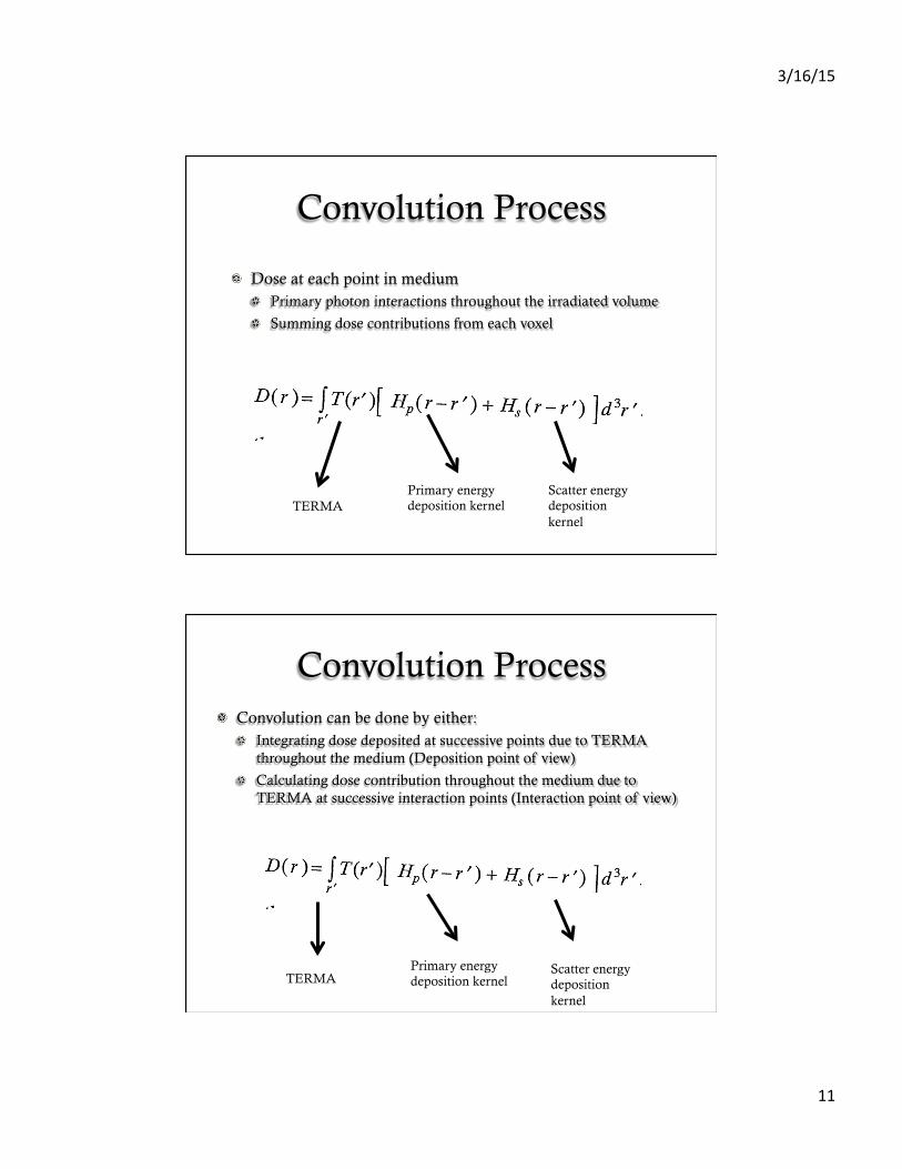
Convolution Process
! Dose at each point in medium ! Primary photon interactions throughout the irradiated volume
! Summing dose contributions from each voxel
TERMA Primary energy deposition kernel
Scatter energy deposition kernel
Convolution Process ! Convolution can be done by either:
! Integrating dose deposited at successive points due to TERMA throughout the medium (Deposition point of view)
! Calculating dose contribution throughout the medium due to TERMA at successive interaction points (Interaction point of view)
TERMA Primary energy deposition kernel
Scatter energy deposition kernel

3/16/15
12
Convolution process
Deposition point of view Interaction point of view
Convolution process
! Deposition point of view ! Best if dose is calculated only in a subset of the irradiated
volume
! TERMA in each voxel must be stored in an array
! Interaction point of view ! Only a single TERMA value needs to be stored at any time
3/16/15
13
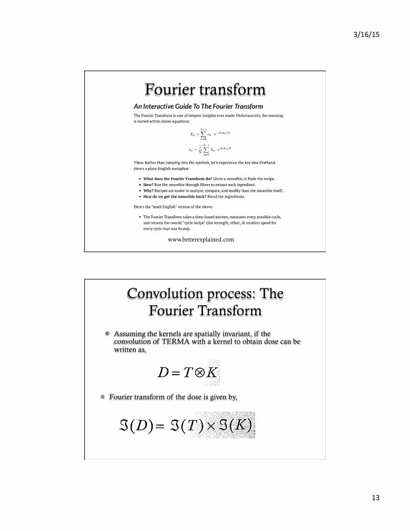
Fourier transform
www.betterexplained.com
Convolution process: The Fourier Transform
! Assuming the kernels are spatially invariant, if the convolution of TERMA with a kernel to obtain dose can be written as,
! Fourier transform of the dose is given by,

3/16/15
14
Convolution in Inhomogeneous medium
! Kernels are functions of displacement only
! In Inhomogeneous media, the fractional energy contribution will depend on both distance between interaction site and deposition site as well as densities at interaction and deposition sites
Convolution in Inhomogeneous medium
! First approximation ! Energy loss by secondary electrons dependent on effective path
length (average density through ray tracing)
! Incorrect for primary kernel ! Electron scattering ! No and energy of electrons depends not only on avg density but also on density
distribution
! Good for scatter kernel ! Fluence of onee-scattered photons is proportional to average density
! Range of electrons ejected by these photons is very small

3/16/15
15
Convolution in Inhomogeneous medium
! Since rate of energy deposition in each voxel is proportional to the density within voxel, kernel value can be obtained by
! Substituting into the equation for dose,
Variations of convolution
! Original Real-Space Convolution (Mackie et al, 1985)
! Kernels separated into primary, truncated first scatter (TFS) and residual first and multiple scatter (RFMS) arrays
! TFS – First scatter dose, relatively close to the interaction site
! RFMS – Multiple scatter and first scatter not included in TFS
! Scatter separation allowed for smaller higher resolution kernel arrays
! Average density scaling for a range of densities between interaction and deposition sites for primary and TFS
! Avg density of phantom in kernel scaling for RFMS

3/16/15
16
Variations of convolution
! Differential pencil beam method (Mohan et al, 1986, Ahnesjo et al., 1987)
! Infinitesimal segment of a pencil beam
! Equivalent to a convolution kernel except, ! Dose deposited in water per unit primary photon collision
density, instead of per unit energy imparted by primary photons
Variations of convolution
! Collapsed cone convolution (Ahnesjo et al., 1989)
! Polyenergetic TERMA and kernel
! Kernel represented analytically and combines primary and scatter contributions
! Functions used to characterize kernel are,

3/16/15
17
Variations of convolution ! Collapsed cone convolution (Ahnesjo et al., 1989)
! Finite number of polar angles w.r.t. primary beam along which the function is defined
! Interaction site – apex of a set of radially directed lines spreading out in 3D
! Each line is further considered the axis of a cone
! Kernel function along each line – energy deposited within the entire cone at radius ‘r’ collapsed onto the line
! TERMA is calculated and represented in a cartesian array
! Inhomogeneities are accounted for in TERMA array
! Reduced computation time when compared to conventional convolution