basic research biology c1 silencing ... - tulane university
TRANSCRIPT

SIGNIFICANCE
Atp6v1c1 silencing attenuatesalveolar bone resorption andprotects the periodontalligament from destructioncaused by inflammation inendodontic disease. Insightsresulting from this study mayassist in developing noveltreatments for endodonticdisease and other osteolyticand inflammatory diseases.
From the *Department of Orthodontics,School and Hospital of Stomatology,Tongji University, Shanghai EngineeringResearch Center of Tooth Restorationand Regeneration, Shanghai, China;†Department of Pathology, School ofMedicine, University of Alabama atBirmingham, Birmingham, Alabama; and‡Oral Biomedical Engineering Laboratory,Shanghai Stomatological Hospital, FudanUniversity, Shanghai, China
Address requests for reprints to Dr Yi-PingLi, Department of Pathology, The Schoolof Medicine, University of Alabama atBirmingham, SHEL 810, 1825 UniversityBlvd, Birmingham, AL 35294-2182, orDr Yuehua Liu, Oral BiomedicalEngineering Laboratory, ShanghaiStomatological Hospital, FudanUniversity, 356 Beijing East Road,Shanghai 20001, China.E-mail addresses: [email protected] [email protected]/$ - see front matter
Copyright © 2019 Published by ElsevierInc. on behalf of American Association ofEndodontists.https://doi.org/10.1016/j.joen.2019.02.024
Yuhui Wang, DDS,*† Wei Chen,
MD,† Liang Hao, DDS, PhD,†
Abigail McVicar,† Jinjin Wu, MS,†
Ning Gao, DDS, PhD,†
Yuehua Liu, DDS, PhD,‡ andYi-Ping Li, PhD†
898 Wang et al.
Downloaded for Anonymous UsFor personal use o
BASIC RESEARCH – BIOLOGY
C1 Silencing AttenuatesInflammation and AlveolarBone Resorption in EndodonticDisease
ABSTRACT
Introduction: Endodontic disease, 1 of the most prevalent chronic infectious diseasesworldwide, occurs when the dental pulp becomes infected and inflamed, leading to bonedestruction around the tooth root, severe pain, and tooth loss. Although many studies havetried to develop therapies to alleviate the bone erosion and inflammation associated withendodontic disease, there is an urgent need for an effective treatment. Methods: In thisstudy, we used a gene-based therapy approach by administering recombinant adeno-associated virus (AAV)-mediated Atp6v1c1 knockdown to target both periapical boneresorption and inflammation in the mouse model of endodontic disease.Results: The resultsshowed that Atp6v1c1 knockdown is simultaneously capable of reducing bone resorption by70% through impaired osteoclast activation, inhibiting inflammation by decreasing T-cellinfiltration in the periapical lesion by 75%, and protecting the periodontal ligament fromdestruction caused by inflammation. Notably, AAV-mediated gene therapy of Atp6v1c1knockdown significantly reduced proinflammatory cytokine expression, including tumor ne-crosis factor a, interleukin 1a, interleukin 17, interleukin 12, and interleukin 6 levels in periapicaltissues caused by bacterial infection. Quantitative real-time polymerase chain reactionrevealed that Atp6v1c1 knockdown reduced osteoclast-specific functional genes (ie, Ctsk) inperiapical tissues. Conclusions: Our results showed that AAV-mediated Atp6v1c1 knock-down in periapical tissues slowed endodontic disease progression, prevented bone erosion,and alleviated inflammation in the periapical tissues and periodontal ligament potentiallythrough regulation of toll-like receptor signaling, indicating that targeting Atp6v1c1 mayfacilitate the design of novel therapeutic approaches to reduce inflammation and bone erosionin endodontic disease. (J Endod 2019;45:898–906.)
KEY WORDS
Atp6v1c1; RNA interference knockdown; gene therapy; gingival inflammation; oral boneresorption; periapical disease
Dental plaque bacteria is the main cause of dental caries. If left untreated, the bacteria progresses to thedental pulp in the root canal, leading to pulp inflammation and necrosis and further bone destructionaround the tooth root, severe pain, and tooth loss. The periodontal ligament (PDL) is comprised ofspecialized connective tissue fibers that attach the tooth to the alveolar bone. During endodontic diseaseprogression, inflammation in the periapical area and PDL leads to tooth loss. Periapical bone destructionis mainly induced by enhanced osteoclast formation and function initiated by extracellular acidificationand following the degradation of the organic constituent of bone. The bacteria increases T- and B-cellactivation, which promotes both inflammation and osteoclast differentiation1. Currently, endodonticdisease is treated by mechanical removal of the infected pulp tissue followed by obturation of the rootcanal space with an inert filling material2; however, periapical bone regeneration may take as long as 2years, and in some cases complete healing is never achieved. Thus, there is urgent need for a novelosteoclast-specific inhibitor that can simultaneously target bone loss and inflammation in periapicaldisease.
Atp6v1c1, a subunit of vacuolar-type H1-ATPase (V-ATPase), is highly expressed in osteoclastsand is mainly localized on the ruffled border of activated osteoclasts3. Functioning as a subunit of
JOE � Volume 45, Number 7, July 2019
er (n/a) at University of Alabama at Birmingham from ClinicalKey.com by Elsevier on October 09, 2019.nly. No other uses without permission. Copyright ©2019. Elsevier Inc. All rights reserved.

osteoclast proton pump Atp6i, Atp6v1c1 canbe induced by receptor activator of nuclearfactor kappa-B ligand (RANKL) andmacrophage colony-stimulating factor(M-CSF) during osteoclast differentiation3. Themature osteoclasts attach to the bone surfaceto deliver the proton pump V-ATPase and thenrelease extracellular matrix–digesting acidproteases, such as cathepsin K (Ctsk), for thedegradation of bone matrices4–6. Our previousresearch showed that osteoclast acidificationactivity and bone resorption are impairedthrough silencing of Atp6v1c13, suggesting itstherapeutic potential for diseases of excessiveosteoclastic bone resorption. Furthermore, ourprevious studies have revealed that silencing ofAtp6v1c1 can prevent cancer progression andmetastasis, indicating its potential function inimmune responses7. As a subunit of Atp6i,Atp6v1c1 is expressed in immune cells suchas macrophages and dendritic cells, as well asosteoclasts3,8, and may play an osteoimmunerole during endodontic disease pathogenesis.The regulation of both osteoclast differentiationand the immune response is crucial for themaintenance of alveolar bone volume becausedisruptions may result in pathologicosteoclastic diseases such as endodonticdisease.
We hypothesized that local Atp6v1c1silencing in the periapical lesion could inhibitosteoclastic activity and inflammationsimultaneously. To investigate the potentialrole of C1 in periapical disease, we used apolymicrobial-induced periapical mouse modelin conjunction with the AAV knockdownsystem to investigate the effect of Atp6v1c1silencing in periapical disease. We proposeAAV-mediated knockdown of Atp6v1c1 as anovel therapeutic target for the treatment ofendodontic disease.
MATERIALS AND METHODS
Study ApprovalAll animal experimentation was performedaccording to the legal requirements of theAssociation for Assessment and Accreditationof the Laboratory Animal Care Internationaland the University of Alabama at BirminghamInstitutional Animal Care and Use Committeeand followed all recommendations of AnimalResearch: Reporting of In Vivo Experimentsguidelines.
AAV RNA Interferences ViralProduction and PurificationWe purchased the AAV Helper-Free System(AAV Helper-Free System Catalog #240071;Stratagene, San Diego, CA). Viral productionwas accomplished using a triple-transfection,
JOE � Volume 45, Number 7, July 2019
Downloaded for Anonymous User (n/a) aFor personal use only. No
helper-free method and purified with amodified version of a published protocol9.
Pulp Exposure, Bacterial Infection,and Transduction of AAV VectorsThe periapical disease mouse model wasproduced as we previously described1,10.Bacterial culture, infection, and viral vectortransduction in a site-specific manner wasperformed as described1,10.
Statistical AnalysisExperimental data are reported as the mean6
standard deviation of triplicate independentsamples. The figures are representative ofthe data (n 5 15). Data quantification analyseswere performed using ImageJ (NationalInstitutes of Health, Bethesda, MD) asdescribed1,10,11.
RESULTS
Atp6v1c1 Knockdown ImpairedOsteoclast Function IncludingOsteoclast-mediated BoneResorption and ExtracellularAcidification In VitroTo enable knockdown of Atp6v1c1, wegenerated short hairpin RNA that targetedAtp6v1c1. We used the AAV2 serotype andsubcloned short hairpin RNA targetingAtp6v1c1 into the AAV.H1 vector (courtesy ofDr. Sergei Musatov, Weill Cornell MedicalCollege, New York City, New York) (Fig. 1A).To confirm the effect of Atp6v1c1 silencing, weexamined the expression of Atp6v1c1 inmouse bonemarrow (MBM) isolated fromwild-type BALB/cJ mice, cultured with M-CSF andRANKL to generate osteoclasts12, andtransduced with AAV-sh-Atp6v1c1 orAAV-luc-YFP. Western blot analysis revealedthat osteoclasts transduced withAAV-sh-Atp6v1c1 have an 80% reduction inAtp6v1c1 expression compared withuntreated osteoclasts (mock) or osteoclaststransduced with AAV-luc-YFP (Fig. 1B).Overall, our results indicate thatAAV-sh-Atp6v1c1 efficiently knocked downAtp6v1c1 expression. To investigate howAtp6v1c1 knockdown can affect osteoclastdifferentiation and function, tartrate-resistantacid phosphatase (TRAP) staining, wheatgerm agglutinin staining, and scanningelectron microscopy were performed. Notably,osteoclast number was not significantlychanged after transduction withAAV-sh-Atp6v1c1, as shown by TRAP staining(Fig. 1C and F). We further sought toinvestigate the effects of AAV-sh-Atp6v1c1 onosteoclast function by examining boneresorption. Wheat germ agglutinin stainingshowed that compared with AAV-luc-YFP
AAV-sh-At
t University of Alabama at Birmingham from ClinicalKey.coother uses without permission. Copyright ©2019. Elsevier Inc
treatment, AAV-mediated Atp6v1c1knockdown reduced osteoclast-mediatedbone resorption by 80% (Fig. 1D and G).Consistently, visualization of the resorptionlacunae through scanning electron microscopyshowed that AAV-sh-Atp6v1c1 completelyimpaired bone resorption in vitro (Fig. 1E andH). To further investigate how Atp6v1c1knockdown can affect osteoclast function, weexamined osteoclast extracellular acidification.Osteoclasts were induced from wild-typeMBM stimulated by M-CSF and RANKL for 3days. After transduction by lentivirus-mediatedAtp6v1c1 knockdown, acridine orangestaining was conducted to evaluate osteoclastextracellular acidification (Fig. 1I). We foundthat lentivirus-sh-Atp6v1c1 severely impairedextracellular acidification compared with thescrambled control (Fig. 1I and J). The actin ringis formed during osteoclast maturation and is akey structure for osteoclast extracellularacidification. Thus, to further investigate thecause of impaired acidification ability, wedetected actin ring formation in mock C1knockdown and AAV-luc-YFP osteoclasts(Fig. 1K). The results showed that C1-depletedosteoclasts fail to form a normal actin ring. Ourresults demonstrated that Atp6v1c1 silencingresults in impaired osteoclast extracellularacidification and bone resorption but notdifferentiation.
Atp6v1c1 Depletion ReducedInfection-stimulated PeriapicalBone Resorption through ReducedOsteoclast DifferentiationIn order to determine the efficacy of AAV-sh-Atp6v1c1 in improving the health of oraltissues affected by endodontic disease, weused a model of periapical lesion induction1,10.Radiographic imaging of the distal root of themandibular first molar was performed tocompare the periapical bone resorption inuninfected normal mice and infected micetreated with either AAV-luc-YFP or AAV-sh-Atp6v1c1 (Fig. 2A). We found that the infectedgroup treated with AAV-luc-YFP hadsignificantly more bone resorption comparedwith the normal control, whereas AAV-sh-Atp6v1c1 treatment in infected mice protectedperiapical bone against resorption as shownby X-ray (Fig. 2A, red arrows) and micro–computed tomographic (mCT) analysis(Fig. 2B, red arrows). The percentage of bonevolume/total volume was increased by 25% inthe AAV-sh-Atp6v1c1–treated mice comparedwith the AAV-luc-YFP treatment group(Fig. 2C). In order to further examine how AAV-sh-Atp6v1c1 treatment attenuates bonedestruction in vivo, tooth sections fromnormal and infected mice treated with
p6v1c1 Prevents Periapical Tissue Damage 899
m by Elsevier on October 09, 2019.. All rights reserved.

FIGURE 1 – AAV-sh-Atp6v1c1 simultaneously targeted and efficiently knocked down the expression of Atp6v1c1. (A) A diagram of loci illustrating the Atp6v1c1 zone of homology andshort hairpin RNA specific for Atp6v1c1 messenger RNA. (B ) Western blot and quantification analysis of Atp6v1c1 expression in MBM stimulated with M-CSF/RANKL for 3 days andtransduced with AAV-luc-YFP or AAV-sh-Atp6v1c1 or left untreated (mock). (C ) TRAP staining of the mock and AAV-sh-Atp6v1c1–treated group. (D) Wheat germ agglutinin (WGA) stainof the bone resorption pit of the untreated and AAV-sh-Atp6v1c1 groups. (E ) Resorption lacunae visualized by scanning electron microscopy (SEM). (F) Quantification of the percentage ofTRAP-positive multinucleated cells in C. (G) Quantification of WGA staining in D. (H) Quantification of bone resorption pits in E. (I) Acridine orange staining of wild-type MBM-inducedosteoclasts treated with or without AAV-sh-Atp6v1c1 in vitro. (J) Quantification of the percentage of red multinucleated cells in I. (K) F-actin ring formation assay shows disrupted ringedstructures of F-actin dots (actin rings) in M-CSF/RANKL-induced AAV-sh-Atp6v1c1 osteoclasts compared with mock and AAV-luc-YFP control. ***P, .001. NS, not significant. n5 9.
AAV-sh-Atp6v1c1 or AAV-luc-YFP werestained with TRAP, which indicated thatAAV-sh-Atp6v1c1 treatment impaired thenumber of activated osteoclasts in vivo by 70%(Fig. 2D and E). Although our in vitro results didnot show any significant changes in osteoclastdifferentiation after Atp6v1c1 silencing, wefound that osteoclast differentiation wassignificantly reduced after Atp6v1c1 silencingin an endodontic disease model because ofattenuated inflammation compared with theinfected AAV-luc-YFP–treated mice. Underinflammatory conditions, activated T cellscan induce osteoclastogenesis viaRANKL-dependent and RANKL-independentmechanisms13. Thus, upon Atp6v1c1
900 Wang et al.
Downloaded for Anonymous User (n/a)For personal use only. No
knockdown, the T-cell–mediated immuneresponse was inhibited, impairingRANKL-stimulated osteoclast differentiation.Collectively, these data showed thatAAV-mediated Atp6v1c1 knockdownprevented periapical bone resorption in vivoby impairing osteoclast differentiation.
Atp6v1c1 Knockdown AttenuatesInflammation in the PeriodontalLigament and Periapical Lesionsthrough Inhibiting Immune CellInfiltrationTo further examine how Atp6v1c1 knockdownattenuates bone destruction in vivo, toothsections from normal and infected mice
at University of Alabama at Birmingham from ClinicalKey.c other uses without permission. Copyright ©2019. Elsevier In
treated with AAV-sh-Atp6v1c1 or AAV-luc-YFP were stained with hematoxylin-eosin(H&E) (Fig. 3A). We found that immune cell(monocyte, macrophage, and granulocyte)infiltration in the periapical lesion wassignificantly increased in the infected grouptreated with AAV-luc-YFP as shown by H&Estaining, whereas immune cell infiltration wasdramatically reduced in the periapical lesions ofthe AAV-sh-Atp6v1c1 group (Fig. 3A). Notably,treatment with AAV-sh-Atp6v1c1 reduced thewidth of the PDL by 50% compared with theAAV-luc-YFP–treated disease group and wassimilar to the normal control (Fig. 3A).Consistent with the mCT results, quantificationanalysis of the bone resorption area showed
JOE � Volume 45, Number 7, July 2019
om by Elsevier on October 09, 2019.c. All rights reserved.

FIGURE 2 – AAV-sh-Atp6v1c1–impaired osteoclast-mediated bone resorption in the periapical lesion area in vivo through reduced osteoclast differentiation. (A) Representativefigures of X-ray from the normal, AAV-luc-YFP, and AAV-sh-Atp6v1c1 groups. Red arrows indicate the bone resorption area in the X-ray images. (B) Two-dimensional and3-dimensional mCT analysis of the periapical lesion area. Red arrows indicate the bone defect area in mCT imaging. (C) Quantification of bone volume/total volume from the untreated,AAV-luc-YFP–treated, and AAV-sh-Atp6v1c1–treated mice in B. (D) TRAP staining of sections from normal mice and infected mice treated with AAV-luc-YFP or AAV-sh-Atp6v1c1.(E) Quantification of TRAP-positive cells in the periapical lesion area in D. *P , .05. **P , .01. ***P , .001. NS, not significant. n 5 9.
that the AAV-sh-Atp6v1c1–treated mice had a50% increase in bone area compared with thedisease group treated with AAV-luc-YFP(Fig. 3B). To further investigate the role ofAtp6v1c1 in inflammation,immunofluorescence andimmunohistochemical staining for CD3-positive T cells was performed in uninfectednormal mice and infected mice treated with
JOE � Volume 45, Number 7, July 2019
Downloaded for Anonymous User (n/a) aFor personal use only. No
AAV-luc-YFP or AAV-sh-Atp6v1c1 (Fig. 3Cand E). The data showed that infiltration ofCD3-positive T cells in the AAV-sh-Atp6v1c1group was reduced by 75% compared withthat of the AAV-luc-YFP group (Fig. 3D and F),indicating that AAV-sh-Atp6v1c1 knockdownattenuates inflammatory responses in theperiapical lesions through inhibiting immunecell infiltration.
AAV-sh-At
t University of Alabama at Birmingham from ClinicalKey.coother uses without permission. Copyright ©2019. Elsevier Inc
AAV-mediated Atp6v1c1Knockdown Reduced theExpression of InflammatoryCytokines in the Periapical Lesionsand PDLWe confirmed Atp6v1c1 knockdown in vivo byimmunohistochemical stain in the periapicallesion area and found that the expression ofAtp6v1c1 had been efficiently knocked down
p6v1c1 Prevents Periapical Tissue Damage 901
m by Elsevier on October 09, 2019.. All rights reserved.
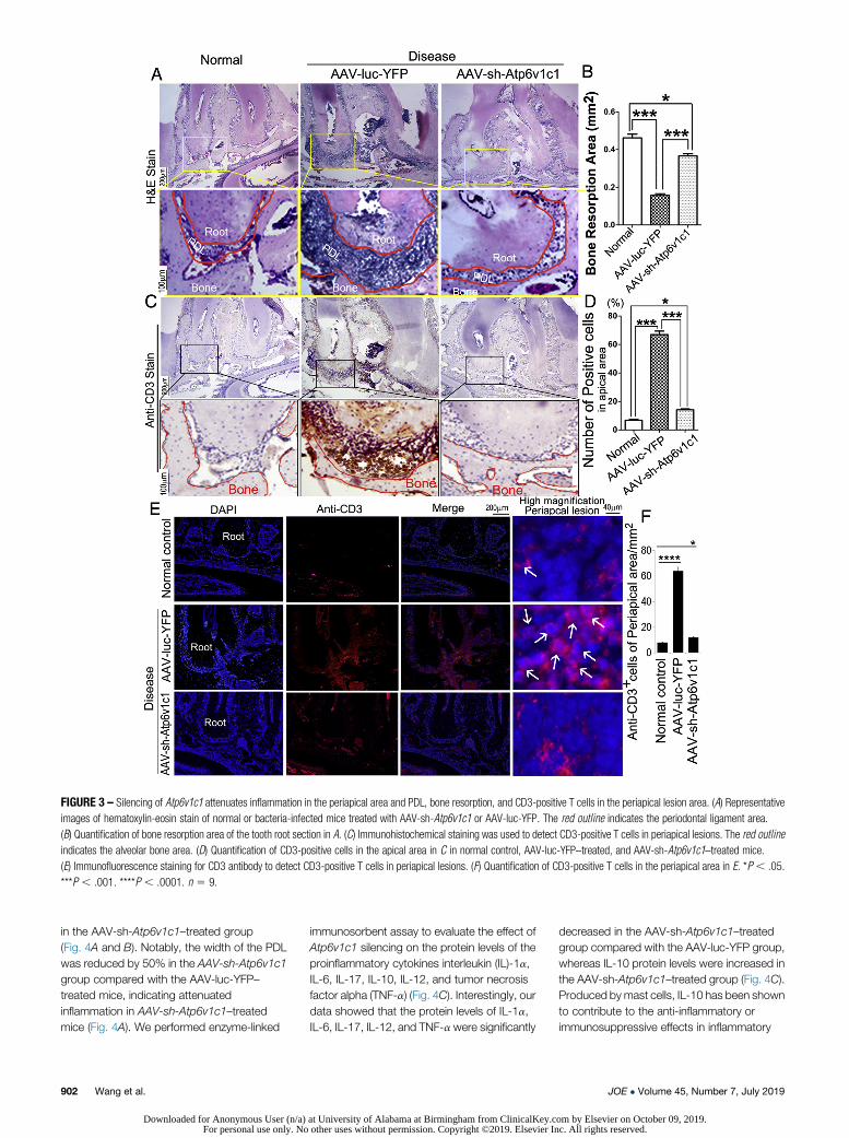
FIGURE 3 – Silencing of Atp6v1c1 attenuates inflammation in the periapical area and PDL, bone resorption, and CD3-positive T cells in the periapical lesion area. (A) Representativeimages of hematoxylin-eosin stain of normal or bacteria-infected mice treated with AAV-sh-Atp6v1c1 or AAV-luc-YFP. The red outline indicates the periodontal ligament area.(B) Quantification of bone resorption area of the tooth root section in A. (C) Immunohistochemical staining was used to detect CD3-positive T cells in periapical lesions. The red outlineindicates the alveolar bone area. (D) Quantification of CD3-positive cells in the apical area in C in normal control, AAV-luc-YFP–treated, and AAV-sh-Atp6v1c1–treated mice.(E) Immunofluorescence staining for CD3 antibody to detect CD3-positive T cells in periapical lesions. (F) Quantification of CD3-positive T cells in the periapical area in E. *P, .05.***P , .001. ****P , .0001. n 5 9.
in the AAV-sh-Atp6v1c1–treated group(Fig. 4A and B). Notably, the width of the PDLwas reduced by 50% in the AAV-sh-Atp6v1c1group compared with the AAV-luc-YFP–treated mice, indicating attenuatedinflammation in AAV-sh-Atp6v1c1–treatedmice (Fig. 4A). We performed enzyme-linked
902 Wang et al.
Downloaded for Anonymous User (n/a)For personal use only. No
immunosorbent assay to evaluate the effect ofAtp6v1c1 silencing on the protein levels of theproinflammatory cytokines interleukin (IL)-1a,IL-6, IL-17, IL-10, IL-12, and tumor necrosisfactor alpha (TNF-a) (Fig. 4C). Interestingly, ourdata showed that the protein levels of IL-1a,IL-6, IL-17, IL-12, and TNF-a were significantly
at University of Alabama at Birmingham from ClinicalKey.c other uses without permission. Copyright ©2019. Elsevier In
decreased in the AAV-sh-Atp6v1c1–treatedgroup compared with the AAV-luc-YFP group,whereas IL-10 protein levels were increased inthe AAV-sh-Atp6v1c1–treated group (Fig. 4C).Produced by mast cells, IL-10 has been shownto contribute to the anti-inflammatory orimmunosuppressive effects in inflammatory
JOE � Volume 45, Number 7, July 2019
om by Elsevier on October 09, 2019.c. All rights reserved.

FIGURE 4 – AAV-mediated Atp6v1c1 knockdown reduced the expression of inflammatory cytokines in the periapical lesion. (A) Immunohistochemical staining was used to verify theeffectiveness of AAV-sh-Atp6v1c1 knockdown Atp6v1c1 in periapical tissues. The red outline indicates the PDL area. (B) Quantification of Atp6v1c1-positive cells in the PDL area innormal control, AAV-luc-YFP–treated, and AAV-sh-Atp6v1c1–treated mice in A. (C) IL-6, IL-1a, IL-17, IL-10, IL-12, and TNF-a levels in the periapical tissues were detected byenzyme-linked immunosorbent assay. (D) Quantitative real-time polymerase chain reaction of osteoclast marker gene (ie, Ctsk) and proinflammatory cytokines (ie, TNF-a, IL-6, andIL-17) in periapical tissues of uninfected mice (normal) or bacteria-infected mice treated with AAV-luc-YFP or with AAV-sh-Atp6v1c1. Expression levels were normalized tohypoxanthine guanine phosphoribosyl transferase. *P , .05. **P , .01. ***P , .001. NS, not significant. n 5 9.
JOE � Volume 45, Number 7, July 2019 AAV-sh-Atp6v1c1 Prevents Periapical Tissue Damage 903
Downloaded for Anonymous User (n/a) at University of Alabama at Birmingham from ClinicalKey.com by Elsevier on October 09, 2019.For personal use only. No other uses without permission. Copyright ©2019. Elsevier Inc. All rights reserved.

conditions14. IL-6 is secreted by osteoblasts inresponse to bone resorption and is importantfor osteoclast differentiation15. The relativemessenger RNA expression level of theosteoclast marker gene Ctsk was decreasedby 60% in the AAV-sh-Atp6v1c1–treatedgroup compared with the AAV-luc-YFPtreatment group (Fig. 4D). In addition, theexpression levels of proinflammatory markersTNF-a and IL-17 were decreased by 50% and75%, in the AAV-sh-Atp6v1c1–treated group,respectively, AAV-sh-Atp6v1c1 also reducedthe expression of IL-6, which is important forosteoclast differentiation, by 50% (Fig. 4D).Previous studies have shown that V-ATPasesare required for mammalian target ofrapamycin complex 1 activation stimulated byamino acids16; thus, we tested colocalizationof Atp6v1c1 and mammalian target ofrapamycin (mTOR) in 4T1 epithelial cells(Supplemental Fig. S1 is available online atwww.jendodon.com). Our results showedcoexpression of Atp6v1c1 and mTOR inepithelial cells, indicating its relationship withmTOR signaling (Supplemental Fig. S1 isavailable online at www.jendodon.com). Thus,Atp6v1c1 may regulate inflammation thoughthe mTOR and/or toll-like receptor (TLR)pathways. Furthermore, our data show thatAtp6v1c1 is involved, not only in osteoclastsbut also in dendritic cells, T cells, andmacrophages. It is also possible that mTORsignaling is mediated by Atp6v1c1 in bothosteoclasts and immune cells. TNF-a andIL-10 are downstream cytokines in TLR2/4signaling, which is involved in the periapicaldisease pathogenesis17. Our results showedthat the expression of TNF-a was down-regulated in the Atp6v1c1 depletion group,whereas IL-10 was up-regulated. Furthermore,knockdown of Atp6v1c1 inhibited the levels ofIL-12 and IL-6, which are regulated by TLR9.These results suggested that Atp6v1c1 mayregulate TLR signaling in endodontic disease.In conclusion, we found that Atp6v1c1knockdown significantly reducedproinflammatory cytokine expression,indicating that Atp6v1c1 may regulate TLRsignaling in endodontic disease.
DISCUSSION
To investigate the role of Atp6v1c1 in periapicaldisease, we characterized the efficacy of agene therapy using recombinant AAV-mediated Atp6v1c1 knockdown to targetperiapical bone resorption and inflammationsimultaneously. Depletion of Atp6v1c1 in themouse model of periapical disease reducedbone destruction by 70%, impaired osteoclastactivation, decreased T-cell infiltration in theperiapical lesion by 75%, and protected the
904 Wang et al.
Downloaded for Anonymous User (n/a)For personal use only. No
PDL from destruction caused by inflammation.Furthermore, AAV-mediated Atp6v1c1knockdown also reduced bacterial infection-stimulated proinflammatory cytokineexpression. Notably, our data also showedthat osteoclast extracellular acidification wasimpaired because of Atp6v1c1 silencing. Ourresults showed that AAV-mediated Atp6v1c1knockdown in periapical tissues can slowendodontic disease progression, prevent boneerosion, and alleviate inflammation, indicatingthat targeting Atp6v1c1 may result in noveltherapeutic approaches for diseases ofosteoclast overactivation such as endodonticdisease.
In this study, we found that depletion ofAtp6v1c1 in the mouse model of endodonticdisease reduced bone destruction. Wepreviously showed that Atp6i is an osteoclast-specific proton pump that is essential forosteoclast-mediated extracellular acidificationduring bone resorption18. As a subunit ofAtp6i, Atp6v1c1 is located in the V1 domain ofV-ATPase and is considered to be directlyresponsible for regulating the dissociativemechanism of the V-ATPase19,20. Moreover,Atp6v1c1 regulates intracellular andintraorganellar pH together with othersubunits3,21. Our results demonstrated thatlocal administration of AAV-sh-Atp6v1c1 in theapical area inhibited bone resorption,potentially resulting from osteoclastmalfunction caused by Atp6v1c1 knockdown.Biochemical analysis revealed that Atp6v1c1stabilizes the V-ATPase complex assemblyand increases proton pump activity, whichpromotes bone resorption22-24. AAV-sh-Atp6v1c1 impairs the proton exchange; thus,the acidic microenvironment that favors boneresorption cannot be maintained. AAV-sh-Atp6v1c1 rescued bone resorption andrestored the bone surrounding the tooth root;therefore, the reduced bone resorption mightbe related to the malfunction of osteoclastsafter Atp6v1c1 knockdown.
Notably, Atp6v1c1 not only protectsagainst bone resorption in endodontic diseasebut also protected the periapical tissues andPDL from destruction caused by inflammation;yet, the mechanism underlying how Atp6v1c1regulates inflammation is still unknown.Inflammatory signals mediated by immunecells and cytokines in endodontic disease havea significant influence over osteoclastdifferentiation and function through direct orindirect effects on osteoclast precursors in thebony microenvironment25. Similarly,osteoclasts can express numerous immunereceptors26,27. Our data show that AAV-sh-Atp6v1c1 decreased CD3-positive T cells andinflammatory cytokines, indicating thatAtp6v1c1 knockdown not only impairs
at University of Alabama at Birmingham from ClinicalKey.c other uses without permission. Copyright ©2019. Elsevier In
osteoclast function but also the immuneresponse. TLR signaling is critical for cytokinesecretion and the T-cell–mediated immuneresponse. Activated T cells can induceosteoclastogenesis via RANKL-dependentand RANKL-independent mechanisms underinflammatory conditions13. Thus, uponAtp6v1c1 knockdown, the T-cell–mediatedimmune response was inhibited, impairingRANKL-stimulated osteoclast differentiation. Aprevious study reported that activated Ctsk isresponsible for the cleavage of TLR9 and theactivation of TLR9 signaling responsible forinflammatory responses28. Under a low pHmicroenvironment, activated Ctsk is secretedeither from immune cell lysosomes or afterosteoclast extracellular acidification. Becauseof osteoclast malfunction after Atp6v1c1silencing, the acidic environment for boneresorption is interrupted, thus inhibiting Ctskactivation. Our data show that Atp6v1c1knockdown impairs osteoclast extracellularacidification and may block Ctsk activation,thus inhibiting TLR9 signaling.
We further evaluated the effects ofAtp6v1c1 silencing on the levels ofproinflammatory cytokines in periapical tissuesand found that IL-6 expression was reducedafter Atp6v1c1 knockdown. IL-6 is secreted byosteoblasts in response to bone resorptionand is important for osteoclast differentiation15.We found similar decreases in the expressionof the classic proinflammatory mediatorsTNF-a and IL-12 after Atp6v1c1 knockdown.Although further studies are needed todetermine the exact mechanism underlyinghow C1 depletion can inhibit inflammation,TLR signaling may be involved. TLRs canrecognize the microorganisms and theircomponents, which stimulate the productionof inflammatory cytokines. TLR2/TLR4 hasbeen shown to be expressed in endodonticdisease29, and although TLR4 knockout miceshowed reduced bone destruction inperiapical lesions, TLR2-deficient miceshowed increased periapical lesion size andincreased osteoclast numbers. TLR9 signalinghas been shown to stimulate IL-6 and IL-12production28. We found that IL-6 and IL-12protein levels were decreased in the Atp6v1c1knockdown group, suggesting that Atp6v1c1may mediate the immune response throughTLR9 signaling. Cintra et al30 showed thatapical periodontitis increased serum levels ofIL-17. AAV-sh-Atp6v1c1 significantlydecreased the levels of IL-1a and IL-17 in themouse model of periapical disease.Furthermore, IL-10, which can suppressproinflammatory responses in inflammatoryconditions14, was increased after Atp6v1c1silencing. Thus, the loss of extracellularacidification by AAV-sh-Atp6v1c1 may inhibit
JOE � Volume 45, Number 7, July 2019
om by Elsevier on October 09, 2019.c. All rights reserved.

Ctsk activation and inhibit TLR9 signaling inperiapical disease, suggesting a critical role ofAtp6v1c1 in mediating immune responses.The PDL is comprised of specializedconnective tissue fibers that attach the tooth tothe alveolar bone, and in endodontic disease,inflammation of the PDL leads to tooth loss.Interestingly, our data show that AAV-sh-Atp6v1c1 protected the PDL from destructioncaused by inflammation although themechanism underlying how Atp6v1c1regulates inflammation warrants further study.
In conclusion, we investigated thetherapeutic effect of AAV-sh-Atp6v1c1 inperiapical disease to inhibit osteoclastic boneresorption and inflammation and revealed thatAAV-sh-Apt6v1c1 protected the PDL fromdestruction caused by inflammation byimpairing osteoclast extracellular acidificationand potentially through regulating TLRsignaling. This work provides important
JOE � Volume 45, Number 7, July 2019
Downloaded for Anonymous User (n/a) aFor personal use only. No
insights into the role of Apt6v1c1 in osteolyticand inflammatory diseases such as periapicaldisease and the mechanisms underlying howApt6v1c1 modulates osteoclast-mediatedbone resorption and inflammation. Insightsresulting from this study may assist indeveloping novel treatments for periapicaldisease and other osteolytic and inflammatorydiseases.
ACKNOWLEDGMENTS
Yi-Ping Li and Yuehua Liu contributed equallyto this study.
We thank Drs. Sergei Musatov andSonoko Ogawa (School of Human Sciences,University of Tsukuba, Tsukuba, Ibaraki,Japan) for kindly providing the AAV-H1-luc-YFP and AAV-H1 vectors and helpfulsuggestions. We are grateful for the assistanceof the Small Animal Phenotyping Core,
AAV-sh-At
t University of Alabama at Birmingham from ClinicalKey.coother uses without permission. Copyright ©2019. Elsevier Inc
Metabolism Core, and NeuroscienceMolecular Detection Core Laboratory at theUniversity of Alabama at Birmingham (P30NS0474666).
Supported by the National Institutes ofHealth (grant nos. DE-028264 and DE-023813to Y.P.L. and AR-070135 and AG-056438 toW.C.) and the National Nature ScienceFoundation of China (grant no. 81470768 toY.L.).
The authors deny any conflicts ofinterest related to this study.
SUPPLEMENTARY MATERIAL
Supplementary material associated with thisarticle can be found in the online version atwww.jendodon.com (https://doi.org/10.1016/j.joen.2019.02.024).
REFERENCES
1. Ma J, Chen W, Zhang L, et al. RNA interference-mediated silencing of Atp6i prevents bothperiapical bone erosion and inflammation in the mouse model of endodontic disease. InfectImmun 2013;81:1021–30.
2. Nilsson E, Bonte E, Bayet F, Lasfargues JJ. Management of internal root resorption on permanentteeth. Int J Dent 2013;2013:929486.
3. Feng S, Deng L, Chen W, et al. Atp6v1c1 is an essential component of the osteoclast protonpump and in F-actin ring formation in osteoclasts. Biochem J 2009;417:195–203.
4. Li YP, Alexander M, Wucherpfennig AL, et al. Cloning and complete coding sequence of a novelhuman cathepsin expressed in giant cells of osteoclastomas. JBoneMiner Res 1995;10:1197–202.
5. Li YP, Chen W, Stashenko P. Molecular cloning and characterization of a putative novel humanosteoclast-specific 116-kDa vacuolar proton pump subunit. Biochem Biophys Res Commun1996;218:813–21.
6. Chen W, Yang S, Abe Y, et al. Novel pycnodysostosis mouse model uncovers cathepsin Kfunction as a potential regulator of osteoclast apoptosis and senescence. Hum Mol Genet2007;16:410–23.
7. Feng S, Zhu G, Mcconnell M, et al. Silencing of Atp6v1c1 prevents breast cancer growth andbone metastasis. Int J Biol Sci 2013;9:853.
8. Mabbott NA, Kenneth Baillie J, Kobayashi A, et al. Expression of mesenchyme-specific genesignatures by follicular dendritic cells: insights from the meta-analysis of microarray data frommultiple mouse cell populations. Immunology 2011;133:482–98.
9. Hommel JD, Sears RM, Georgescu D, et al. Local gene knockdown in the brain using viral-mediated RNA interference. Nat Med 2003;9:1539–44.
10. Gao B, ChenW, Hao L, et al. Inhibiting periapical lesions through AAV-RNAi silencing of cathepsinK. J Dent Res 2013;92:180–6.
11. Yang S, Hao L, McConnell M, et al. Inhibition of Rgs10 expression prevents immune cell infiltrationin bacteria-induced inflammatory lesions and osteoclast-mediated bone destruction. Bone Res2013;1:267–81.
12. Yang S, Li YP. RGS10-null mutation impairs osteoclast differentiation resulting from the loss of[Ca21]i oscillation regulation. Genes Dev 2007;21:1803–16.
13. Weitzmann MN, Cenci S, Rifas L, et al. T cell activation induces human osteoclast formation viareceptor activator of nuclear factor kappaB ligand-dependent and -independent mechanisms. JBone Miner Res 2001;16:328–37.
p6v1c1 Prevents Periapical Tissue Damage 905
m by Elsevier on October 09, 2019.. All rights reserved.

14. Grimbaldeston MA, Nakae S, Kalesnikoff J, et al. Mast cell-derived interleukin 10 limits skinpathology in contact dermatitis and chronic irradiation with ultraviolet B. Nat Immunol2007;8:1095–104.
15. Yoshitake F, Itoh S, Narita H, et al. Interleukin-6 directly inhibits osteoclast differentiation bysuppressing receptor activator of NF-kappaB signaling pathways. J Biol Chem2008;283:11535–40.
16. Zoncu R, Bar-Peled L, Efeyan A, et al. mTORC1 senses lysosomal amino acids through aninside-out mechanism that requires the vacuolar H(1)-ATPase. Science 2011;334:678–83.
17. Hirai K, Furusho H, Kawashima N, et al. Serum amyloid A contributes to chronic apicalPeriodontitis via TLR2 and TLR4. J Dent Res 2019;98:117–25.
18. Li YP, Chen W, Liang YQ, et al. Atp6i-deficient mice exhibit severe osteopetrosis due to loss ofosteoclast-mediated extracellular acidification. Nat Genet 1999;23:447–51.
19. P�erez-Say�ans M, Reboiras-L�opez MD, Somoza-Martín JM, et al. Measurement of ATP6V1C1expression in brush cytology samples as a diagnostic and prognostic marker in oral squamouscell carcinoma. Cancer Biol Ther 2010;9:1057–64.
20. Inoue T, Forgac M. Cysteine-mediated cross-linking indicates that subunit C of the V-ATPase isin close proximity to subunits E and G of the V1 domain and subunit a of the V0 domain. J BiolChem 2005;280:27896–903.
21. HussM,Wieczorek H. Inhibitors of V-ATPases: old and new players. J Exp Biol 2009;212:341–6.
22. Yuan FL, Li X, Lu WG, et al. The vacuolar ATPase in bone cells: a potential therapeutic target inosteoporosis. Mol Biol Rep 2010;37:3561–6.
23. Wang Y, Toei M, Forgac M. Analysis of the membrane topology of transmembrane segments inthe C-terminal hydrophobic domain of the yeast vacuolar ATPase subunit a (Vph1p) by chemicalmodification. J Biol Chem 2008;283:20696–702.
24. Petzoldt AG, Gleixner EM, Fumagalli A, et al. Elevated expression of the V-ATPase C subunittriggers JNK-dependent cell invasion and overgrowth in a Drosophila epithelium. Dis ModelMech 2013;6:689–700.
25. Wu Y, Humphrey MB, Nakamura MC. Osteoclasts - the innate immune cells of the bone.Autoimmunity 2008;41:183–94.
26. Takayanagi H. The unexpected link between osteoclasts and the immune system. Adv Exp MedBiol 2010;658:61–8.
27. Nakashima T, Takayanagi H. Osteoclasts and the immune system. J Bone Miner Metab2009;27:519–29.
28. Asagiri M, Hirai T, Kunigami T, et al. Cathepsin K-dependent toll-like receptor 9 signaling revealedin experimental arthritis. Science 2008;319:624–7.
29. Rider D, Furusho H, Xu S, et al. Elevated CD14 (cluster of differentiation 14) and toll-like receptor(TLR) 4 signaling deteriorate periapical inflammation in TLR2 deficient mice. Anat Rec (Hoboken)2016;299:1281–92.
30. Cintra LT, Samuel RO, Azuma MM, et al. Apical periodontitis and periodontal disease increaseserum IL-17 levels in normoglycemic and diabetic rats. Clin Oral Investig 2014;18:2123–8.
906 Wang et al. JOE � Volume 45, Number 7, July 2019
Downloaded for Anonymous User (n/a) at University of Alabama at Birmingham from ClinicalKey.com by Elsevier on October 09, 2019.For personal use only. No other uses without permission. Copyright ©2019. Elsevier Inc. All rights reserved.