basic principles of em watermark
TRANSCRIPT

Electron Microscopy: Basic Methods Workshop 1
SIR WILLIAM DUNN SCHOOL OF PATHOLOGY
Basic Principles of Electron Microscopy (EM)
Dr Errin Johnson
Head of the Dunn School EM Facility
Basic PrinciplesBrief history of EM
1924 – Louis de Broglie theorised the wave/particle duality of electrons
1926 – Hans Busch demonstrated that magnetic lenses can manipulate the path of electrons in the same way as
optical lenses do with light
For a detailed history of EM, see:
1. Haguenau et al. (2003) Key events in the history of electron microscopy.
Microscopy & Microanalysis, 9(2): 96-138.
2. Masters, B (2009) History of the electron microscope in cell biology. eLS, Wiley.
1932 – Ernst Ruska & Max Knoll invent the TEM
1873 – Hermann von Helmholtz & Ernst Abbe show that the wavelength of light affects optical resolution
1937 – Manfred von Ardenne builds the first SEM
1951 – Erwin Muller develops the field emission microscope for atomic resolution
1951 – Albert Claude et al publish the first TEM image of an intact cell
1934 – Ladisalus Marton publishes the first biological EM micrograph
1932
Today

Electron Microscopy: Basic Methods Workshop 2
Basic PrinciplesResolution
• Resolution is the smallest distance at
which two neighbouring points can be
distinguished and is dependent on
wavelength
• The wavelength of accelerated electrons
(6 pm) is several orders of magnitude
shorter than that of light (600 nm)
www.ammrf.org
Light
Electrons
Basic Principles Resolution

Electron Microscopy: Basic Methods Workshop 3
Basic PrinciplesResolution and Contrast
Radiolarian imaged with both SEM (top) and light microscopy (bottom). From: General Chemistry: Principles, Patterns, and Applications, B. Averill & P. Elderege
Ligh
t mic
rosc
opy
Ele
ctro
n m
icro
scop
y
TEM image of fibroblast cell (E Johnson). Bottom: Confocal image of a kidney cell stained with DAPI (blue) and MitoTracker (red) (Hammamatsu.magnet.fsu.edu)
• EM images are monochromatic and are essentially intensity maps of the number of
electrons that are detected from a given point. False colour may be added during
post-processing of the image, if desired.
Hydrothermal worm (x525) by Philippe Crassous, FEI.com
Original SEM image False-coloured SEM image
Basic PrinciplesSignal detection

Electron Microscopy: Basic Methods Workshop 4
Features of Electron MicroscopesOverview
Scanning Electron Microscopy
Transmission Electron Microscopy
Features of Electron MicroscopesOverview
The main components of an
electron microscope are:
• An electron gun
• Electromagnetic lens system
• Vacuum system
• Camera/detector
• Computer

Electron Microscopy: Basic Methods Workshop 5
• The gun consists of an electron source, electrode, Wenhelt assembly and anode
• Electron sources are typically Tungsten or LaB6 and can be thermionic or field
emission (FEG). A current is run through the filament/crystal to heat it or, a field
is applied to the tip, resulting in the emission of electrons from the tip. The high
voltage difference between the cap and the anode causes the electrons to
accelerate and form a beam
www.wikipedia.orgwww.ammrf.org
Features of Electron MicroscopesThe Electron gun
• EM lenses are electromagnetic, creating precise, circular magnetic fields that
manipulate the electron beam, much the same way that optical lenses focus and
direct light
www.ammrf.org
Features of Electron MicroscopesElectromagnetic Lenses
www.ammrf.org

Electron Microscopy: Basic Methods Workshop 6
• EMs have elaborate pumping systems
to ensure that the microscope is
operated under a high vacuum (10-4 Pa)
• Maintains the integrity of the electron
beam, as any interaction with gas atoms
will cause the beam to scatter
• Avoids arcing between the cathode and
ground (and damage to the filament)
Overview of vacuum system on the Tecnai12 TEM
Features of Electron Microscopes Vacuum systems
Transmission Electron Microscopy (TEM)

Electron Microscopy: Basic Methods Workshop 7
The TEM
120 kV
• Resolution depends on a number of factors, including the accelerating voltage, the
type of electron source used and how you setup the microscope
• Accelerating voltage (kV) is typically 80-300 kV for biological specimens
300 kV 1000 kV
The TEM
Electron Microscopy: Basic Methods Workshop 8
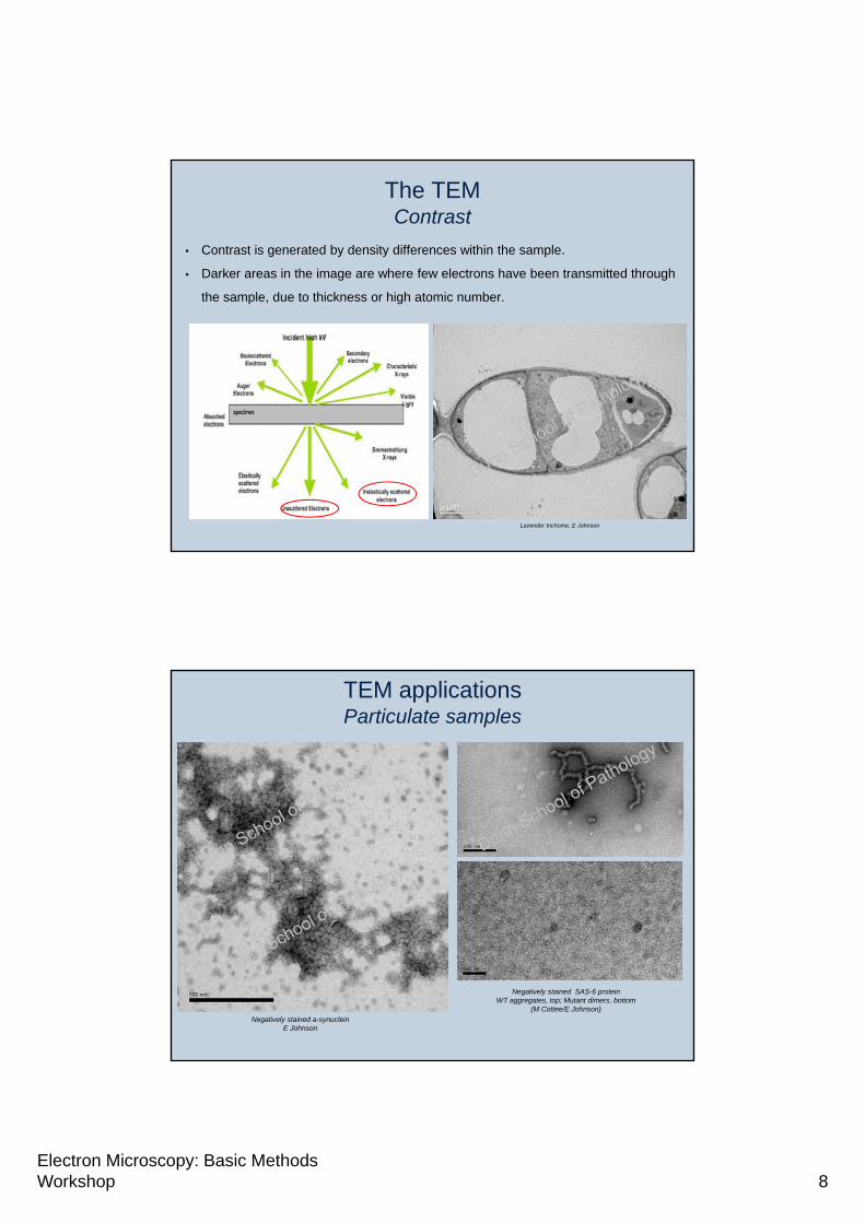
The TEMContrast
• Contrast is generated by density differences within the sample.
• Darker areas in the image are where few electrons have been transmitted through
the sample, due to thickness or high atomic number.
Lavender trichome, E Johnson
TEM applicationsParticulate samples
Negatively stained a-synucleinE Johnson
Negatively stained SAS-6 protein WT aggregates, top; Mutant dimers, bottom
(M Cottee/E Johnson)

Electron Microscopy: Basic Methods Workshop 9
TEM imagingParticulate samples - Viruses
Negatively stained virus-like particles (D Leneghan/E Johnson)
Negatively stained virus-like particles (Ebola) (E Johnson)
TEM imagingParticulate samples - Bacteria
Negatively stained Neisseria sp. (R Exley/EJohnson)
500 nm 200 nm

Electron Microscopy: Basic Methods Workshop 10
TEM ApplicationsUltrastructure - Cells
Nucleus Golgi
ER Centrioles
Mammalian culture cells (E Johnson)
TEM ApplicationsUltrastructure - Tissue
Mouse liver tissue, untreated (left) and under ER stress (right) (V Liebe & E Johnson)

Electron Microscopy: Basic Methods Workshop 11
TEM ApplicationsAdvanced techniques
Multi-lysosomal body (A.J. Koster and W.J.C. Geerts, Utrecht University)
3D to
mo
grap
hy
Cry
o-E
MC
orrelative
micro
scop
y
Gatan.com
Correlative light and electron microscopy of cryo-sections (Vicidomini et al. Traffic 9:1828–1838, 2008)
Pro
tein
lo
cali
sati
on
Whole mount immunolabelled Trypanosome cytoskeleton (S Dean, Dunn School)
Scanning Electron Microscopy (SEM)

Electron Microscopy: Basic Methods Workshop 12
The SEM
The SEM Signal detection
Secondary electrons (SEs) provides surface morphology and topology information.
SEs are captured by the Everhart-Thornley detector
www.ammrf.orgwww.ammrf.org Dept Biological Sciences, Smith College Northampton USA
Electron Microscopy: Basic Methods Workshop 13
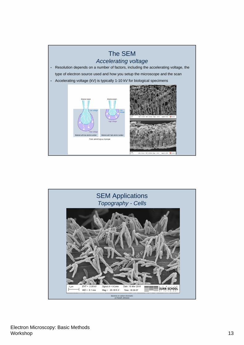
The SEMAccelerating voltage
Low kV
High kV
• Resolution depends on a number of factors, including the accelerating voltage, the
type of electron source used and how you setup the microscope and the scan
• Accelerating voltage (kV) is typically 1-10 kV for biological specimens
SEM ApplicationsTopography - Cells
Bacteria on carbon electrodes(s Putra/E Johnson)
Electron Microscopy: Basic Methods Workshop 14
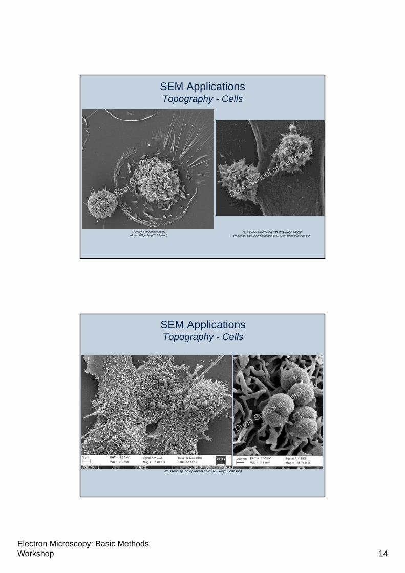
SEM ApplicationsTopography - Cells
Monocyte and macrophage (B van Wilgenburg/E Johnson)
HEK 293 cell interacting with streptavidin coated dynabeads plus biotinylated anti-EPCAM (M Brenner/E Johnson)
SEM ApplicationsTopography - Cells
Neisseria sp. on epithelial cells (R Exley/EJohnson)

Electron Microscopy: Basic Methods Workshop 15
SEM ApplicationsTopography - Organisms
Drosophila rough eye phenotype (M Elschami, NDCN)
C. elegans(E Johnson/A Moloney, Dunn School)
The SEMDiverse imaging capabilities

Electron Microscopy: Basic Methods Workshop 16
Biological ApplicationsSEM - Advanced
Protein localisation Correlative microscopy
Ch
emical co
mp
ositio
n
BSE
SE
Microtubules (D Barton, University of Sydney)Microtubules (D Barton, University of Sydney)
EDS analysis of cheese (M Foley, University of Sydney)
3D
SE
M
Moritz Helmstaedter, Max-Planck Institute for Medical Research
Questions?
A happy Leishmania nucleus(J Valli)