basic fibroblast growth factor elicits formation of interstitial axonal branches via enhanced
TRANSCRIPT

Molecular Biology of the CellVol. 21, 334–344, January 15, 2010
Basic Fibroblast Growth Factor Elicits Formation ofInterstitial Axonal Branches via Enhanced Severingof MicrotubulesLiang Qiang, Wenqian Yu, Mei Liu, Joanna M. Solowska, and Peter W. Baas
Department of Neurobiology and Anatomy, Drexel University College of Medicine, Philadelphia, PA 19129
Submitted September 30, 2009; Revised November 6, 2009; Accepted November 12, 2009Monitoring Editor: Paul Forscher
The formation of interstitial axonal branches involves the severing of microtubules at sites where new branches form.Here we wished to ascertain whether basic fibroblast growth factor (bFGF) enhances axonal branching throughalterations in proteins involved in the severing of microtubules. We found that treatment of cultured hippocampalneurons with bFGF heightens expression of both katanin and spastin, which are proteins that sever microtubules inthe axon. In addition, treatment with bFGF enhances phosphorylation of tau at sites expected to cause it to dissociatefrom microtubules. This is important because tau regulates the access of katanin to the microtubule. In live-cellimaging experiments, axons of neurons treated with bFGF displayed greater numbers of dynamic free ends ofmicrotubules, as well as greater numbers of short mobile microtubules. Entirely similar enhancement of axonalbranching, short microtubule transport, and frequency of microtubule ends was observed when spastin wasoverexpressed in the neurons. Depletion of either katanin or spastin with siRNA diminished but did not eliminatethe enhancement in branching elicited by bFGF. Collectively, these results indicate that bFGF enhances axonalbranch formation by augmenting the severing of microtubules through both a spastin-based mode and a katanin-based mode.
INTRODUCTION
A typical vertebrate neuron extends a single axon thatbranches extensively in order to innervate multiple targets.This can occur either via bifurcation of the growth cone atthe tip of the parent axon, or via the formation of interstitial(also called “collateral”) branches along the length of theparent axon. Although the former predominates in culturesof PNS neurons, interstitial branching appears to be theprimary mode by which axons arborize during developmentand in cultures of CNS neurons. The formation of an inter-stitial branch involves dynamic interplay between the mi-crotubules in the parent axon and the cortical actin cytoskel-eton (Dent and Kalil, 2001). The most dramatic cytoskeletalevent that occurs during branch formation appears to be thefocal severing of the long microtubules that dominate theaxonal shaft into a concentration of numerous very shortmicrotubules that are able to transit into the newly formingbranch (Yu et al., 1994; Dent et al., 1999). This is criticalbecause the newly forming branch needs a robust supply ofincoming microtubules (Yu et al., 1994), because only veryshort microtubules in the axon are mobile (Wang andBrown, 2002) and because an enrichment of plus ends ofmicrotubules is important for interacting with a variety ofproteins and cortical structures relevant to axonal branching(Kornack and Giger, 2005).
There are two microtubule-severing enzymes that havebeen studied extensively in neurons, namely katanin and
spastin (Yu et al., 2007; Falnikar and Baas, 2009). Recently,we and others documented that overexpression of spastin incultured rat hippocampal neurons markedly enhances theformation of axonal branches, whereas depletion of spastinmarkedly diminishes branch frequency (Yu et al., 2008;Riano et al., 2009). The story with katanin was not as simple,as overexpression of katanin did not enhance branch forma-tion (Yu et al., 2005). However, we found that katanin’saccess to the microtubule is regulated by tau (Qiang et al.,2006) and that depletion of tau can cause enhancement ofaxonal branching through a katanin-based mechanism (Yuet al., 2008). Therefore we proposed that axons can branchthrough either a katanin-based mode involving tau or aspastin-based mode that appears to be independent of tau.The existence of both modes provides redundancy but alsomay provide different levels of control, in response togrowth factors that regulate axonal branching at differentdevelopmental stages or under different circumstances.
Basic fibroblast growth factor (bFGF) is one of the mostpotent inducers of axonal branch formation (Szebenyi et al.,2001; Aletsee et al., 2003; Klimaschewski et al., 2004). Whenapplied to neuronal cultures, bFGF causes a marked increasein branch frequency that is apparent within a day of appli-cation. Little is known, however, about the specific changeselicited by bFGF that give rise to branching. Here, wewanted to test the hypothesis that bFGF alters the levelsand/or regulation of proteins relevant to the severing ofmicrotubules. We also wanted to ascertain whether bFGFstimulates axonal branching through the katanin-basedmode, the spastin-based mode, or both. Finally, we investi-gated whether experimental augmentation of microtubulesevering produces similar effects on microtubule behaviorsas treatment of the neurons with bFGF.
This article was published online ahead of print in MBC in Press(http://www.molbiolcell.org/cgi/doi/10.1091/mbc.E09–09–0834)on November 25, 2009.
Address correspondence to: Peter W. Baas ([email protected]).
334 © 2010 by The American Society for Cell Biology

MATERIALS AND METHODS
Cell Culture and TransfectionCultures of rat hippocampal neurons were prepared as previously described(Qiang et al., 2006; Yu et al., 2008). DNA constructs were transfected into thedissociated neurons before plating, using the Amaxa Nucleofector (AmaxaBiosystems, Koln, Germany). After nucleofection, the neurons were platedonto poly-l-lysine–treated glass coverslips as previously described (Qiang etal., 2006; Yu et al., 2008). For some experiments, the coverslips were alsocoated with laminin after the polylysine treatment because this was found topromote faster and straighter growth of axons. bFGF (Promega, Madison, WI,Cat. no. 9PIG507) was applied to some of the neuronal cultures at a concen-tration of 20 ng/ml (Szebenyi et al., 2001). DNA constructs included enhancedgreen fluorescent protein (EGFP)-�-tubulin (Clontech, Palo Alto, CA), EGFP-EB3 (provided by Dr. Niels Galjart, Erasmus Medical Center), and mCherry-spastin (prepared in our laboratory). For the spastin construct, we used theM85 isoform of spastin, which is the predominant isoform expressed indeveloping neurons (Claudiani et al., 2005; Connell et al., 2008; Mancuso andRugarli, 2008; Solowska et al., 2008; Yu et al., 2008). For controls, we used theappropriate constructs for EGFP or/and mCherry. For studies involvingsiRNA, neurons were transfected with a pool of four sequences specific to ratspastin, p60-katanin, or p80-katanin (purchased as “smartpools” from Dhar-macon, Boulder, CO, Accession numbers: XM-343018, L-080249-01, andM-092761-00, respectively) or with a nonspecific control sequence (Dharma-con, Cat. no. D-001206-03-20). Transfections with the siRNA smartpools wereperformed using the Nucleofector as previously described (Yu et al., 2005,2008; Qiang et al., 2006). After 2 d in culture to permit protein depletion, theneurons treated with siRNA were replated so that neurites could grow anewin the near absence of the relevant protein targeted by the siRNA.
Microtubule Transport AssayTo image the transport of microtubules in axons, the neurons were transfectedwith EGFP-�-tubulin. In some experiments, control neurons (not treated withbFGF) were compared with neurons treated with bFGF. In other cases, controlneurons (expressing mCherry) were compared with neurons expressingmCherry-spastin. The latter was to ascertain any potential changes elicited byspastin overexpression. In other experiments, neurons were transfected withcontrol or spastin siRNA and then transfected with the EGFP-�-tubulinconstruct 2 d later and then replated. To assist in monitoring transport ofmicrotubules over relatively long and straight trajectories, the neurons wereplated onto poly-l-lysine with laminin (see above). Imaging was conducted24–48 h after plating or replating. Individual unfasciculated axons withclearly identifiable directionality of growth from the cell body were chosen foranalysis so that anterograde and retrograde movements could be identifiedwith certainty. We used the Zeiss inverted Observer Z1 microscope (CarlZeiss, Jena, Germany), with a Zeiss incubation chamber, interfaced with theMicroPoint Mosaic Digital Diaphragm system from Photonics Instruments(St. Charles, IL), coupled to an argon ion laser. The Zeiss incubation chamberprovides optimal culture conditions of 37°C and 5% CO2. The photonic laserMosaic system was used to photobleach regions of the axons in similarmanner to our earlier studies on microtubule transport (Hasaka et al., 2004; Heet al., 2005; Ahmad et al., 2006; Myers and Baas, 2007). Only one photo-bleached region was made on each axon analyzed. This region was always40–50 �m in length and always was located roughly equidistant from the cellbody and the tip of the axon. A 100� 1.3 NA Plan Apo oil immersion objectiveand a GFP filter set (Chroma Technology, Brattleboro, VT) were used foracquiring fluorescence images. The CCD camera and most aspects of themicroscope were controlled by Axiovision 4.1 software (Zeiss) running on anIntel Xeon processor-based computer (Fujitsu Siemens, Berlin, Germany) withthe Windows XP Professional operating system (Microsoft, Seattle, WA). Theintensity of epifluorescence illumination was attenuated to 30% to reducephotobleaching and photodamage during acquisition; 2 � 2 binning wasapplied to increase signal intensity. Images were acquired at 1.8-s intervals,with �280–320-ms exposure time. Time-lapse images were saved as “zvi”files and analyzed using Axiovision 4.1 software. Motion analysis was per-formed only on axons that displayed at least one microtubule that was visiblewithout digital processing. Additional analysis was conducted using digitalcontrast adjustments within the “Image Properties” application of Axiovision4.1. Statistics were performed using the Student’s t test.
EB3 Live-Cell ImagingTo image the assembly of microtubules from their plus ends, we expressedfluorescently tagged EB3, which is a microtubule “end binding protein” thattracks with the plus ends of microtubules during bouts of rapid assembly(Stepanova et al., 2003). We have used this approach in a number of ourstudies on microtubule behaviors in cultured neurons (Hasaka et al., 2004;Ahmad et al., 2006; Myers et al., 2006; Nadar et al., 2008). To visualize EB3comets within the axon of the cultured neurons in the experiments involvingbFGF, we transfected neurons with EGFP-EB3 and cultured in medium con-taining bFGF. To image EGFP-EB3 within the axons of spastin-overexpressingneurons, we cotransfected neurons with both EGFP-EB3 and mCherry-spas-tin. Images were acquired at a rate of 1 per second excluding camera exposure
times to time-lapse movies. Depending on the fluorescence intensity of EB3,exposure time ranged from 250 to 350 ms so that the real-time imaging ofindividual axons ranged from 2 to 4 min. As with the microtubule transportassay described above, we used the Zeiss inverted Observer Z1 microscopewith the Zeiss incubation chamber, and we analyzed only one region per axonthat always located roughly equidistant from the cell body and the tip of theaxon. The region we analyzed was always 50–70 �m in length, a bit longerthan the length analyzed in the microtubule transport studies. Details on theobjective, camera, software, illumination, and binning were also the same asdescribed above for the microtubule transport studies. EB3 quantification wasperformed using Axiovision “measure” module. Numbers of EB3 comets,average speeds of the comets and longevities of the comets were analyzed indetails. Frames extracted for still images were exported from Axiovision asTIFF files and processed in Photoshop 7 (Adobe Photosystems, San Jose, CA).Statistics were performed using the Student’s t test.
Primary Antibodies Used for Western Blotting andImmunofluorescence AnalysesPrimary antibodies included the following: monoclonal: tau1 (anti-dephos-phorylated tau, obtained from Dr. Lester Binder of Northwest University;Binder et al., 1985), tau5 (anti-total tau, also obtained from Binder; Papasozo-menos and Binder, 1987), anti-� tubulin directly conjugated with Cy3 (Sigma,St. Louis, MO, Cat. no. C4585), anti-GAPDH (Ambion, Austin, TX, Cat. no.AM4300); Polyclonal: anti-spastin (AAA spastin antibody; Solowska et al.,2008), tauR1 (anti-total tau, obtained from Binder; Berry et al., 2004), anti-P60-katanin (Yu et al., 2005), anti-P80-katanin (Yu et al., 2005), and anti-GFP(Abcam, Cambridge, MA, Cat. no. ab6556).
Western BlottingWestern blotting was conducted as in our previous studies for siRNA exper-iments, to ensure that over 95% of the spastin protein had been depletedbefore the replating step (Qiang et al., 2006; Yu et al., 2008). Western blottingwas also used for experiments aimed at ascertaining changes in the levels ofvarious proteins during treatment of neurons with bFGF. For the latter,Western blotting was conducted on cultures that had been exposed to bFGFfor 4, 24, or 72 h.
Immunofluorescence AnalysesProcedures of the immunofluorescence studies were performed as previouslydescribed (Yu et al., 2005, 2008; Qiang et al., 2006). For the experiments onmicrotubule distribution after experimental manipulations, the cultures weresimultaneously fixed and extracted 24–48 h after plating. For the experimentsrelevant to the ratio images of tau1/tauR1, the cultures were fixed and thenextracted after fixation 24–72 h after plating. Primary antibodies againstproteins of interest (same as used for the Western blotting, see above; exceptthat we used the polyclonal antibody tauR1 for staining total tau) wereapplied followed by appropriate secondary antibodies. Images (except thoseusing tau1 and tauR1 antibodies) were obtained on an Axiovert 200M micro-scope (Carl Zeiss) equipped with a high-resolution CCD (Orca, Hamamatsu,Japan). The images were obtained using identical camera, microscope, andimaging criteria such as gain, brightness and contrast, and exposure time. Insome cases, fluorescence images were subjected to the “invert” function inAdobe Photoshop (San Jose, CA) that converts blacks to whites and whites toblacks and inverts gray levels proportionally, because we found that finedetails were more clearly visualized. The resulting images are referred to as“inverted images.” In one set of studies, ratio images were generated ofcultures double-labeled with tau1 and tauR1 antibodies using the Pascalconfocal microscope. Statistics were performed using the Student’s t test.
RESULTS
bFGF Increases the Frequency of Microtubule Transportin the AxonThe goal of the first set of experiments was to test whetherthe axons of neurons treated with bFGF display a greaterfrequency of microtubule transport. This would presumablyoccur if there were higher numbers of microtubules (result-ing from microtubule severing) short enough to undergotransport. Live-cell imaging studies of microtubules in theaxons of cultured rat sympathetic neurons (from the supe-rior cervical ganglia of newborn rat pups) have consistentlyshown that only very short microtubules, roughly 7 �m orless, are mobile (Wang and Brown, 2002; Ahmad et al., 2006).The present studies are the first to use the photobleach assayfor microtubule transport on cultured hippocampal neu-rons. We have found that when using this assay on sympa-thetic neurons, it is highly advantageous to grow them on
FGF, Axon Branches, Microtubule Severing
Vol. 21, January 15, 2010 335
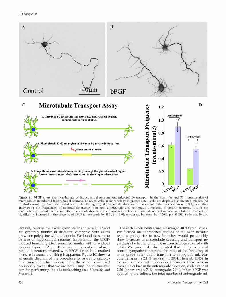
laminin, because the axons grow faster and straighter andare generally thinner in diameter, compared with axonsgrown on polylysine without laminin. We found the same tobe true of hippocampal neurons. Importantly, the bFGF-induced branching effect remained similar with or withoutlaminin. Figure 1, A and B, show examples of control neu-rons and neurons treated with bFGF for 48 h; a markedincrease in axonal branching is apparent. Figure 1C shows aschematic diagram of the procedure for assaying microtu-bule transport, which is essentially the same as we usedpreviously except that we are now using the Mosaic sys-tem for performing the photobleaching (see Materials andMethods).
For each experimental case, we imaged 40 different axons.We focused on unbranched regions of the axon becauseregions giving rise to new branches would presumablyshow increases in microtubule severing and transport re-gardless of whether or not the neuron had been treated withbFGF. We previously documented that, in the axons ofcontrol sympathetic neurons, the ratio of the frequency ofanterograde microtubule transport to retrograde microtu-bule transport is 2:1 (Hasaka et al., 2004; He et al., 2005). Inthe axons of control hippocampal neurons, there was aneven greater bias in the anterograde direction, with a ratio of2.5:1 (anterograde, 71%; retrograde, 29%). When bFGF wasapplied to the culture, the total number of anterograde mi-
Figure 1. bFGF alters the morphology of hippocampal neurons and microtubule transport in the axon. (A and B) Immunostains ofmicrotubules in cultured hippocampal neurons. To reveal cellular morphology in greater detail, cells are displayed as inverted images. (A)Control neuron. (B) Neurons treated with bFGF (20 ng/ml). (C) Schematic diagram of the microtubule transport assay. (D) Quantitativeanalyses of the frequencies of microtubule transport in both anterograde and retrograde directions. In control neurons, 71% of themicrotubule transport events are in the anterograde direction. The frequencies of both anterograde and retrograde microtubule transport aresignificantly increased in the presence of bFGF (anterograde by 45%, p � 0.01; retrograde by more than 120%, p � 0.001). Scale bar, 40 �m.
L. Qiang et al.
Molecular Biology of the Cell336

crotubule transport events was significantly increased. Incontrol axons there were 0.618 � 0.039 transport events/minin the anterograde direction, whereas in bFGF-treated axonsthere were 0.898 � 0.071 transport events/min in the antero-grade direction. This is a 45% increase in transport events(p � 0.01). Retrograde transport was displayed as 0.250 �0.031 and 0.551 � 0.038 transport events/min (p � 0.01) incontrol and bFGF-treated axons, respectively (120% in-crease, Figure 1D). Thus, the ratio of anterograde to retro-grade microtubule transport dropped to �1.6:1 in bFGF-treated hippocampal neurons.
In pursuing these experiments, we noticed that not onlydoes bFGF elicit more axonal branches, but also it signifi-cantly enhances the number of immature neurites (“minorprocesses”) that grow from the cell body (Figure 1, A and B).This is relevant to the issue of microtubule severing becausewe have documented in previous studies that manipulationof proteins relevant to microtubule severing can affect thenumber of immature neurites as well as the branching of theaxon (Yu et al., 2005, 2008). These observations may reflect amechanistic and perhaps evolutionary link between processnumber and axonal branching as means for enhancing thecomplexity of the neuritic arbor of a developing neuron.
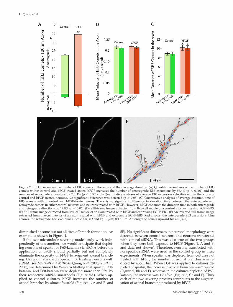
bFGF Increases the Number and Duration of MicrotubulePlus-End Assembly Events in the AxonWe would expect the severing of microtubules not only tocreate more short mobile microtubules but would also tocreate more dynamic plus ends of microtubules within theaxonal shaft. To explore the effects of bFGF on the number,distribution and dynamic behavior of microtubule plus endsin the axon, we visualized fluorescent “comets” at the plusends of microtubules generated by expression of EGFP-EB3in the neurons. EB3 is a member of a category of proteinscalled TIPs that associate with microtubule plus ends duringbouts of rapid assembly (Stepanova et al., 2003; Kornack andGiger, 2005; Wu et al., 2006). The excursion of the EGFP-EB3at the plus end of the microtubule appears as a movingcomet because the EB3 gradually dissociates from the tubu-lin subunits after their addition to the plus end of the mi-crotubule, thus producing a comet-shaped burst of fluores-cence with its tail toward the minus end of the microtubule.The number of comets was quantified per 100 �m of axonallength. In control axons, the average numbers of EB3 cometswere 22.37 � 0.497 in the anterograde direction and 1.78 �0.129 in the retrograde direction, respectively. Both numberswere significantly augmented when bFGF was added intothe culture (p � 0.001). The average numbers were 34.31 �0.938 (53.4% increase relative to control) in the anterogradedirection and 6.78 � 0.267 (281.1% increase relative to con-trol) in the retrograde direction (Figure 2, A, D, and E,Movies 1–3). Interestingly, the greater increase in the retro-grade comets was even more accentuated at sites within theparent axon that were giving rise to new branches. How-ever, in the newly forming branches themselves, EB3 cometsonly moved anterogradely (Figure 2F and Movie 3).
Regarding the velocity of EB3 comets, we did not detectany significant difference between anterograde and retro-grade comets in each group (p � 0.05, Figure 2B, Movies 1and 2). However, we detected a slight increase in the dura-tion of both anterograde and retrograde EB3 comets inbFGF-treated axons (10.14 � 0.610 s, anterograde; 10.07 �0.485 s, retrograde; p � 0.05), comparing to those in controls(8.83 � 0.361 s, anterograde; 8.77 � 0.435 s, retrograde,Figure 2C, Movies 1 and 2). The longer durations suggestthat bFGF-treatment not only generates more microtubule
plus ends but it also affects the dynamic properties of themicrotubules as well.
bFGF Impacts Proteins Related to Microtubule SeveringAs noted earlier, application of bFGF results in higher num-bers of immature processes as well as higher numbers ofaxonal branches (Figure 1). Given that experimental manip-ulation of proteins relevant to microtubule severing canhave these same two effects on neurons (Yu et al., 2005, 2008),this observation provides provocative initial evidence that akey effect of bFGF may be to alter the expression or activityof these proteins. To investigate this, we analyzed the effectsof bFGF on spastin and P60-katanin. In addition, we in-cluded two other proteins in these analyses, namely tau andP80-katanin. Tau binds along the surface of the microtubuleand suppresses the capacity of P60-katanin (and spastin, toa much lesser extent) to access the microtubule lattice (Qianget al., 2006; Yu et al., 2008). P80-katanin is a protein thatinteracts with P60-katanin in a manner that can augment itsmicrotubule-severing properties (McNally et al., 2000). Toinvestigate the effects of bFGF on these four proteins, cul-tured hippocampal neurons were allowed to grow axons for24–48 h, after which they were rinsed three to four timeswith serum-free medium. After rinsing, bFGF-containingserum-free medium was added for 4, 24, and 72 h beforeharvesting the cultures for Western blotting. With regard totau, we used two monoclonal different antibodies: one calledtau5, which recognizes all tau, and the other called tau1,which recognizes tau only when it is dephosphorylated atcertain sites that are relevant to its binding to microtubules.When tau is phosphorylated at these sites, its binding affin-ity to microtubules is diminished and it is no longer recog-nized by the tau1 antibody (Papasozomenos and Binder,1987; Mandell and Banker, 1996).
As shown in Figure 3, the addition of bFGF causes eleva-tions in the levels of both spastin and P60-katanin after 24 h,but does not increase the levels of P80-katanin or tau (Figure3, A–C). However, with regard to tau, it is clear that itbecomes markedly more phosphorylated as early as 4 h afteraddition of bFGF. Twenty-four hours later, it reaches itshighest point and remains at this higher phosphorylationlevel even after 72 h (Figure 3, A and C). These results areconsistent with a scenario whereby bFGF enhances micro-tubule severing first by increasing the phosphorylation oftau in a manner that causes it to dissociate from microtu-bules and then by increasing the levels of both P60-kataninand spastin. Expression of P80-katanin does not appear to bemodified by bFGF (Figure 3, B and C).
In a recent publication, we speculated that there are two“modes” by which the neuron augments microtubule sev-ering to elicit branch formation. We called these the ka-tanin-mode and the spastin-mode (Yu et al., 2008). Thekatanin-mode relies on the local dissociation of tau frommicrotubules at a site of impending branch formation, whichenables P60-katanin to focally sever microtubules in thatvicinity. The spastin-mode is not dependent on changes intau, but rather upon the accumulation of sufficient levels ofspastin protein. If correct, this would mean that any onebranch could form via either one of these two modes or acombination of both. Consistent with this view, spastin wasobserved to accumulate at some sites of branch formation,but not all (Yu et al., 2008). Here, we used immunofluores-cence-based ratio imaging to analyze axons with regard totau phosphorylation. As predicted by the existence of twodifferent modes for branching, we observed that the ratio oftau1 to total tau (tau1/tauR1; see Materials and Methods) was
FGF, Axon Branches, Microtubule Severing
Vol. 21, January 15, 2010 337
diminished at some but not all sites of branch formation. Anexample is shown in Figure 4.
If the two microtubule-severing modes truly work inde-pendently of one another, we would anticipate that deplet-ing neurons of spastin or P60-katanin via siRNA before theapplication of bFGF should partially but not completelyeliminate the capacity of bFGF to augment axonal branch-ing. Using our standard approach for treating neurons withsiRNA (see Materials and Methods; Qiang et al., 2006; Yu et al.,2008), we determined by Western blotting that spastin, P60-katanin, and P80-katanin were depleted more than 95% bytheir respective siRNA smartpools (Figure 5A). When ap-plied to control cultures, bFGF increases the number ofaxonal branches by almost fourfold (Figures 1, A and B, and
5F). No significant differences in neuronal morphology weredetected between control neurons and neurons transfectedwith control siRNA. This was also true of the two groupswhen they were both exposed to bFGF (Figure 1, A and B,and data not shown). Therefore, neurons transfected withnonspecific siRNA were used as the control group in theseexperiments. When spastin was depleted from cultures nottreated with bFGF, the number of axonal branches was re-duced by about half. When FGF was applied to cultures de-pleted of spastin, the increase in axonal branches was 2.52-fold(Figure 5, Bb and F), whereas in the cultures depleted of P60-katanin, the increase was 1.70-fold (Figure 5, Cc and F). Thus,each of the two severing proteins contributes to the augmen-tation of axonal branching produced by bFGF.
Figure 2. bFGF increases the number of EB3 comets in the axon and their average duration. (A) Quantitative analyses of the number of EB3comets within control and bFGF-treated axons. bFGF increases the number of anterograde EB3 excursions by 53.4% (p � 0.001) and thenumber of retrograde excursions by 281.1% (p � 0.001). (B) Quantitative analyses of average EB3 excursion velocities within the axons ofcontrol and bFGF-treated neurons. No significant difference was detected (p � 0.05). (C) Quantitative analyses of average duration time ofEB3 comets within control and bFGF-treated axons. There is no significant difference in duration time between the anterograde andretrograde comets in either control neurons and neurons treated with bFGF. However, bFGF enhances the duration time in both anterogradeand retrograde directions by 14.8% (p � 0.05). (D) Still-frame image extracted from live-cell movie of a control axon expressing EGFP-EB3.(E) Still-frame image extracted from live-cell movie of an axon treated with bFGF and expressing EGFP-EB3. (F) An inverted still-frame imageextracted from live-cell movies of an axon treated with bFGF and expressing EGFP-EB3. Red arrows, the anterograde EB3 excursions; bluearrows, the retrograde EB3 excursions. Scale bar, (D and E) 12 �m; (F) 5 �m. Anterograde equals upward for all (D–F).
L. Qiang et al.
Molecular Biology of the Cell338

The results on immature neurites were not completely thesame as the results on branching. When applied to thecultures, bFGF increased the number of immature neuritesby about twofold (Figures 1, A and B, and 5E). When spastinor P60-katanin was depleted from cultures not treated withbFGF the number of immature neurites remained the same
(Figure 5, B, C, and E). Moreover, depletion of spastin orP60-katanin had no impact on bFGF’s augmentation of neu-rite number (Figure 5, Bb, Cc, and E). Interestingly, deple-tion of P80-katanin had no apparent effect on the formationof axonal branches or immature neurites (Figure 5, Dd, E,and F). Thus, although overexpression of either spastin or
Figure 3. Profile of proteins relevant to mi-crotubule severing is changed in the presenceof bFGF. (A and B) Western blots of whole cellextracts probed with antibodies for dephos-phorylated tau (tau1), total tau (tau5), P80-katanin, P60-katanin, spastin, and GAPDH.A, Tau becomes more phosphorylated withbFGF treatment (20 ng/ml), as early as 4 h. (B)Both P60-katanin and spastin are elevated af-ter 24 h in bFGF. The levels of P80-katanin arenot affected by bFGF. (C) Quantitative analy-sis based on the densitometry values of im-munopositive bands using the Syngene imag-ing system. The values are expressed asarbitrary units. Final data were normalized todensitometry values of either tau5 (for studiesin A) or GAPDH (for studies in B). Each barrepresents the average measurements of threeexperiments.
Figure 4. Tau phosphorylation is elevated in regions of impending axonal branching. (A) Overlay fluorescence images of tau1 (green) andtauR1 (red) in the axonal shaft and in a region of impending axonal branch formation. (B) Ratio image of tau1/tauR1 of panel A generatedby Pascal software in pseudocolor. Low ratio is black (see bar at bottom right). (C) Quantitative intensity of a line profile through the ratioimage shown in B. The site of impending branch formation displays dramatically lower ratio of tau1/tauR1 than adjacent regions of theaxonal shaft. Scale bar, 4 �m.
FGF, Axon Branches, Microtubule Severing
Vol. 21, January 15, 2010 339

Figure 5. Morphological analyses of hippocampal neurons exposed to bFGF and depleted of spastin, P60-katanin or P80-katanin. (A)Western blots of whole cell extracts probed with antibodies for spastin, P60-katanin, and P80-katanin. Each protein was successfully knockeddown in cultured of hippocampal neurons by more than 95% by the relevant siRNA smartpool. (Bb–Dd) Immunostains of microtubules incultured hippocampal neurons. To reveal cellular morphology in greater detail, cells were displayed as inverted images. (B) Spastin-depletedneuron. (b) Spastin-depleted treated with bFGF. (C) P60-katanin–depleted neuron. (c) P60-katanin–depleted neuron treated bFGF. (D)P80-katanin–depleted neuron. (d) P80-katanin–depleted neuron treated with bFGF. (E) Quantitative analyses of the number of immatureneurites of the different experimental groups. There is no significant difference between the control and the spastin-depleted group (B) (p �0.05), between the control and the P60-katanin–depleted group (C) (p � 0.05) and between the control and the P80-katanin–depleted group
L. Qiang et al.
Molecular Biology of the Cell340

P80-katanin dramatically increases neurite number (Yu et al.,2005, 2008), the presence of these proteins does not appear tobe required to achieve the typical neurite number displayedeither with or without bFGF.
Overexpression of Spastin Alters the Frequency ofMicrotubule Transport and the Behavior of EB3Comets within the AxonTo test whether changes in microtubule transport and thebehavior of EB3 comets induced by bFGF are attributed tomicrotubule-severing activity, we analyzed microtubuletransport and the behavior of EB3 comets in axons withexperimentally enhanced microtubule-severing activity. Forthese experiments, we overexpressed spastin rather thanp60-katanin because the activity of the latter is complicatedby its regulation by tau. For each experimental case, weimaged 40 different axons. When spastin was overexpressedin cultured hippocampal neurons (using mCherry wild-typespastin), the number of anterograde microtubule transportevents was significantly increased compared with controlaxons from 0.618 � 0.039 to 0.845 � 0.040 events/min (37%elevation; p � 0.001), and the number of retrograde trans-port was also significantly increased from 0.250 � 0.031–0.508 � 0.053 events/min (103% elevation; p � 0.001, Figure6). In addition, we used siRNA for spastin to evaluatewhether a reduction in microtubule-severing activity wouldresult in diminution of microtubule transport frequencies.When spastin was depleted by siRNA (which depletes over95% of the protein, as determined by Western blotting; seeYu et al., 2008 and Figure 5), the total number of anterogrademicrotubule transport events (0.419 � 0.050 transport events/min, p � 0.01), as well as the retrograde ones (0.172 � 0.021transport events/min, p � 0.01, Figure 6), was significantlyreduced by 32 and 31%, respectively.
With regard to the behavior of EB3 comets within theaxon, numbers of both anterograde and retrograde cometswere significantly augmented when spastin was overex-pressed (p � 0.001). Per 100 �m of axon, there was anaverage of 33.73 � 1.287 (50.8% increase) in the anterogradedirection and 7.25 � 0.395 (307.3% increase) in the retro-
grade direction (Figure 7, A, D, and E, and Movies 4 and 5).Similar to the bFGF-treated group, the average velocity ofEB3 comets was not significantly different with the overex-pression of spastin in either direction (p � 0.05, Figure 7Band Movies 4 and 5). However, the duration was slightlygreater of both anterograde and retrograde EB3 comets inthe axons overexpressing spastin (10.00 � 0.428 s, antero-grade; 10.08 � 0.363 s, retrograde; p � 0.05), compared withthose in controls (8.83 � 0.361 s, anterograde; 8.77 � 0.435 s,retrograde, Figure 7C and Movies 4 and 5).
Thus, both with regard to microtubule transport and thebehavior of EB3 comets, overexpression of spastin results inchanges that are remarkably similar to those observed whenneurons are treated with bFGF.
DISCUSSION
In order for an interstitial branch to form, there must be alocal concentration of microtubule free ends as well as anabundance of short microtubules that are able to transit intothe new branch (Joshi and Baas, 1993). It is now well estab-lished that such effects are produced by microtubule sever-ing (Dent et al., 1999; Yu et al., 2005, 2008; Riano et al., 2009)and that P60-katanin and spastin have somewhat differentsevering properties (Yu et al., 2008). P60-katanin chops mi-crotubules rather unevenly, resulting in a mixture of longand short polymers, whereas spastin produces a more even
Figure 5 (cont). (D) (p � 0.05). Although bFGF significantly in-creased the number of immature neurites in the control siRNA (p �0.01), spastin-depleted (p � 0.01), P60-katanin–depleted (p � 0.01)and P80-katanin–depleted groups (p � 0.01), no significant differ-ence was detected between control neurons treated with bFGF andspastin-depleted neurons treated with bFGF (b) (p � 0.05), betweencontrol neurons treated with bFGF and P60-katanin–depleted neu-rons treated with bFGF (c) (p � 0.05) and between control neuronstreated with bFGF and P80-katanin–depleted neurons treated withbFGF (d) (p � 0.05). (F) Quantitative analyses of the number ofprimary branches per 100 �m of axon of different groups under theconditions described in B, b, C, c, D, and d compared with thecontrol. bFGF significantly increased the number of axonal branchesin the control siRNA neurons (p � 0.001), spastin-depleted neurons(between B and b; p � 0.01), P60-katanin–depleted neurons (be-tween C and c; p � 0.01), and P80-katanin–depleted neurons (be-tween D and d; p � 0.001). There is a significant decrease of axonalbranches after the depletion of spastin (B) or P60-katanin (C) inhippocampal neurons compare to control (p � 0.01). After beingtreated with bFGF, the number of axonal branches is also signifi-cantly decreased in spastin siRNA neurons compared with control(b, p � 0.001), as is the case with P60-katanin–depleted neuronscompared with control (c, p � 0.001). No significant difference wasdetected between control and P80-katanin–depleted neurons (D,p � 0.05) or between control neurons treated with bFGF and P80-katanin–depleted neurons treated with FGF (d, p � 0.05). Scale bar,30 �m.
Figure 6. The frequency of microtubule transport changes whenmicrotubule severing is experimentally enhanced or suppressed.Quantitative analyses of the frequencies of microtubule transport inboth anterograde and retrograde directions. In control neurons, 71%of the microtubule transport events are in the anterograde direction.The frequencies of both anterograde and retrograde microtubuletransport are significantly increased (anterograde by 37%; retro-grade by over 103%; p � 0.001) as a result of spastin overexpression.When spastin is depleted, the frequency of anterograde microtubuletransport is reduced by 32% (p � 0.01), as well as that of theretrograde transport by 31% (p � 0.05). The ratios of anterograde toretrograde transport are altered accordingly (see Results).
FGF, Axon Branches, Microtubule Severing
Vol. 21, January 15, 2010 341

chopping, resulting in a concentration of short microtubules.Severing of microtubules by P60-katanin is attenuated by thebinding of tau to the microtubule lattice (Qiang et al., 2006),but spastin is less affected by tau microtubules (Yu et al.,2008). On this basis, we have proposed that there is a ka-tanin-mode and a spastin-mode by which axons can severmicrotubules to give rise to a branch. The former is depen-dent on tau, whereas the latter is not. There may be othermodes as well, based on other severing proteins (Zhang etal., 2007; Lee et al., 2009).
It makes sense that neurons would have more than oneoption for severing microtubules. One advantage is redun-dancy. It is known, for example, that animals lacking spastinor tau do not display major flaws in axonal branching (Dawsonet al., 2001; Tarrade et al., 2006). Another advantage is thateach option could be utilized differently during develop-ment; for example, various growth factors relevant toaxonal branching (such as slit proteins, SDF-1, or netrin-1;Wang et al., 1999; Dent et al., 2004; Pujol et al., 2005) could
elicit their effects specifically through one option or an-other. Here we sought to determine if altering in micro-tubule severing is a primary means by which bFGF elicitsits effects on axonal branching, and if so, whether bFGFdoes so by impacting the spastin-mode, the katanin-mode,or both.
Our biochemical analyses indicate that treatment of neu-rons with bFGF results in a notable increase in the levels ofboth spastin and P60-katanin. There is no detectable increasein P80-katanin, which suggests that the activity of P60-katanin is not augmented by changes in the levels of thiscofactor. However, there is a notable increase in the phos-phorylation of tau at sites on the molecule that cause it todissociate from the microtubule. Less tau bound to the mi-crotubules would make them more sensitive to severing byP60-katanin. The conclusion that bFGF augments bothmodes is supported by observations that bFGF is still able toenhance branching when either spastin or katanin was de-pleted from the neurons.
Figure 7. Overexpression of spastin alters the behavior of EB3 comets in a manner similar to bFGF. (A) Quantitative analyses of the numberof EB3 comets within the axons of control neurons and neurons overexpressing spastin. Overexpression of spastin increases the number ofanterograde EB3 excursion by 50.8% (p � 0.001), and that of retrograde by 307.3% (p � 0.001) relative to the control. (B) Quantitative analysesof average EB3 excursion velocities within the axons of control neurons and neurons overexpressing spastin. No significant difference wasdetected between each group (p � 0.05). (C) Quantitative analysis of average duration time of EB3 comets in the axons of control andspastin-overexpressing neurons. There is no significant difference in duration time between the anterograde and retrograde comets in eithercontrol neurons or neurons overexpressing spastin. However, overexpression of spastin enhances the duration time in the anterogradedirection (by 13.3%, p � 0.05) and the retrograde direction (by 13.6%, p � 0.05). (D) Still-frame image extracted from a live-cell movie of acontrol axon expressing EGFP-EB3. (E) Still-frame image extracted from a live-cell movie of an axon expressing EGFP-EB3 and mCherry-spastin. Scale bar, 15 �m. The anterograde direction is toward the right for D and E.
L. Qiang et al.
Molecular Biology of the Cell342

It should be noted that, although not eliminated entirely,branching was diminished when either spastin or P60-ka-tanin was depleted, indicating a role for each of the twoproteins in regulating branching. It is curious that thedepletion of P60-katanin resulted in a relatively mildphenotype compared with previous studies in which ax-onal formation was severely compromised by injection of afunction-blocking antibody to P60-katanin (Ahmad et al.,1999) or by expression of a p60-katanin dominant/negativeconstruct (Karabay et al., 2004; Yu et al., 2005). One possibil-ity is that these other experimental tools may affect othersevering proteins as well as P60-katanin. For example, inorder to function, P60-katanin must form a hexamer (Hart-man and Vale, 1999). If other microtubule-severing proteinscohexamerize with P60-katanin, the dominant-negative ap-proach or the function-blocking antibody would be expectedto inhibit other severing proteins as well as P60-katanin.Alternatively, the milder phenotype observed with thesiRNA-based depletion may be due to upregulation of othersevering proteins during the gradual depletion of P60-katanin.
The live-cell imaging studies of microtubule behaviorswere undertaken in light of what we call the “cut and run”model for microtubules (Baas et al., 2005, 2006). The premiseof this model is that molecular motor proteins are constantlytugging on microtubules of all lengths, but only transportmicrotubules that are sufficiently short. If this is correct, thedeterminative step in microtubule mobility is the severing oflong microtubules into pieces short enough to be trans-ported. This model predicts that more microtubule severingin the axon should lead to more frequent microtubule trans-port events, whereas less severing should lead to less fre-quent microtubule transport events. This prediction wasborn out in our observations on neurons in which spastinwas either depleted or overexpressed. In the former casemicrotubule transport was less frequent, whereas in thelatter case it was more frequent. We also observed morefrequent assembly excursions with the EB3 imaging as aresult of spastin overexpression, which is consistent with theexpectation that enhanced severing would create more as-sembly-active plus ends of microtubules. Particularly inter-esting was the fact that treatment of neurons with bFGFcaused qualitatively similar changes in microtubule behav-iors as observed with spastin overexpression. Specifically,bFGF treatment enhanced the frequency of microtubuletransport as well as the numbers of EB3 comets. Thus, al-though bFGF undoubtedly affects a variety of proteins thatwe have not studied here, it is notable that the phenotypeproduced by bFGF treatment can be closely matched byoverexpression of just one protein.
The EB3 (or in some studies, EB1 has been used) approachfor visualizing microtubule behaviors in the axon also re-veals information on the polarity orientation of the micro-tubules. Anterogradely-moving comets indicate plus-end-distal microtubules, whereas retrogradely-moving cometsindicate minus-end-distal microtubules. Before the introduc-tion of this method (Stepanova et al., 2003), the polarityorientation of axonal microtubules was thought to be per-fectly or almost perfectly uniform (Baas et al., 1988; Heide-mann, 1991). The EB3 method has revealed that developingaxons in culture can sometimes display surprising numbersof minus-end-distal microtubules. The existence of someminus-end-distal of microtubules in shorter axons of cul-tured neurons probably reflects their immaturity, as longermore established axons in culture actually do display over90% plus-end-distal microtubules (see Hasaka et al., 2004;where 96.3% of axonal microtubules were plus-end-distal).
Moreover, in studies in which the comets were observed inan intact animal, the microtubules in the axon were revealedto be completely plus-end-distal (Zheng et al., 2008). In lightof these observations, it is interesting that addition of bFGFto our cultures resulted in a notable uptick in the numbers ofretrogradely-moving comets. This was particularly accentu-ated in the parent axon at sites giving rise to new branches,although no minus-end-distal microtubules were observedin the newly forming branches themselves. We suspect thatthe transient presence of higher numbers of minus-end-distal microtubules in axons represents moments of en-hanced plasticity, when the axon is undergoing rapid mor-phological changes, such as branch formation.
We recognize that axonal branch formation involves crit-ical changes in membrane trafficking and the actin cytoskel-eton, not just microtubules. However, it has been reportedthat local reduction in microtubule mass results in the ad-dition of new membrane to the axon (Zakharenko andPopov, 1998), and it is known that plus ends of microtubulesinteract with a wide variety of proteins that impact thecortical actin cytoskeleton (Kornack and Giger, 2005). Thus,we would contend that alterations in microtubule severingmay be upstream to the various other cellular events impor-tant for interstitial branches to form. If this is correct, alter-ations in microtubule severing would be the seminal eventin the formation of axonal branches.
ACKNOWLEDGMENTS
We thank Dr. Niels Galjart for the EB3 construct and Dr. Lester Binder for thetau antibodies. This work was supported by grants to P.W.B. from theNational Institutes of Health, the National Science Foundation, the SpasticParaplegia Foundation, the State of Pennsylvania Tobacco Settlement Funds,and the Craig H. Neilsen Foundation and also by a grant to W.Y. from theCraig H. Neilsen Foundation.
REFERENCES
Ahmad, F. J., He, Y., Myers, K. A., Hasaka, T. P., Francis, F., Black, M. M., andBaas, P. W. (2006). Effects of dynactin disruption and dynein depletion onaxonal microtubules. Traffic 7, 524–537.
Ahmad, F. J., Yu, W., McNally, F. J., and Baas, P. W. (1999). An essential rolefor katanin in severing microtubules in the neuron. J. Cell Biol. 145, 305–315.
Aletsee, C., Brors, D., Mlynski, R., Ryan, A. F., and Dazert, S. (2003). Branch-ing of spiral ganglion neurites is induced by focal application of fibroblastgrowth factor-1. Laryngoscope 113, 791–796.
Baas, P. W., Deitch, J. S., Black, M. M., and Banker, G. A. (1988). Polarityorientation of microtubules in hippocampal neurons: uniformity in the axonand nonuniformity in the dendrite. Proc. Natl. Acad. Sci. USA 85, 8335–8339.
Baas, P. W., Karabay, A., and Qiang, L. (2005). Microtubules cut and run.Trends Cell Biol. 15, 518–524.
Baas, P. W., Vidya Nadar, C., and Myers, K. A. (2006). Axonal transport ofmicrotubules: the long and short of it. Traffic 7, 490–498.
Berry, R. W., et al. (2004). Tau epitope display in progressive supranuclearpalsy and corticobasal degeneration. J. Neurocytol. 33, 287–295.
Binder, L. I., Frankfurter, A., and Rebhun, L. I. (1985). The distribution of tauin the mammalian central nervous system. J. Cell Biol. 101, 1371–1378.
Claudiani, P., Riano, E., Errico, A., Andolfi, G., and Rugarli, E. I. (2005).Spastin subcellular localization is regulated through usage of different trans-lation start sites and active export from the nucleus. Exp. Cell Res. 309,358–369.
Connell, J. W., Lindon, C., Luzio, J. P., and Reid, E. (2008). Spastin couplesmicrotubule severing to membrane traffic in completion of cytokinesis andsecretion. Traffic 10, 42–56.
Dawson, H. N., Ferreira, A., Eyster, M. V., Ghoshal, N., Binder, L. I., andVitek, M. P. (2001). Inhibition of neuronal maturation in primary hippocam-pal neurons from tau deficient mice. J. Cell Sci. 114, 1179–1187.
Dent, E. W., Barnes, A. M., Tang, F., and Kalil, K. (2004). Netrin-1 andsemaphorin 3A promote or inhibit cortical axon branching, respectively, byreorganization of the cytoskeleton. J. Neurosci. 24, 3002–3012.
FGF, Axon Branches, Microtubule Severing
Vol. 21, January 15, 2010 343

Dent, E. W., Callaway, J. L., Szebenyi, G., Baas, P. W., and Kalil, K. (1999).Reorganization and movement of microtubules in axonal growth cones anddeveloping interstitial branches. J. Neurosci. 19, 8894–8908.
Dent, E. W., and Kalil, K. (2001). Axon branching requires interactions be-tween dynamic microtubules and actin filaments. J. Neurosci. 21, 9757–9769.
Falnikar, A., and Baas, P.W. (2009). Critical roles for microtubules in axonaldevelopment and disease. Results Probl. Cell Differ. 48, 47–64.
Hartman, J. J., and Vale, R. D. (1999). Microtubule disassembly by ATP-dependent oligomerization of the AAA enzyme katanin. Science 286, 782–785.
Hasaka, T. P., Myers, K. A., and Baas, P. W. (2004). Role of actin filaments inthe axonal transport of microtubules. J. Neurosci. 24, 11291–11301.
He, Y., Francis, F., Myers, K. A., Yu, W., Black, M. M., and Baas, P. W. (2005).Role of cytoplasmic dynein in the axonal transport of microtubules andneurofilaments. J. Cell Biol. 168, 697–703.
Heidemann, S. R. (1991). Microtubule polarity determination based on for-mation of protofilament hooks. Methods Enzymol. 196, 469–477.
Joshi, H. C., and Baas, P. W. (1993). A new perspective on microtubules andaxon growth. J. Cell Biol. 121, 1191–1196.
Karabay, A., Yu, W., Solowska, J. M., Baird, D. H., and Baas, P. W. (2004).Axonal growth is sensitive to the levels of katanin, a protein that seversmicrotubules. J. Neurosci. 24, 5778–5788.
Klimaschewski, L., Nindl, W., Feurle, J., Kavakebi, P., and Kostron, H. (2004).Basic fibroblast growth factor isoforms promote axonal elongation andbranching of adult sensory neurons in vitro. Neuroscience 126, 347–353.
Kornack, D. R., and Giger, R. J. (2005). Probing microtubule �TIPs: regulationof axon branching. Curr. Opin. Neurobiol. 15, 58–66.
Lee, H. H., Jan, L. Y., and Jan, Y. N. (2009). Drosophila IKK-related kinase Ik2and Katanin p60-like 1 regulate dendrite pruning of sensory neuron duringmetamorphosis. Proc. Natl. Acad. Sci. USA 106, 6363–6368.
Mancuso, G., and Rugarli, E. I. (2008). A cryptic promoter in the first exon ofthe SPG4 gene directs the synthesis of the 60-kDa spastin isoform. BMC Biol.6, 31.
Mandell, J. W., and Banker, G. A. (1996). A spatial gradient of tau proteinphosphorylation in nascent axons. J. Neurosci. 16, 5727–5740.
McNally, K.P., Bazirgan, O.A., and McNally, F.J. (2000). Two domains of p80katanin regulate microtubule severing and spindle pole targeting by p60katanin. J. Cell Sci. 113(Pt 9), 1623–1633.
Myers, K. A., and Baas, P. W. (2007). Kinesin-5 regulates the growth of theaxon by acting as a brake on its microtubule array. J. Cell Biol. 178, 1081–1091.
Myers, K. A., Tint, I., Nadar, C. V., He, Y., Black, M. M., and Baas, P. W. (2006).Antagonistic forces generated by cytoplasmic dynein and myosin-II duringgrowth cone turning and axonal retraction. Traffic 7, 1333–1351.
Nadar, V. C., Ketschek, A., Myers, K. A., Gallo, G., and Baas, P. W. (2008).Kinesin-5 is essential for growth-cone turning. Curr. Biol. 18, 1972–1977.
Papasozomenos, S. C., and Binder, L. I. (1987). Phosphorylation determinestwo distinct species of Tau in the central nervous system. Cell Motil. Cytoske-let. 8, 210–226.
Pujol, F., Kitabgi, P., and Boudin, H. (2005). The chemokine SDF-1 differen-tially regulates axonal elongation and branching in hippocampal neurons.J. Cell Sci. 118, 1071–1080.
Qiang, L., Yu, W., Andreadis, A., Luo, M., and Baas, P. W. (2006). Tau protectsmicrotubules in the axon from severing by katanin. J. Neurosci. 26, 3120–3129.
Riano, E., et al. (2009). Pleiotropic effects of spastin on neurite growth depend-ing on expression levels. J. Neurochem. 108, 1277–1288.
Solowska, J. M., Morfini, G., Falnikar, A., Himes, B. T., Brady, S. T., Huang, D.,and Baas, P. W. (2008). Quantitative and functional analyses of spastin in thenervous system: implications for hereditary spastic paraplegia. J. Neurosci.28, 2147–2157.
Stepanova, T., Slemmer, J., Hoogenraad, C. C., Lansbergen, G., Dortland, B.,De Zeeuw, C. I., Grosveld, F., van Cappellen, G., Akhmanova, A., and Galjart,N. (2003). Visualization of microtubule growth in cultured neurons via theuse of EB3-GFP (end-binding protein 3-green fluorescent protein). J. Neurosci.23, 2655–2664.
Szebenyi, G., Dent, E. W., Callaway, J. L., Seys, C., Lueth, H., and Kalil, K.(2001). Fibroblast growth factor-2 promotes axon branching of cortical neu-rons by influencing morphology and behavior of the primary growth cone.J. Neurosci. 21, 3932–3941.
Tarrade, A., et al. (2006). A mutation of spastin is responsible for swellings andimpairment of transport in a region of axon characterized by changes inmicrotubule composition. Hum. Mol. Genet. 15, 3544–3558.
Wang, K. H., Brose, K., Arnott, D., Kidd, T., Goodman, C. S., Henzel, W., andTessier-Lavigne, M. (1999). Biochemical purification of a mammalian slitprotein as a positive regulator of sensory axon elongation and branching. Cell96, 771–784.
Wang, L., and Brown, A. (2002). Rapid movement of microtubules in axons.Curr. Biol. 12, 1496–1501.
Wu, X., Xiang, X., and Hammer, J. A., 3rd (2006). Motor proteins at themicrotubule plus-end. Trends Cell Biol. 16, 135–143.
Yu, W., Ahmad, F. J., and Baas, P. W. (1994). Microtubule fragmentation andpartitioning in the axon during collateral branch formation. J. Neurosci. 14,5872–5884.
Yu, W., Qiang, L., and Baas, P. W. (2007). Microtubule-severing in the axon:implications for development, disease, and regeneration after injury. J. Envi-ron. Biomed. 1, 1–7.
Yu, W., Qiang, L., Solowska, J. M., Karabay, A., Korulu, S., and Baas, P. W.(2008). The microtubule-severing proteins spastin and katanin participatedifferently in the formation of axonal branches. Mol. Biol. Cell 19, 1485–1498.
Yu, W., Solowska, J. M., Qiang, L., Karabay, A., Baird, D., and Baas, P. W.(2005). Regulation of microtubule severing by katanin subunits during neu-ronal development. J. Neurosci. 25, 5573–5583.
Zakharenko, S., and Popov, S. (1998). Dynamics of axonal microtubulesregulate the topology of new membrane insertion into the growing neurites.J. Cell Biol. 143, 1077–1086.
Zhang, D., Rogers, G. C., Buster, D. W., and Sharp, D. J. (2007). Threemicrotubule severing enzymes contribute to the “Pacman-flux” machinerythat moves chromosomes. J. Cell Biol. 177, 231–242.
Zheng, Y., Wildonger, J., Ye, B., Zhang, Y., Kita, A., Younger, S. H., Zimmer-man, S., Jan, L. Y., and Jan, Y. N. (2008). Dynein is required for polarizeddendritic transport and uniform microtubule orientation in axons. Nat. CellBiol. 10, 1172–1180.
L. Qiang et al.
Molecular Biology of the Cell344