baltimore, mid.) addison's disease may be the result of an autoimmune phenomenon. methods the...
TRANSCRIPT

Journal of Clinical InvestigationVol. 42, No. 10, 1963
STUDIES OF THE ADRENALANTIGENS ANDANTIBODIES INADDISON'S DISEASE *
By R. M. BLIZZARD AND M. KYLE
(From. the Department of Pediatrics, The Johns Hopkins School of Medicine and Hospital,Baltimore, Mid.)
(Submitted for publication March 26, 1963; accepted July 8, 1963)
Certain criteria (1, 2) must be met in order toprove that autosensitization or autoimmune dis-ease is related to observed pathological findings.These are: 1) the direct demonstration of specificantibodies of the circulating type, or the demon-stration by indirect means of the cell-bound type ofantibody; 2) the characterization or isolation ofthe specific antigen against which these antibodiesare directed; 3) the production of antibodiesagainst the same type of antigen in experimentalanimals; and 4) the appearance of pathologicalchanges that are basically similar to those in thehuman disease in the corresponding tissues of anactively sensitized animal.
All the criteria have been met in Hashimoto'sthyroiditis and spontaneous hypothyroidism, andthese are now regarded as autoimmune diseases.Since idiopathic Addison's disease results fromatrophy of the adrenal gland similar to the thy-roidal atrophy occurring in spontaneous myx-edema, it is reasonable to postulate that an auto-immune process may be related to the developmentof idiopathic Addison's disease.
In the present study, a high incidence of cir-culating adrenal antibodies was demonstrated inthe sera of 71 Addisonian patients. The organand species specificities of the adrenal antigen(s)have also been determined, and some physicalcharacteristics of the circulating antibodies havebeen elucidated. The findings further satisfythe criteria above and suggest that some cases ofidiopathic Addison's disease may be the result ofan autoimmune phenomenon.
METHODS
The diagnosis of Addison's disease was clearly estab-lished in all instances. All patients required mineralcorticoid therapy, with one exception (Table I, no. 27).
*Work supported by U. S. Public Health Servicegrant A-4499, National Institutes of Health, Bethesda,Md.
The duration of Addison's disease for 26 of these pa-tients has been published previously (3). Since no cor-relation between onset and the presence of circulatingantibodies was found, no further tabulation has beenmade.
The methods of collecting and storing sera and tis-sue have previously been described (3). The indirectCoons test was performed with unfixed thyroid (4, 5)and adrenal tissue (3). Salivary, liver, kidney, andpheochromocytoma tissues were also used for testing ofserum by the indirect Coons technique.
Complement-fixing (CF) antibodies to adrenal tissuewere measured by the procedure of Blizzard and associ-ates (6) for determining thyroidal CF antibodies, butwith the following modifications: 1) extracts of adrenaltissue were used without addition of saturated ammoniumsulfate and without dialysis, and 2) in the final reading,the extent of hemolysis was determined by noting thesize of the red erythrocyte pellet after gentle centrifuga-tion of the tubes for 1 minute. Positive results, or fail-ures of hemolysis to occur, were recorded 1 to 4+. All1+ reactions were considered equivocal and were there-fore recorded as negative in Tables I and II. Identicaltechniques were used for measuring CF antibodies tokidney tissue.
Tissue extracts for absorption or inhibition studieswere prepared like those for the CF test. A 0.1-ml volof the extract was added to 0.1 ml of appropriately di-luted serum. The monilial extract was prepared from aculture from a patient with generalized moniliasis. Thefungus colonies were scraped from the Sabaroud's me-dium and -washed twice with 5 to 10 times as much nor-mal saline as packed monilial cells. The cells were re-suspended in normal saline to form a 50%o suspension(wt: vol) divided into two portions A and B. PortionB of this material, while kept close to 0° C, was exposedto ultrasonic vibration for 15 minutes at about 20,000cycles per minute and was then centrifuged in an In-ternational centrifuge for 5 minutes at 3,200 rpm to sepa-rate the cellular debris. A 0.1-ml vol of A and 0.1 ml ofsupernatant fluid from B were then added to separatetubes containing 0.1 ml of undiluted and doubling dilu-tions of positive serum. This mixture was incubated forat least 30 minutes at 370 C before use in the Coons test.Comparably diluted positive serums were used simul-taneously as controls.
The microsomes and mitochondria used in the inhibi-tion tests were prepared from trimmed, weighed, andminced tissue. A 10%o homogenate was made in 0.25 M
1653

R. M. BLIZZARD AND M. KYLE
TABLE I
Antibodies in sera of Addisonian patients*
Additional diagnosis
TTTT, DMSMSM, DMGoiter
SM, DM, hypogonadismIHIHIHIHIH, monil.IH, monil.IH., monil., PA
IH, monil.Monil.IHIH, monil.IH, monil.IH, monil.
Calcified adrenalsTBC
1 TT1 SM, 1 Hash., 1 DM1 SM, 1 Hypopituitarism, 1 histoplasmosis
1 TBC, 1 Schilder's disease
Coons test (unfixed tissue)
Sali- Thy-vary roidalt Adrenal
- - + [NTMt- - + [lit
_ - + [2]_ - + [32]
+ [NT]+ + E64]_ - + [8J
- + [2]- + [8]
+ + + [1]+ + [1]
- + + [4]+ + + [4]
+ + [8]+ + + [16]
+ + [2]+ + + [8]
+ + [1]+ + [32]
_ + [1]
+ + ENT]_ - +[ 1]
- + + [4]_ + + [2]_ - + [8]+ + [4]
+ + 1]
_ - + [2]+ - + [161
+ + [16]+ [2]
- + [4]_ - + [8]+ - + [16]
+ [16]
+ + [2]
+
DM
* Symbols: Superscript letters indicate groupings of siblings. TT, thyrotoxicosis; DM, diabetes mellitus; SM, spon-taneous myxedema; IH, idiopathic hypoparathyroid; monil., moniliasis; PA, pernicious anemia; TBC, tuberculosis;
Hash., Hashimoto's thyroiditis. +, Positive to test; -, negative to test. Titers are given in brackets; NT, not titered.
QI, quantity insufficient.t Reciprocal of titer positive to test.I Anticomplementary.
1654
Patient no.
1
23456789
101112131415161 7a18a1920212223242526b
27b
28293031c32c33d34d35d36e37e
38f394041-43944-46h47-4950-5354-67
68-7071
Complement-fixation test
Adre- Kid-nal ney
Ql QI
+ _Q
+ QI_ QI
+ +
+ -
_F -
+ Ql
+ -
QI +
+ +- +I
- QI
+ +
- +
+- -

ADRENALANTIGENS AND ANTIBODIES IN ADDISON'S DISEASE
sucrose with a Potter-Elvejhem homogenizer. This sus-pension was centrifuged in a refrigerated model PR2-International centrifuge at 2,500 rpm for 10 minutes toseparate the nuclear debris. The supernatant fluid was putin an appropriate centrifuge tube, which was filled to ca-pacity with 0.25 Msucrose and spun at 10,500 rpm for 10minutes in a Spinco preparative model L ultracentrifugeto obtain the mitochondrial fraction. The mitochondriawere resuspended in 0.25 M sucrose at least once andrecentrifuged at the same speed for 10 minutes. In ex-periment 1, Table VII, the adrenal mitochondria werewashed, resuspended, and recentrifuged 3 times in anattempt to eliminate microsomal contamination. Toseparate the microsomes, the supernatant fluid obtainedafter the first mitochondrial separation was centrifugedat 35,000 rpm for 1 hour. After removal of the re-sulting supernatant fluid, the microsomal layer wasgently overlaid with sucrose and recentrifuged for 15minutes at 35,000 rpm. The microsomal and mitochon-drial fractions were resuspended in saline after wash-ing. Appropriate dilutions of the cell fractions thenwere added to the test serum, and the mixture was in-cubated overnight at 40 C. Nitrogen determinationswere done by the micro-Kjeldahl technique (7), andthe results multiplied by a factor, 6.25, to determinethe average protein concentration.
Immunoelectrophoresis of sera was performed with aNIL-agafor microimmunoelectrophoresis apparatus 1 in1.5%o Noble agar gel in a barbital buffer at pH 8.2 andof ionic strength 0.05. After electrophoresis of the sera,dilutions of adrenal extracts of 1: 1, 1: 2, and 1: 4 (wt:vol) wsere placed in the troughs and permitted to dif-fuse toward the serum for 24 to 48 hours. Precipitatingantibodies to adrenal antigen were investigated by theagar slant technique (6) with adrenal extract as antigen.
RESULTS
Incidence of adrenal and other antibodies insera of Addisonian patients. Sera of 11 of 21children and 25 of 50 adults, or 36 of 71 patients,with Addison's disease contained circulatingadrenal antibodies, as measured by the indirectCoons technique (Table I). Serum titers variedbetween 1: 1 (undiluted) and 1: 64. When these36 sera were tested by the CF technique, 24 werepositive, 10 were negative, and there was insuffi-cient serum for this test from two patients. Ingeneral, sera with high antibody titers by theCoons test usually had demonstrable CF anti-bodies, and low-titered serums usually had no CFantibodies. Sera 33 and 34, from siblings, wereexceptional; they had titers of 1: 8 and 1: 16 inthe Coons test but did not have demonstrable
1 National Instruments Laboratories, Inc., Washing-ton, D. C.
CF antibodies. Only two sera, no. 39 and 40, hadCF antibodies when no antibodies were found bythe Coons technique. Patients 39 and 40 were theonly two in this series believed to have Addison'sdisease of tuberculous rather than idiopathicorigin.
Twenty-two of the Addisonian patients hadcirculating antithyroid antibodies, as measured bythe indirect Coons technique with unfixed thyroidslices. There was, however, no correlation be-tween the presence of circulating thyroid andadrenal antibodies. Eight of the 36 patients withcirculating adrenal antibodies (by the Coons test)had circulating antibodies to the intercalated ductsof submaxillary and parotid salivary tissue. Thetiters of adrenal antibodies and the presence of apositive test to salivary tissue could not be cor-related. None of these patients had Sjdgren'ssyndrome or any other known disease involvingsalivary tissue.
Of the 24 sera with adrenal antibodies demon-strated by both the Coons and CF methods, 6 haddemonstrable CF antibodies against kidney tis-sue, 15 had no antibodies, and 3 were not tested.The presence or absence of CF antibodies to renaltissue could not be correlated with the titer ofserum giving a positive Coons test for adrenalantibodies. The failure of correlation of adrenalCF antibody titers with renal CF antibodies, andthe occasional positive test for CF antibodies tokidney tissue when no such antibodies to adrenaltissue were demonstrable, suggest that personswhose sera fixed complement when placed withkidney tissue had erythrocyte antibodies. Theo-retically, the renal extracts could have containedsubstance A or B, since the patients from whomkidneys were removed were not always type 0.This possibility is unlikely, however, since the re-sults represent repeated observations with differ-ent extracts of renal tissue.
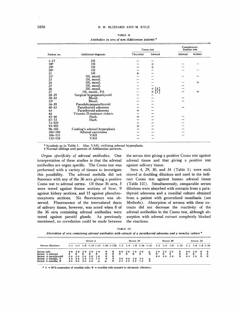
Four of 124 persons without Addison's dis-ease had serum adrenal antibodies demonstrableby the fluorescein technique, and none had CFantibodies (Table II). All four of these sub-jects had idiopathic hypoparathyroidism. Sera of23 patients with idiopathic hypoparathyroidism,29 patients (no. 96 to 124) with adrenal diseaseother than Addison's disease, and 68 subjectswith neither adrenal nor parathyroid disease gavenegative results to testing.
1655
R. M. BLIZZARD AND M. KYLE
TABLE II
Antibodies in sera of non-Addisonian patients *
Complement-Coons test fixation test
Patient no. Additional diagnosis Thryoidal Adrenal Adrenal Kidney
1-17 IH - -18b IH - + - -19b IH - +20b IH - - - -21 IH + -22f IH, monil. - - -
23 IH, monil. - -
24 IH, monil. - - - +25 IH, monil. - - -
26 IH, monil. - + [4]27 IH, monil., PA - + [1] - +28-29 Surgical hypoparathyroid - -
30-32 Monil. - -
33c Monil. - -
34-39 Pseudohypoparathyroid - -
40-42 Parathyroid adenoma - -
43 Parathyroid adenoma + -
44 Vitamin D-resistant rickets45-66 Hash. +67-72 Hash.73-92t93-95t + -96-101 Cushing's adrenal hyperplasia - -102-103 Adrenal carcinoma104-121 VAH - -122-124 VAH - -
* Symbols as in Table 1. Also, VAH, virilizing adrenal hyperplasia.t Normal siblings and parents of Addisonian patients.
Organ specificity of adrenal antibodies. Oneinterpretation of these studies is that the adrenalantibodies are organ specific. The Coons test was
performed with a variety of tissues to investigatethis possibility. The adrenal medulla did notfluoresce with any of the 36 sera giving a positiveCoons test to adrenal cortex. Of these 36 sera, 5were tested against frozen sections of liver, 9against kidney sections, and 15 against pheochro-mocytoma sections. No fluorescence was ob-served. Fluorescence of the intercalated ductsof salivary tissue, however, was noted when 8 ofthe 36 sera containing adrenal antibodies were
tested against parotid glands. As previouslymentioned, no correlation could be made between
the serum titer giving a positive Coons test againstadrenal tissue and that giving a positive testagainst salivary tissue.
Sera 4, 29, 30, and 34 (Table I) were eachtitered at doubling dilutions and used in the indi-rect Coons test against human adrenal tissue(Table III). Simultaneously, comparable serum
dilutions were absorbed with extracts from a para-
thyroid adenoma and a monilial culture obtainedfrom a patient with generalized moniliasis (seeMethods). Absorption of serums with these ex-
tracts did not decrease the reactivity of theadrenal antibodies in the Coons test, although ab-sorption with adrenal extract completely blockedthe reactions.
TABLE III
Absorption of sera containing adrenal antibodies with extracts of a parathyroid adenoma and a monilia culture *
Serum 4 Serum 29 Serum 30 Serum 34
Serum dilutions: 1:2 1:4 1:8 1:16 1:32 1:64 1:128 1:2 1:4 1:8 1:16 1:32 1:2 1:4 1:8 1:16 1:2 1:4 1:8 1:16
Serum only 4+ 3+ 2+ 2+ 1+ 0 0 4+ 3+ 1+ 2+ 4 2+ 2+ 2+ 0 4+ 2+ 0 0Serum +adrenal 0 0 0 0 0 0 0 0 0 0 0 0 0 0 0 0 0 0 0 0Serum +parathryoid 3 + 3+ 2 + 2 + 1+ 0 0 4+ 3 + 2 + 0 4+ 2 + 0 0Serum +monilia, A 4+ 3+ 2+ 2+ 1+ 0 0 3+ 4+ 2+ 2+ 4Serum +monilia, B 4+ 4+ 3+ 2+ :1 4 0 3+ 2+ 2+ 1+ 0
* A = 50%suspension of monilial cells; B = monilial cells exposed to ultrasonic vibration.
1656

ADRENALANTIGENS AND ANTIBODIES IN ADDISON'S DISEASE
Similarly, the adrenal antibodies in serum 4were not absorbed with microsomes from kid-ney, liver, thyroid, or pheochromocytoma tissue.The microsomes from adrenal tissue, however,which weighed only %8 to /14 as much as the tis-sue yielding the microsomes from kidney, liver,thyroid, and pheochromocytoma, blocked the re-
action (Table IV). This experiment was re-
peated, with virtually the same results.Species specificity of adrenal antibodies. The
indirect Coons test also was used to evaluate thespecies specificity of the adrenal antibodies.Eleven sera positive for human adrenal antibodieswere titered and tested against canine adrenalslices (Table V). Four of these sera were testedagainst the adrenal glands of other species. Thetiters giving a positive reaction against adrenaltissue from the human, monkey, guinea pig, cow,
pig, and cat were similar. Fourteen Addisoniansera giving a negative indirect Coons test withhuman adrenal slices and 10 sera from normalpersons failed to produce fluorescence when testedwith bovine adrenal slices. Therefore, no non-
specific fluorescence was observed when animaladrenal tissue was used.
Different types of adrenal antibodies. Since a
positive Coons test may reflect the presence ofone or more antibodies, the CF test was used todetermine if more than one CF antibody couldbe demonstrated. In the CF reaction, serum 23(Table VI) reacted at a high serum dilution, butonly with a high concentration of antigen. Wehave termed the CF antibody in this serum CFAB1. In contrast, serum 26 (Table VI) reactedat a lower serum dilution but with a weak anti-
TABLE IV
Absorption of serum 4 diluted 1:8 withvarious tissues
microsomes from
AdrenalMicro- Coons
Tissue somes* reaction
g tissueControl 0 3+
2.00 0Adrenal 1.00 0
0.50 00.20 2+
Kidney 5.50 3+Liver 5.50 3+Thyroid 7.00 3 +
Pheochromocytoma 4.00 3 +
* Expressed as total grams of tissue from which micro-somes were extracted to absorb 1 ml of diluted serum.
gen concentration. This indicated a separate an-tigen-antibody complex (CF AB2), although theantigen in both systems may be the same. Serum4 reacted at both high serum and weak antigendilutions, indicating a high titer of adrenal CFAB2. The same findings were demonstratedwhen bovine adrenal tissue was used as theantigen.
Both immunoelectrophoresis and agar slantswere used in an attempt to demonstrate the pres-ence of one or more precipitating antigen-anti-body complexes. Sera 4, 6, 20, 23, 26, and 34(Table I) were used in immunoelectrophoresis,and sera 4, 20, and 29 were tested with agarslants. No precipitating antibodies could be dem-onstrated by either technique.
Characteristics of the adrenal antigen. Theantigen responsible for the positive Coons test
TABLE V
Species specificity of adrenal antigen determined by titration of sera containing antiadrenalantibodies against adrenal slices of various species used in Coons test
Serum no.* Human Canine Bovine Porcine Simian Guinea pig Feline
4 1:32t 1:64 1:64 1:128 1:64 1:16 1:6429 1:8 1:16 1:8 1:16 1:16 1:812 1:4 1:8 1:4 1:4 1:8 1:415 1:16 1:32 1:32 1:64 1:32 1:6430 1:32 1:3226+ 1:4 1:219 1:32 1:3210 1:1 1:111 1:1 1:1
2 1:1 1:214 1:8 1:8
* Listed in Table 1.t Weakest serum titer positive to Coons test.tSee Table II.
1657

R. M. BLIZZARD AND M. KYLE
TABLE VI
Complement-fixing titers of adrenal antibodies compared for three sera*
Serum dilutionSerum Antigen
no. dilution Undiluted 1:2 1:4 1:8 1:16 1:33 1:64
23 0.02 4+ 4+ 4+ 4+ 4+ 0 00.01 2+ 2+ 2+ 2+ 1+ 0 00.005 0 0 0 0 0 0 00.0025 0 0 0 0 0 0 00.0012 0 0 0 0 0 0 00.0006 0 0 0 0 0 0 0
26 0.02 4+ 4+ 4+ 1 + 0 0 00.01 4+ 4+ 3+ 1+ 0 0 00.005 4+ 4+ 3+ 0 0 0 00.0025 4+ 4+ 3+ 0 0 0 00.0012 4+ 4+ 4+ 0 0 0 00.0006 not done
4 0.02 4+ 4+ 4+ 4+ 4+ 3+ 00.01 4+ 4+ 4+ 4+ 4+ 1+ 00.005 4+ 4+ 4+ 4+ 4+ 2+ 00.0025 4+ 4+ 4+ 4+ 4+ 2+ 00.0012 4+ 4+ 4+ 4+ 4+ 3+ 00.0006 2+ 2+ 2+ 2+ 1+ 1+ 0
* Interpretation of data is that there are two separate complement-fixing antibodies for adrenal tissue.
was found in both the microsomal and mitochon-drial fractions. After ultracentrifugation of freshadrenal homogenates, serial dilutions of the micro-somal and mitochondrial fractions were made.The protein concentration was determined, andinhibition of the Coons test against adrenal tissuewith microsomes and mitochondria was evaluated(Table VII). Both adrenal microsomes and mito-chondria in appropriate dilutions could block theindirect Coons test. Serum 4 was used in allexperiments.
The antigenic concentration, as related to pro-tein concentration, was slightly greater in the
TABLE VII
Localization of adrenal antigens in both microsomes andmitochondria of adrenal tissue*
Microsomes Mitochondria
Adrenal Coons Coonsgland Proteint Test Proteint Test
mg mgNo. 1 0 3+ 0 3+
1.08* 0 1.25* 00.54 0 0.95 00.27 0 0.63 00.13 2+ 0.32 2+
No. 2 0 3+ 0 3+1.00 0 1.23 00.50 0 0.62 1 +0.25 1+ 0.32 2+0.13 3+ 0.15 3+
* Serum 4 (Table I) diluted 1:8 was absorbed with microsomes andmitochrondria from two separate adrenal glands .
t Milligrams protein to absorb 1 ml of diluted serum.
microsomal than in the mitochondrial fraction.The antigenic potency of the nuclear fraction wasnot evaluated. Since only one serum was evalu-ated, and this serum may contain only one anti-body, it is not known whether all adrenal antigensoccur in both microsomes and mitochondria.
The effect of extreme temperature on micro-somal antigen was evaluated. For this purpose,0.54 mg of microsomal protein per ml of dilutedserum was used, an amount shown to be in ex-cess of that necessary to block completely theCoons reaction to adrenal antigen (Table VII,experiment 1). Boiling the microsomes in waterfor 5 minutes destroyed the antigen. After re-peated slow freezing and thawing for 10 times, theantigen retained its potency, and the Coons testwas inhibited. Therefore, heat lability but coldstability of the antigen were demonstrated.
To determine whether hydrocortisone was anadrenal antigen involved in the positive Coonstest, cortisol was added to a concentration of 500,ug per 100 ml to serum 4. The titer of this serumboth with and without added cortisol was 1: 32,indicating no inhibition of the antibodies withhydrocortisone.
DISCUSSION
By the indirect Coons technique, circulatingadrenal antibodies have been found in sera of 51 %of 71 patients with Addison's disease. In a previ-
1658

ADRENALANTIGENS AND ANTIBODIES IN ADDISON'S DISEASE
ous report (3) concerning 31 of these 71, thisincidence was similar (53 %). Significantly, noneof 29 patients with adrenal tumors, idiopathicCushing's syndrome, or virilizing adrenal hyper-plasia had demonstrable adrenal antibodies.Equally important was the finding that the seraof only 4 of 124 subjects without Addison's dis-ease had adrenal antibodies demonstrable by theindirect fluorescein method, and one of these four(no. 18, Table II) was known to have limited cor-
tisol excretion secondary to adrenocortical stimu-lation (8). Therefore, it is probable that the dem-onstration of circulating adrenal antibodies by thefluorescein technique reflects the presence of anatrophic and not hyperplastic adrenal disease.
Since adrenal antibodies have been shown to beorgan specific, the first criterion in establishingAddison's disease as an autoimmune process hasbeen met, i.e., specific antibodies of the circulatingtype have been directly demonstrated. The isola-tion of specific antigens in the adrenal corticalmicrosomes and mitochondria meets the secondcriterion. The third criterion is that antibodiesmust be produced in animals with the same typeof antigen. Witebsky and Milgrom (9) and Mil-cou, Pop, Lupulescou, and Taga (10) have suc-cessfullv induced the formation of circulating anti-bodies to homologous adrenal tissue in animals.In guinea pigs, the adrenal antibodies were bothorgan specific and species specific. In rabbits, asnot in guinea pigs, but as for the circulating anti-bodies of Addisonian patients, the antibodies werespecies specific but not organ specific (9).Witebsky and MIilgrom (1, 9) found that theadrenal antigen inducing antibody formation inanimals was thermolabile, like that in the micro-somes of the human adrenal gland, and probablylocated in the adrenal cortex.
As for the fourth criterion to establish Addison'sdisease as an autoimmune process, investigators(9-12) have attempted to produce adrenal atrophyin animals by immunizing them with homologousadrenal extracts. Milgrom and Witebsky (9)reported that lymphocytic infiltration occurredthroughout the adrenal cortex, although most of itoccurred at the corticomedullary border zone andextended into the inner medulla. Rabbits im-munized with extracts of other organs andFreund's adjuvant occasionally had similar le-sions. Mlcou and associates (10) noted multiple
lesions in both zones of the adrenal glands of iso-immunized rabbits. In the cortex, the histopathol-ogy consisted of lymphocytic infiltration, granu-locytic and fibroblastic infiltration, interstitialhemorrhage, and thrombotic obstruction of thecapillaries and arterioles. The changes reportedin the cortex of the animals immunized by Coloverand Glynn (12) possibly were more marked thanthose produced by other investigators. In the pa-renchymal cells of the cortex, degenerative changesin the cytoplasm and nuclei were observed, andthere was gross infiltration of the cortex withhistiocytic elements, macrophages, lymphocytes,occasional plasma cells, and eosinophils. Insome glands, the zona reticularis was particularlyaffected, and its parenchymal cells were largely re-placed by infiltrating cells. The medullas of manyglands were also severely affected. Although noneof the investigators have reported adrenal atrophyoccurring secondary to immunization, the histo-pathology of the adrenal glands of immunizedanimals and that recorded for adrenal atrophy inhumans have some characteristics in common;i.e., Guttman (13) and Duff and Bernstein (14)reported that in the adrenal remnants of someAddisonian patients, lymphocytic infiltration oc-curs in the medulla and in the remaining remnantsof cortical cells. Therefore, the fourth criterionhas been partially met, although not so completelyas the other criteria.
Although several criteria of idiopathic Addi-son's disease as an autoimmune process have beenestablished, the role of the circulating antibodieshas not been determined. These antibodies mayonly reflect the disease process and not be re-sponsible for it, and the possible role of cellularantibodies must be considered. Experiments ofFelix-Davies and Waksman (15) and of Mc-Master, Lerner, and Exum (16) suggest thatcellular antibodies are responsible for lympho-cytic infiltration of the thyroid in autoimmune thy-roidal disease. Since lymphocytic thyroiditis haslong been recognized as frequently associatedwith Addison's disease (17), further support islent to the concept that Addison's disease may alsoresult from an autosensitization process, possiblycaused by cellular antibodies.
The high incidence of thyroidal antibodies insera of Addisonian patients (22 of 71) suggeststhat in some persons multiple endocrine deficien-
1659

R. M. BLIZZARD AND M. KYLE
cies occur, and all of these deficiencies may beassociated with autoimmune processes. As notedabove, the coincident occurrence of lymphocyticthyroiditis, with or without hypothyroidism, andAddison's disease was first recognized in 1930(17). The triad of Addison's disease, hypopara-thyroidism, and moniliasis has been recordedfrequently (8, 18). Twelve of our 71 Addisonianpatients had associated hypoparathyroidism, and 7of the 12 also had moniliasis. The sera of 10 ofthese 12 were positive when tested for adrenalantibodies. Since the adrenal antibodies are or-gan specific, multiple circulating antibody-antigencomplexes probably are involved in the autoim-mune processes. The possibility, however, of acommon cellular antibody-antigen system betweenmultiple endocrine glands must be considered;further studies are necessary to elucidate it.
SUMMARY
Circulating adrenal antibodies, tested by the in-direct Coons test, were found in 36 of 71 serafrom Addisonian patients. Complement-fixingantibodies were found in 24 of these 36 sera. Thecirculating antibodies were organ specific but notspecies specific. There was no cross-reactivitywith tissue of the adrenal medulla, pheochromo-cytoma, thyroid, salivary gland, liver, or kidney,and none with monilial organisms. The anti-gen(s) to which the circulating antibodies areproduced is in both the microsomal and mitochon-drial fractions. Some of the physical character-istics of the antigen(s) have been studied. Fur-ther work is indicated to clarify the significanceof circulating adrenal antibodies in Addison'sdisease.
ACKNOWLEDGMENTS
We gratefully acknowledge the cooperation of themany physicians who assisted in the collection of sera,the review and criticisms of the manuscript by Dr. IreneSolomon, and the secretarial assistance of Mrs. MaryWestervelt.
REFERENCES
1. Witebsky, E. The question of self-recognition by thehost and problems of auto-antibodies and theirspecificity. Cancer Res. 1961, 21, 216.
2. Witebsky, E., N. R. Rose, K. Terplan, J. R. Paine,and R. W. Egan. Chronic thyroiditis and auto-
immunization. J. Amer. med. Ass. 1957, 164,1439.
3. Blizzard, R. M., M. A. Kyle, R. W. Chandler, andW. Hung. Adrenal antibodies in Addison's dis-ease. Lancet 1962, 2, 901.
4. Holborow, E. J., P. C. Brown, I. Mf. Roitt, and D.Doniach. Cytoplasmic localization of "comple-ment-fixing" auto-antigen in human thyroid epi-thelium. Brit. J. exp. Path. 1959, 40, 583.
5. Chandler, R. W., R. M. Blizzard, Wkr. Hung, and M.Kyle. Incidence of thyrocytotoxic factor andother antithyroid antibodies in the mothers of cre-tins. New Engl. J. Med. 1962, 267. 376.
6. Blizzard, R. M., R. WV. Chandler, B. H. Landing,M. D. Pettit, and C. D. West. 'Maternal autoim-munization to thyroid as a probable cause ofathyrotic cretinism. New Engl. J. Med. 1960, 263,327.
7. Kabat, E. A., and M. M. Mayer. Experimental Im-munochemistry. Springfield, Ill., Charles CThomas, 1961, p. 476.
8. Morse, W. I., W. A. Cochrane, and P. L. Landrigan.Familial hypoparathyroidism with pernicious ane-mia, steatorrhea, and adrenocortical insufficiency.New Engl. J. Med. 1961, 264, 1021.
9. Witebsky, E., and F. Milgrom. Immunological stud-ies on adrenal glands. II. Immunization withadrenals of the same species. Immunology 1962,5, 67.
10. Milcou, S. M., A. Pop, A. Lupulescou, and M. Taga.L'auto-immunisation experimentale de la surrenalechez le lapin. Ann. d'Endocr. (Paris) 1959, 20,799.
11. Steiner, J. W., B. Langer, D. L. Schatz, and R.Volpe. Experimental immunologic adrenal injury.A response to inj ections of autologous and homolo-gous adrenal antigens in adjuvant. J. exp. Med.1960, 112, 187.
12. Colover, J., and L. E. Glynn. Experimental iso-im-mune adrenalitis. Immunology 1958, 1, 172.
13. Guttman, P. H. Addison's disease. A statisticalanalysis of five hundred and sixty-six cases and astudy of the pathology. Arch. Path. 1930, 10, 742.
14. Duff, G. L., and C. Bernstein. Five cases of Addi-son's disease with so-called atrophy of adrenalcortex. Bull. Johns Hopk. Hosp. 1933, 52, 67.
15. Felix-Davies, D., and B. H. WNaksman. Passivetransfer of experimental immune thyroiditis in theguinea pig. Arthr. and Rheum. 1961, 4, 416.
16. McMaster, P. R. B., E. M. Lerner II, and E. D.Exum. The relationship of delayed hypersensitivityand circulating antibody to experimental allergicthyroiditis in inbred guinea pigs. J. exp. Med.1961, 113, 611.
17. Wells, H. G. Addison's disease with selective de-struction of the suprarenal cortex ("suprarenalcortex atrophy"). Arch. Path. 1930, 10, 499.
18. Craig, J. M., L. H. Schiff, and J. E. Boone. Chronicmoniliasis associated with Addison's disease. Amer.J. Dis. Child. 1955, 89, 669.
1660