balancing image quality and radiation dose in radiography...
TRANSCRIPT
7/22/2014
1
Balancing Image Quality and Radiation Risk in Radiography
Clinical/Physical/Exam/Cost
Considerations
Alexander S. Pasciak, PhD
University of Tennessee Medical Center
Some observations
• Radiography receives little public publicity in regard to risk; secondary to CT and nuclear medicine
• Many radiologists, while they are now forced to strongly consider radiation dose in CT, don’t consider radiation dose in radiography – Resulting from publicity in recent years as well as new
TJC requirements – Pediatric radiography is one exception
• Dose “too low” to worry about
Some observations, continued
• Of course, because of the lackadaisical view on the importance of dose in conventional radiography, it is often one area that can benefit substantially from improvement – Dose creep?
– Babygrams?
– Poor pediatric protocol choices?
– Portables and technique refinement?
– Appropriate techniques based on exam type?
7/22/2014
2
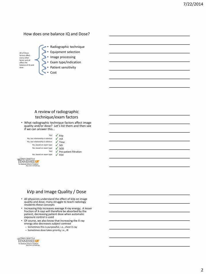
How does one balance IQ and Dose?
• Radiographic technique
• Equipment selection
• Image processing
• Exam type/indication
• Patient sensitivity
• Cost
All of these factors affect every other factor and all affect the balance of IQ and dose
A review of radiographic technique/exam factors
• What radiographic technique factors affect image quality and/or dose? Let’s list them and then see if we can answer this…
kVp mA Time SID SOD Pre-patient filtration FOV
Yes, but relationship is obvious
Yes, but relationship is obvious
Yes, based on exam type
Yes, based on exam type
Yes, based on exam type
Yes!
Yes!
kVp and Image Quality / Dose
• All physicists understand the effect of kVp on image quality and dose; many struggle to teach radiology residents these concepts
• Increasing kVp increases average X-ray energy; A lesser fraction of X-rays will therefore be absorbed by the patient, decreasing patient dose when automatic exposure control is used
• Of course, we also know that increasing the X-ray energy also decreases subject contrast – Sometimes this is purposeful, i.e., chest X-ray – Sometimes dose takes priority, i.e., IR
7/22/2014
3
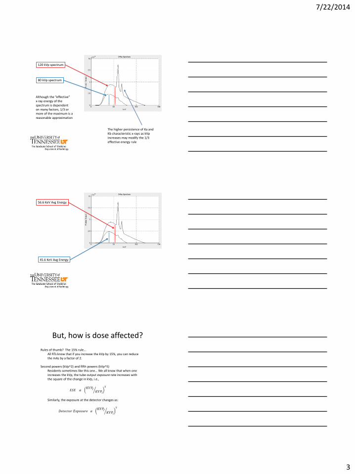
120 kVp spectrum
80 kVp spectrum
Although the “effective” x-ray energy of the spectrum is dependent on many factors, 1/3 or more of the maximum is a reasonable approximation
The higher persistence of Ka and Kb characteristic x-rays as kVp increases may modify the 1/3 effective energy rule
56.6 KeV Avg Energy
45.6 KeV Avg Energy
But, how is dose affected?
Rules of thumb? The 15% rule… All RTs know that if you increase the kVp by 15%, you can reduce the mAs by a factor of 2.
Second powers (kVp^2) and fifth powers (kVp^5)
Residents sometimes like this one… We all know that when one increases the kVp, the tube output exposure rate increases with the square of the change in kVp, i.e.,
𝐸𝑆𝐸 𝛼 𝐾𝑉𝑃𝑓
𝐾𝑉𝑃𝑖
2
Similarly, the exposure at the detector changes as:
𝐷𝑒𝑡𝑒𝑐𝑡𝑜𝑟 𝐸𝑥𝑝𝑜𝑠𝑢𝑟𝑒 𝛼 𝐾𝑉𝑃𝑓
𝐾𝑉𝑃𝑖
5
7/22/2014
4
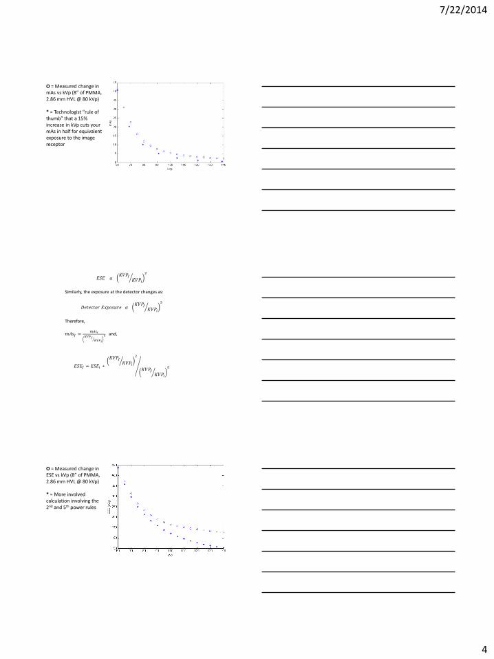
O = Measured change in mAs vs kVp (8” of PMMA, 2.86 mm HVL @ 80 kVp) * = Technologist “rule of thumb” that a 15% increase in kVp cuts your mAs in half for equivalent exposure to the image receptor
𝐸𝑆𝐸 𝛼 𝐾𝑉𝑃𝑓
𝐾𝑉𝑃𝑖
2
Similarly, the exposure at the detector changes as:
𝐷𝑒𝑡𝑒𝑐𝑡𝑜𝑟 𝐸𝑥𝑝𝑜𝑠𝑢𝑟𝑒 𝛼 𝐾𝑉𝑃𝑓
𝐾𝑉𝑃𝑖
5
Therefore,
𝑚𝐴𝑠𝑓 =𝑚𝐴𝑠𝑖
𝐾𝑉𝑃𝑓𝐾𝑉𝑃𝑖
5 and,
𝐸𝑆𝐸𝑓 = 𝐸𝑆𝐸𝑖 ∗
𝐾𝑉𝑃𝑓𝐾𝑉𝑃𝑖
2
𝐾𝑉𝑃𝑓𝐾𝑉𝑃𝑖
5
O = Measured change in ESE vs kVp (8” of PMMA, 2.86 mm HVL @ 80 kVp) * = More involved calculation involving the 2nd and 5th power rules
7/22/2014
5
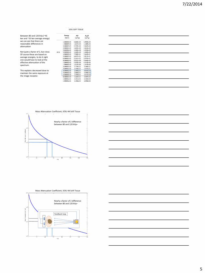
ICRU SOFT TISSUE
Between 80 and 120 kVp (~45 kev and ~55 kev average energy) we can see that there are reasonable differences in attenuation Not quite a factor of 2, but close. Of course these are based on average energies, to do it right one would have to look at the effective attenuation of the spectrum. This explains decreased dose to maintain the same exposure at the image receptor.
Mass Attenuation Coefficient, ICRU 44 Soft Tissue
Nearly a factor of 2 difference between 80 and 120 kVp–
Mass Attenuation Coefficient, ICRU 44 Soft Tissue
Nearly a factor of 2 difference between 80 and 120 kVp–
Feedback loop
7/22/2014
6
*Pre-patient filtration has a similar effect to increasing the kVp on patient dose, as it increases the average energy of the beam. There are some exceptions to the purpose of this, notably in angiography
But what about contrast?
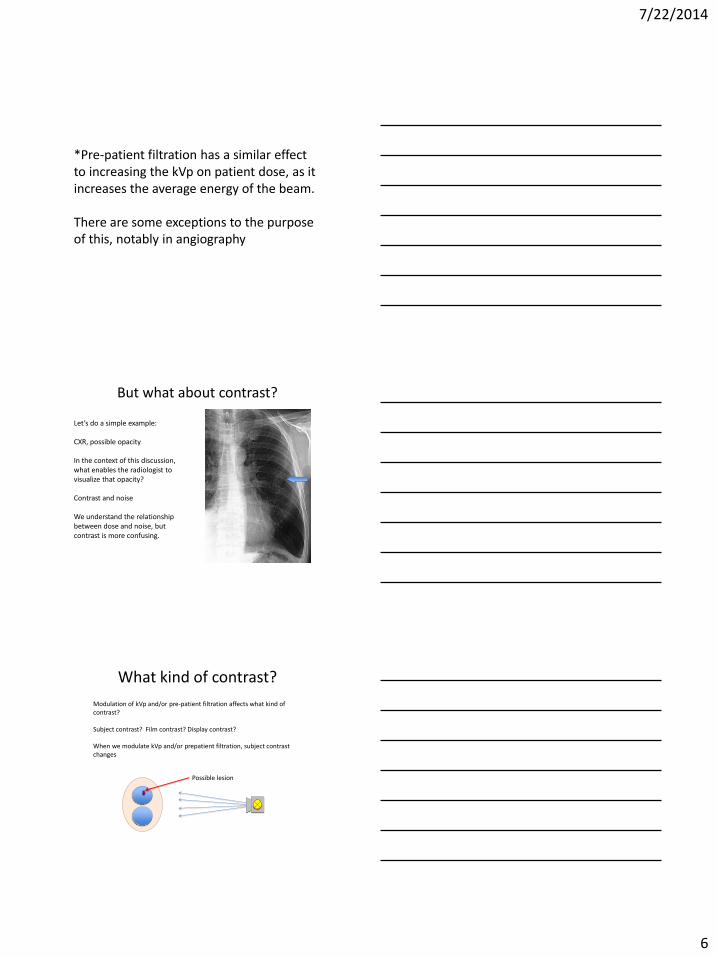
Let’s do a simple example: CXR, possible opacity In the context of this discussion, what enables the radiologist to visualize that opacity? Contrast and noise We understand the relationship between dose and noise, but contrast is more confusing.
What kind of contrast?
Modulation of kVp and/or pre-patient filtration affects what kind of contrast? Subject contrast? Film contrast? Display contrast? When we modulate kVp and/or prepatient filtration, subject contrast changes
Possible lesion
7/22/2014
7
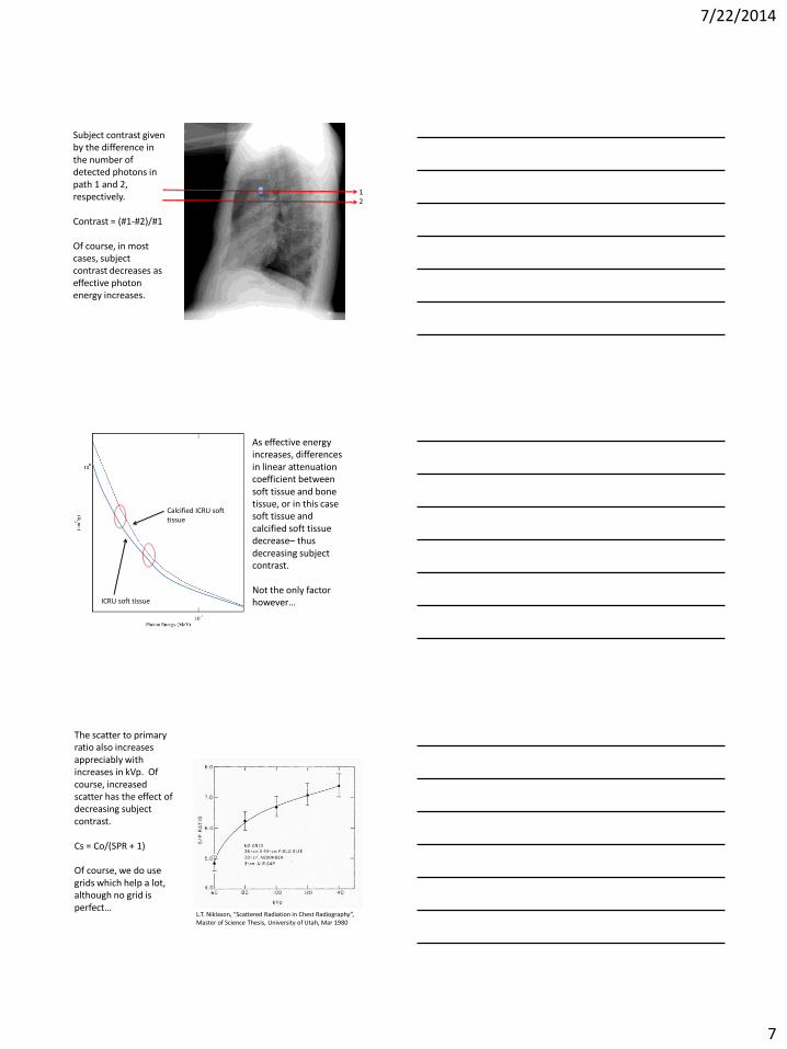
1 2
Subject contrast given by the difference in the number of detected photons in path 1 and 2, respectively. Contrast = (#1-#2)/#1 Of course, in most cases, subject contrast decreases as effective photon energy increases.
Calcified ICRU soft tissue
ICRU soft tissue
As effective energy increases, differences in linear attenuation coefficient between soft tissue and bone tissue, or in this case soft tissue and calcified soft tissue decrease– thus decreasing subject contrast. Not the only factor however…
L.T. Niklason, “Scattered Radiation in Chest Radiography”, Master of Science Thesis, University of Utah, Mar 1980
The scatter to primary ratio also increases appreciably with increases in kVp. Of course, increased scatter has the effect of decreasing subject contrast. Cs = Co/(SPR + 1) Of course, we do use grids which help a lot, although no grid is perfect…
7/22/2014
8
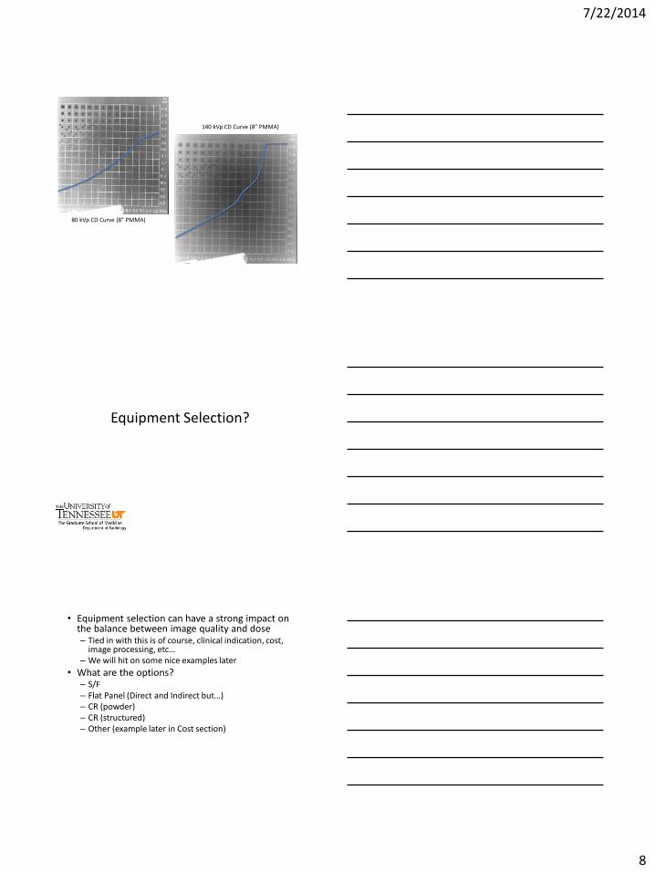
80 kVp CD Curve (8” PMMA)
140 kVp CD Curve (8” PMMA)
Equipment Selection?
• Equipment selection can have a strong impact on the balance between image quality and dose – Tied in with this is of course, clinical indication, cost,
image processing, etc… – We will hit on some nice examples later
• What are the options? – S/F – Flat Panel (Direct and Indirect but…) – CR (powder) – CR (structured) – Other (example later in Cost section)
7/22/2014
9
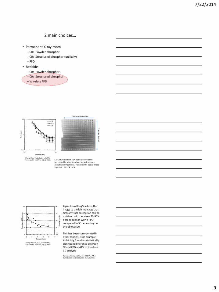
2 main choices…
• Permanent X-ray room
– CR: Powder phosphor
– CR: Structured phosphor (unlikely)
– FPD
• Bedside
– CR: Powder phosphor
– CR: Structured phosphor
– Wireless FPD
X. Rong, Shaw CC, Liu X, Lemacks MR, Thompson SK. Med Phys 28(11), 2001;
Co
ntrast lim
ited
Resolution limited
CD Comparisons of FP, CR and SF have been performed by several authors, as well as more analytical comparisons. However, the above image says it all. FP > CR ~= SF
Again from Rong’s article, the image to the left indicates that similar visual perception can be obtained with between 70-90% dose reduction with a FPD compared to SF depending on the object size. This has been corroborated in other reports. One example, Aufrichtig found no statistically significant difference between SF and FPD at 41% of the dose. CD analysis Richard Aufrichtig and Ping Xue 2000 Phys. Med. Biol. 45 2653. doi:10.1088/0031-9155/45/9/316
X. Rong, Shaw CC, Liu X, Lemacks MR, Thompson SK. Med Phys 28(11), 2001;
7/22/2014
10
*Dobbins JT, Samei E, Chotas HG, Warp RJ, Baydush AH, Floyd CE, et al. Chest radiography: optimization of X-ray spectrum for cesium iodide-amorphous silicon flat-panel detector. Radiology 2003;226:221–30.
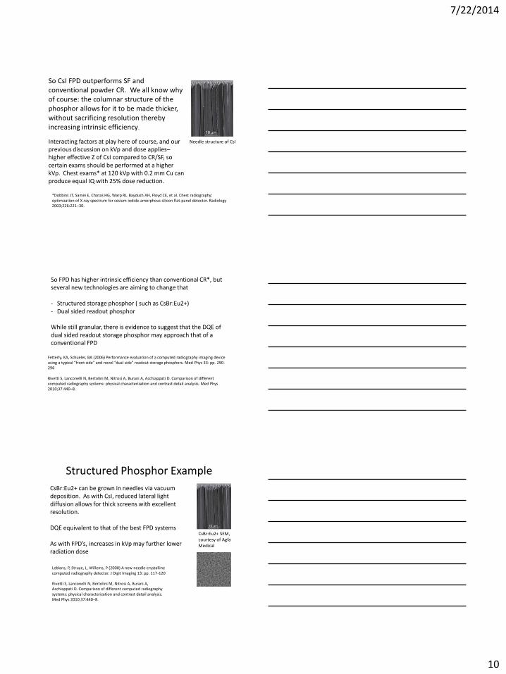
Needle structure of CsI
So CsI FPD outperforms SF and conventional powder CR. We all know why of course: the columnar structure of the phosphor allows for it to be made thicker, without sacrificing resolution thereby increasing intrinsic efficiency.
Interacting factors at play here of course, and our previous discussion on kVp and dose applies– higher effective Z of CsI compared to CR/SF, so certain exams should be performed at a higher kVp. Chest exams* at 120 kVp with 0.2 mm Cu can produce equal IQ with 25% dose reduction.
Fetterly, KA, Schueler, BA (2006) Performance evaluation of a computed radiography imaging device using a typical “front side” and novel “dual side” readout storage phosphors. Med Phys 33: pp. 290-296 Rivetti S, Lanconelli N, Bertolini M, Nitrosi A, Burani A, Acchiappati D. Comparison of different computed radiography systems: physical characterization and contrast detail analysis. Med Phys 2010;37:440–8.
So FPD has higher intrinsic efficiency than conventional CR*, but several new technologies are aiming to change that - Structured storage phosphor ( such as CsBr:Eu2+) - Dual sided readout phosphor
While still granular, there is evidence to suggest that the DQE of dual sided readout storage phosphor may approach that of a conventional FPD
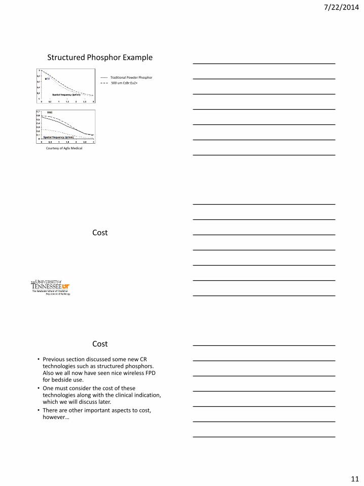
Structured Phosphor Example
CsBr:Eu2+ SEM, courtesy of Agfa Medical
Leblans, P, Struye, L, Willems, P (2000) A new needle-crystalline computed radiography detector. J Digit Imaging 13: pp. 117-120 Rivetti S, Lanconelli N, Bertolini M, Nitrosi A, Burani A, Acchiappati D. Comparison of different computed radiography systems: physical characterization and contrast detail analysis. Med Phys 2010;37:440–8.
CsBr:Eu2+ can be grown in needles via vacuum deposition. As with CsI, reduced lateral light diffusion allows for thick screens with excellent resolution. DQE equivalent to that of the best FPD systems As with FPD’s, increases in kVp may further lower radiation dose
7/22/2014
11
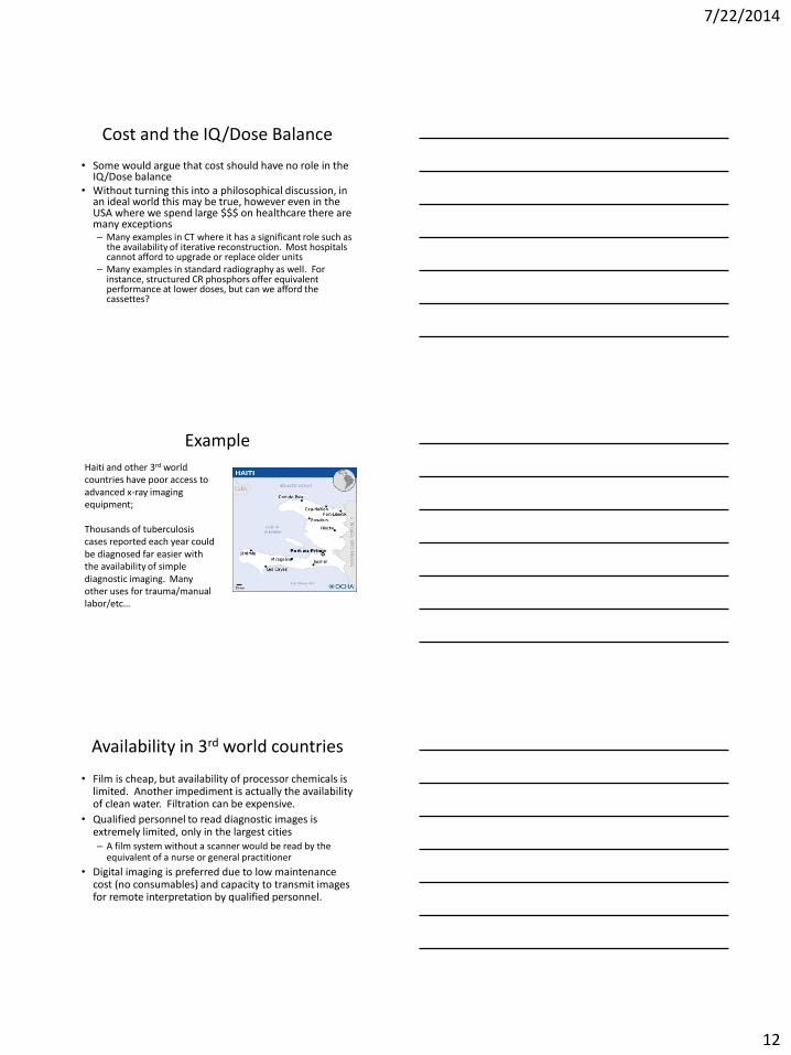
Structured Phosphor Example
Traditional Powder Phosphor
500 um CsBr:Eu2+
Courtesy of Agfa Medical
Cost
Cost
• Previous section discussed some new CR technologies such as structured phosphors. Also we all now have seen nice wireless FPD for bedside use.
• One must consider the cost of these technologies along with the clinical indication, which we will discuss later.
• There are other important aspects to cost, however…
7/22/2014
12
Cost and the IQ/Dose Balance
• Some would argue that cost should have no role in the IQ/Dose balance
• Without turning this into a philosophical discussion, in an ideal world this may be true, however even in the USA where we spend large $$$ on healthcare there are many exceptions – Many examples in CT where it has a significant role such as
the availability of iterative reconstruction. Most hospitals cannot afford to upgrade or replace older units
– Many examples in standard radiography as well. For instance, structured CR phosphors offer equivalent performance at lower doses, but can we afford the cassettes?
Example
Haiti and other 3rd world countries have poor access to advanced x-ray imaging equipment; Thousands of tuberculosis cases reported each year could be diagnosed far easier with the availability of simple diagnostic imaging. Many other uses for trauma/manual labor/etc…
Availability in 3rd world countries
• Film is cheap, but availability of processor chemicals is limited. Another impediment is actually the availability of clean water. Filtration can be expensive.
• Qualified personnel to read diagnostic images is extremely limited, only in the largest cities – A film system without a scanner would be read by the
equivalent of a nurse or general practitioner
• Digital imaging is preferred due to low maintenance cost (no consumables) and capacity to transmit images for remote interpretation by qualified personnel.
7/22/2014
13
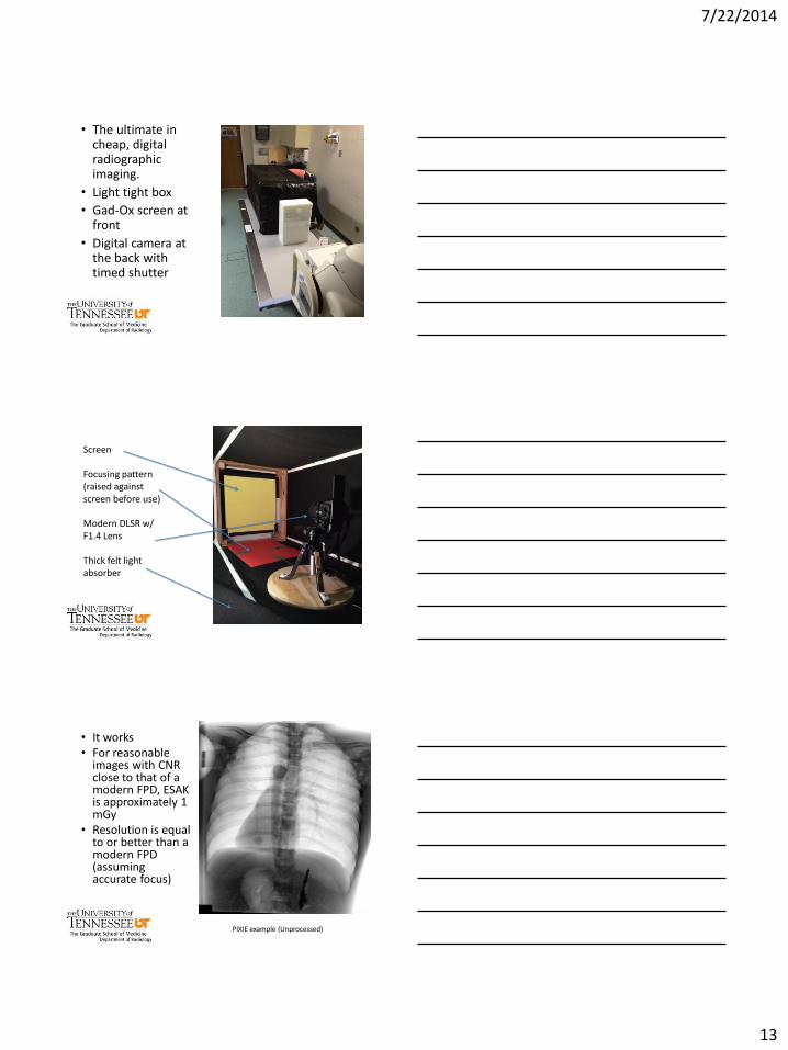
• The ultimate in cheap, digital radiographic imaging.
• Light tight box
• Gad-Ox screen at front
• Digital camera at the back with timed shutter
Screen Focusing pattern (raised against screen before use) Modern DLSR w/ F1.4 Lens Thick felt light absorber
• It works • For reasonable
images with CNR close to that of a modern FPD, ESAK is approximately 1 mGy
• Resolution is equal to or better than a modern FPD (assuming accurate focus)
PIXIE example (Unprocessed)
7/22/2014
14
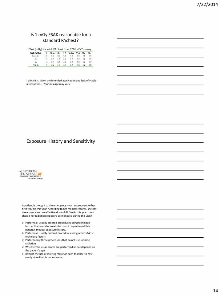
Is 1 mGy ESAK reasonable for a standard PAchest?
ESAK (mGy) for adult PA chest from 2001 NEXT survey
I think it is, given the intended application and lack of viable alternatives… Your mileage may vary.
Exposure History and Sensitivity
A patient is brought to the emergency room subsequent to her fifth trauma this year. According to her medical records, she has already received an effective dose of 48.5 mSv this year. How should her radiation exposure be managed during this visit? a) Perform all usually ordered procedures using technique
factors that would normally be used irrespective of this patient’s medical exposure history.
b) Perform all usually ordered procedures using reduced dose technique factors.
c) Perform only those procedures that do not use ionizing radiation
d) Whether the usual exams are performed or not depends on the patient’s age
e) Restrict the use of ionizing radiation such that her 50 mSv yearly dose limit is not exceeded.
7/22/2014
15
Sensitivity
Sensitivity may be dependent on other factors including: - Type of imaging procedure - Age of the patient - Sex of the patient - Rare conditions such as AT - Previous exams?
Dose and Risk?
Dose
Ris
k
LNT model endorsed by BEIR committee for estimating mortality and morbidity from solid tumors. What does the LNT suggest about considering previous exposure history?
Eisenberg JD, Harvey HB, Moore DA, Gazelle GS, Pandharipande PV. Falling prey to the sunk cost bias: a potential harm of patient radiation dose histories. Radiology 2012;263:626–8.
Dose and Risk?
Dose
Ris
k
However, we don’t use LNT for everything. We know that leukemia is more appropriately represented by a linear-quadratic risk model. In the case of leukemia, the linear quadratic model might indicate previous exposure history could have a role in clinical decision making.
7/22/2014
16
Dose and Risk?
Dose
Ris
k
However, as pointed out by Eisenburg*, at low doses common in diagnostic imaging, and in particular in diagnostic radiology, the linear quadratic model looks like LNT, and so one can arrive at the same conclusion; i.e., that is risk to the patient from today’s procedure is the same regardless of previous medical exposure.
*Eisenberg JD, Lewin SO, Pandharipande PV. The Fisherman's Cards: How to Address Past and Future Radiation Exposures in Clinical Decision Making. AJR Am J Roentgenol 2014;202:362–7.
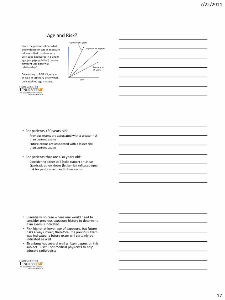
Age and Risk?
Dose
Ris
k
LNT model is to describe risk vs. dose to a specific population; We all understand that it is only linear within the confines of a single population— - How about age? Eisenburg* does not consider varying populations
*Eisenberg JD, Lewin SO, Pandharipande PV. The Fisherman's Cards: How to Address Past and Future Radiation Exposures in Clinical Decision Making. AJR Am J Roentgenol 2014;202:362–7.
Age and Risk?
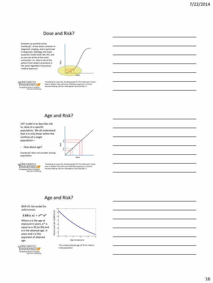
𝑬𝑨𝑹 𝒆, 𝒂 = 𝒆𝜸𝒆∗𝒂𝒏
BEIR VII risk model for solid tumors
Where e is the age at exposure in years, e* is equal to e-30 (e<30) and a is the attained age, in years and n is the exponent of attained age. Age at exposure
Exce
ss c
ases
per
10,
000
Py-
Sv
*For a fixed attained age of 70 Yrs- Risk to male population
7/22/2014
17
Age and Risk?
From the previous slide, what dependence on age at exposure tells us is that risk does vary with age; Exposures in a single age group (population) carry a different LNT dose/risk relationship*. *According to BEIR VII, only up to an e of 30 years, after which only attained age matters.
Dose
Exposure at 5 years
Exposure at 10 years
Exposure at 15 years
• For patients <30 years old: – Previous exams are associated with a greater risk
than current exams
– Future exams are associated with a lesser risk than current exams
• For patients that are >30 years old: – Considering either LNT (solid tumor) or Linear
Quadratic at low doses (leukemia) indicates equal risk for past, current and future exams
• Essentially no case where one would need to consider previous exposure history to determine if an exam is indicated.
• Risk higher at lower age of exposure, but future risks always lower; therefore, if a previous exam was indicated, a future exam will certainly be indicated as well
• Eisenberg has several well written papers on this subject—useful for medical physicists to help educate radiologists
7/22/2014
18
Clinical Indication
“The job of the diagnostic medical physicist is to solve
problems for physicians”
Charles Willis, PhD, DABR, FAAPM
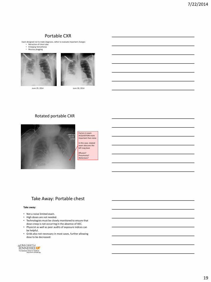
Common Exams: Portable chest
What is the radiologist looking for when they read a bedside chest exam? The indication is drastically different than for a conventional 2-view chest, and radiologists typically look for:
• Large issues- exams not performed for differential diagnosis– not for differentiating atelectasis from pleural effusion
• Looking for large changes from previous day- intended to prevent catastrophe
7/22/2014
19
Portable CXR Exam designed not to make diagnosis, rather to evaluate important changes
• Retraction of chest tube • Enlarging hemothorax • Mucous plugging
June 29, 2014 June 30, 2014
Rotated portable CXR
Factors in exam ACQUISITION more important than noise In this case, rotated exam obscures the left lung base Effusion? Pneumonia? Atelectasis?
Take Away: Portable chest
Take away: • Not a noise limited exam. • High doses are not needed. • Technologists must be closely monitored to ensure that
dose-creep is not occurring in the absence of AEC. • Physicist as well as peer audits of exposure indices can
be helpful. • Grids also not necessary in most cases, further allowing
dose to be decreased.
7/22/2014
20
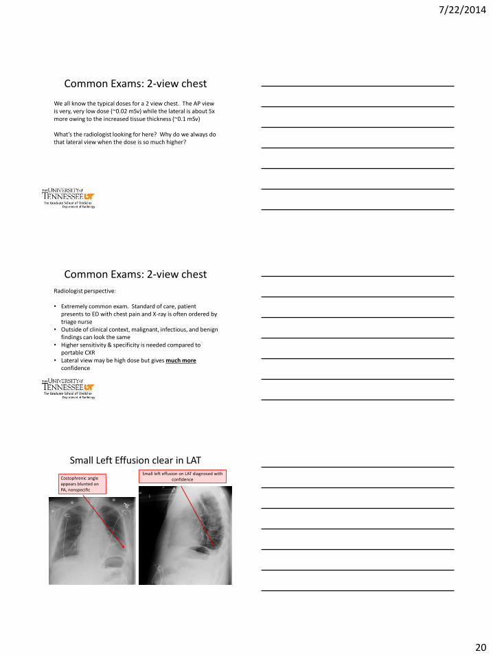
Common Exams: 2-view chest
We all know the typical doses for a 2 view chest. The AP view is very, very low dose (~0.02 mSv) while the lateral is about 5x more owing to the increased tissue thickness (~0.1 mSv) What’s the radiologist looking for here? Why do we always do that lateral view when the dose is so much higher?
Common Exams: 2-view chest
Radiologist perspective: • Extremely common exam. Standard of care, patient
presents to ED with chest pain and X-ray is often ordered by triage nurse
• Outside of clinical context, malignant, infectious, and benign findings can look the same
• Higher sensitivity & specificity is needed compared to portable CXR
• Lateral view may be high dose but gives much more confidence
Small Left Effusion clear in LAT
Costophrenic angle appears blunted on PA, nonspecific
Small left effusion on LAT diagnosed with confidence
7/22/2014
21
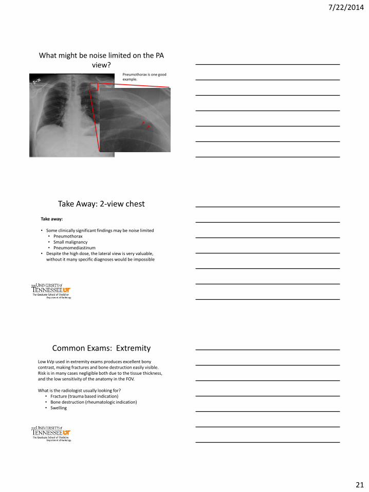
What might be noise limited on the PA view?
Pneumothorax is one good example.
Take Away: 2-view chest
Take away: • Some clinically significant findings may be noise limited
• Pneumothorax • Small malignancy • Pneumomediastinum
• Despite the high dose, the lateral view is very valuable, without it many specific diagnoses would be impossible
Common Exams: Extremity
Low kVp used in extremity exams produces excellent bony contrast, making fractures and bone destruction easily visible. Risk is in many cases negligible both due to the tissue thickness, and the low sensitivity of the anatomy in the FOV. What is the radiologist usually looking for?
• Fracture (trauma based indication) • Bone destruction (rheumatologic indication) • Swelling
7/22/2014
22
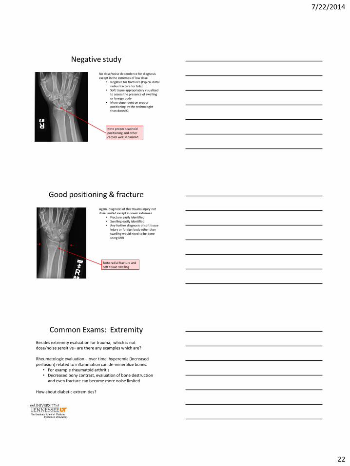
Negative study
No dose/noise dependence for diagnosis except in the extremes of low dose.
• Negative for fractures (typical distal radius fracture for falls)
• Soft tissue appropriately visualized to assess the presence of swelling or foreign body
• More dependent on proper positioning by the technologist than dose/IQ
Note proper scaphoid positioning and other carpals well separated
Good positioning & fracture
Again, diagnosis of this trauma injury not dose limited except in lower extremes
• Fracture easily identified • Swelling easily identified • Any further diagnosis of soft tissue
injury or foreign body other than swelling would need to be done using MRI
Note radial fracture and soft tissue swelling
Common Exams: Extremity
Besides extremity evaluation for trauma, which is not dose/noise sensitive– are there any examples which are? Rheumatologic evaluation - over time, hyperemia (increased perfusion) related to inflammation can de-mineralize bones.
• For example rheumatoid arthritis • Decreased bony contrast, evaluation of bone destruction
and even fracture can become more noise limited
How about diabetic extremities?
7/22/2014
23
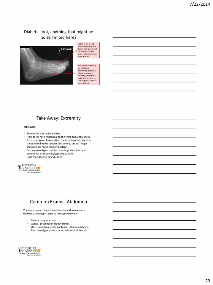
Diabetic foot, anything that might be noise limited here?
Notice there is also demineralization here due to poor peripheral circulation– studies begin to become more noise limited
Also notice soft tissue gas indicating necrotizing fasciitis, a clinical emergency requiring immediate surgical debridement. This diagnosis can be noise limited.
Take Away: Extremity
Take away: • Extremities not radiosensitive • High doses not needed due to the small tissue thickness • For many types of exams (i.e., fracture, trauma) diagnosis
is not noise limited (proper positioning, proper image processing is much more important)
• Certain exam types may be more important (diabetic extremities or rheumatologic evaluation)
• Dose may depend on indication!
Common Exams: Abdomen
There are many clinical indications for abdominal x-ray. However, radiologists tend to focus primarily on:
• Bones - bony anatomy • Stones - presence of kidney stones • Mass - abnormal organ contours (splenomegaly, etc) • Gas - bowel gas pattern or intraabdominal free air
7/22/2014
24
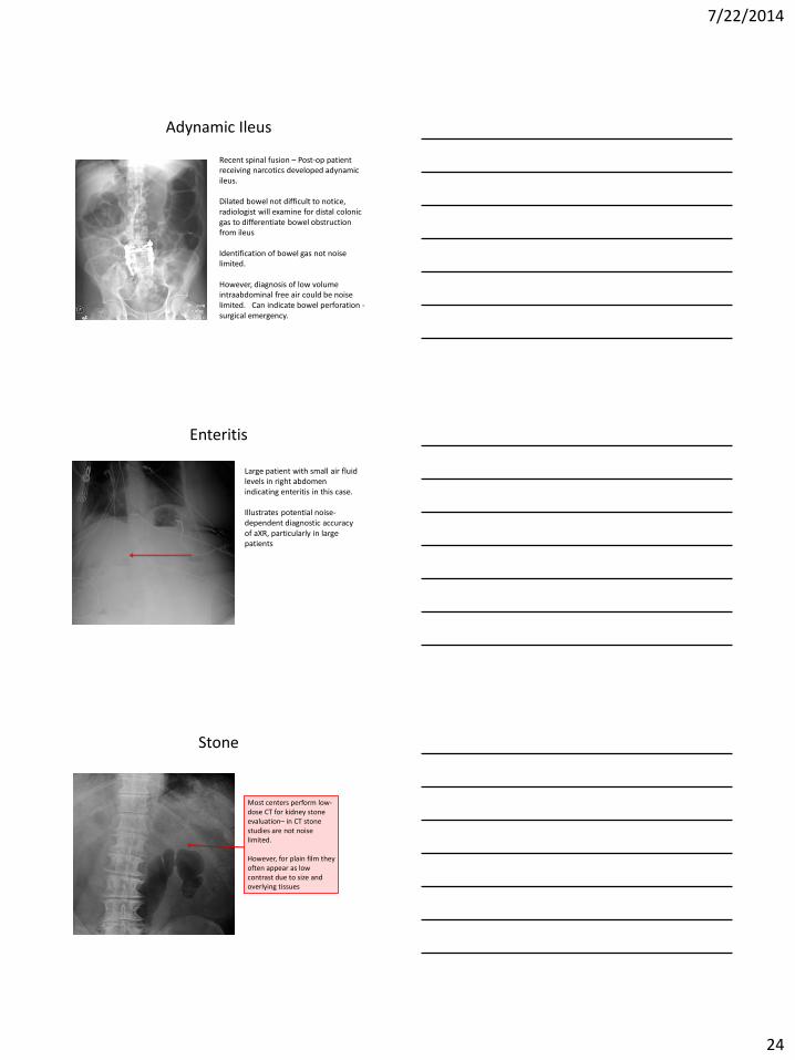
Adynamic Ileus
Recent spinal fusion – Post-op patient receiving narcotics developed adynamic ileus. Dilated bowel not difficult to notice, radiologist will examine for distal colonic gas to differentiate bowel obstruction from ileus Identification of bowel gas not noise limited. However, diagnosis of low volume intraabdominal free air could be noise limited. Can indicate bowel perforation - surgical emergency.
Enteritis
Large patient with small air fluid levels in right abdomen indicating enteritis in this case. Illustrates potential noise-dependent diagnostic accuracy of aXR, particularly in large patients
Stone
Most centers perform low-dose CT for kidney stone evaluation– in CT stone studies are not noise limited. However, for plain film they often appear as low contrast due to size and overlying tissues
7/22/2014
25
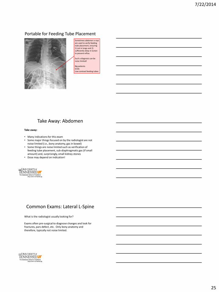
Sometimes abdomen x-rays are used to verify feeding tube placement, ensuring 1) not in lungs and 2) sufficiently deep in lumen to prevent reflux. Such a diagnosis can be noise limited! Big patients Grids Low contrast feeding tubes
Portable for Feeding Tube Placement
Take Away: Abdomen
Take away:
• Many indications for this exam • Some major things focused on by the radiologist are not
noise limited (i.e., bony anatomy, gas in bowel) • Some things are noise limited such as verification of
feeding tube placement, sub-diaphragmatic gas (if small amount) and, surprisingly, small kidney stones
• Dose may depend on indication!
Common Exams: Lateral L-Spine
What is the radiologist usually looking for? Exams often pre-surgical to diagnose changes and look for fractures, pars defect, etc. Only bony anatomy and therefore, typically not noise limited.
7/22/2014
26
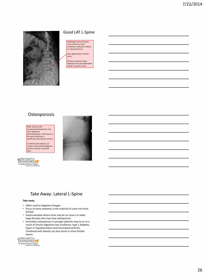
Good LAT L-Spine
Radiologist will commonly trace coutour of each vertebrae, looking for defects to indicate fracture Also degeneration of facet joints All bony anatomy makes diagnosis not noise dependent except in specific cases
Osteoporosis
Older women with osteopenia/osteoporosis may have significant demineralization of the bones in the spine resulting in significantly decreased contrast. Combined with obesity can create a noise limited diagnosis and the need for increased dose.
Take Away: Lateral L-Spine Take away:
• Often used to diagnose changes • Focus on bony anatomy, in the majority of cases not noise
limited • Good examples where noise may be an issue is in older,
large females who may have osteoporosis • Secondary osteoporosis in younger patients may occur as a
result of chronic digestive tract conditions, type 1 diabetes, hyper or hypothyroidism and rheumatoid arthritis. Combined with obesity can also result in noise-limited exams.
7/22/2014
27
Conclusion
• IQ and dose balance in radiography is not as straight forward as it may seem
• Many interacting factors at play. • Some equipment is better than others; however, one must
carefully consider the clinical indication for the exams performed, as well as the age of the patient to determine if its worth it
• Prior dose history should not be considered under any circumstances
• Medical physicists need to understand the job of the radiologist in order to assist them at reducing patient dose
Acknowledgements
Austin C. Bourgeois, MD Rebecca M. Marsh, PhD