bacteriological and serological studies...
TRANSCRIPT
Cairo University Faculty of Veterinary Medicine
Microbiology Department
BACTERIOLOGICAL AND SEROLOGICAL STUDIES ON AVIAN MYCOPLASMAS IN MENOFIA GOVERNORATE
Thesis Presented
By
EMAN MAHMOUD FARIED AHMAD SHARAF
(B.V.Sc., Zagazig University, Benha Branch, 1990) (M.V.Sc., Zagazig University, Benha Branch, 2000)
FOR The Degree Of Doctor Of Philosophy
In Veterinary Medical Science (Bacteriology, Immunology and Mycology)
2004

Under Supervision of
PROF. DR. MOHAMED KAMAL REFAI Prof. of Microbiology
Faculty of Veterinary Medicin, Cairo University
DR. AMAL MOHAMED RASHWAN Chief Researcher of Mycoplasma
Department, Animal Health Research Institute
Dokki - Giza

INTRODUCTION Mycoplama gallisepticum (MG) infection in chickens is still an important veterinary problem causing decreased egg production, growth rate and feed conversion rate; ncreased mortality and condemnation rate of carcasses as well as indirect losses due to increased sensitivity of infected birds to management failures and associating agents, for example infectious bronchitis, laryngotracheitis, or Newcastle disease viruses and Escherichia coli (Carpenter et al., 1981).

INTRODUCTION
Successful control of the disease, including eradication of M. gallisepticum depends very much on reliable diagnosis of infection. This can be done by: culturing the agent and by detecting antibodies against M. gallisepticum by serological tests, such as serum plate agglutination test (SPA).
This test is rapid and sensitive but often gives false positive reactions connected with antigen preparation techniques (Opitz and Cyr, 1986 and Ahmad et al., 1989).

INTRODUCTION The use of enzyme - linked immunosorbent assay
(ELISA) has been proposed because of higher sensitivity (Ansari et al., 1983; Avakian et al., 1987 and Stipkovites et al.,1993).
Sodium dodecyl sulfate - polyacrylamide gel electrophoresis (SDA - PAGE) was used for identification of different strains of MG (Khan et al., 1987; Barbour and Newman , 1989 and Thongkamkoon et al., 1996).
Western - blot technique was used for
identification of different strains of MG (Avakian and kleven, 1990; Levisohn et al.,1995; Ellakany et al.,1997 and Salisch et al.,1999).

INTRODUCTION Polymerase chain reaction (PCR) was
chosen due to many advantages that were reported for this technique as it is very specific and sensitive (Kempf et al., 1993; Fan et al.,1995 and Ren et al.,2000).

Aim of the work The aim of this work was to determine whether these recent techniques could overcome the previously mentioned disadvantages of the traditional techniques,
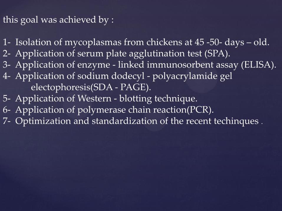
this goal was achieved by : 1- Isolation of mycoplasmas from chickens at 45 -50- days – old. 2- Application of serum plate agglutination test (SPA). 3- Application of enzyme - linked immunosorbent assay (ELISA). 4- Application of sodium dodecyl - polyacrylamide gel electophoresis(SDA - PAGE). 5- Application of Western - blotting technique. 6- Application of polymerase chain reaction(PCR). 7- Optimization and standardization of the recent techinques .

MATERIAL AND METHODS
Samples 200 Samples including airsacs and lungs were collected from100 chickens (at 45-50 days-old) which showed respiratory manifestations as nasal discharge, respiratory rules and cough from different farms at Menofia Governorate .

MATERIAL AND METHODS Mycoplasma strains and antisera : Mycoplasma gallisepticum (S6 and F strains) were kindly supplied by Dr. Amal Rashwan , Mycoplasma Department , Animal Health Research Institute , Dokki, Giza. Markers used in PCR : QX 174 DNA - Hae III Digest (molecular weights:1353,1078,872,603,310,276,234,194,118 and 72 bp , FiNN Zymes Oy ,Fin Land) Protein Standard Molecular Weight marker (BioRad)

MATERIAL AND METHODS
1- PCR primers according to EL-Shater et al. (1995) and Innis and Gelfand (1990) : Two oligonucleotide primers were selected as (one right assigned (1) and one left assigned (2) . The sequence of primer (1) was 5 - TAA GAA TCC AGG GTG AGC AAT -3. The sequence of primer (2) was 5 - TCC TCC ACT AAA TAA ATT GAC CCG -3. Synthesis of these primers was done by (MWG - Biotech. AG, Germany).

MATERIAL AND METHODS 2- PCR primers according to Kempf et al. (1993) : Two oligonucleotide primers were selected as (one right assigned (1) and one left assigned (2) The sequence of primer (1) was 5 -TAAC TAT CGC ATG AGAAT AAC -3. The sequence of primer (2) was 5- GTT ACT TAT TCAAA TGG TACAG -3. Synthesis of these primers was done by (MWG - Biotech.AG, Germany).

Methods • Isolation and identification of members of the genus
Mycoplasma (Razin & Tully,1983)
• Differentiation of Mycoplasma and Acholeplasma isolates by digitonin sensitivity test (Erno and Stipkovitis,1973)
• Identification of M. gallisepticum isolates
1-Biochemical characterization a) Glucose fermentation test (Erno and Stipkovitis, 1973) b) Arginine deamination test (Erno and Stipkovitis, 1973) 2-Growth inhibition test (Clyde, 1964) :
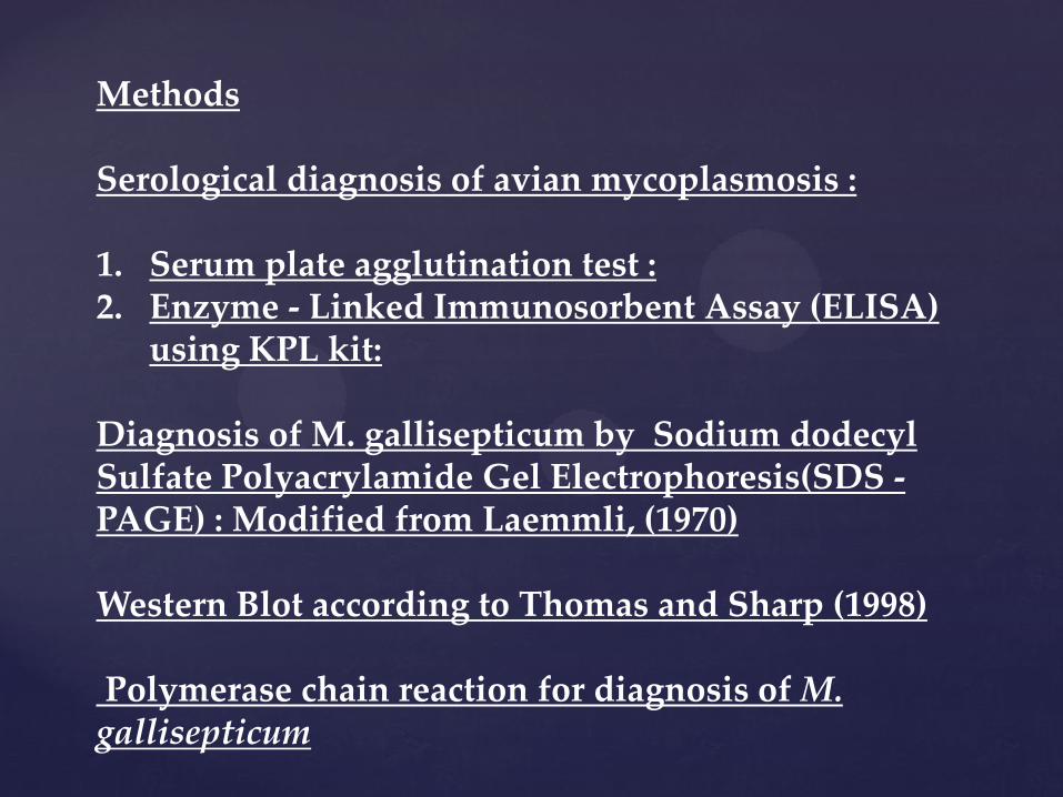
Methods Serological diagnosis of avian mycoplasmosis : 1. Serum plate agglutination test : 2. Enzyme - Linked Immunosorbent Assay (ELISA)
using KPL kit: Diagnosis of M. gallisepticum by Sodium dodecyl Sulfate Polyacrylamide Gel Electrophoresis(SDS -PAGE) : Modified from Laemmli, (1970) Western Blot according to Thomas and Sharp (1998) Polymerase chain reaction for diagnosis of M. gallisepticum

RESULTS

Samples Number
examined
Number
positive
Differentiation isolates by digition in
sensitivity test
Mycoplasma % Acholeplasma %
Air sac 100 35 34 97 1 29
Lung 100 22 20 90 2 9
Total No. of
isolates
200 57 54 94 3 5.3
Table (1) Results of isolation and identification of Mycoplasma and
Acholeplasma from chickens at 45-50-days-old .

Samples
Number of
examined
isolates
Biochemical test
Digitonin G+
A-
G -
A+
G+
A+
G –
A - + -
Air sac 35 34 1 32 2
_
_
Lung 22 20 2 20 _
_
_
Total 57 54 3 52 2
_
_
Table (2) Results of isolation and biochemical identification
of M.gallisepticum isolated from chickens at 45-50- days-old
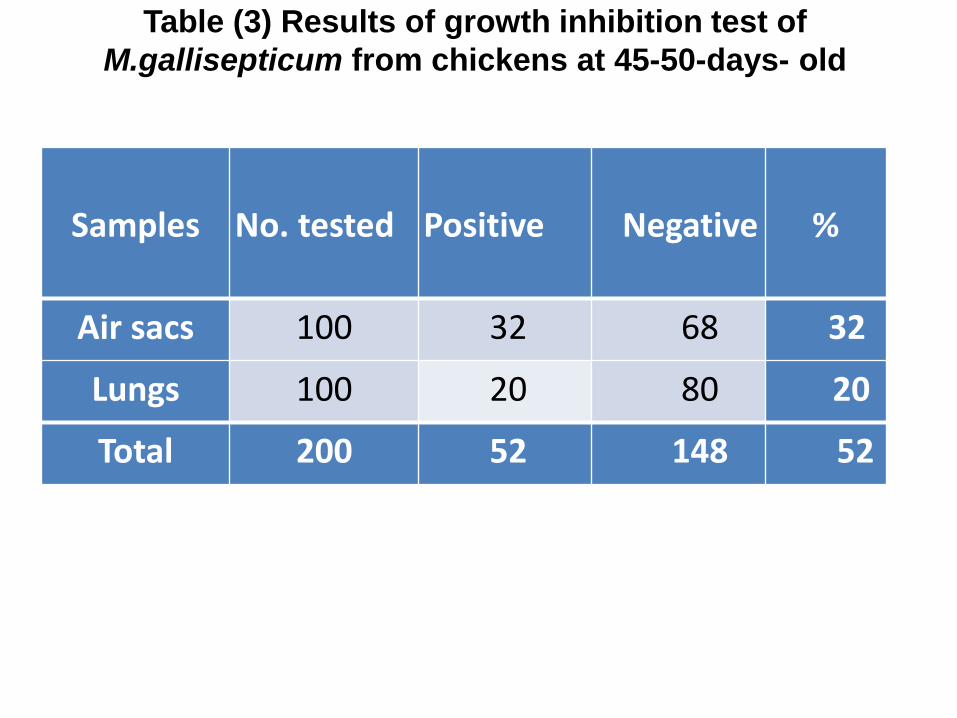
Samples No. tested Positive Negative %
Air sacs 100 32 68 32
Lungs 100 20 80 20
Total 200 52 148 52
Table (3) Results of growth inhibition test of
M.gallisepticum from chickens at 45-50-days- old

Total No.
of
samples
No. of
positive
No.of
suspected
No. of
negative
100 65 9 26
% 65 9 26
Table (4) serum plate agglutination test for sera collected
from chickens at 45 –50 -days – old using
M. gallispeticum antigen

ELISA
KITS
1 2 3 4 5 6 7 8 9 10 11 12
A 1.262 0.183 1.204 0.607 0.495 0.581 0.652 0.3 0.453 0.786 0.433 0.671
B 1.264 0.82 0.711 1.204 1.015 1.146 1.305 2.28 1.282 0.719 1.164 1.432
C 1.140 1.24 1.939 1.049 1.831 1.282 0.884 1.337 1.921 1.237 1.569 1.31
D 1.104 1.495 1.197 1.171 1.054 1.447 1.15 1.199 1.235 1.502 0.627 0.854
E 1.172 1.231 1.297 1.074 0.821 0.781 1.12 1.101 0.982 1.291 0.232 0.226
F 0.221 0.273 0.991 1.101 0.121 0.087 0.279 1.192 1.421 0.086 0.088 0.087
G 0.098 0.97 1.261 0.981 0.093 0.223 0.273 0.791 0.962 0.869 0.674 0.841
H 0.198 0.292 1.12 1.32 0.852 0.247 0.213 0.217 0.187
Table (5) ELISA optical density for sera collected from
chickens at 45 –50- days – old using
M.gallisepticum coated plate (KPL kit)

ELISA
KITS 1 2 3 4 5 6 7 8 9 10 11 12
A 412.11 263.02 375.65 477.69 62.57 212.85 690.14 190.17 506.29
B 1622.0 747.85 568.18 1491.9 1105.6 1369.5 1712.9 4279.3 1661.7 580.83 1407.2 2004.1
C 1357.0 1569.6 3300.1 1172.4 3007.3 1661.7 860.37 1784.9 3250.7 1563.1 2333.9 1724.1
D 1283.0 2153.8 1477.0 1421.9 1182.3 2039.4 1377.9 1481.3 1558.7 2170.6 440.88 807.0
E 1424.0 1550.1 1695.0 1222.2 749.57 1111.2 1987.4 1934.2 1611.5 2486.4 113.47 106.70
F 101.20 162.90 1635.3 1934.2 14.17 0.1168 170.60 2193.2 2888.6 0.0153 0.2651 0.1160
G 3.0351 1580.0
3
2396.3
4
1608.9
3 1.4176 103.40 162.90
1134.7
0 1559.2 1323.0 870.08 1254.3
H 77.05 187.65 1987.4 2574.4 1281.0 130.90 92.56 96.85 66.26
Table (6) ELISA titres for sera collected from chickens at 45 –50- days – old
using M.gallisepticum coated plate (KPL kit )
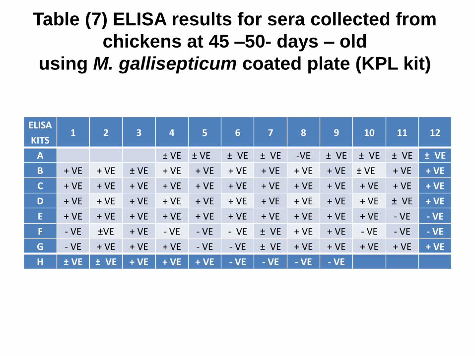
ELISA
KITS 1 2 3 4 5 6 7 8 9 10 11 12
A ± VE ± VE ± VE ± VE -VE ± VE ± VE ± VE ± VE
B + VE + VE ± VE + VE + VE + VE + VE + VE + VE ± VE + VE + VE
C + VE + VE + VE + VE + VE + VE + VE + VE + VE + VE + VE + VE
D + VE + VE + VE + VE + VE + VE + VE + VE + VE + VE ± VE + VE
E + VE + VE + VE + VE + VE + VE + VE + VE + VE + VE - VE - VE
F - VE ±VE + VE - VE - VE - VE ± VE + VE + VE - VE - VE - VE
G - VE + VE + VE + VE - VE - VE ± VE + VE + VE + VE + VE + VE
H ± VE ± VE + VE + VE + VE - VE - VE - VE - VE
Table (7) ELISA results for sera collected from
chickens at 45 –50- days – old
using M. gallisepticum coated plate (KPL kit)
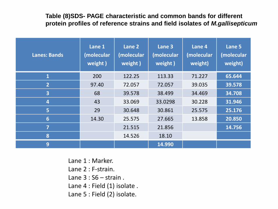
Lanes: Bands
Lane 1
(molecular
weight )
Lane 2
(molecular
weight )
Lane 3
(molecular
weight )
Lane 4
(molecular
weight)
Lane 5
(molecular
weight)
1 200 122.25 113.33 71.227 65.644
2 97.40 72.057 72.057 39.035 39.578
3 68 39.578 38.499 34.469 34.708
4 43 33.069 33.0298 30.228 31.946
5 29 30.648 30.861 25.575 25.176
6 14.30 25.575 27.665 13.858 20.850
7 21.515 21.856 14.756
8 14.526 18.10
9 14.990
Table (8)SDS- PAGE characteristic and common bands for different
protein profiles of reference strains and field isolates of M.gallisepticum
Lane 1 : Marker. Lane 2 : F-strain. Lane 3 : S6 – strain . Lane 4 : Field (1) isolate . Lane 5 : Field (2) isolate.

Lanes: Bands Lane 1
(amount)
Lane 2
(amount)
Lane 3
(amount)
Lane 4
(amount)
Lane 5
(amount)
1 2.4187 12.610 21.171 35.823 1.4996
2 0.85856 22.139 15.884 43.290 69.890
3 12.166 37.801 30.778 1.7609 5.9491
4 24.063 3.4126 4.0786 1.3764 4.7539
5 41.977 1.6688 5.7743 10.299 5.2414
6 18.517 13.694 5.2524 7.1394 10.996
7 8.1268 0.56013 0.62325
8 0.23553 3.3819
9 12.841
Sum 100 99.688 99.722 99.688 98.954
In lane 100 100 100 100 100
Table ( 9 ) SDS - PAGE characteristic and percentage of amount of the
different proteins of reference strains and field isolates of M.gallisepticum
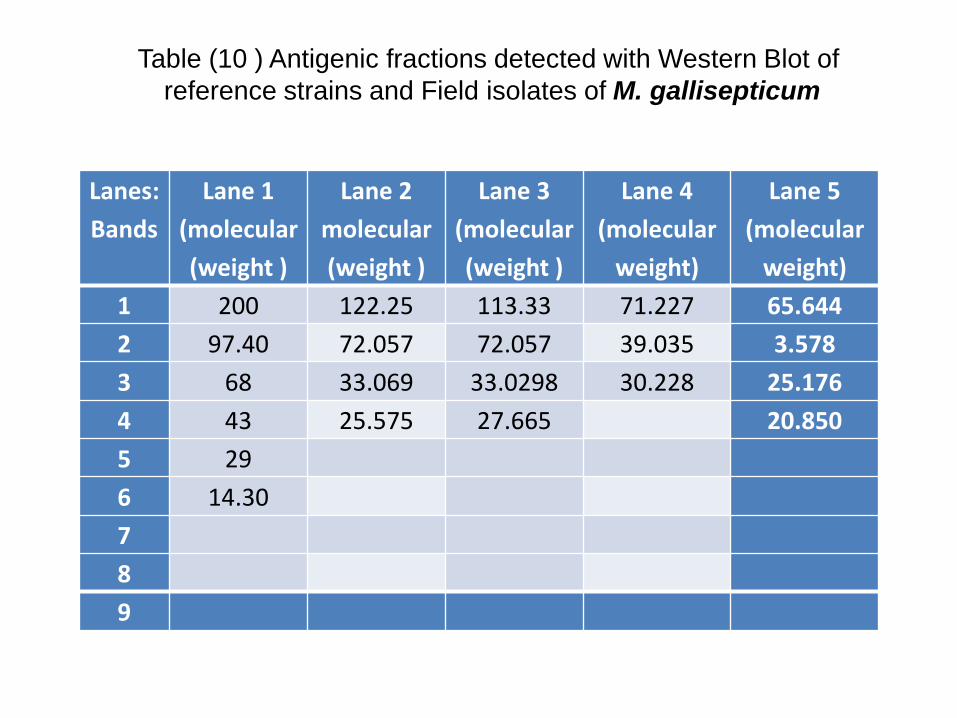
Lanes:
Bands
Lane 1
(molecular
(weight )
Lane 2
molecular
(weight )
Lane 3
(molecular
(weight )
Lane 4
(molecular
weight)
Lane 5
(molecular
weight)
1 200 122.25 113.33 71.227 65.644
2 97.40 72.057 72.057 39.035 3.578
3 68 33.069 33.0298 30.228 25.176
4 43 25.575 27.665 20.850
5 29
6 14.30
7
8
9
Table (10 ) Antigenic fractions detected with Western Blot of
reference strains and Field isolates of M. gallisepticum
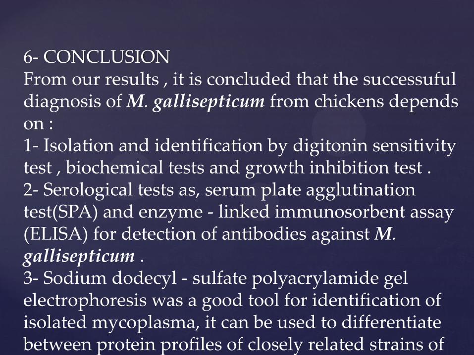
6- CONCLUSION From our results , it is concluded that the successuful diagnosis of M. gallisepticum from chickens depends on : 1- Isolation and identification by digitonin sensitivity test , biochemical tests and growth inhibition test . 2- Serological tests as, serum plate agglutination test(SPA) and enzyme - linked immunosorbent assay (ELISA) for detection of antibodies against M. gallisepticum . 3- Sodium dodecyl - sulfate polyacrylamide gel electrophoresis was a good tool for identification of isolated mycoplasma, it can be used to differentiate between protein profiles of closely related strains of M.gallisepticum .

4- Western - blot was also a good tool for differentiation of immunoreactivity of M. gallisepticum strains . 5- Polymerase chain reaction (PCR) is a recent technique which can be used to compare between strains of M. gallisepticum , also for detection of mycoplasma infection but it is very sensitive and has some disadvantages , may gives false- negative results due to inhibitors in extracted DNA, faulty reagents or procedure, it may be too sensitive yielding results due to contamination of PCR reagents with DNA. However, the use of the correct specific primer and almost care in applying technique would helpful in the rapid diagnosis of M. gallisepticum.