bacteriological analytical manual (bam) > bam ... bam e. coli o157-h7.pdf · transfer suspicious...
TRANSCRIPT
BAM: Diarrheagenic Escherichia coli
July 2009
Bacteriological Analytical Manual Chapter 4a Diarrheagenic Escherichia coliAuthors
Escherichia coli is one of the predominant species of facultative anaerobes in the human gut and usually harmless to the host; however, a group of pathogenic E. coli has emerged that causes diarrheal disease in humans. Referred to as Diarrheagenic E. coli (28) or commonly as pathogenic E. coli, these groups are classified based on their unique virulence factors and can only be identified by these traits. Hence, analysis for pathogenic E. coli often requires that the isolates be first identified as E. coli before testing for virulence markers. The pathogenic groups includes enterotoxigenic E. coli (ETEC), enteropathogenic E. coli (EPEC), enterohemorrhagic E. coli (EHEC), enteroinvasive E. coli (EIEC), enteroaggregative E. coli (EAEC), diffusely adherent E. coli (DAEC) and perhaps others that are not yet well characterized (21, 28). Of these, only the first 4 groups have been implicated in food or water borne illness. Some properties and symptoms of these 4 subgroups are discussed below and summarized in Table 1.
ETEC is recognized as the causative agent of travelers' diarrhea and illness is characterized by watery diarrhea with little or no fever. ETEC infections occurs commonly in under-developed countries but, in the U.S., it has been implicated in sporadic waterborne outbreaks as well as due to the consumption of soft cheeses, Mexican-style foods and raw vegetables. Pathogenesis of ETEC is due to the production of any of several enterotoxins. ETEC may produce a heat-labile enterotoxin (LT) that is very similar in size (86 kDa), sequence, antigenicity, and function to the cholera toxin (CT). ETEC may also produce a heat stable toxin (ST) that is of low molecular size (4 kDa) and resistant to boiling for 30 min. There are several variants of ST, of which ST1a or STp is found in E. coli isolated from both humans and animals, while ST1b or STh is predominant in human isolates only. The infective dose of ETEC for adults has been estimated to be at least 108 cells; but the young, the elderly and the infirm may be susceptible to lower levels. Because of its high infectious dose, analysis for ETEC is usually not performed unless high levels of E. coli have been found in a food. Also, if ETEC is detected, levels should also be enumerated to assess the potential hazard of the contaminated food. Production of LT can be detected by Y-1 adrenal cell assays (28) or serologically by commercial reverse passive latex agglutination assay and ELISA (see Appendix 1). The production of ST canalso be detected by ELISA or by infant mouse assay (35). Both LT and ST genes have also been sequenced and PCR (37, 41) and gene probe assays (see chapter 24) are available. Analysis of colonies on plating media using gene probe/colony hybridization also allows enumeration of ETEC in foods.
EIEC closely resemble Shigella and causes an invasive, dysenteric form of diarrhea in humans (7). Like Shigella, there are no known animal reservoirshence the primary source for EIEC appears to be infected humans. Although the infective dose of Shigella is low and in the range of 10 to few hundredcells, volunteer feeding studies showed that at least 106 EIEC organisms are required to cause illness in healthy adults. Unlike typical E. coli, EIEC are non-motile, do not decarboxylate lysine and do not ferment lactose, so they are anaerogenic. Pathogenicity of EIEC is primarily due its ability to invadeand destroy colonic tissue. The invasion phenotype, encoded by a high molecular weight plasmid, can be detected by invasion assays using HeLa or Hep-2 tissue culture cells (7, 25) or by PCR and probes specific for invasion genes (see chapter 24).
EPEC causes a profuse watery diarrheal disease and it is a leading cause of infantile diarrhea in developing countries. EPEC outbreaks have been linkedto the consumption of contaminated drinking water as well as some meat products. Through volunteer feeding studies the infectious dose of EPEC in healthy adults has been estimated to be 106 organisms. Pathogenesis of EPEC involves intimin protein (encoded by eae gene) that causes attachment and effacing lesions (14); but it also involves a plasmid-encoded protein referred to as EPEC adherence factor (EAF) that enables localized adherence obacteria to intestinal cells (36). Production of EAF can be demonstrated in Hep-2 cells and the presence of eae gene can be tested by PCR assays (28).
EHEC are recognized as the primary cause of hemorrhagic colitis (HC) or bloody diarrhea, which can progress to the potentially fatal hemolytic uremic syndrome (HUS). EHEC are typified by the production of verotoxin or Shiga toxins (Stx). Although Stx1 and Stx2 are most often implicated in human illness, several variants of Stx2 exist. There are many serotypes of Stx-producing E. coli (STEC), but only those that have been clinically associated with HC are designated as EHEC. Of these, O157:H7 is the prototypic EHEC and most often implicated in illness worldwide (3, 13, 19, 28). The infectious dose for O157:H7 is estimated to be 10 - 100 cells; but no information is available for other EHEC serotypes. EHEC infections are mostly fooor water borne and have implicated undercooked ground beef (3, 13), raw milk (31), cold sandwiches (19), water (34), unpasteurized apple juice (2) and sprouts and vegetables (4, 17). EHEC O157:H7 are phenotypically distinct from E. coli in that they exhibit slow or no fermentation of sorbitol and do not have glucuronidase activity (see chapter 4. LST-MUG for details); hence, these traits are often used to isolate this pathogen from foods. The production of Stx1 and Stx2 can be tested by cytotoxicity assays on vero or HeLa tissue culture cells or by commercially available ELISA or RPLA kits (see Appendix 1). Gene probes (see chapter 24) and PCR assays specific for stx1 and stx2 and other trait EHEC markers are also available (12, 15) (and see below).
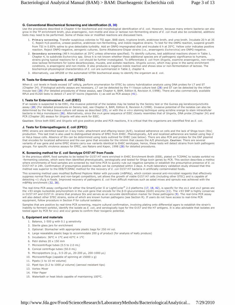
Properties/Symptoms ETEC EPEC EHEC EIECToxin LT/STa - Shiga or Vero toxin (Stx or VT) -Invasive - - - +Intimin - + + -Enterohemolysin - - + -Stool Watery Watery, Bloody Watery, very bloody Mucoid, bloodyFever Low + - +Fecal leukocytes - - - +Intestine involved Small Small Colon Colon, lower smallSerology VariousO26, O111 & others O157:H7, O26, O111 & others VariousID
b High High Low High
Table 1. Some properties and symptoms associated with pathogenic E. coli subgroups.
a LT, labile toxin; ST, stable toxin. b ID, infective dose.
Isolation and Identification of Pathogenic Escherichia coli- Except EHEC of serotype O157:H7Since pathogenic E. coli are identified based on its unique virulence properties, the analytical procedure for these pathogens in foods generally requiresthe isolation and identification of the organisms as E. coli before testing for the specific virulence traits. Following is a general procedure for enrichmenand isolation of pathogenic E. coli from food (25).
Food
Home> Food> Science & Research> Laboratory Methods
Page 1 of 13Bacteriological Analytical Manual (BAM) > BAM: Diarrheagenic Escherichia coli
7/29/2010http://www.fda.gov/Food/ScienceResearch/LaboratoryMethods/BacteriologicalAnalytical...

A. Equipment and materialsBalance, ≥ 2 kg with 0.1 g sensitivity1.Blender, Waring or equivalent model with low speed operation at 8000 rpm, with 1 liter glass or metal jar2.Incubators, 35 ± 0.5°C and 44 ± 1°C3.Petri dishes 20 x 150 mm4.Pipets, Pasteur5.pH test paper, range 6.0-8.06.
B. Media
Tryptone phosphate (TP) broth (M1621)1.
Brain heart infusion (BHI) broth (M242)2.
Levine's eosin-methylene blue (L-EMB) agar (M803)3.
MacConkey agar (M914)4.
Triple sugar iron (TSI) agar (M1495)5.
Blood agar base (BAB) (M216)6.
Tryptone (tryptophane) broth (M1647)7.
Bromcresol purple broth (M268) supplemented individually with 0.5% (w/v) of each: glucose, adonitol, cellobiose, sorbitol, arabinose, mannitoland lactose
8.
Urea broth (M1719)9.
Lysine decarboxylase broth, Falkow (M8710).10.
Potassium cyanide (KCN) broth (M12611)11.
MR-VP broth (M10412)12.
Indole nitrite medium (tryptic nitrate) (M6613)13.
Acetate agar (M314)14.
Mucate broth (M10515)15.
Mucate control broth (M10616)16.
Malonate broth (M9217)17.
Koser's citrate broth (M7218)18.
C. Reagents, inorganic, organic, and biological
Sodium bicarbonate solution, 10%, aqueous (sterile) (R7019)1.
ONPG (o-nitrophenyl-β-D-galactopyranoside) disks (R5320)2.
Phosphate buffered saline solution, (sterile) (PBS) (R6021), or Butterfield's Phosphate-buffered dilution water (BPBW) (R1122).3.
Kovac's reagent (R3823)4.
VP reagents (R8924)5.
Oxidase test reagent (R5425)6.
Nitrite detection reagents (R4826)7.
Mineral oil, heavy sterile (R4627)8.
Gram stain reagents (R3228)9.
D. Enumeration.Refrigerate samples promptly after receipt. Do not freeze except to hold frozen products until just prior to analysis. Analyze samples as soon as possible. If enumeration is required, prepare a homogenate of 25 g in 225 mL of PBS or BPBW. Perform decimal dilutions (in PBS or BPBW) from the homogenate and direct plate onto MacConkey agar to yield isolated colonies. After incubating the plates for 20 h at 35°C, perform colony lifts and hybridization with specific gene probes for virulence genes (see chapter 24). This enumeration procedure is most effective if E. coli constitutes at least 10% of the microbial growth on isolation agars and it is present at a level of >1,000 cells/g.
E. Enrichment for Pathogenic E. coliThe approach recommended here permits qualitative determination of the presence of pathogenic E. coli. Aseptically weigh 25 g of sample into 225 mlof BHI broth (dilution factor of 1:10). If necessary, sample size may deviate from 25 g depending on availability of the sample, as long as the diluent iadjusted proportionally. Blend or stomach briefly. Incubate the homogenate for 10 min at room temperature with periodic shaking then allow the sample to settle by gravity for 10 min. Decant medium carefully into a sterile container and incubate for 3 h at 35°C to resuscitate injured cells. Transfer contents to 225 mL double strength TP broth in a sterile container and incubate 20 h at 44.0 ± 0.2°C. After incubation, streak to L-EMB and MacConkey agars. Incubate these agars for 20 h at 35°C.
F. Selection.Typical lactose-fermenting colonies on L-EMB agar appear dark centered and flat, with or without metallic sheen. Typical colonies on MacConkey agar appear brick red. Lactose non-fermenting biotypes on both agars produce colorless or slightly pink colonies.
NOTE: EIEC do not ferment lactose and there may also be atypical non-lactose fermenting strains in the other pathogenic E. coli groups; therefore, asmany as 20 colonies (10 typical and 10 atypical) should be picked for further characterization.
Page 2 of 13Bacteriological Analytical Manual (BAM) > BAM: Diarrheagenic Escherichia coli
7/29/2010http://www.fda.gov/Food/ScienceResearch/LaboratoryMethods/BacteriologicalAnalytical...
G. Conventional Biochemical Screening and identification (8, 30)Use the procedures described in Chapter 4 for biochemical and morphological identification of E. coli. However, because many enteric bacteria can alsogrow in the TP enrichment broth, plus anaerogenic, non-motile and slow or lactose non-fermenting strains of E. coli must also be considered, additionatests may need to be performed. Some of these new or modified reactions are discussed here.
Primary screening. Transfer suspicious colonies to TSI agar, BAB slant, tryptone broth, arabinose broth, and urea broth. Incubate 20 h at 35C. Reject H2S-positive, urease-positive, arabinose non-fermenting, and indole-negative strains. To test for the ONPG reaction, suspend growth from TSI in 0.85% saline to give detectable turbidity. Add an ONPG-impregnated disk and incubate 6 h at 35°C. Yellow color indicates positive reaction. Reject ONPG-negative, aerogenic cultures. Some Alkalescens-Dispar strains (i.e., anaerogenic Escherichia) are ONPG-negative.
1.
Secondary screening (48 h incubation at 35°C unless otherwise specified). To identify cultures, test additional reactions shown in Table 1, Chapter 4, to subdivide Escherichia spp. Since it is not known whether these additional species are of pathogenic significance to humans, strains giving typical reactions for E. coli should be further investigated. To differentiate E. coli from Shigella, examine anaerogenic, non-motileslow lactose fermenters for lysine decarboxylase, mucate, and acetate reactions. Shigella sonnei, which may grow in the same enrichment conditions, is anaerogenic and non-motile. It also produces a negative indole reaction and shows slow or non-fermentation of lactose. The biochemical-physiological characteristics of E. coli are summarized in Table 2, chapter 4.
2.
Alternatively, use API20E or the automated VITEK biochemical assay to identify the organism as E. coli.3.
H. Tests for Enterotoxigenic E. coli (ETEC)
When E. coli levels in foods exceed 104 cells/g, perform enumeration for ETEC by colony hybridization analysis using DNA probes for LT and ST (Chapter 24). If biological activity assays are necessary, LT can be detected by the Y-l tissue culture test (28) and ST can be detected by the infant mouse test (28) (For detailed procedures of these assays, see Chapter 4, BAM, Edition 8, Revision A /1998). There are also commercially available RPLA and ELISA tests to detect LT and ST toxins (Appendix 1) as well as PCR assays (41).
I. Tests for Enteroinvasive E. coli (EIEC)If an isolate is suspected to be EIEC, the invasive potential of the isolates may be tested by the Sereny test or the Guinea pig keratoconjunctivitis assay (28) (For detailed procedures on Sereny test, see Chapter 4, BAM, Edition 8, Revision A /1998). Invasive potential of the isolates can also be determined by the HeLa tissue culture cell assay as described (25), or with the in vitro staining technique using acridine orange to stain intracellular bacteria in HeLa monolayers (26). Alternatively, since the invA gene sequence of EIEC closely resembles that of Shigella, DNA probe (Chapter 24) and PCR (Chapter 28) assays for Shigella will also work for EIEC.
Caution: Since both EIEC and Shigella will give positive probe and PCR reactions, it is critical that the organisms are identified first as E. coli.
J. Tests for Enteropathogenic E. coli (EPEC)EPEC strains are identified based on 3 key traits: attachment and effacing lesion (A/E), localized adherence on cells and the lack of Shiga toxin (Stx) production. This last trait is also used to distinguished strains of EPEC from EHEC. Phenotypically, A/E and localized adherence are tested using Hep-2 or HeLa tissue cells. Absence of Stx can be determined using tests outlined for EHEC (see below). There are also PCR and probes for the EAF plasmid that encodes for localized adherence and the eae gene that encodes for the intimin that causes the A/E phenotype. Caution: There are several variants of eae gene and some EPEC strains carry eae variants identical to EHEC serotypes; hence, these tests will detect strains from both pathogenicgroups. For specific virulence assays for EPEC, see Nataro and Kaper, 1998 (28) for detailed procedures.
K. Screening method for E. coli Serotype O157:H7 from FoodsIn the previous eBAM, food samples to be tested for O157:H7 were enriched in EHEC Enrichment Broth (EEB), plated on TCSMAC to isolate sorbitol no-fermenting colonies, which were then identified phenotypically, serologically and tested for Shiga toxin genes by PCR. This section describes a methodwhere enrichments of food samples are screened by real-time PCR to quickly rule out negative samples or establish the presumptive presence of E. colO157:H7 in 24h. Confirmation of presumptive positive results can be completed within 3 days. A multi-laboratory validation study showed that this method was superior to the EEB/TCSMAC method for the recovery of E. coli O157:H7 bacteria in artificially contaminated foods.
This screening method uses modified Buffered Peptone Water with pyruvate (mBPWp), which contain several anti-microbial reagents that effectively suppress normal flora growth and non-target competitors, yet allows the growth of viable O157:H7 cells (including other STEC) and is capable of detecting <1 cfu/g in foods. Improved recovery of pathogenic E. coli from difficult matrices such as salad mixes and sprouts was achieved with the mBPWp enrichment (32, 38).
The real-time PCR assay configured for either the SmartCycler II or LightCycler® 2.0 platforms (17, 18, 42), is specific for the stx1 and stx2 genes andthe +93 single nucleotide polymorphism in the uidA gene that encode for the β-D-glucuronidase (GUD) enzyme (11). The +93 SNP is highly conservedin O157:H7 and O157:H- strains that produce Stx and serves as an accurate identification marker for these pathogens (9). The real-time PCR assay will also detect other STEC strains, some of which are known human pathogens (see Section R). If users do not have access to real-time PCR equipment, follow procedure in Section P for cultural isolation.
Samples that are positive by real-time PCR screening, require cultural confirmation, involving plating onto differential agars to establish the strain's inability to ferment sorbitol, identify the isolate as E. coli, and serologically type for the O157 and the H7 antigens. It is also advisable that isolates aretested again by PCR for stx1 and stx2 genes to confirm their toxigenic potential.
L. Equipment and materialsBalance, 1-500 g with 0.1 g sensitivity1.Sterile glass jars for enrichment2.Optional: Stomacher with appropriate plastic bags for 250 ml vol.3.Large resealable plastic bags to accommodate 200 g of product (for analysis of leafy produce)4.Incubators: 36°C ± 1°C and 42°C ± 1°C5.Petri dishes 20 x 150 mm6.Microcentrifuge tubes (0.5 to 2.0 mL)7.Conical centrifuge tubes (50.0 mL)8.Micropipettors (e.g., 0.5-20 µL, 20-200 µL, 200-1000 µL)9.Microcentrifuge (capable of spinning at 15000 x g)10.Pipets (1 to 10 ml volume)11.Pipet tips (0.2 to 1000 µl volume) (aerosol resistant tips)12.Vortex Mixer13.Filter Paper14.Waterbath or heat block capable of maintaining 100°C.15.
Page 3 of 13Bacteriological Analytical Manual (BAM) > BAM: Diarrheagenic Escherichia coli
7/29/2010http://www.fda.gov/Food/ScienceResearch/LaboratoryMethods/BacteriologicalAnalytical...
SmartCycler II PCR Thermalcycler (Cepheid, Sunnyvale, CA) capable of performing cycling parameters described below and simultaneous real-time sequence detection for FAM, TET, Texas Red and Cy5 dyes.
16.
SmartCycler PCR reaction tubes (minimum reaction volume of 25 µl) and racks compatible with PCR thermalcycler.17.Microcentrifuge with capillary adapters (#11909312001) or LC Carousel Centrifuge 2.0 (#03 709 507 001) (Roche Applied Science, Indianapolis, IN).
18.
LightCycler 2.0 real-time PCR instrument (Roche Applied Science, Indianapolis, IN) capable of performing cycling parameters described below and simultaneous detection of LightCycler Dyes LC610, LC670 and LC705 as described in this protocol.
19.
LightCycler PCR capillary tubes (minimum reaction volume of 20 µl, #04929292001) and cooling block (#11909339001) compatible with PCR thermalcycler.
20.
Ice bucket and ice21.Sterile tongs22.Latex gloves or equivalent.23.
M. Media and Reagents
Modified Buffered Peptone water with pyruvate (mBPWp) and Acriflavin-Cefsulodin-Vancomycin (ACV) Supplement (M192a29)1.STEC/O157 primers and probes listed in Tables 2 and 3 are specific to real-time PCR platform being used. 2.
Primers - 10 µM working solution of each primer listed in Table 2 or 3. Stock and Working solutions can be prepared from commercially synthesized primers with basic desalt purification (Fisher/Genosys or equivalent) by rehydrating with sterile distilled water to appropriate concentrations. Store at -20°C to -70°C in a non-frost-free freezer.
a.
Probes - 10 µM working solution of each probe listed in Tables 2 or 3. Dual Hybridization Probes should be purchased as RP HPLC-purified and labeled as indicated in Tables 2 and 3. Stock and working solutions can be prepared from commercially synthesized probes with molecular grade sterile distilled water. Working solutions should be aliquoted in small amounts and stored frozen (-20 to -70°C) and away from light until use to avoid fluorophore degradation.
b.
Note: Alternatively the primers, probes and internal control assay components are also available as a lyophilized bead product for use othe SmartCycler II.
Primers1 GenBank # Bases 5' → 3' Sequence1F934 M19473 26 gTg gCA TTA ATA CTg AAT TgT CAT CA1R1042 M19473 21 gCg TAA TCC CAC ggA CTC TTC2F1218 X07865 24 gAT gTT TAT ggC ggT TTT ATT TgC2R1300 X07865 26 Tgg AAA ACT CAA TTT TAC CTT Tag CAAF241 AF305917 21 CAg TCT ggA TCg CgA AAA CTgAR383 AF305917 22 ACC AgA CgT TgC CCA CAT AAT T
Table 2. Primer/probe sequences for use on SmartCycler II platform.
Probes1 GenBank # Bases 5' → 3' Sequence1P990 M19473 31 TxRd-TgA TgA gTT TCC TTC TAT gTg TCC ggC AgA T-BHQ22P1249 X07865 25 6FAM-TCT gTT AAT gCA ATg gCg gCg gAT T-BHQ1AP266 AF305917 15 TET-ATT gAg CAg CgT Tgg-MGB/NFQimer/Probe name composed of target gene (stx1, stx2 or uidA), forward primer (F), reverse primer (R) or probe (P), 5' base position of oligonucleotide he respective gene sequence specified in column 2.
Primers1 GenBank # Bases 5' → 3' SequenceA F 241 AF305917 21 CAg TCT ggA TCg CgA AAA CTgA R 383 AF305917 22 ACC AgA CgT TgC CCA CAT AAT T1 F 406 M19573 25 gAg gAA ggg Cgg TTT AAT AAT CTA C1 R 667 M19573 27 CAC TAT gCg ACA TTA AAT CCA gAT AAg2 F 492 X07865 28 gTT TTg ACC ATC TTC gTC TgA TTA TTg A2 R 737 X07865 22 ACT CCA TTA ACg CCA gAT ATg A
Table 3. Primer and probe sequences for use on the LightCycler 2.0 platform.
Probes1 GenBank # Bases 5' → 3' SequenceA 705 LC 288 AF305917 27 LCRed705-CTT TCC CAC CAA CgC TgC TCA ATT CCA-PhosphateA FL P 319 AF305917 29 CAC AgC AAT TgC CCg gCT TTC TTg TAA Cg-Fluoroscein1 610 LC 552 M19573 32 LCRed610-gTC Tgg TgA CAg TAg CTA TAC CAC gTT ACA gC-Phosphate1 FL P 524 M19573 26 CCT TTC CAg gTA CAA CAg Cgg TTA CA-Fluoroscein2 670 LC 670 X07865 28 LCRed670-gCT ggA ACg TTC Cgg AAT gCA AAT CAg T-Phosphate2 FL P 642 X07865 26 gTT ATA CCA CTC TgC AAC gTg TCg CA-Fluorosceinmer/Probe name composed of target gene (stx1, stx2 or uidA), forward primer (F), reverse primer (R) or probe (610LC, 670LC, 705LC, or FL P), 5' baseition of oligonucleotide in the respective gene sequence specified in column 2.
Real-time PCR additional reagents depending on real-time platform being used: 3.OmniMix-HS or SmartMix HM PCR Reagent Beads (Cepheid, Sunnyvale, CA. Also available through Fisher).a.LightCycler FastStart DNA Master Hybridization Probes M-Grade (Roche Applied Science, Catalog #3 383 393, Kit for 96 reactions, or equivalent).
b.
Tellurite Cefixime – Sorbitol MacConkey Agar (TC-SMAC) (M19430)4.Chromogenic selective agar: 5.
R&F® E. coli O157:H7 agar (R&F Laboratories, Downers Grove, IL), prepared according to manufacturer instructions using 10 mg/L novobiocin plus 0.8 mg/L potassium tellurite.
a.
Rainbow® Agar O157 (BIOLOG, Hayward, CA) prepared according to manufacturer instructions for high background flora containing 10 mg/L novobiocin plus 0.8 mg/L potassium tellurite.
b.
Levine's Eosin-Methylene Blue (L-EMB) agar (M8031) (Section R only)6.
Trypticase Soy Agar with Yeast Extract (TSAYE) (M15332).7.
Butterfield's Phosphate Buffer (R1133) (pH 7.2 ± 0.2. Sterilize by autoclaving)8.Sterile distilled water, molecular grade water or equivalent9.Physiological saline (0.85% NaCl)10.
Kovac's Reagent (R3834)11.
Page 4 of 13Bacteriological Analytical Manual (BAM) > BAM: Diarrheagenic Escherichia coli
7/29/2010http://www.fda.gov/Food/ScienceResearch/LaboratoryMethods/BacteriologicalAnalytical...
ColiComplete Discs - contains fluorogenic MUG substrate for GUD and chromogenic X-gal for GAL (BioControl, Bellevue, WA)12.Anti-O157 and anti-H7 latex reagent (Remel, Lenexa, KS, or equivalent)13.API20E or VITEK GNI (BioMerieux, St. Louis, MO).14.
N. Sample Preparation and Enrichment ProcedureSample Preparation 1.
Leafy Produce - add an equal weight of Butterfield's phosphate buffer to at least 200 g of product in a sterile re-sealable plastic bag andgently agitate by hand for 5 min. Weigh 125 g of produce rinsate into 125 ml double strength (2X) mBPWp.
a.
Juice, milk or other turbid beverage samples - aseptically centrifuge 200 ml of sample at 10,000 x g for 10 min. After decanting the supernatant, resuspend the pellet material in 225 ml of mBPWp.
b.
Bottled water or other non-turbid liquids - weigh 125 ml into 125 ml of 2X mBPWp. Also, use this approach with liquids in which a visiblepellet would not be produced after centrifugation.
c.
All other foods - weigh 25 g of food into 225 ml of mBPWp. Blend or stomach briefly as necessary.d.
Enrichment Incubate homogenate at 37°C ± 1°C static for 5 hours, then add 1ml each of the ACV supplements (see section M2) and incubate at 42°C ± 1 °C static overnight (18-24 h).
2.
Enrichment Control Strains - 465-97 USDA (stx1-, stx2- uidA+). Alternatively, use ATCC43890 (stx1+, stx2-, uidA+), ATCC 43888 (stx1-, stx2- uidA+or equivalent if 465-97 is not available. Note: these strains do not have both stx genes and should not be used as controls in PCR.
OPTIONAL: When O157:H7 contamination is suspected, particularly in foods with high levels of competing microbial flora, such as sprouts or raw meats, the use of immunomagnetic separation (IMS) prior to screening can be helpful. Several IMS procedures are available, including Dynabeads® anti-E.coli O157 (Invitrogen Corp., Carlsbad,CA) and Pathatrix Immunocapture system E.coli O157 kit (Matrix MicroScience Ltd., UK) and some of these have been tested on selected foods. Perform IMS on the 5 hr or overnight enrichment (depending on the IMS system used). From the final IMS bead suspension (approximately 100 µl), plate and assay by real-time PCR as described below in section O. For specific details on performing IMS on enrichment samples, contact: Karen Jinneman, FDA-PRLNW.
O. Real-time PCR ScreeningReal-time PCR assembly and data analysis protocols are described below for two instrument platforms; SmartCycler II and Light Cycler 2.0. Use of other platforms and protocols must first be validated. Alternately, if equipment and/or reagents are not available for real-time PCR screening of enrichment broths, refer to section P for cultural analysis of all enrichments.
DNA Template Preparation: 1.Transfer 1ml of overnight enrichment to a microcentrifuge tube and centrifuge 12,000 x g for 3 min.a.Remove supernatant and completely resuspend pellet in 1 ml 0.85% NaCl.b.Centrifuge 12,000 x g for 3 min.c.Remove supernatant and completely resuspend pellet in 1 ml sterile water.d.Place in waterbath or heat block capable of maintaining 100°C for 10 min.e.Centrifuge 12,000 x g for 1 minute, remove and save supernatant as DNA template (This may be frozen, minimum -20 °C, for future PCR tests).
f.
Make a 1:10 dilution of this template and use 1 µl for testing by real time-PCR.g.For pure cultures (including control cultures), 1 ml of broth culture or colony growth from agar plate suspended in 0.85% saline maybe prepared as in steps a-g above. Templates may be frozen at minimum -20°C for future use.
h.
PCR Controls: 2.For a positive PCR control include template prepared from E. coli O157:H7, such as ATCC strain 43895 (EDL 933) or ATCC strain 43894 both possessing all three gene targets (stx1+, stx2+ and uidA+).
a.
If no internal amplification control is incorporated into the reaction, prepare a reaction tube including 1 µl of a 1:10 dilution of EDL 933 control template and 1 µl of a 1:10 dilution of the food sample enrichment template.
b.
Always include a no template (water) negative control tube in every run.c.
Smart Cycler II - reaction assembly and data analysis protocol: 3.Reaction assembly a.
Prepare a PCR Master Mix from the reaction components and final concentrations for STEC/O157 listed in Table 4. Keep all thawed reagents and reactions on ice. Alternatively a PCR Master Mix may be prepared by following package insert for STEC/O157 CSR bead and OmmiMix HS or SmartMix HM bead.
1.
Add 24 µl of Master Mix to each SmartCycler tube and cap loosely.2.Add 1 µl of sample or control template and snap cap tightly.3.Briefly centrifuge to bring all liquid to bottom of tube and place in thermalcycler.4.Create a "run" on SmartCycler II. Give each run a unique run name, select Dye set FTTC25, select 2-step PCR protocol as described below and assign appropriate sites on SC block.
5.
Initial Activation 60 sec at 95°C
40 cycles10 sec at 94°C, (optics off)40 sec at 63°C, (optics on).
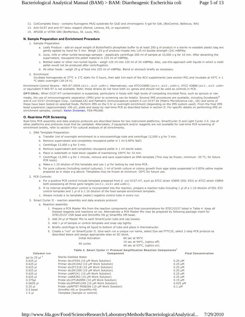
Volume/rxn Component Final Concentrationqsi to 25 µl 2 Sterile Distilled Water 0.625 µl Primer stx1F934 (10 µM Work Solution) 0.25 µM0.625 µl Primer stx1R1042 (10 µM Work Solution) 0.25 µM0.625 µl Primer stx2F1218 (10 µM Work Solution) 0.25 µM0.625 µl Primer stx2R1300 (10 µM Work Solution) 0.25 µM0.625 µl Primer uidAF241 (10 µM Work Solution) 0.25 µM0.625 µl Primer uidAR383 (10 µM Work Solution) 0.25 µM0.375µl Probe stx1PTxRd990 (10 µM Work Solution) 0.15 µM0.0625 µl Probe stx2PFAM1249 (10 µM Work Solution) 0.025 µM0.25 µl Probe uidAPTET-MGB266 (10 µM Work Solution) 0.1 µM0.5 bead OmniMix-HS or SmartMix-HS 1-5 µl Template (Sample or control)
Table 4. Smart Cycler II Protocol Amplification Reaction Components1
Page 5 of 13Bacteriological Analytical Manual (BAM) > BAM: Diarrheagenic Escherichia coli
7/29/2010http://www.fda.gov/Food/ScienceResearch/LaboratoryMethods/BacteriologicalAnalytical...
1All primers, probes, and IC DNA primers and probes are lyophilized in the STEC/O157 CSR Bead 2Appropriate amount of sterile distilled water is added depending on sample template volume being used.
Qualitative data analysis. On the SmartCycler II Instrument, set the following Analysis Settings for FAM, TET, TxRd and Cy5 channels (32). Update analysis settings if they are changed before recording results. Note: Internal Control (IC) cycle threshold (Ct) is CSR Lot specific (Ct = 25-35). ICt is consistently not present for the IC (Cy5, channel 4) target in the negative control tube only at 15 fsu, contact method authors. Lotspecific recommendations for Cy5 manual threshold setting may be issued.
b.
Usage: AssayCurve Analysis: PrimaryThreshold Setting: ManualManual Threshold Fluorescence Units: 15.0Auto Min Cycle: 5Auto Max Cycle: 10Valid Min Cycle: 3Valid Max. Cycle: 60Background subtraction: ONBoxcar Avg. Cycles: 0Background Min. Cycle: 5Background Max. Cycle: 40
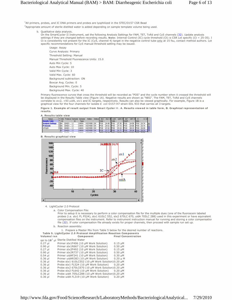
Primary fluorescence curves that cross the threshold will be recorded as "POS" and the cycle number when it crossed the threshold will be displayed in the Results Table view (Figure 1A). Negative results are shown as "NEG". The FAM, TET, TxRd and Cy5 channels correlate to stx2, +93 uidA, stx1 and IC targets, respectively. Results can also be viewed graphically. For example, Figure 1B is a graphical view for the four channels for isolate E. coli O157:H7 strain EDL 933 that carries all 3 targets.
Figure 1. Example of result output from Smart Cycler II. A. Results viewed in table form, B. Graphical representation of results.
A. Results table view
B. Results graphical view
LightCycler 2.0 Protocol 4.Color Compensation File: Prior to setup it is necessary to perform a color compensation file for the multiple dyes (one of the fluoroscein labeled probes (i.e. stx1 FL P524), stx1 610LC 552, stx2 670LC 670, uidA 705LC 288) used in this experiment or have equivalent compensation files on the instrument. Refer to instrument instruction manual for running and storing a color compensationfile (22). If color compensation file already exists for proper channels, then proceed with sample run set up.
a.
Reaction assembly: b.Prepare a Master Mix from Table 5 below for the desired number of reactions. 1.
Volume/rxn Component Final Concentrationqsi to 181 µl Sterile Distilled Water 0.27 µl Primer stx1F406 (10 µM Work Solution) 0.15 µM0.90 µl Primer stx1R667 (10 µM Work Solution) 0.50 µM0.27 µl Primer stx2F492 (10 µM Work Solution) 0.15 µM0.90 µl Primer stx2R737 (10 µM Work Solution) 0.50 µM0.54 µl Primer uidAF241 (10 µM Work Solution) 0.30 µM0.36 µl Primer uidAR383 (10 µM Work Solution) 0.20 µ M0.36 µl Probe stx1 610LC552 (10 µM Work Solution)0.20 µM0.36 µl Probe stx1 FL524 (10 µM Work Solution) 0.20 µM0.36 µl Probe stx2 670LC670 (10 µM Work Solution)0.20 µM0.36 µl Probe stx2 FL642 (10 µM Work Solution) 0.20 µM0.36 µl Probe uidA 705LC288 (10 µM Work Solution)0.20 µM0.36 µl Probe uidA FL319 (10 µM Work Solution) 0.20 µM
Table 5. LightCycler 2.0 Protocol Amplification Reaction Components
Page 6 of 13Bacteriological Analytical Manual (BAM) > BAM: Diarrheagenic Escherichia coli
7/29/2010http://www.fda.gov/Food/ScienceResearch/LaboratoryMethods/BacteriologicalAnalytical...
1.44 µl MgCl2 (25 mM) 3.0 mM1.8 µl FastStart DNA Master2 1-5 µl Template (Sample or control) 1Reaction volume is made up to 18 µl instead of the 20 µl standard. Appropriate amount of sterile distilled water is added depending on sample template volume used. 2FastStart DNA Master Hybridization Probes M-Grade mix made up as specified by manufacturer.
Ensure tube is mixed completely by gentle pipeting or slow vortex, and quickly spin tube to bring contents to bottom.
2.
Pipet 17 µl of Master Mix to desired number glass capillary tubes (20 µl capacity) including controls.3.Add 1 µl of Template DNA to each of the capillaries for a total volume of 18 µl and cap.4.Create New LightCycler Experiment.5.Program the 4 segment run (Table 6) into your LightCycler Instrument. 6.
Program NameCyclesAnalysis ModeTemperature Targets
Target (°C)
Hold (m or s)
Ramp Rate (°C/sec)
Sec Target (°C)
Step Size (°C)
Step Delay (cycles) Acquisition Mode
Activation 1 none 95 10m 20 0 0 0 none
Amplification 40 quantification 95 10s 20 0 0 0 none63 40s 20 0 0 0 single
Melt 1melting 95 0s 20 0 0 0 none
curves 55 15s 20 0 0 0 none95 0s 0.1 0 0 0 continuous
Cooling 1 none 40 30s 20 0 0 0 none
Table 6. LightCycler run parameters
Enter the Setup information as follows: 7.Default Channel: 530Seek Temperature: 30Max. Seek Pos: Number of samples being analyzed in run Instrument Type: 6 Ch.Capillary Size: 20 µl
Load capillaries into 20 µl tube carousel and place carousel in centrifuge. Spin down samples. Note: Alternatively, thtubes can be individually spun down using the centrifuge adapters from the cooling block.
8.
Place carousel in the LightCycler and under "Run" module "Start Run".9.Enter a unique run name and continue. Under the "Samples" module, enter a sample identifier into capillary location10.
Qualitative Data Analysis: c.Select "Analysis" from the top menu bar. Choose "Qualitative Detection" under the "Amplification Analysis" heading.1.Under the "Color Compensation" drop down menu, select color compensation file previously created for this assay.2.Select Channel 610 under the Channel drop down menu which will correspond to the stx1 gene target. Click on the "Advanced" tab and expand the window bar which will allow you to see the CP (crossing point) values and score assigned to the call. Data can be recorded by taking screen shot and importing into Excel or Word, exported via manufacturer's instructions or following established procedures.
3.
Select Channel 670 (stx2) under the drop down menu and repeat step 3.4.Select Channel 705 (uidA) under the drop down menu and repeat step 3.5.
Melt Curve Analysis for stx1, stx2, and uidA +93 SNP (O157:H7): d.Select "Analysis" from the top menu bar. Choose "Tm Calling" under the "Melt Curve Analysis" heading.1.Under the "Color Compensation" drop down menu, select color compensation file previously created for this assay.2.Select Channel 610 under the Channel drop down menu which will correspond to the stx1 gene target. Under the "Display" drop down tab select the "Peak Height" option. Adjust window to see data if necessary.
3.
A predominant melt peak around 68°C is indicative of stx1.4.Select Channel 670 (stx2) under the drop down menu. stx2 exhibits a predominant melt peak around 70°C.5.Select Channel 705 (uidA) under the drop down menu. Predominant melt peak are as follows: 6.E. coli Tm ≈ 65°C E. coli O157:H7 Tm ≈ 71°CNote: If there is a melt peak at ~ 71°C, it's positive for O157:H7. There may also be a melt peak at ~ 65°C, since other E. coli may be present.
If the sample contains stx1 and stx2 gene, it is called "positive" in the qualitative detection module for channel 610 and 670, respectively. Neither genes are present if "negative" on both channels.
The sample is positive for the +93 uidA SNP indicative of O157:H7, if the qualitative detection module for channel 705 shows "positive" and a melt peak is observed at ~ 71°C. Other E. coli will be "positive" in the qualitative detection module channel 705 but will not exhibit a melt peak at 71°C.
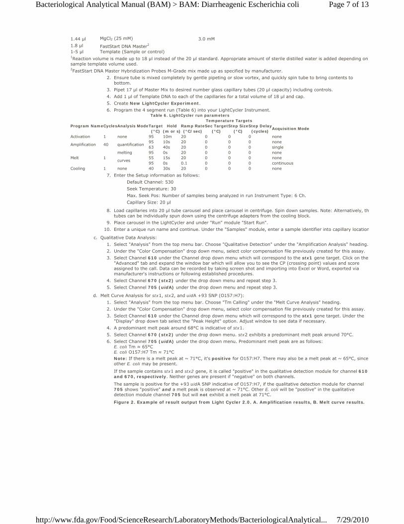
Figure 2. Example of result output from Light Cycler 2.0. A. Amplification results, B. Melt curve results.
Page 7 of 13Bacteriological Analytical Manual (BAM) > BAM: Diarrheagenic Escherichia coli
7/29/2010http://www.fda.gov/Food/ScienceResearch/LaboratoryMethods/BacteriologicalAnalytical...
A. Result call, crossing point (CP) and amplification curve graphic for stx2 (channel 670). Similar result screens available for stx1 result in channel 610.
B. Result view of melting curve analysis for uidA +93 SNP, (channel 705). O157:H7 have a melt temperature (Tm) of ~ 71°C compared to a Tm of ~ 65° C for E. coli.
Interpretation of real-time PCR Results (Smart Cycler II and LightCycler 2.0) 5.Negative samples: All three targets – stx1, stx2, and +93 uidA SNP are "negative" and the spiked PCR control is positive for one or more targets. If an internal control (IC) is incorporated in the reaction as in the CSR bead, and is positive, this would also indicate that the reactions worked correctly.
a.
No further analysis is needed.■
Probable positive O157 samples: If the +93 uidA SNP target is positive by itself or in combination with positive in either stx1, stx2 or both. Proceed to section P for culture isolation and confirmation
b.
Probable positive STEC samples: If the +93 uidA SNP target is negative but either or both stx1 and stx2 are positive, the sample may contain a non-O157 STEC or possibly that the +93 uidA SNP target was not amplified above background. Continue with section P. Also see section R, for additional information.
c.
NOTE: It is possible to have an IC be negative when one or more gene targets are positive, as the amplification of the target genes may out compete the Internal Control for available reagents. In those instances, the analysis is still valid as long as the amplification of the target gene has crossed the threshold to indicate successful PCR. Continue with section P for isolation. However, if all the target genes are negative as well as the spiked food control or IC, it invalidates the PCR analysis. Troubleshoot the reaction and rerun the assay or streak enrichments as described in section P.
P. Cultural Isolation and Presumptive Isolate Screening.For overnight enrichment samples that are found probable positive by the real-time PCR assay, cultural confirmation is required. Similarly, for samplesthat have not been screened by real-time PCR follow these procedures for culture isolation.
Isolation procedure. 1.
Serially dilute the overnight sample enrichment in Butterfield's phosphate buffer (R1135) and spread-plate appropriate dilutions (usually 0.05 mL of 10-2 and 10-4 dilutions should yield approximately 100-300 isolated colonies) in duplicate onto TC-SMAC and one chromogenic agar (Rainbow® Agar O157 or R&F® E. coli O157:H7 agar). Optionally, a streak plate may also be included.
a.
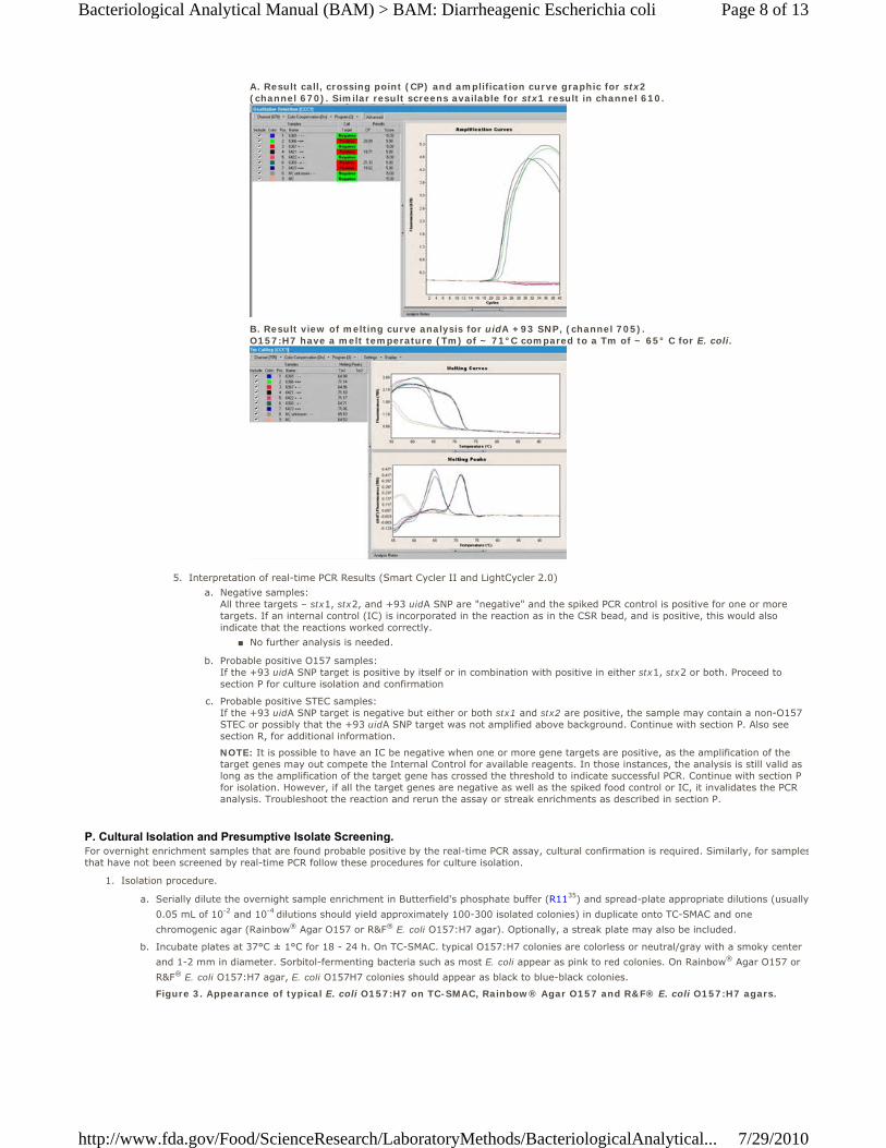
Incubate plates at 37°C ± 1°C for 18 - 24 h. On TC-SMAC. typical O157:H7 colonies are colorless or neutral/gray with a smoky center and 1-2 mm in diameter. Sorbitol-fermenting bacteria such as most E. coli appear as pink to red colonies. On Rainbow® Agar O157 or R&F® E. coli O157:H7 agar, E. coli O157H7 colonies should appear as black to blue-black colonies.
b.
Figure 3. Appearance of typical E. coli O157:H7 on TC-SMAC, Rainbow® Agar O157 and R&F® E. coli O157:H7 agars.
Page 8 of 13Bacteriological Analytical Manual (BAM) > BAM: Diarrheagenic Escherichia coli
7/29/2010http://www.fda.gov/Food/ScienceResearch/LaboratoryMethods/BacteriologicalAnalytical...
Screen typical colonies by picking a portion of each isolated suspect colony from the isolation agar and testing for O157 antigen by latexagglutination (Remel kit).
c.
Pick all typical colonies that screen positive (up to 10, if >10 are present) from isolation agars and streak onto TSAYE plates to check fopurity.
d.
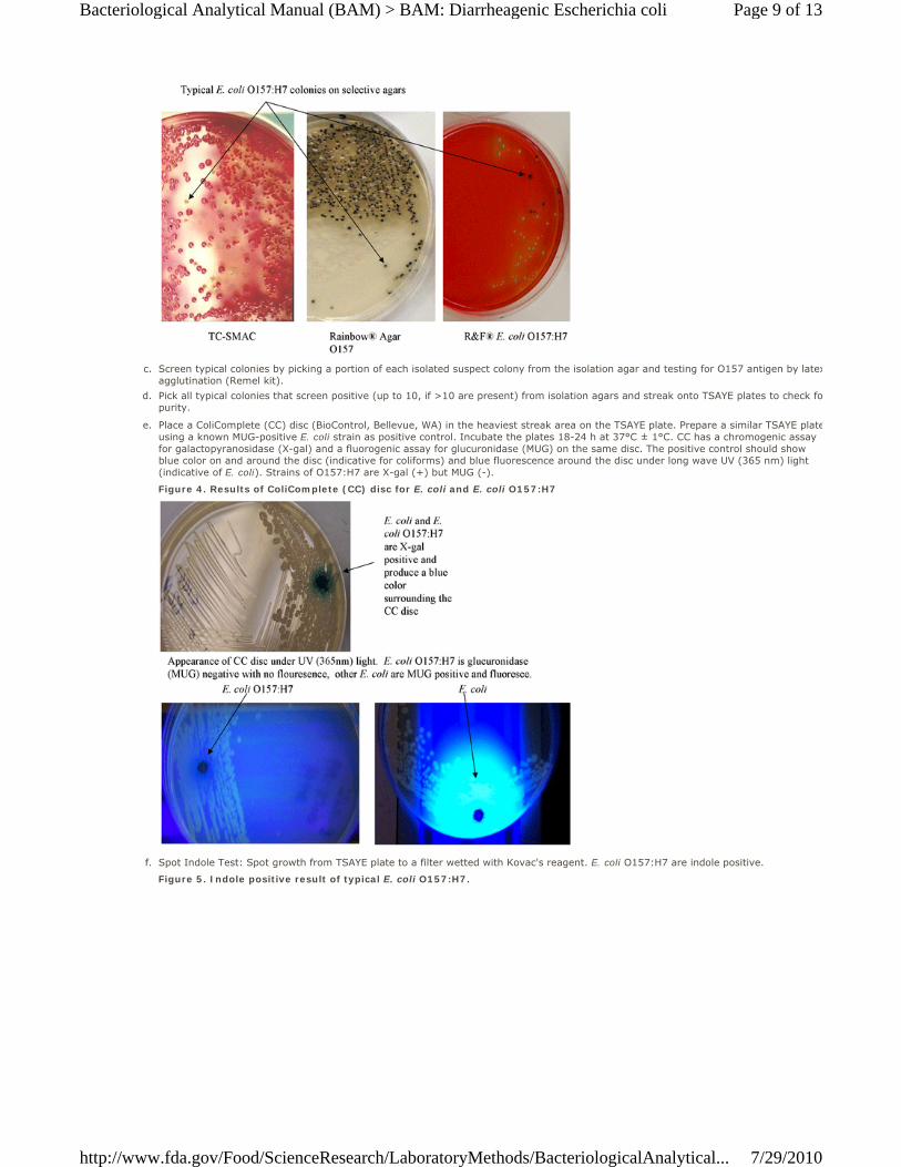
Place a ColiComplete (CC) disc (BioControl, Bellevue, WA) in the heaviest streak area on the TSAYE plate. Prepare a similar TSAYE plateusing a known MUG-positive E. coli strain as positive control. Incubate the plates 18-24 h at 37°C ± 1°C. CC has a chromogenic assay for galactopyranosidase (X-gal) and a fluorogenic assay for glucuronidase (MUG) on the same disc. The positive control should show blue color on and around the disc (indicative for coliforms) and blue fluorescence around the disc under long wave UV (365 nm) light (indicative of E. coli). Strains of O157:H7 are X-gal (+) but MUG (-).
e.
Figure 4. Results of ColiComplete (CC) disc for E. coli and E. coli O157:H7
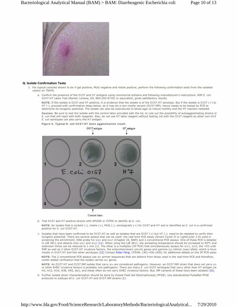
Spot Indole Test: Spot growth from TSAYE plate to a filter wetted with Kovac's reagent. E. coli O157:H7 are indole positive.f.
Figure 5. Indole positive result of typical E. coli O157:H7.
Page 9 of 13Bacteriological Analytical Manual (BAM) > BAM: Diarrheagenic Escherichia coli
7/29/2010http://www.fda.gov/Food/ScienceResearch/LaboratoryMethods/BacteriologicalAnalytical...
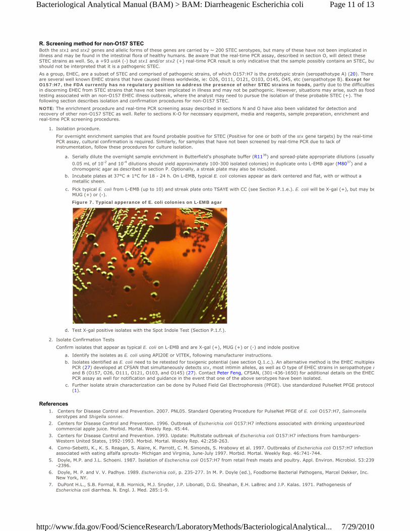
Q. Isolate Confirmation TestsFor typical colonies shown to be X-gal positive, MUG negative and indole positive, perform the following confirmation tests from the isolated colony on TSAYE.
1.
Confirm the presence of the O157 and H7 antigens using commercial antisera and following manufacturer's instructions. RIM E. coli O157:H7 Latex Test (Remel, Lenexa, KS, 800-255-6730) or equivalent, gives satisfactory results.
a.
NOTE: If the isolate is O157 and H7 positive, it is evidence that the isolate is of the O157:H7 serotype. But if the isolate is O157 (+) buH7 (-), proceed with confirmation steps below, as it may be a non-motile variant (O157:NM), hence needs to be tested by PCR to determine its toxigenic potential. The isolate can also be subcultured to blood agar to induce motility and the H7 reaction retested.
Caution: Be sure to test the isolate with the control latex provided with the kit, to rule out the possibility of autoagglutinating strains ofE. coli that will react with both reagents. Also, do not use H7 latex reagent without testing 1st with the O157 reagent as other non-O157E. coli serotypes can also carry the H7 antigen.
Figure 6. Typical E. coli O157:H7 latex agglutination result.
Test O157 and H7 positive strains with API20E or VITEK to identify as E. coli.b.
NOTE: An isolate that is sorbitol (-), indole (+), MUG (-), serologically (+) for O157 and H7 and is identified as E. coli is a confirmed positive for E. coli O157:H7.
Isolates that have been confirmed to be O157:H7 as well as isolates that are O157 (+) but H7 (-), need to be retested to verify their toxigenic potential. There are several assays that can be used: the real-time PCR assay (Smart Cycler II or LightCycler 2.0) used in screening the enrichment; DNA probe for stx1 and stx2 (Chapter 24, BAM) and 2 conventional PCR assays. One of these PCR is detailedin LIB 3811 and detects only stx1 and stx2 (11). When using the LIB 3811, the annealing temperature should be increased to 59°C andextension times can be reduced to 1 min (1). The other is a multiplex (5P PCR) that simultaneously assays for stx1, stx2, the +93 uidA SNP as well as 2 other O157:H7 virulence factors: the enterohemolysin (ehxA) genes and gamma (γ) intimin (eae) allele, which is foundmostly in O157:H7 and few other serotypes (12) Contact Peter Feng, CFSAN, (301-436-1650; for additional details on the 5P PCR assay
c.
NOTE: The 2 conventional PCR assays use stx primer sequences that are distinct from those used in the real-time PCR and therefore, confer added verification that the isolate carries stx genes.
NOTE: An O157:H7 and O157:NM isolate that carry stx are considered pathogenic. However, an O157:NM strain that does not carry stxor other EHEC virulence factors is probably non-pathogenic. There are many E. coli O157 serotypes that carry other than H7 antigen (ieH3, H12, H16, H38, H45, etc), and these often do not carry EHEC virulence factors. But, NM variants of these have been isolated (10).
Further isolate strain characterization should be done by Pulsed Field Gel Electrophoresis (PFGE). Use standardized PulseNet PFGE protocols to subtype all E. coli O157:H7 and O157:NM strains (1).
d.
Page 10 of 13Bacteriological Analytical Manual (BAM) > BAM: Diarrheagenic Escherichia coli
7/29/2010http://www.fda.gov/Food/ScienceResearch/LaboratoryMethods/BacteriologicalAnalytical...
R. Screening method for non-O157 STECBoth the stx1 and stx2 genes and allelic forms of these genes are carried by ~ 200 STEC serotypes, but many of these have not been implicated in illness and may be found in the intestinal flora of healthy humans. Be aware that the real-time PCR assay, described in section O, will detect these STEC strains as well. So, a +93 uidA (-) but stx1 and/or stx2 (+) real-time PCR result is only indicative that the sample possibly contains an STEC, butshould not be interpreted that it is a pathogenic STEC.
As a group, EHEC, are a subset of STEC and comprised of pathogenic strains, of which O157:H7 is the prototypic strain (seropathotype A) (20). There are several well known EHEC strains that have caused illness worldwide, ie: O26, O111, O121, O103, O145, O45, etc (seropathotype B). Except for O157:H7, the FDA currently has no regulatory position to address the presence of other STEC strains in foods, partly due to the difficultiesin discerning EHEC from STEC strains that have not been implicated in illness and may not be pathogenic. However, situations may arise, such as foodtesting associated with an non-O157 EHEC illness outbreak, where the analyst may need to pursue the isolation of these probable STEC (+). The following section describes isolation and confirmation procedures for non-O157 STEC.
NOTE: The enrichment procedure and real-time PCR screening assay described in sections N and O have also been validated for detection and recovery of other non-O157 STEC as well. Refer to sections K-O for necessary equipment, media and reagents, sample preparation, enrichment and real-time PCR screening procedures.
Isolation procedure.1.
For overnight enrichment samples that are found probable positive for STEC (Positive for one or both of the stx gene targets) by the real-time PCR assay, cultural confirmation is required. Similarly, for samples that have not been screened by real-time PCR due to lack of instrumentation, follow these procedures for culture isolation.
Serially dilute the overnight sample enrichment in Butterfield's phosphate buffer (R1136) and spread-plate appropriate dilutions (usually 0.05 mL of 10-2 and 10-4 dilutions should yield approximately 100-300 isolated colonies) in duplicate onto L-EMB agar (M8037) and a chromogenic agar as described in section P. Optionally, a streak plate may also be included.
a.
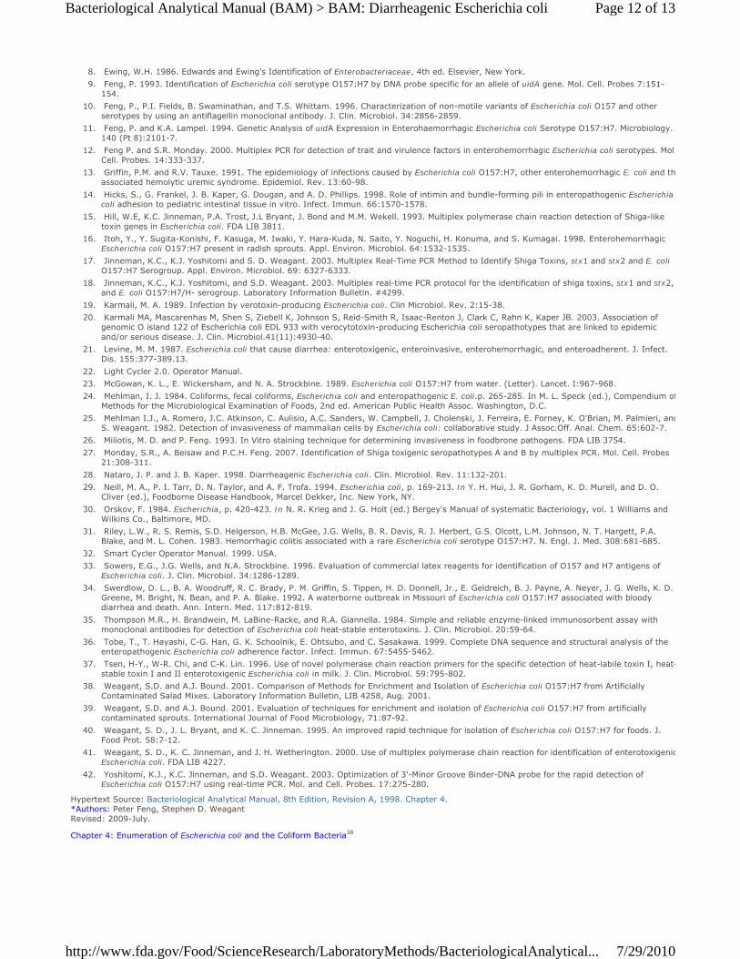
Incubate plates at 37°C ± 1°C for 18 - 24 h. On L-EMB, typical E. coli colonies appear as dark centered and flat, with or without a metallic sheen.
b.
Pick typical E. coli from L-EMB (up to 10) and streak plate onto TSAYE with CC (see Section P.1.e.). E. coli will be X-gal (+), but may beMUG (+) or (-).
c.
Figure 7. Typical apperance of E. coli colonies on L-EMB agar
Test X-gal positive isolates with the Spot Indole Test (Section P.1.f.).d.
Isolate Confirmation Tests2.
Confirm isolates that appear as typical E. coli on L-EMB and are X-gal (+), MUG (+) or (-) and indole positive
Identify the isolates as E. coli using API20E or VITEK, following manufacturer instructions.a.Isolates identified as E. coli need to be retested for toxigenic potential (see section Q.1.c.). An alternative method is the EHEC multiplexPCR (27) developed at CFSAN that simultaneously detects stx, most intimin alleles, as well as O type of EHEC strains in seropathotype Aand B (O157, O26, O111, O121, O103, and O145) (27). Contact Peter Feng, CFSAN, (301-436-1650) for additional details on the EHEC PCR assay as well for notification and guidance in the event that one of the above serotypes have been isolated.
b.
Further isolate strain characterization can be done by Pulsed Field Gel Electrophoresis (PFGE). Use standardized PulseNet PFGE protocols(1).
c.
References Centers for Disease Control and Prevention. 2007. PNL05. Standard Operating Procedure for PulseNet PFGE of E. coli O157:H7, Salmonella serotypes and Shigella sonnei.
1.
Centers for Disease Control and Prevention. 1996. Outbreak of Escherichia coli O157:H7 infections associated with drinking unpasteurized commercial apple juice. Morbid. Mortal. Weekly Rep. 45:44.
2.
Centers for Disease Control and Prevention. 1993. Update: Multistate outbreak of Escherichia coli O157:H7 infections from hamburgers-Western United States, 1992-1993. Morbid. Mortal. Weekly Rep. 42:258-263.
3.
Como-Sebetti, K., K. S. Reagan, S. Alaire, K. Parrott, C. M. Simonds, S. Hrabowy et al. 1997. Outbreaks of Escherichia coli O157:H7 infection associated with eating alfalfa sprouts- Michigan and Virginia, June-July 1997. Morbid. Mortal. Weekly Rep. 46:741-744.
4.
Doyle, M.P. and J.L. Schoeni. 1987. Isolation of Escherichia coli O157:H7 from retail fresh meats and poultry. Appl. Environ. Microbiol. 53:239-2396.
5.
Doyle, M. P. and V. V. Padhye. 1989. Escherichia coli, p. 235-277. In M. P. Doyle (ed.), Foodborne Bacterial Pathogens, Marcel Dekker, Inc. New York, NY.
6.
DuPont H.L., S.B. Formal, R.B. Hornick, M.J. Snyder, J.P. Libonati, D.G. Sheahan, E.H. LaBrec and J.P. Kalas. 1971. Pathogenesis of Escherichia coli diarrhea. N. Engl. J. Med. 285:1-9.
7.
Page 11 of 13Bacteriological Analytical Manual (BAM) > BAM: Diarrheagenic Escherichia coli
7/29/2010http://www.fda.gov/Food/ScienceResearch/LaboratoryMethods/BacteriologicalAnalytical...
Ewing, W.H. 1986. Edwards and Ewing's Identification of Enterobacteriaceae, 4th ed. Elsevier, New York.8. Feng, P. 1993. Identification of Escherichia coli serotype O157:H7 by DNA probe specific for an allele of uidA gene. Mol. Cell. Probes 7:151-154.
9.
Feng, P., P.I. Fields, B. Swaminathan, and T.S. Whittam. 1996. Characterization of non-motile variants of Escherichia coli O157 and other serotypes by using an antiflagellin monoclonal antibody. J. Clin. Microbiol. 34:2856-2859.
10.
Feng, P. and K.A. Lampel. 1994. Genetic Analysis of uidA Expression in Enterohaemorrhagic Escherichia coli Serotype O157:H7. Microbiology. 140 (Pt 8):2101-7.
11.
Feng P. and S.R. Monday. 2000. Multiplex PCR for detection of trait and virulence factors in enterohemorrhagic Escherichia coli serotypes. Mol.Cell. Probes. 14:333-337.
12.
Griffin, P.M. and R.V. Tauxe. 1991. The epidemiology of infections caused by Escherichia coli O157:H7, other enterohemorrhagic E. coli and thassociated hemolytic uremic syndrome. Epidemiol. Rev. 13:60-98.
13.
Hicks, S., G. Frankel, J. B. Kaper, G. Dougan, and A. D. Phillips. 1998. Role of intimin and bundle-forming pili in enteropathogenic Escherichia coli adhesion to pediatric intestinal tissue in vitro. Infect. Immun. 66:1570-1578.
14.
Hill, W.E, K.C. Jinneman, P.A. Trost, J.L Bryant, J. Bond and M.M. Wekell. 1993. Multiplex polymerase chain reaction detection of Shiga-like toxin genes in Escherichia coli. FDA LIB 3811.
15.
Itoh, Y., Y. Sugita-Konishi, F. Kasuga, M. Iwaki, Y. Hara-Kuda, N. Saito, Y. Noguchi, H. Konuma, and S. Kumagai. 1998. Enterohemorrhagic Escherichia coli O157:H7 present in radish sprouts. Appl. Environ. Microbiol. 64:1532-1535.
16.
Jinneman, K.C., K.J. Yoshitomi and S. D. Weagant. 2003. Multiplex Real-Time PCR Method to Identify Shiga Toxins, stx1 and stx2 and E. coli O157:H7 Serogroup. Appl. Environ. Microbiol. 69: 6327-6333.
17.
Jinneman, K.C., K.J. Yoshitomi, and S.D. Weagant. 2003. Multiplex real-time PCR protocol for the identification of shiga toxins, stx1 and stx2, and E. coli O157:H7/H- serogroup. Laboratory Information Bulletin. #4299.
18.
Karmali, M. A. 1989. Infection by verotoxin-producing Escherichia coli. Clin Microbiol. Rev. 2:15-38.19. Karmali MA, Mascarenhas M, Shen S, Ziebell K, Johnson S, Reid-Smith R, Isaac-Renton J, Clark C, Rahn K, Kaper JB. 2003. Association of genomic O island 122 of Escherichia coli EDL 933 with verocytotoxin-producing Escherichia coli seropathotypes that are linked to epidemic and/or serious disease. J. Clin. Microbiol.41(11):4930-40.
20.
Levine, M. M. 1987. Escherichia coli that cause diarrhea: enterotoxigenic, enteroinvasive, enterohemorrhagic, and enteroadherent. J. Infect. Dis. 155:377-389.13.
21.
Light Cycler 2.0. Operator Manual.22. McGowan, K. L., E. Wickersham, and N. A. Strockbine. 1989. Escherichia coli O157:H7 from water. (Letter). Lancet. I:967-968.23. Mehlman, I. J. 1984. Coliforms, fecal coliforms, Escherichia coli and enteropathogenic E. coli.p. 265-285. In M. L. Speck (ed.), Compendium ofMethods for the Microbiological Examination of Foods, 2nd ed. American Public Health Assoc. Washington, D.C.
24.
Mehlman I.J., A. Romero, J.C. Atkinson, C. Aulisio, A.C. Sanders, W. Campbell, J. Cholenski, J. Ferreira, E. Forney, K. O'Brian, M. Palmieri, andS. Weagant. 1982. Detection of invasiveness of mammalian cells by Escherichia coli: collaborative study. J Assoc.Off. Anal. Chem. 65:602-7.
25.
Miliotis, M. D. and P. Feng. 1993. In Vitro staining technique for determining invasiveness in foodbrone pathogens. FDA LIB 3754.26. Monday, S.R., A. Beisaw and P.C.H. Feng. 2007. Identification of Shiga toxigenic seropathotypes A and B by multiplex PCR. Mol. Cell. Probes 21:308-311.
27.
Nataro, J. P. and J. B. Kaper. 1998. Diarrheagenic Escherichia coli. Clin. Microbiol. Rev. 11:132-201.28. Neill, M. A., P. I. Tarr, D. N. Taylor, and A. F. Trofa. 1994. Escherichia coli, p. 169-213. In Y. H. Hui, J. R. Gorham, K. D. Murell, and D. O. Cliver (ed.), Foodborne Disease Handbook, Marcel Dekker, Inc. New York, NY.
29.
Orskov, F. 1984. Escherichia, p. 420-423. In N. R. Krieg and J. G. Holt (ed.) Bergey's Manual of systematic Bacteriology, vol. 1 Williams and Wilkins Co., Baltimore, MD.
30.
Riley, L.W., R. S. Remis, S.D. Helgerson, H.B. McGee, J.G. Wells, B. R. Davis, R. J. Herbert, G.S. Olcott, L.M. Johnson, N. T. Hargett, P.A. Blake, and M. L. Cohen. 1983. Hemorrhagic colitis associated with a rare Escherichia coli serotype O157:H7. N. Engl. J. Med. 308:681-685.
31.
Smart Cycler Operator Manual. 1999. USA.32. Sowers, E.G., J.G. Wells, and N.A. Strockbine. 1996. Evaluation of commercial latex reagents for identification of O157 and H7 antigens of Escherichia coli. J. Clin. Microbiol. 34:1286-1289.
33.
Swerdlow, D. L., B. A. Woodruff, R. C. Brady, P. M. Griffin, S. Tippen, H. D. Donnell, Jr., E. Geldreich, B. J. Payne, A. Neyer, J. G. Wells, K. D. Greene, M. Bright, N. Bean, and P. A. Blake. 1992. A waterborne outbreak in Missouri of Escherichia coli O157:H7 associated with bloody diarrhea and death. Ann. Intern. Med. 117:812-819.
34.
Thompson M.R., H. Brandwein, M. LaBine-Racke, and R.A. Giannella. 1984. Simple and reliable enzyme-linked immunosorbent assay with monoclonal antibodies for detection of Escherichia coli heat-stable enterotoxins. J. Clin. Microbiol. 20:59-64.
35.
Tobe, T., T. Hayashi, C-G. Han, G. K. Schoolnik, E. Ohtsubo, and C. Sasakawa. 1999. Complete DNA sequence and structural analysis of the enteropathogenic Escherichia coli adherence factor. Infect. Immun. 67:5455-5462.
36.
Tsen, H-Y., W-R. Chi, and C-K. Lin. 1996. Use of novel polymerase chain reaction primers for the specific detection of heat-labile toxin I, heat-stable toxin I and II enterotoxigenic Escherichia coli in milk. J. Clin. Microbiol. 59:795-802.
37.
Weagant, S.D. and A.J. Bound. 2001. Comparison of Methods for Enrichment and Isolation of Escherichia coli O157:H7 from Artificially Contaminated Salad Mixes. Laboratory Information Bulletin, LIB 4258, Aug. 2001.
38.
Weagant, S.D. and A.J. Bound. 2001. Evaluation of techniques for enrichment and isolation of Escherichia coli O157:H7 from artificially contaminated sprouts. International Journal of Food Microbiology, 71:87-92.
39.
Weagant, S. D., J. L. Bryant, and K. C. Jinneman. 1995. An improved rapid technique for isolation of Escherichia coli O157:H7 for foods. J. Food Prot. 58:7-12.
40.
Weagant, S. D., K. C. Jinneman, and J. H. Wetherington. 2000. Use of multiplex polymerase chain reaction for identification of enterotoxigenicEscherichia coli. FDA LIB 4227.
41.
Yoshitomi, K.J., K.C. Jinneman, and S.D. Weagant. 2003. Optimization of 3'-Minor Groove Binder-DNA probe for the rapid detection of Escherichia coli O157:H7 using real-time PCR. Mol. and Cell. Probes. 17:275-280.
42.
Hypertext Source: Bacteriological Analytical Manual, 8th Edition, Revision A, 1998. Chapter 4. *Authors: Peter Feng, Stephen D. Weagant Revised: 2009-July.
Chapter 4: Enumeration of Escherichia coli and the Coliform Bacteria38
Page 12 of 13Bacteriological Analytical Manual (BAM) > BAM: Diarrheagenic Escherichia coli
7/29/2010http://www.fda.gov/Food/ScienceResearch/LaboratoryMethods/BacteriologicalAnalytical...
Links on this page:
http://www.fda.gov/Food/ScienceResearch/LaboratoryMethods/BacteriologicalAnalyticalManualBAM/ucm062752.htm1.
http://www.fda.gov/Food/ScienceResearch/LaboratoryMethods/BacteriologicalAnalyticalManualBAM/ucm063362.htm2.
http://www.fda.gov/Food/ScienceResearch/LaboratoryMethods/BacteriologicalAnalyticalManualBAM/ucm064449.htm3.
http://www.fda.gov/Food/ScienceResearch/LaboratoryMethods/BacteriologicalAnalyticalManualBAM/ucm064496.htm4.
http://www.fda.gov/Food/ScienceResearch/LaboratoryMethods/BacteriologicalAnalyticalManualBAM/ucm063699.htm5.
http://www.fda.gov/Food/ScienceResearch/LaboratoryMethods/BacteriologicalAnalyticalManualBAM/ucm063356.htm6.
http://www.fda.gov/Food/ScienceResearch/LaboratoryMethods/BacteriologicalAnalyticalManualBAM/ucm063867.htm7.
http://www.fda.gov/Food/ScienceResearch/LaboratoryMethods/BacteriologicalAnalyticalManualBAM/ucm063364.htm8.
http://www.fda.gov/Food/ScienceResearch/LaboratoryMethods/BacteriologicalAnalyticalManualBAM/ucm062935.htm9.
http://www.fda.gov/Food/ScienceResearch/LaboratoryMethods/BacteriologicalAnalyticalManualBAM/ucm064474.htm10.
http://www.fda.gov/Food/ScienceResearch/LaboratoryMethods/BacteriologicalAnalyticalManualBAM/ucm063514.htm11.
http://www.fda.gov/Food/ScienceResearch/LaboratoryMethods/BacteriologicalAnalyticalManualBAM/ucm064068.htm12.
http://www.fda.gov/Food/ScienceResearch/LaboratoryMethods/BacteriologicalAnalyticalManualBAM/ucm064362.htm13.
http://www.fda.gov/Food/ScienceResearch/LaboratoryMethods/BacteriologicalAnalyticalManualBAM/ucm062237.htm14.
http://www.fda.gov/Food/ScienceResearch/LaboratoryMethods/BacteriologicalAnalyticalManualBAM/ucm064081.htm15.
http://www.fda.gov/Food/ScienceResearch/LaboratoryMethods/BacteriologicalAnalyticalManualBAM/ucm064085.htm16.
http://www.fda.gov/Food/ScienceResearch/LaboratoryMethods/BacteriologicalAnalyticalManualBAM/ucm064500.htm17.
http://www.fda.gov/Food/ScienceResearch/LaboratoryMethods/BacteriologicalAnalyticalManualBAM/ucm064411.htm18.
http://www.fda.gov/Food/ScienceResearch/LaboratoryMethods/BacteriologicalAnalyticalManualBAM/ucm059108.htm19.
http://www.fda.gov/Food/ScienceResearch/LaboratoryMethods/BacteriologicalAnalyticalManualBAM/ucm062262.htm20.
http://www.fda.gov/Food/ScienceResearch/LaboratoryMethods/BacteriologicalAnalyticalManualBAM/ucm062269.htm21.
http://www.fda.gov/Food/ScienceResearch/LaboratoryMethods/BacteriologicalAnalyticalManualBAM/ucm061208.htm22.
http://www.fda.gov/Food/ScienceResearch/LaboratoryMethods/BacteriologicalAnalyticalManualBAM/ucm062242.htm23.
http://www.fda.gov/Food/ScienceResearch/LaboratoryMethods/BacteriologicalAnalyticalManualBAM/ucm061693.htm24.
http://www.fda.gov/Food/ScienceResearch/LaboratoryMethods/BacteriologicalAnalyticalManualBAM/ucm062263.htm25.
http://www.fda.gov/Food/ScienceResearch/LaboratoryMethods/BacteriologicalAnalyticalManualBAM/ucm062257.htm26.
http://www.fda.gov/Food/ScienceResearch/LaboratoryMethods/BacteriologicalAnalyticalManualBAM/ucm062254.htm27.
http://www.fda.gov/Food/ScienceResearch/LaboratoryMethods/BacteriologicalAnalyticalManualBAM/ucm062229.htm28.
http://www.fda.gov/Food/ScienceResearch/LaboratoryMethods/BacteriologicalAnalyticalManualBAM/ucm172352.htm29.
http://www.fda.gov/Food/ScienceResearch/LaboratoryMethods/BacteriologicalAnalyticalManualBAM/ucm172353.htm30.
http://www.fda.gov/Food/ScienceResearch/LaboratoryMethods/BacteriologicalAnalyticalManualBAM/ucm064449.htm31.
http://www.fda.gov/Food/ScienceResearch/LaboratoryMethods/BacteriologicalAnalyticalManualBAM/ucm063785.htm32.
http://www.fda.gov/Food/ScienceResearch/LaboratoryMethods/BacteriologicalAnalyticalManualBAM/ucm061208.htm33.
http://www.fda.gov/Food/ScienceResearch/LaboratoryMethods/BacteriologicalAnalyticalManualBAM/ucm062242.htm34.
http://www.fda.gov/Food/ScienceResearch/LaboratoryMethods/BacteriologicalAnalyticalManualBAM/ucm061208.htm35.
http://www.fda.gov/Food/ScienceResearch/LaboratoryMethods/BacteriologicalAnalyticalManualBAM/ucm061208.htm36.
http://www.fda.gov/Food/ScienceResearch/LaboratoryMethods/BacteriologicalAnalyticalManualBAM/ucm064449.htm37.
http://www.fda.gov/Food/ScienceResearch/LaboratoryMethods/BacteriologicalAnalyticalManualBAM/ucm064948.htm38.
Page 13 of 13Bacteriological Analytical Manual (BAM) > BAM: Diarrheagenic Escherichia coli
7/29/2010http://www.fda.gov/Food/ScienceResearch/LaboratoryMethods/BacteriologicalAnalytical...