bacterial vaginosis - mdlab.com · medical diagnostic laboratories, l.l.c. 877.269.0090...
TRANSCRIPT

Medical Diagnostic Laboratories, L.L.C. • www.mdlab.com • 877.269.0090
The vaginal microflora is a dynamic ecosystem normally inhabited by lactobacilli. These bacteria support healthy vaginal conditions by maintaining an acidic environment that is inhospitable to other pathogenic microorganisms. L. crispatus, L. gasseri, L. jensenii, and L. iners are considered to be the four major vaginal Lactobacillus species (1, 2, 3). Usually, the vaginal flora is dominated by one of these bacteria accompanied by less abundant and less frequently detected minor Lactobacillus species including L. acidophilus, L. johnsoni, L. vaginalis, L. fermentum, and L. reuteri (4). The numerical prevalence of lactobacilli in the vagina prevents its colonization by other pathogens. Many important aspects of women’s sexual and reproductive health rely on the protective role of lactobacilli in the vaginal environment.
The composition of the vaginal microflora is not static but changes over time in response to various endogenous and exogenous influences. The most common alteration in vaginal microflora is a condition named Bacterial Vaginosis (BV) (1, 4). BV is a disorder characterized by an overgrowth of anaerobic bacteria in the vagina leading to a replacement of healthy lactobacilli. Bacterial species including Gardnerella vaginalis, Atopobium vaginae, Megasphaera Type 1 and Type 2, Bacterial Vaginosis-Associated Bacterium 2 (BVAB2), Bacteroides species, Mobiluncus species, Mycoplasma species, and Ureaplasma urealyticum are described as indicative diagnostic markers of BV (5-8). The presence of these microorganisms along with a depletion of the protective lactobacilli suggests that vaginal microbial conditions are abnormal. Clinical symptoms of BV include an increase in vaginal pH, vaginal discharge and an unpleasant fishy odor. BV is associated with an increased risk of sexually transmitted infections and serious pregnancy complications including miscarriage and preterm birth (5, 9-13). BV is very common in women of reproductive age and is one of the most common reasons that women seek treatment from health care providers (14, 15).
Bacterial vaginosis is associated with an increase of vaginal pH from a healthy range (3.8 – 4.2) to > 4.5 in which an overgrowth of Gardnerella vaginalis (A), a modest increase in facultative anaerobes (B) and the
Bacterial Vaginosis(With Lactobacillus Profiling) qPCR Panel
depletion of lactobacilli (C) is observed. A dramatic increase of the obligate anaerobic bacteria (D) such as Atopobium vaginae, Megasphaera species and BVAB2 is also observed during BV and are important indicator organisms associated with the BV state (15).
Figure 1. Changes of the vaginal microflora associated with the development of bacterial vaginosis.
Epidemiology
The prevalence of BV is difficult to determine as the disease can be symptomatic or asymptomatic. Most prevalence studies rely on women experiencing symptoms who seek gynecological care and some have reported that BV affects up to 23% of women of reproductive age (14). The prevalence of BV in the United States is higher among African American women than among women of other racial groups. According to epidemiological studies, up to 46% of African-American women, compared with only 14% of non-African American women, acquire BV (16, 17). The basis for this racial disparity has not yet been determined. Although BV is not considered a sexually transmitted infection, it is rarely observed in women who have not yet engaged in sexual activity (18).
Pathogenesis
The underlying pathogenesis of BV is hypothesized to involve a depletion of lactobacilli from the vaginal flora and consequent overgrowth of Gram-negative anaerobes and facultative anaerobes such as G.

Medical Diagnostic Laboratories, L.L.C. • www.mdlab.com • 877.269.0090
Laboratory BV testing involves Gram staining of vaginal smears, microscopic evaluation and scoring numbers of bacterial morphotypes and clue cells according to Nugent (21) or Ison and Hay (22). Since the majority of bacterial species associated with BV are fastidious anaerobic microorganisms that are either difficult to culture or, in some cases, have yet to be cultured, the use of conventional microbiological techniques other than Gram staining is not appropriate for BV diagnostic purposes (23).
Both the Amsel criteria and Nugent scoring approaches suffer from subjective interpretation and, as it has been shown in multiple studies, do not perfectly agree with one another (23-25). Up to half of all women who meet the diagnostic criteria for BV might not exhibit clinical symptoms (26).
Recently, the utility of molecular techniques, such as quantitative PCR (qPCR), for the quantitation of major bacterial species inhabiting the vaginal environment of healthy women and BV carriers has been demonstrated (1, 8, 19, 27, 28). qPCR assessment of BV-related bacteria correlates significantly with a high sensitivity and specificity to the Nugent score and to a lesser extent with Amsel criteria in the diagnosis of BV (19, 23, 25). Application of molecular methods for characterization of vaginal microflora in BV patients has become an accepted practice and a major trend in laboratory diagnostics (1).
Table 1. The Sensitivity and Specificity of PCR detection of vaginal bacteria in association with BV (19).
Bacterium Sensitivity % (95% CI)
Specificity % (95% CI)
Gardnerella vaginalis 97.3 (90.6 – 99.3) 45.5 (37.5 – 53.6)
Atopobium species 95.9 (88.6 – 98.6) 84.6 (77.8 – 89.6)
Megasphaera Type 1 94.5 (86.7 – 97.9) 94.4 (89.4 – 97.1)
Megasphaera Type 2 6.9 (3.0 – 15.1) 100 (97.4 – 100)
BVAB2 80.8 (70.3 – 88.2) 96.5 (92.1 – 98.5)
Either Megasphaera Type 1 or BVAB2 95.9 (88.6 – 98.6) 93.7 (88.5 – 96.7)
Lactobacillus crispatus 8.2 (3.8 – 16.8) 6.3 (3.4 – 11.5)
Lactobacillus iners 94.5 (86.7 – 97.9) 11.9 (7.6 – 18.2)
vaginalis and Mobiluncus species. The development of culture-independent molecular diagnostics has greatly increased the number of bacterial species associated with BV to include A. vaginae, Eggerthella species, Leptotrichia amnionii, Megasphaera species, Mycoplasma species, U. urealyticum, and BVAB2 (6, 8, 19). The particular ways a woman acquires an infection remain unknown. Since microorganisms associated with BV are shown to have their natural habitat in the gastrointestinal tract, BV might be an endogenous infection (4). Nevertheless, the prevalence of BV is higher in sexually active women having sexual contacts with new and/or multiple partners. Decreasing the number of unprotected sexual encounters may reduce the overall incidence rate as well as the rate of re-infection (18).
The precise mechanism of how lactobacilli are depleted and replaced by BV-associated bacteria during BV is also still unknown. One possibility is that a change in vaginal pH is the underlying cause of BV. Lactic acid is produced by Lactobacillus which maintains the vaginal pH between 3.8 and 4.2. This environment favors the growth of Lactobacillus and inhibits the growth of pathogenic bacteria. Risk factors for BV include douching, menstruation, and unprotected sexual contact, all of which raise the vaginal pH above the optimum for Lactobacillus growth (2).
Gynecological Complications
Women with BV have a three- to four-fold higher risk for acquisition of Chlamydia trachomatis and Neisseria gonorrhoeae (13) and are also at higher risk for HIV infection (10). Women with BV who undergo invasive surgery such as a hysterectomy or abortion suffer from post-operative infections two- to three- times more often than women without BV (11). BV is also associated with pelvic inflammatory disease (12) and endometritis (9). Accurate diagnosis and appropriate treatment of BV is especially critical for pregnant women, as the disease is associated with a five-fold increased risk for late miscarriage and pre-term birth (5, 11).
Diagnosis
There are two major approaches in conventional BV diagnostics: clinical and laboratory. Clinical BV diagnosis is based on the fulfillment of at least 3 out of 4 criteria described by Amsel (20):
Increased vaginal pH > 4.5
The presence of clue cells (exfoliated vaginal epithelial cells with attached bacteria) by wet-mount microscope.
Positive “whiff test” or amine test (fishy odor after addition of 10% KOH).
The presence of a thin, non-clumping gray-white adherent vaginal discharge.
◦
◦
◦
◦

Medical Diagnostic Laboratories, L.L.C. • www.mdlab.com • 877.269.0090
PCR assays detecting vaginal microflora were performed on 216 subjects also characterized using Nugent criteria to define BV status. The BV associated organisms included in the BV panel (A. vaginae, Megasphaera Type 1 and 2, and BVAB2) demonstrated high sensitivity and specificity for BV. Conversely, the healthy Lactobacilli (L. crispatus) demonstrated a negative correlation with BV with low sensitivity and specificity for BV. Although G. vaginalis detection displayed high sensitivity for BV, the specificity was lower due to the fact that this organism can be found in low numbers in normal vaginal microflora and often in intermediate or transitional vaginal microflora. Like G. vaginalis, L. iners can be detected in both healthy and disturbed vaginal microflora including BV and along with the depletion of other Lactobacillus species, may be indicative of an intermediate or transitional stage between normal and abnormal microflora. Megasphaera Type 2 was not found to be highly prevalent in this patient cohort, however the presence of this organism is highly specific for BV.
Characteristics of microorganisms, associated with normal, transitional and abnormal vaginal microflora (BV)
Lactobacillus crispatus, Lactobacillus gasseri, and Lactobacillus jensenii are Gram-positive, rod-shaped, facultative anaerobic non-spore forming bacteria and common members of healthy vaginal bacterial microflora. Along with L. iners, they are classified as the four major vaginal Lactobacillus species. L. crispatus, L. gasseri, and L. jensenii promote a healthy vaginal microenvironment by supporting an acidic pH, producing hydrogen peroxide, and preventing colonization by other microbial pathogens (2, 4, 27, 29). Vaginal microflora dominated by any of these species is considered to be normal (1, 4). The amount of L. crispatus, L. gasseri, and/or L. jensenii bacteria is reduced in certain vaginal conditions, including BV. There is a negative association between the presence of these Lactobacillus species and BV (3, 8).
Lactobacillus iners is a rod-shaped facultative anaerobic non-spore forming Gram-positive bacteria. It is a common member of human-associated bacterial microflora. Along with L. gasseri, L. crispatus, and L. jensenii, it is considered to be one of the four major vaginal Lactobacillus species. Physiologically, L. iners is different from other vaginal lactobacilli as it is less prone to hydrogen peroxide production and also is the
most fastidious microorganism compared with the other Lactobacillus species (1, 4). L. iners can be detected in both healthy and disturbed vaginal microflora including BV (6, 8, 19) (Table 1). L. iners’ dominance, along with the depletion of other Lactobacillus species, indicates that the vaginal microflora may be in a transitional stage between abnormal and normal (3, 29, 30). The medical relevance of transitional or intermediate vaginal microflora is currently an issue of debate in the literature.
Gardnerella vaginalis is a Gram-variable facultative anaerobic bacterium and was one of the first organisms to be associated with BV. The abilities of G. vaginalis to form a biofilm and produce prolidase, sialidase, β-galactosidase, and vaginolysin may play a role in the pathogenesis of this condition (4, 5). In recent years, the application of culture-independent techniques has revealed the ubiquitous nature of G. vaginalis. Due to the common occurrence of this microorganism in healthy women, the role of G. vaginalis as a BV diagnostic marker has been challenged (1, 6, 8, 7, 25). Even though the presence of G. vaginalis bacteria in the vaginal milieu signifies an occurring disturbance, the concomitant detection of G. vaginalis with other BV-associated microorganisms is more indicative of BV (27, 28, 31).
Atopobium vaginae is a Gram-positive anaerobic bacterium that has more recently become associated with BV. Similar to G. vaginalis, the presence of the organism at a high concentration is highly sensitive and specific for the diagnosis of BV (7, 19, 28). In addition, the presence of G. vaginalis and A. vaginae together is associated with disease recurrence (31). Some A. vaginae isolates exhibit reduced susceptibility to metronidazole in vitro which could be a contributing factor to disease recurrence (32). A. vaginae triggers an inflammatory response from vaginal epithelial cells, which may contribute to the pathogenesis of BV (33).
Megasphaera species are Gram-negative fastidious anaerobic organisms. A recent study has identified 16S rDNA present in BV patient samples as belonging to a Megasphaera species that has not yet been cultured (34). Detection of Megasphaera species DNA provides high sensitivity and specificity compared to Amsel criteria and Nugent score (7, 19). Successful antibiotic treatment of BV reduces the vaginal concentration of Megasphaera species (7) and persistence of this organism is associated with chronic BV (35).

Medical Diagnostic Laboratories, L.L.C. • www.mdlab.com • 877.269.0090
Bacterial Vaginosis-Associated Bacterium 2 (BVAB2) was identified through the molecular characterization of 16S rDNA sequences of the vaginal flora of women suffering from BV, which revealed three uncultured species belonging to the order Clostridiales associated with the disease, named Bacterial Vaginosis-Associated Bacterium 1, 2, and 3 (BVAB1, BVAB2, BVAB3) (7). Similar to Megasphaera species detection of BVAB2 16S rDNA provides high sensitivity and specificity for the diagnosis of BV (7). In addition, elimination of BVAB2 by antibiotic therapy is associated with disease resolution (19) and its persistence is associated with chronic disease (35).
MDL Bacterial Vaginosis (with Lactobacillus profiling) qPCR Panel
MDL has developed a new state-of-the-art qPCR molecular assay targeting a set of bacteria commonly found in normal healthy vaginal microflora and microflora associated with BV. The new MDL Bacterial Vaginosis (with Lactobacillus profiling) qPCR Panel includes tests for four major vaginal Lactobacillus species: L. crispatus, L. gasseri, L. jensenii, and L. iners, and five BV-associated pathogens: Gardnerella vaginalis, Atopobium vaginae, Megasphaera Type 1 and Type 2, and BVAB2. Even though all of the qPCR tests in the panel can be ordered and performed separately, only the combination of the assays in a single panel allows for the accurate relative quantitative evaluation of the bacterial species composition in a given clinical sample.
The MDL Bacterial Vaginosis (with Lactobacillus profiling) qPCR Panel test results are reported in two formats: text-based and graphical. The text format has a standard layout of diagnostic qualitative test reporting. The graphic format is a representation of the results of all the quantitative tests included in the panel. The relative ratios of DNA species in the given sample in proportion to one another is a reflection of the relative concentrations of different bacteria in vaginal specimens. This user-friendly test report simplifies data interpretation and analysis. The single pie chart graph provides the physician with a snapshot of the vaginal bacterial microflora accompanied by a summary suggestive of vaginal microflora state: either normal or affected by BV (Figure 2).
Normal vaginal microflora. Not Suggestive of Bacterial Vaginosis.
Abnormal vaginal microflora. Suggestive of Bacterial Vaginosis.
Lactobacillus crispatus
Lactobacillus gasseri
Lactobacillus jensenii
Lactobacillus iners
Gardnerella vaginalis
Atopobium vaginae
BVAB2
Megasphaera Type 1
Megasphaera Type 2
Figure 2. Graphic representations of MDL Bacterial Vaginosis (with Lactobacillus profiling) qPCR Panel test results for normal and abnormal vaginal microflora.
Generally, molecular diagnostic tests for BV are focused on the detection of recognized pathogenic markers of the disease. Incorporation of the Lactobacillus qPCR assays makes our test considerably more comprehensive and greatly extends the diagnostics available for assessment of vaginal health. The new MDL Bacterial Vaginosis (with Lactobacillus profiling) qPCR Panel is a significant advancement beyond the qualitative identification of BV-associated microorganisms since it now covers microbial markers of the normal vaginal environment. It can be used successfully for the determination of relative vaginal microflora composition and bacterial loads, which might facilitate monitoring of the response to antibiotic therapy.
Treatment
The Centers for Disease Control and Prevention (CDC) has recommended treatment for BV (36) (Tables 2). Treatment is recommended for all patients exhibiting symptomatic BV as defined by Amsel criteria and/or Nugent score, and asymptomatic patients should be treated prior to invasive procedures such as an abortion or hysterectomy in order to reduce the possibility of complicating infections. In addition, asymptomatic BV patients who are pregnant and at-risk for pre-term labor

Medical Diagnostic Laboratories, L.L.C. • www.mdlab.com • 877.269.0090
should also be treated, as this regimen may increase their chance of carrying their pregnancy to term.
Table 2. CDC’s 2015 Sexually Transmitted Diseases Summary of 2015 CDC Treatment Guidelines Pocket Guide (36).
Recommended Regimens
Metronidazole orala 500 mg orally twice a day for 7 days ORMetronidazole gel 0.75%a one applicator (5 g) intravaginally, once a day for 5 days ORClindamycin cream 2%a, b one applicator (5 g) intravaginally at bedtime for 7 daysRecommended Regimens (Alternatives)
Tinidazole 2 g orally once daily for 2 days OR
Tinidazole 1 g orally once daily for 5 days OR
Clindamycin 300 mg orally twice daily for 7 days ORClindamycin ovules 100 mg intravaginally once at bedtime for 3 daysTreatment is recommended for all symptomatic pregnant womena The recommended regimens are equally efficacious.b These creams are oil-based and may weaken latex condoms and
diaphragms. Refer to product labeling for further information.
Treatment of women who have symptomatic BV and are at high-risk for pre-term labor can reduce the frequency of labor complications. Patients should abstain from alcohol consumption during metronidazole treatment and for 24 hours thereafter. Patients should also be advised that intravaginal clindamycin treatment can weaken condoms and diaphragms. Intravaginal clindamycin cream is recommended for patients who are allergic to, or intolerant of, metronidazole (36).
Monitoring of Treatment Efficacy
The restoration of normal microflora is the final result physicians strive to achieve with antimicrobial therapy for BV. Successful treatment of BV with antimicrobials results in a three- to four-log decrease in the vaginal concentrations of BV-associated microorganisms followed by the rise in lactobacilli concentration of about the same magnitude (6, 7). Eradication of BV-related bacteria and their replacement with Lactobacillus species suggests a complete BV cure. Meanwhile, failure of BV antibiotic therapy is associated with only minor changes in the composition of the vaginal bacteria (6, 7, 35) (Table 4).
The MDL Bacterial Vaginosis (with Lactobacillus profiling) qPCR Panel offers an opportunity for physician to monitor the efficacy of antimicrobial therapy. Dynamic changes in bacterial composition during the course of treatment and post-treatment may be observed to help physicians assess treatment success with the return of Lactobacilli or treatment failure with persistence or recurrence of the BV associated organisms. Treatment regimens can therefore be appropriately adjusted in an effort to achieve an efficient cure.
BacteriumCured of BV (n=24)
Pretreatment Post treatmentConcentration per swab No. PCR-positive isolates % PCR-positive isolates Concentration per swab No. PCR-positive isolates % PCR-positive isolates
G.vaginalis 2.77E + 08 24 100.0 1.88E + 04 15 62.5Atopobium species 5.36E + 07 24 100.0 < 750 5 20.8Megasphaera species 3.37E + 07 22 91.7 < 1000 3 12.5BVAB2 5.11E + 06 16 66.7 < 1000 1 4.2
BacteriumPersistent BV (n=24)
Pretreatment Post treatmentConcentration per swab No. PCR-positive isolates % PCR-positive isolates Concentration per swab No. PCR-positive isolates % PCR-positive isolates
G.vaginalis 1.40E + 08 24 100.0 2.00E + 08 24 100.0Atopobium species 4.96E + 07 24 100.0 4.65E + 07 24 100.0Megasphaera species 4.53E + 07 24 100.0 2.66E + 07 23 95.8BVAB2 3.50E + 06 24 100.0 2.07E + 06 22 91.7
Table 4. Changes in vaginal bacterial concentrations with intravaginal metronidazole therapy for bacterial vaginosis as assessed by PCR (7). Bacterial concentrations and percent positive for BV associated bacteria and the healthy Lactobacillus crispatus were determined by qPCR using vaginal fluid collected at initial diagnosis (pre-treatment) and one month after metronidazole antibiotic treatment (post-treatment) in women with cured and persistent BV. Post-treatment patients cured of BV demonstrated a depletion of the BV-associated bacteria and a replacement with healthy Lactobacilli. Conversely, patients with persistent BV maintained the same bacterial profile between pre- and post-treatment.

Medical Diagnostic Laboratories, L.L.C. • www.mdlab.com • 877.269.0090
19. Fredricks DN, Fiedler TL, Thomas KK, et al. 2007. Targeted PCR for detection of vaginal bacteria associated with bacterial vaginosis. J Clin Microbiol 45:3270-3276.
20. Amsel R, Totten PA, Spiegel CA, et al. 1983. Nonspecific vaginitis. Diagnostic criteria and microbial and epidemiologic associations. Am J Med 74(1):14-22.
21. Nugent RP, Krohn MA, Hillier SL. 1991. Reliability of diagnosing bacterial vaginosis is improved by a standardized method of Gram stain interpretation. J Clin Microbiol 29(2):297-301.
22. Ison CA, Hay PE. 2002. Validation of a simplified grading of Gram stained vaginal smears for use in genitourinary medicine clinics. Sex Transm Infect 78(6): 413-5.
23. Forsum U, Hallen A, Larsson PG. 2005. Bacterial vaginosis – a laboratory and clinical diagnostics enigma. APMIS 113(3):153-61.
24. Schwebke JR, Hillier SL, Sobel JD, et al. 1996. Validity of the vaginal Gram stain for the diagnosis of bacterial vaginosis. Obstet Gynecol 88(4 Pt 1):573-6.
25. Sha BE, Chen HY, Wang QJ, et al. 2005. Utility of Amsel criteria, Nugent score, and quantitative PCR for Gardnerella vaginalis, Mycoplasma hominis, and Lactobacillus spp. for diagnosis of bacterial vaginosis in human immunodeficiency virus-infected women. J Clin Microbiol 43(9):4607-12.
26. Schwebke JR. 2000. Asymptomatic bacterial vaginosis: response to therapy. Am J Obstet Gynecol 183(6):1434-9.
27. De Backer E, Verhelst R, Verstraelen H, et al. 2007. Quantitative determination by Real-Time PCR of four vaginal Lactobacillus species, Gardnerella vaginalis and Atopobium vaginae indicates an inverse relationship between L. gasseri and L. iners. BMC Microbiol 19;7:115.
28. Menard JP, Mazouni C, Fenollar F, et al. 2010. Diagnostic accuracy of quantitative Real-Time PCR assay versus clinical and Gram stain identification of bacterial vaginosis. Eur J Clin Microbiol Infect Dis 29(12):1547-52.
29. Verstraelen H, Verhelst R, Claeys G, et al. 2009. Longitudinal analysis of the vaginal microflora in pregnancy suggests that L. crispatus promotes the stability of the normal vaginal microflora and that L. gasseri and/or L. iners are more conducive to the occurrence of abnormal vaginal microflora. BMC Microbiol 2;9:116.
30. Jakobsson T, Forsum U. 2007. Lactobacillus iners: a marker of changes in the vaginal flora? J Clin Microbiol 45(9):3145.
31. Bradshaw CS, Tabrizi SN, Fairley CK, et al. 2006. The association of Atopobium vaginae and Gardnerella vaginalis with bacterial vaginosis and recurrence after oral metronidazole therapy. J Infect Dis 194:828-836.
32. De Backer E, Verhelst R, Verstraelen H, et al. 2006. Antibiotic susceptibility of Atopobium vaginae. BMC Infect Dis 6:51.
33. Libby EK, Pascal KE, Mordechai E, et al. 2008. Atopobium vaginae triggers an innate immune response in an in vitro model of bacterial vaginosis. Microbes Infect 10:439-446.
34. Zozaya-Hinchliffe M, Martin DH, Ferris MJ. 2008. Prevalence and abundance of uncultivated Megasphaera-like bacteria in the human vaginal environment. Appl Environ Microbiol 74:1656-1659.
35. Marrazzo JM, Thomas KK, Fiedler TL, et al. 2008. Relationship of specific vaginal bacteria and bacterial vaginosis treatment failure in women who have sex with women. Ann Intern Med 149:20-28.
36. CDC. 2015. Sexually Transmitted Diseases, Treatment Guidelines, 2015. MMWR 64:72-75.
References:1. Lamont RF, Sobel JD, Akins RA, et al. 2011. The vaginal
microbiome: new information about genital tract flora using molecular based techniques. BJOG 118(5):533-49.
2. Redondo-Lopez V, Cook RL, Sobel JD. 1990. Emerging role of lactobacilli in the control and maintenance of the vaginal bacterial microflora. Rev Infect Dis 12:856-872.
3. Tamrakar R, Yamada T, Furuta I, et al. 2007. Association between Lactobacillus species and bacterial vaginosis-related bacteria, and bacterial vaginosis scores in pregnant Japanese women. BMC Infect Dis 7;7:128.
4. Forsum U, Holst E, Larsson PG, et al. 2005. Bacterial vaginosis – a microbiological and immunological enigma. APMIS 113(2):81-90.
5. Donati L, Di Vico A, Nucci M, et al. 2010. Vaginal microbial flora and outcome of pregnancy. Arch Gynecol Obstet 281(4):589-600.
6. Srinivasan S, Liu C, Mitchell CM, et al. 2010. Temporal variability of human vaginal bacteria and relationship with bacterial vaginosis. PLoS One 15;5(4):e10197.
7. Fredricks DN, Fiedler TL, Thomas KK, et al. 2009. Changes in vaginal bacterial concentrations with intravaginal metronidazole therapy for bacterial vaginosis as assessed by quantitative PCR. J Clin Microbiol 47:721-726.
8. Zozaya-Hinchliffe M, Lillis R, Martin DH, et al. 2010. Quantitative PCR assessments of bacterial species in women with and without bacterial vaginosis. J Clin Microbiol 48(5):1812-9.
9. Andrews WW, Hauth JC, Cliver SP, et al. 2006. Association of asymptomatic bacterial vaginosis with endometrial microbial colonization and plasma cell endometritis in nonpregnant women. Am J Obstet Gynecol 195:1611-1616.
10. Atashili J, Poole C, Ndumbe PM, et al. 2008. Bacterial vaginosis and HIV acquisition: a meta-analysis of published studies. AIDS 22:1493-1501.
11. Larsson PG, Bergstrom M, Forsum U, et al. 2005. Bacterial vaginosis. Transmission, role in genital tract infection and pregnancy outcome: an enigma. APMIS 113:233-245.
12. Ness R B, Kip KE, Hillier SL, et al. 2005. A cluster analysis of bacterial vaginosis-associated microflora and pelvic inflammatory disease. Am J Epidemiol 162:585-590.
13. Wiesenfeld HC, Hillier SL, Krohn MA, et al. 2003. Bacterial vaginosis is a strong predictor of Neisseria gonorrhoeae and Chlamydia trachomatis infection. Clin Infect Dis 36:663-668.
14. Allsworth J, Peipert JF. 2007. Prevalence of bacterial vaginosis: 2001-2004 National Health and Nutrition Examination Survey data. Obstet Gynecol 109:114-120.
15. Faro S. 2003. Vaginitis: Differential Diagnosis and Management. Parthenon Publishing Group, Hilshire Village, Texas.
16. Royce RA, Jackson TP, Thorp JM Jr., et al. 1999. Race/ethnicity, vaginal flora patterns, and pH during pregnancy. Sex Transm Dis 26:96-102.
17. Uscher-Pines L, Hanlon AL, Nelson DB. 2009. Racial differences in bacterial vaginosis among pregnant women: the relationship between demographic and behavioral predictors and individual BV-related microorganism levels. Matern Child Health J 13(4):512-9.
18. Fethers K A, Fairley CK, Hocking JS, et al. 2008. Sexual risk factors and bacterial vaginosis: a systematic review and meta-analysis. Clin Infect Dis 1;47(11):1426-35.
Medical Diagnostic Laboratories, L.L.C. • www.mdlab.com • 877.269.0090
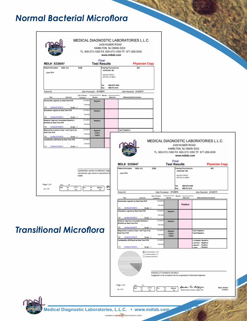
Normal Bacterial Microflora
Transitional Microflora

Medical Diagnostic Laboratories, L.L.C. • www.mdlab.com • 877.269.0090
Abnormal Bacterial Microflora:Indicative of Bacterial Vaginosis