bacterial determinants of importance in the virulence of
TRANSCRIPT

VETERINARY RESEARCHPersson and Bojesen Veterinary Research (2015) 46:57 DOI 10.1186/s13567-015-0206-z
REVIEW Open Access
Bacterial determinants of importance in thevirulence of Gallibacterium anatis in poultry
Gry Persson and Anders M Bojesen*Abstract
Gallibacterium anatis, a member of the Pasteurellaceae family, constitute a part of the normal micro-flora of theupper respiratory tract and the lower genital tract in chickens. However, increasing evidence indicate that G. anatisis also associated with a wide range of pathological changes, particularly in the reproductive organs, which leads todecreased egg production, lowered animal welfare and increased mortality. As a recently defined opportunisticpathogen limited focus has been placed on the pathogenesis and putative virulence factors permitting G. anatis tocause disease. One of the most studied virulence determinants is a large RTX-like toxin (GtxA), which has beendemonstrated to induce a strong leukotoxic effect on avian macrophages. A number of fimbria of different sizes andshapes has been described. Particularly fimbriae belonging to the F17-like family appears to be common in a diverseselection of G. anatis strains. Mutants lacking the FlfA fimbria were severely attenuated in experimentally infectedchickens. Additional characteristics including the ability to express capsular material possibly involved in serum resistance;secretion of metalloproteases capable of degrading immunoglobulins, and hemagglutinins, which may promote biofilmformation are all factors likely linked to the virulence of G. anatis. A major advantage for the study of how G. anatisinteract with its host is the ability to perform biologically relevant experimental infections where natural routes ofexposure allows reproduction of lesions observed during spontaneous infections. This review summarizes thecurrent understanding of the G. anatis pathogenesis and discusses the contribution of the established and putativevirulence factors described for this bacterium to date.
Table of contents
1. Introduction2. Pathogenesis
* CDeUn
2.1 Transmission2.2 Experimental infections
3. Virulence factors of Gallibacterium anatis3.1 The RTX-like toxin GtxA3.2 Fimbriae3.3 Outer membrane vesicles3.4 Capsule3.5 Metalloproteases3.6 Biofilm formation3.7 Haemagglutination3.8 Other potential factors involved in virulence
4. Conclusions5. Competing interests
orrespondence: [email protected] of Veterinary Disease Biology, Faculty of Health Sciences,iversity of Copenhagen, 1870 Frederiksberg C, Denmark
© 2015 Persson and Bojesen. This is an OpenLicense (http://creativecommons.org/licenses/any medium, provided the original work is pr(http://creativecommons.org/publicdomain/ze
6. Authors’ contributions7. Acknowledgements8. References
1. IntroductionGallibacterium is a genus within the Pasteurellaceaefamily [1,2] and associated with a range of avian hostspecies. The bacterium was first described in 1950 byKjos-Hansen as a hemolytic “cloaca bacterium” normallyoccurring in the cloaca of healthy chickens and cocks,but also isolated in pure cultures from numerous casesof acute salpingitis and peritonitis [3]. Since then similarbacteria, reported as Actinobacillus salpingitidis, avianPasteurella haemolytica-like organisms or Pasteurellaanatis were isolated and described from a number ofclinical cases in chickens [4–12], before Gallibacteriumwas established as an independent genus in 2003 [2].Colonies of Gallibacterium are 1–2 mm greyish, smooth,semitransparent, slightly raised and circular with an entiremargin when incubated for 24 h at 37 °C on nutrient-richplates containing blood. The genus comprises four named
Access article distributed under the terms of the Creative Commons Attributionby/4.0), which permits unrestricted use, distribution, and reproduction inoperly credited. The Creative Commons Public Domain Dedication waiverro/1.0/) applies to the data made available in this article, unless otherwise stated.

Persson and Bojesen Veterinary Research (2015) 46:57 Page 2 of 11
species; Gallibacterium anatis, Gallibacterium melopsit-taci sp. nov., Gallibacterium trehalosifermentans sp. nov.,and Gallibacterium salpingitidis sp. nov., and three geno-mospecies. G. anatis can be further sub-divided into twophenotypically distinct biovars; biovar haemolytica andthe non-hemolytic biovar anatis (Figure 1) [2]. Strains ofG. anatis biovar haemolytica and genomospecies 1 and 2form β-hemolytic zones (1–2 mm) around the colonies onagar plates with blood from calf, horse, swine, sheep,rabbit or chicken [3,10,13,14].Gallibacterium anatis is commonly isolated from chick-
ens but has also been reported from a wide range of bothdomestic and non-domestic birds, including turkeys,geese, ducks, pheasants, partridges, cage birds and wildbirds [1–4,15–17]. Gallibacterium anatis infections inhumans have only very rarely been reported and herethe bacterium merely seem to affect severely immuno-compromised individuals [18,19]. In the chicken, G.anatis is frequently found in the upper respiratory tractand lower genital tract of healthy animals. However, G.anatis has also been associated with a wide rangeof pathological lesions, especially in the reproductiveorgans of the egg-laying chicken and is considered amajor cause of salpingitis and peritonitis in chickens,leading to lowered egg-production and increased mortality[20–22]. Moreover, G. anatis is globally distributed, havingbeen isolated from poultry in countries within Europe[1,2,4,23], Africa [24], Asia [25], Australia [6] and theAmericas [7,26–29].The role of G. anatis as a cause of disease has been
debated, yet during the more recent years increasing evi-dence seems to support that this organism is a likelycause of disease and lowered animal welfare. Thecurrent report aims at summarizing past and presentknowledge within this area.
Figure 1 Biovars of Gallibacterium anatis show difference in hemolyti12656–12 and G. anatis bv. anatis strain F149. Hemolysis is seen as a clearin2 mm. greyish, smooth, semitransparent, slightly raised and circular with an econtaining blood. Strains of G. anatis biovar haemolytica and genomospeciesplates with blood from calf, horse, swine, sheep, rabbit or chicken [3,10,12,13,1
2. PathogenesisGallibacterium anatis can be persistently isolated from thetrachea and cloaca of healthy birds, showing that it con-stitutes a part of the normal microflora in the upperrespiratory tract and lower genital tract of healthy chick-ens in commercial flocks [3,4,7,17,29–31]. Although G.anatis has been associated with a wide range of differentpathological lesions, including septicemia, pericarditis,hepatitis, oophoritis, follicle degeneration, enteritis, upperrespiratory tract lesions, salpingitis and peritonitis[4–12,21,24,25,27,32], the importance of G. anatis as apathogen has remained controversial. No clinical picture isspecifically associated with G. anatis and lesions cannot bedistinguished from those caused by avian pathogenicEscherichia coli [22]. In addition, G. anatis is often isolatedtogether with E. coli [22,33], whose importance in salpin-gitis is well defined [20,34,35]. However, G. anatis has alsobeen isolated in pure culture from chickens suffering fromdifferent lesions [3,5,11,21,22,25,27,33,36], and a studyshowed that G. anatis was the most common single-bacterial infection in chickens with reproductive tract disor-ders [21], suggesting its potential as an important poultrypathogen.Based on previous pathological findings from which G.
anatis has been isolated, and recent investigations by Pau-del et al., it seems that G. anatis is able to colonize theupper respiratory tract without causing clinical signs,whereas it may cause severe lesions in the reproductivetract [29,37]. This suggests a role of G. anatis as an oppor-tunistic pathogen that given the right circumstances is ableto cause disease. Predisposing factors such as simultaneousinfection with other microorganisms [6,14,27], hormonalinfluences [5,11], age [4,10], seasonal changes [21], stress[38], low immunological status [39], and probably also hostgenetic predisposition, could explain the contradictory
c properties. Colonies of G. anatis biovar (bv.) haemolytica straing zone around the colonies of G. anatis b haemolytica. Colonies are 1–ntire margin when incubated for 24 h at 37 °C on nutrient-rich plates1 and 2 shows β-hemolytic zones (1–2 mm) around the colonies on agar4].

Persson and Bojesen Veterinary Research (2015) 46:57 Page 3 of 11
results obtained by experimental infection studies and bac-teriological findings in naturally infected chickens.While hens and pullets have been the main focus of pre-
vious pathogenesis research, a recent investigation byPaudel et al. [40] was aimed at elucidating the effect of ex-perimental infections in cockerels. Interestingly, cockerelsinfected by intra-nasal inoculation became culture-positivein the testis and epididymis within a week post infection.The infection affected the semen quality significantly by in-ducing lowered sperm density, decrease in total motilityand progressive motility, and reduced membrane integrity,thus clearly indicating a negative effect on fertility.
2.1 TransmissionThe natural mode of transmission is currently being dis-cussed. It has been widely accepted that horizontal trans-fer of Gallibacterium was the main transmission route, asthe bacterium has not been isolated from chickens youn-ger than four weeks [4,17], and that ascending infectionsfrom the cloaca appeared the most probable route to thereproductive organs [22,30]. However, one previous studyshowed trans-ovarian transfer at low level [41], and isola-tion of G. anatis from the egg yolk has also been observed[10,11,29,33], both indicating the possibility for verticaltransmission. Moreover, quantitative (q) PCR targetinggtxA allowed detection of G. anatis in samples from chick-ens as young as four days [42], suggesting that the lack ofsensitive methods for identification could have been thereason as to why G. anatis has not previously been de-tected in chickens younger than four weeks [4]. The factthat cockerels also seemed to get infected in the repro-ductive organs and semen suggests that the males poten-tially may play an important role in transmission betweenadult birds and possibly to their offspring [40]. In addition,simultaneous isolation of G. anatis from the trachea andinternal organs of chickens suffering from salpingitis andperitonitis also indicate that given the right circumstances,the bacterium could enter the systemic circulation fromits natural habitats and spread to other sites of the body[7,22,27,29,37,43]. This suggests that tissue tropism ratherthan a more mechanistic ascend through the oviduct, aspreviously anticipated, might account for the observed as-sociation of G. anatis with reproductive tract disorders.The level of biosecurity seems to be important for trans-
mission, as the bacterium is much more prevalent in pro-duction systems with low biosecurity level [30]. So,although horizontal transfer may be the main mode oftransmission within a flock, it appears plausible that verti-cal transfer may be an important link between flocks.
2.2 Experimental infectionsThe circumstances that shift G. anatis from being a nor-mal inhabitant of the mucosal surfaces to a pathogen
causing severe lesions are not known. Most early experi-mental infection studies aimed at elucidating the exactrole of G. anatis as a pathogen have not allowed firmconclusions due to varying study designs and use of dif-ferent strains. The results ranged from no obviouspathogenicity [4,17,32] to disease resembling the naturalinfection including re-isolation of bacteria from multipleorgans, reduced egg-production and increased mortality[3,8,10,11,14,29,44–46].As previously discussed, it is possible that the presence
of predisposing factors, such as impaired immunity, isnecessary for establishment of a consistent clinical pic-ture of experimental infections. The opportunistic po-tential of G. anatis was shown by Bojesen et al., whodemonstrated that experimental infection of 15-week oldheterophil-depleted chickens resulted in lesions similarto those induced during natural infections from whichG. anatis had been isolated, whereas non-depleted chick-ens showed less pronounced lesions [39].Other important factors that probably play a signifi-
cant role for the contradictory results are the differencein virulence between strains and the uncertain identifica-tion and classification of the organism in the early stud-ies [4,5,37,43,47]. The pronounced genetic diversity maythus be reflected in differences in the possession and ex-pression of different virulence factors [48,49].An alternative explanation to the different results
could also lie in the applied in vivo infection model. Itseems obvious that depending on the aim of the investi-gations, it is crucial to use the most biologically relevantmodel. Gallibacterium anatis is an inhabitant of theupper respiratory tract and lower reproductive tract mu-cosa in the chicken. Thus intramuscular, sub-cutaneousor intravenous injections of Gallibacterium might notgive a realistic picture of the natural course of the infec-tion. Recent studies using intra-nasal instillation of spe-cific pathogen free (SPF) chickens have shown that thebacteria were able to spread to internal organs, mostparticular colonizing organs of the reproductive tract,resulting in lesions similar to natural infections [29,37].Intra-nasal instillation could be a good model for study-ing colonization of the airway [37,43]. However, by usingintra-nasal instillation it might be difficult to quantifythe exact number of bacteria that actually enter the host.This also appears to be reflected in the varying degree ofre-isolation of G. anatis, even with the same strain [37],which makes it difficult to evaluate and compare the exactvirulence of different strains of G. anatis. Moreover, as G.anatis is considered an opportunistic bacterium, the simul-taneous presence of other bacterial species could have anunidentified impact on the pathogenesis of Gallibacterium.Vazquez et al. showed that experimental infection with G.anatis had a more severe effect on egg production in com-mercial chickens than SPF chickens [46].

Persson and Bojesen Veterinary Research (2015) 46:57 Page 4 of 11
Several studies point at G. anatis being particularly in-volved in infections of the reproductive tract and periton-eum [3,46] and this view has been supported by Mirleet al. who showed that G. anatis was among the mostcommon bacterial agents isolated from lesions in the re-productive organs [21] during natural infections. There-fore, to study the pathogenic nature of the bacteriumwithin these organs it would be feasible to use a modelspecifically targeting these organs. Intra-peritoneal injec-tion bypasses some of the early stages of the immune re-sponse and has been used in numerous cases to study thevirulence of Gallibacterium [3,4,10,11,14,32,39]. Thismodel may imitate a natural infection of the peritoneum,while one of the draw-backs is that the bacteria quicklyenters systemic circulation and thus might affect multipleorgans not related to the natural course of infection, giv-ing an inaccurate and complicated clinical picture thatmight make it more difficult to control the course of dis-ease. Recently, a new animal model has enabled experi-mental infections of the oviduct, in which an avianpathogenic strain of E. coli are injected into the lumen ofthe oviduct by a simple surgical procedure [50]. Further-more, the controlled deposition of the inoculum makes iseasier to control and study the infection. The inoculationwith E. coli resulted in peritonitis, salpingitis, oophoritisand necrotic hepatitis – all in line with the clinical pictureseen during the natural course of infection with this strain[50]. Thus, this in vivo model might resemble the naturalinfection of the reproductive tract more than the intra-peritoneal infection model.Using an appropriate infection model to study viru-
lence factors and host-pathogen interaction could leadus to a deeper understanding of the pathogenic natureof G. anatis, and thus aid in the development of newprevention and treatment strategies.
3. Virulence factors of Gallibacterium anatisVirulence factors are defined as components of an or-ganism that give it the ability to cause disease, and thusdetermine the pathogenicity of the organisms, but aredispensable for its viability. Virulence factors are in-volved in many aspects of the host-pathogen interface,including colonization, nutrient acquisition, immune-evasion and immunosuppression, and include toxins, en-zymes and adhesion molecules. Limited knowledge hasbeen obtained about the mechanisms involved in thepathogenesis of G. anatis and few virulence factors havebeen characterized in depth.
3.1 The RTX-like toxin GtxAOne of the main characteristics used for identification ofG. anatis biovar haemolytica is its ability on blood-agarplates to form a broad β-hemolytic zone around the col-ony. The protein responsible is the secreted toxin named
GtxA (Gallibacterium toxin A), which is the most welldescribed virulence factor of G. anatis [13,48]. The GtxAprotein expressed by G. anatis possesses hemolytic activ-ity against erythrocytes from a wide variety of hosts, andleukotoxic activity against the chicken macrophage cellline HD11 [13].RTX-toxins are pore forming exoproteins that are se-
creted via a type I secretion system (T1SS) in Gram-negative bacteria. Their name originates from a region,generally found in the carboxyl terminal of the protein,which contains a series of Ca2+ binding glycine-rich nona-peptide repeats (repeats in the structural toxin) [51,52].RTX toxins are expressed by many members of Pasteurel-laceae where they are responsible for the hemolytic andalso the leukotoxic phenotype of these bacteria [51]. TheRTX toxins are usually transcribed from a four-gene op-eron, comprising the genes rtxC, rtxA, rtxB and rtxD intranscribed order, respectively. The gene rtxC encodes anactivation protein that acetylates the toxin, encoded byrtxA. The genes rtxB and rtxD encode translocator pro-teins that together with the outer membrane proteinnamed TolC comprise the T1SS for the functional RTXtoxin. The homologue toxin, HlyA, from E. coli is oftenused as the model for describing the typical RTX toxin, al-though variation across species does exist. A schematicpresentation of HlyA and GtxA domain organization isshown in Figure 2. GtxA is unusually large; with its 2038amino acids it is almost twice the size of HlyA. It consistsof two domains: a C-terminal domain with homology toother RTX toxins responsible for the hemolytic function;and a N-terminal domain of approximately 950 aminoacids with unknown function and no obvious homologs,but which is required for full hemolytic activity and theleukotoxic activity of the toxin (Figure 2) [13]. A similarstructure is seen for the recently identified 238 kDa RTX-like toxin named AvxA from Avibacterium paragalli-narum, in which only the C-terminal part of the proteinshow homology to RTX toxins, while the N-terminal func-tion as a serine protease [53]. Moreover, at the geneticlevel an atypical organization for gtxA is seen, while gtxAand gtxC is located together, the genes gtxB, −D and theE. coli tolC homologue gtxE are located elsewhere in thegenome [13].The role of GtxA in the pathogenesis is not fully
understood, although a gtxA knockout mutant is clearlyattenuated in virulence (Pors, S., unpublished data). Thehemolytic effect seems unspecific as hemolysis is observedagainst a wide range of blood cells from different animalspecies [3,23,54], indicating that lysis of erythrocytesmight not be the main target for GtxA. The N-terminuscontains a domain with weak homology to Talin, a proteininvolved in the linkage of the cytoplasmic portion of integ-rins to the actin cytoskeleton by interactions with vinculinand alpha-actinin [55,56]. Actin plays many physiologically

Figure 2 GtxA is a novel RTX-like protein with an unusual domain organization. Schematic presentation of the domain organization ofGtxA from G. anatis biovar haemolytica strain 12656–12 [RefSeq:WP_013746567] compared to the typical domain organization of RTX toxinsrepresented by HlyA from E. coli [PRF:225074]. HlyA has 34% coverage and 29% identity with GtxA. Regions of homology are shown with dotted lines.The GtxA toxin is comprised of 2038 amino acids (aa). In the N-terminal part of GtxA (approximately aa 1 – 950), two overlapping regions with weakhomology to a membrane protein with unknown function (COG1511) is present. The remaining C-terminal part of GtxA has homology to HlyAby having a RTX N-terminal domain (pfam02382), the Ca2+-binding repeats (COG2931) and a domain classified as peptidase M10 serralysin C terminal(pfam08548). GtxA also contains the lysine residues, Lys1484 and Lys1607, required for activation of the toxin by acetylation by GtxC. The homologuelysine residues in HlyA are found at Lys564 and Lys690. In addition to the described domains, HlyA also contains a RTX C-terminal domain (pfam08339).Domains were identified using NCBI conserved domain search with default settings.
Persson and Bojesen Veterinary Research (2015) 46:57 Page 5 of 11
important roles in the cell, some of which also involve theregulation of immune cell recognition and adherence, pro-duction and release of immune cell signaling moleculesand phagocytosis. It is therefore not surprising that severalclasses of bacterial toxins target the actin cytoskeleton ofthe host cells as an immune evasion strategy [57]. Somehigh molecular weight toxins belonging to the RTX family,called MARTX (multifunctional autoprocessing RTX),have been show to bind to and modulate actin [57]. Itcould therefore be speculated that GtxA represents a novelform of RTX-like toxin, with a proposed function in im-mune evasion.
3.2 FimbriaeAn important aspect of colonization is the ability to ad-here to and invade host tissue. It has been shown that G.anatis has the ability to adhere to chicken epithelial cells[58] and inert surfaces, and short fimbria-like structureson the bacteria have been observed [59]. Fimbriae arehair-like structures expanding from the surface of thebacteria, and are often involved in adhesion to host cells[60]. Recently, several F-17 like fimbriae clusters wereidentified in the genomes of three different G. anatisstrains [49]. F-17 like fimbriae belong to a group of fim-briae that bind N-acetyl-D-glucosamine (Glc-NAc) con-taining receptors on the surface of host cells, and isthus, thought to be involved in adhesion of the bacteriato the mucosal surfaces within the host [61]. This typeof fimbria is expressed by several pathogenic types ofEscherichia coli, including strains of avian pathogenic E.coli (APEC) [61,62]. In G. anatis the F17-like fimbria isencoded by a four-gene cluster comprising a gene en-coding a chaperone, an usher protein, an adhesion pro-tein and a structural subunit protein designated as flfD,flfC, flfG and flfA, respectively [63]. The chaperone and
the usher protein facilitate folding, assembly and secre-tion of the structural subunit protein, which make upthe stem of the fimbriae. The adhesin is located at thetip of the fimbrial structure and is responsible for recep-tor recognition and binding [61,64]. A recent compre-hensive study of the fimbriae genes in the genome of 22G. anatis strains showed that F17-like fimbriae are verycommon with strains encoding between 1–3 differentfimbrial clusters [65]. Based on the structural genes, theseclusters could be divided into five phylogenetic types (Flf,Flf1-4), with the cluster designated Flf1 being the mostfrequently occurring fimbrial cluster present in 74% of thegenomes investigated. Interestingly, none of the strainsencoding Flf1 appeared to express the structural protein,FlfA1, in vitro. In contrast, the Flf cluster, encoding thepreviously described 20.5 kDa fimbrial subunit proteinFlfA (Figure 3) [63], was encoded in 65% of the strainsand expressed in vitro by 79% of these strains [65].The genetic variation was greatest in the genes encod-
ing the structural proteins and the adhesin. Polyclonalantibodies raised against the three most common typesof the structural subunit proteins did not cross-reactwith the other Flf-types [65]. This indicates that thestructural subunit and the adhesin are under selectivepressure for immune recognition, and thereby could in-dicate the importance of fimbriae during the pathogen-esis of G. anatis. This has been supported by Bageret al., who showed that the FlfA fimbria is important forvirulence in vivo, as a knockout mutant of flfA was at-tenuated. From this it was also suggested that fimbrialexpression may govern the tissue tropism observed forG. anatis [22,63]. The presence of several fimbrial clus-ters could be a result of an immunogenic pressure favor-ing duplication events and increased affinity for differenttargets in the host tissue, or could reflect a functional

Figure 3 The F17-like fimbria, FlfA, is exposed on the surface ofG. anatis. FlfA on the surface of G. anatis strain 12656–12 is shown byimmunogold electron microscopy using anti-FlfA serum, followed by asecondary antibody conjugated to 10-nm gold particles. Picturemodified from Copyright © American Society for Microbiology, [63].
Figure 4 Outer membrane vesicles (OMVs) are naturallysecreted from G. anatis. OMVs are seen as spherical structures insizes ranging from 20 nm and up to 160 nm. The OMVs were isolatedfrom G. anatis strain 12656–12 and viewed by transmission electronmicroscopy with 2% (w/v) phosphotungstic acid staining [69].
Persson and Bojesen Veterinary Research (2015) 46:57 Page 6 of 11
diversity, where different fimbria are expressed at differ-ent time points during infection. The exact role, targetcells and regulation of the identified fimbriae in G. ana-tis still need to be determined.
3.3 Outer membrane vesiclesOuter membrane vesicles (OMV) are spherical, bilayeredmembrane structures, which have been associated withan enormous functional diversity. OMVs are released byvirtually all Gram-negative bacteria where they are pro-duced by budding of the outer membrane and thereforemainly consist of outer membrane components, such asmembrane associated proteins and LPS, although, theyhave also been shown to contain periplasmic compo-nents and even compounds of cytoplasmic origin suchas DNA [66–68]. Recently it was demonstrated that G.anatis produce OMVs (Figure 4) with a protein contentthat varied depending on growth condition [69], suggest-ing that OMV production could serve multiple functionsand be a way to cope with changing environments, e.g.within the host. To study the formation of OMVs, anOMV-overproducing mutant of G. anatis was created byknocking out the gene tolR, encoding a protein within theTol-Pal system, which is crucial for membrane stabilityand integrity [69,70]. The mutant released considerably
higher amounts of OMVs, supporting that membrane sta-bility is essential for OMV formation [69].The functions of Gallibacterium OMVs have not been
determined. OMVs could act as a means to get rid ofmisfolded or excess proteins or enable replacement oflipids in the outer membrane during growth. However,the presence of specific molecules, e.g. proteins, lipids orpolysaccharides, under different circumstances indicatesan active role of the OMVs. In other species OMVs havebeen ascribed importance for biofilm formation [71] andfor carrying quorum sensing molecules involved in cell-to-cell communication [72]. Microvesicles produced byG. anatis adhering to glass has previously been observed[59]. However, increased release of OMVs by the tolRknockout mutant does not seem to have any effect onthe level of biofilm formation (unpublished data). TheΔtolR mutant OMVs are much more uniform and lessaffected by environmental changes compared to wild-type OMVs [69]. This could be due to the more artificialnature of the mutant OMVs, which may lead to a lackof specific molecules within the vesicles, e.g. factorsrequired for biofilm formation. OMVs have also beenshown to act as transportation vehicles for the deliveryof lipids, membrane proteins, insoluble compounds orcompounds that are easily degraded, including toxins andDNA. The transport of proteases and hemolysins byOMVs has been shown for Actinobacillus pleuropneumo-niae and Avibacterium paragallinarum [73,74]. However,vesicles from G. anatis do not seem to be involved in ei-ther hemolysis or proteolysis (unpublished data). A fewproteins have been identified as being a part of the OMVs,including a possible hemagglutinin with sequence similar-ity to the filamentous hemagglutinin protein precursor

Figure 5 Capsular material is present on the surface of G.anatis. A thin capsular structure can be seen on the surface of G. anatisstrain 12656–12 using uranyl acetate staining followed bytransmission electron microscopy [80].
Persson and Bojesen Veterinary Research (2015) 46:57 Page 7 of 11
FhaB from Bordetella pertussis, which has been found im-portant for colonization of the host mucosa [69,75]. An-other OMV associated protein in G. anatis is MDN1, anAAA ATPase containing a vWA domain, which in otherspecies has been suggested to be important for the bacter-ial stress response, cell adhesion and/or biofilm formation[69,76]. From this, it could be speculated that OMVs fromG. anatis could act as a possible virulence factor import-ant for adherence and colonization of the host. Other pos-sible functions of OMVs include modulating the hostimmune response, acting as target for phages or being in-volved in the binding and removal of anti-bacterial sub-stances including different antibiotics, all aiding in thesurvival of the microorganism [67,68]. Despite the manyspeculations, the role of G. anatis OMVs has not been de-termined. As they seem affected by the surroundings, it islikely that additional host factors, such as those found inserum, might play a role in regulation and function of theOMVs. The amount of OMVs produced as well as theprotein profile dramatically changes when G. anatis is in-cubated in the presence of serum (unpublished data), sup-porting the hypothesis that the OMVs might play a role inthe bacterium’s interaction with its natural environment,the chicken host.
3.4 CapsuleBacterial capsules are composed of extracellular polysac-charide, and are found in a diverse array of both Gram-negative and Gram-positive pathogens [77]. Functionalimportance has been documented in relation to adhe-sion, cell-cell interactions and immune evasion [78,79].The presence of a thin capsule on G. anatis has been ob-served by transmission electron microscopy (Figure 5)[80] and Kjos-Hansen observed the presence of a cap-sule in primary culture, which however, disappearedafter sub-cultivation. Moreover, the capsule was notpresent in isolates from healthy chickens [3]. Analysis ofthe genomes of three G. anatis strains revealed a capsu-lar locus [49] in one of the strains. The function of thecapsule of Gallibacterium is not known. Interestingly, acapsule-knockout mutant (ΔgexD) proved to be morevirulent than its wild-type counterpart [80]. Althoughfurther studies are needed to elucidate this, it could bespeculated that removal of the capsule led to the expos-ure of some normally hidden antigens or pathogen-associated molecular patterns (PAMPs), resulting in anincreased immune response against G. anatis.
3.5 MetalloproteasesProteases are enzymes that catalyze the hydrolysis ofpeptide bonds in proteins or peptides. These enzymesseem to catalyze many essential functions of pathogenicbacteria. Metalloproteases are one class of proteases thatplay many important functions in virulence, including
colonization, nutrient acquisition, immune evasion, andbacterial invasion into the systemic circulation [81].Modulation of the host immune response can be facili-tated by e.g. acting on serum components, such as im-munoglobulins and proteins of the complement system[81]. G. anatis expresses metalloproteases capable of de-grading avian immunoglobulin IgG [82] suggesting apossible role in immune evasion. Although, the geneticsbehind this function have not been determined, severalmetalloproteases are encoded within the genome of G.anatis, including an extracellular protein with a metal-dependent endonuclease domain, a zinc metalloproteaseand an ATP-dependent metalloprotease [49]. One ormore of these proteins could be responsible for the pro-teolytic capability of G. anatis. The specificity or role inpathogenesis of the IgG-degrading metalloproteases isnot known, but it has been speculated that this proteincould be responsible for the host-specific pathogenicityobserved for some strains [43].
3.6 Biofilm formationBacterial biofilms are structured cell communities em-bedded in a polymeric matrix that allows adherence tosurfaces and live tissue. Clinically, biofilm formation is
Persson and Bojesen Veterinary Research (2015) 46:57 Page 8 of 11
associated with persistent and chronic infections and in-creased resistance to antimicrobials [83,84]. It has beenshown that G. anatis is capable of binding to inert sur-faces, which is regarded as a first step towards biofilm for-mation [59]. The ability to form biofilm varies betweenisolates, and Johnson et al. was able to divide strains of G.anatis into groups of weak, moderate and strong biofilmproducers [49]. Although no clear correlation betweenformation of biofilm and pathogenesis could be noted[59], this grouping of strains according to the level of bio-film formed, revealed a pattern corresponding to the ap-parent evolutionary decent of the strains [49]. This couldindicate that biofilm formation might play an importantrole for certain clades within G. anatis.
3.7 HemagglutinationSome strains of G. anatis have been found capable of ag-glutinating avian erythrocytes [85,86]. Hemagglutinationis linked with the expression of hemagglutinins or adhe-sins capable of binding receptors on the surface of redblood cells. Genome analysis revealed the presence of anumber of putative hemagglutinins, and the presence ofa potential hemagglutinin in OMVs released from G.anatis has been observed [49,69]. Some of these hemag-glutinins could be responsible for the observed agglutin-ating activity seen for some strains [49,69,87]. The roleand importance of these proposed hemagglutinins hasnot been determined, yet hemagglutinating activity havebeen shown to be important for other poultry patho-gens, such as Avibacterium paragallinarum [88,89].
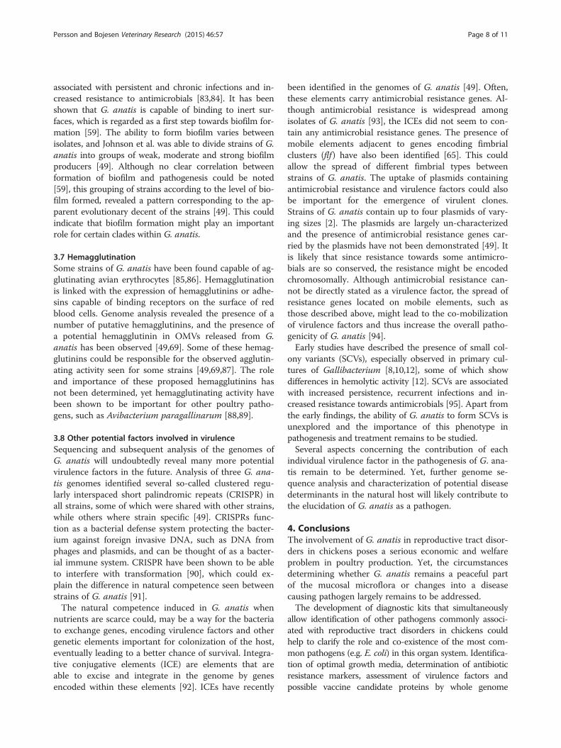
3.8 Other potential factors involved in virulenceSequencing and subsequent analysis of the genomes ofG. anatis will undoubtedly reveal many more potentialvirulence factors in the future. Analysis of three G. ana-tis genomes identified several so-called clustered regu-larly interspaced short palindromic repeats (CRISPR) inall strains, some of which were shared with other strains,while others where strain specific [49]. CRISPRs func-tion as a bacterial defense system protecting the bacter-ium against foreign invasive DNA, such as DNA fromphages and plasmids, and can be thought of as a bacter-ial immune system. CRISPR have been shown to be ableto interfere with transformation [90], which could ex-plain the difference in natural competence seen betweenstrains of G. anatis [91].The natural competence induced in G. anatis when
nutrients are scarce could, may be a way for the bacteriato exchange genes, encoding virulence factors and othergenetic elements important for colonization of the host,eventually leading to a better chance of survival. Integra-tive conjugative elements (ICE) are elements that areable to excise and integrate in the genome by genesencoded within these elements [92]. ICEs have recently
been identified in the genomes of G. anatis [49]. Often,these elements carry antimicrobial resistance genes. Al-though antimicrobial resistance is widespread amongisolates of G. anatis [93], the ICEs did not seem to con-tain any antimicrobial resistance genes. The presence ofmobile elements adjacent to genes encoding fimbrialclusters (flf ) have also been identified [65]. This couldallow the spread of different fimbrial types betweenstrains of G. anatis. The uptake of plasmids containingantimicrobial resistance and virulence factors could alsobe important for the emergence of virulent clones.Strains of G. anatis contain up to four plasmids of vary-ing sizes [2]. The plasmids are largely un-characterizedand the presence of antimicrobial resistance genes car-ried by the plasmids have not been demonstrated [49]. Itis likely that since resistance towards some antimicro-bials are so conserved, the resistance might be encodedchromosomally. Although antimicrobial resistance can-not be directly stated as a virulence factor, the spread ofresistance genes located on mobile elements, such asthose described above, might lead to the co-mobilizationof virulence factors and thus increase the overall patho-genicity of G. anatis [94].Early studies have described the presence of small col-
ony variants (SCVs), especially observed in primary cul-tures of Gallibacterium [8,10,12], some of which showdifferences in hemolytic activity [12]. SCVs are associatedwith increased persistence, recurrent infections and in-creased resistance towards antimicrobials [95]. Apart fromthe early findings, the ability of G. anatis to form SCVs isunexplored and the importance of this phenotype inpathogenesis and treatment remains to be studied.Several aspects concerning the contribution of each
individual virulence factor in the pathogenesis of G. ana-tis remain to be determined. Yet, further genome se-quence analysis and characterization of potential diseasedeterminants in the natural host will likely contribute tothe elucidation of G. anatis as a pathogen.
4. ConclusionsThe involvement of G. anatis in reproductive tract disor-ders in chickens poses a serious economic and welfareproblem in poultry production. Yet, the circumstancesdetermining whether G. anatis remains a peaceful partof the mucosal microflora or changes into a diseasecausing pathogen largely remains to be addressed.The development of diagnostic kits that simultaneously
allow identification of other pathogens commonly associ-ated with reproductive tract disorders in chickens couldhelp to clarify the role and co-existence of the most com-mon pathogens (e.g. E. coli) in this organ system. Identifica-tion of optimal growth media, determination of antibioticresistance markers, assessment of virulence factors andpossible vaccine candidate proteins by whole genome

Persson and Bojesen Veterinary Research (2015) 46:57 Page 9 of 11
sequence analysis would further aid in the process of devel-oping new treatment or prevention strategies.A deeper insight into the basic biology of G. anatis is
also likely to improve the understanding of this organ-ism. Many of the early papers indicate that the bacteriagrow best under micro-aerophillic conditions, and havereported different size variants with different morph-ology and biochemical properties [3,12]. A standardizedcultivation scheme does not necessarily take this into ac-count, and the repeated sub-cultivation of laboratorystrains might lead to the loss of expression of virulencefactors initially important for the strains when originallyisolated from diseased chickens. One evident example ofthis is the loss of capsule when sub-cultivating primaryisolates [3]. In order to be able to understand the exacthost-pathogen interplay, and the switch from a normalinhabitant of the healthy chicken to a pathogen causingdisease and mortality, further insight into the expressionof virulence factors under more biologically relevant cir-cumstances is desirable.
5. Competing interestsThe authors declare that they have no competing interests.
6. Authors’ contributionsGP wrote and drafted the manuscript. AMB participated in the design andhelped draft the manuscript. Both authors read and approved the final manuscript.
7. AcknowledgementsGP is supported by a PhD stipend from the Faculty of Health and MedicalSciences, Copenhagen University.
Received: 9 January 2015 Accepted: 11 May 2015
8. References1. Bisgaard M, Korczak BM, Busse HJ, Kuhnert P, Bojesen AM, Christensen H (2009)
Classification of the taxon 2 and taxon 3 complex of Bisgaard within Gallibacteriumand description of Gallibacterium melopsittaci sp. nov., Gallibacteriumtrehalosifermentans sp. nov. and Gallibacterium salpingitidis sp. nov. Int JSyst Evol Microbiol 59:735–744
2. Christensen H, Bisgaard M, Bojesen AM, Mutters R, Olsen JE (2003) Geneticrelationships among avian isolates classified as Pasteurella haemolytica,'Actinobacillus salpingitidis' or Pasteurella anatis with proposal of Gallibacteriumanatis gen. nov., comb. nov. and description of additional genomospecies withinGallibacterium gen. nov. Int J Syst Evol Microbiol 53:275–287
3. Kjos-Hansen B (1950) Egglederperitonitt forårsaket av patogen "kloakkbakterie"hos høns. Nord Vet Med 2:523–531
4. Bisgaard M (1977) Incidence of Pasteurella haemolytica in the respiratorytract of apparently healthy chickens and chickens with infectious bronchitis.Characterisation of 213 strains. Avian Pathol 6:285–292
5. Gerlach H (1977) Die Bedeutung von Pasteurella haemolytica in Hühnerbeständen.Prakt Tierarzt 5:324–325
6. Gilchrist P (1963) A survey of avian respiratory diseases. Aust Vet J 39:140–1447. Hacking WC, Pettit JR (1974) Pasteurella hemolytica in pullets and laying
hens. Avian Dis 18:483–4868. Harbourne J (1962) A haemolytic cocco-bacillus recovered from poultry. Vet
Rec 74:566–5679. Hinz K (1970) Bakteriologische Befunde bei Erkrankung der Atmungsorgane
von Junghennen. Congress of the World Veterinary Poultry Association10. Janetschke P, Risk G (1970) Über gehäuftes Auftreten von Pasteurella hämolytica
beim Haushuhn in Syrien. Monatsh Veterinarmed 25:23–27 (in German)11. Kohlert R (1968) Untersuchungen zur ätiologie der Eileiterentzündung beim
Huhn. Monatsh Veterinarmed 23:392–395 (in German)
12. Greenham L, Hill T (1962) Observations on an avian strain of Pasteurellahaemolytica. Vet Rec 74:861–863
13. Kristensen BM, Frees D, Bojesen AM (2010) GtxA from Gallibacterium anatis,a cytolytic RTX-toxin with a novel domain organisation. Vet Res 41:25
14. Matthes S, Löliger H, Schubert H (1969) Enzootic in chicken due topasteurella haemolytica. Dtsch Tieraerztl Wochenschr 76:88 (in German)
15. Bisgaard M (1993) Ecology and significance of Pasteurellaceae in animals.Zentbl Bakteriol 279:7–26
16. Gregersen RH, Neubauer C, Christensen H, Korczak B, Bojesen AM, Hess M,Bisgaard M (2010) Characterization of Pasteurellaceae-like bacteria isolatedfrom clinically affected psittacine birds. J Appl Microbiol 108:1235–1243
17. Mushin R, Weisman Y, Singer N (1980) Pasteurella haemolytica found in therespiratory tract of fowl. Avian Dis 162–168
18. Aubin GG, Haloun A, Treilhaud M, Reynaud A, Corvec S (2013)Gallibacterium anatis Bacteremia in a Human. J Clin Microbiol 51:3897–3899
19. Gautier A-L, Dubois D, Escande F, Avril J-L, Trieu-Cuot P, Gaillot O (2005)Rapid and accurate identification of human isolates of Pasteurella and relatedspecies by sequencing the sodA gene. J Clin Microbiol 43:2307–2314
20. Jordan F, Williams N, Wattret A, Jones T (2005) Observations on salpingitis,peritonitis and salpingoperitonitis in a layer breeder flock. Vet Rec 157:573–577
21. Mirle C, Schöngarth M, Meinhart H, Olm U (1991) Untersuchungen zu Auftretenund Bedeutung von Pasteurella haemolytica-Infektionen bei Hennen unterbesonderer Berücksichtigung von Erkrankungen des Legesapparates. MonatshVeterinarmed 46:545–549 (in German)
22. Neubauer C, De Souza-Pilz M, Bojesen AM, Bisgaard M, Hess M (2009) Tissuedistribution of haemolytic Gallibacterium anatis isolates in laying birds withreproductive disorders. Avian Pathol 38:1–7
23. Mráz O, Vladík P, Bohácek J (1976) Actinobacilli in domestic fowl. ZentralblBakteriol Orig A 236:294–307
24. Addo P, Mohan K (1985) Atypical Pasteurella haemolytica type A frompoultry. Avian Dis 29:214–217
25. Suzuki T, Ikeda A, Shimada J, Yanagawa Y, Nakazawa M, Sawada T (1996)Isolation of "Actinobacillus salpingitidis"/avian Pasteurella haemolytica-likeorganisms group from diseased chickens. J Japan Vet Med Assoc 49:800–809
26. Mendoza K, Zavaleta AI, Koga Y, Rodríguez J, Alvarado A, Tinico R (2014)Variabilidad genética de cepas de Gallibacterium anatis aisladas de avescomerciales del Perú con infecciones respiratorias. Rev Investig Vet Peru25:233–244 (in Spanish)
27. Shaw DP, Cook DB, Maheswaran SK, Lindeman CJ, Halvorson DA (1990)Pasteurella haemolytica as a co-pathogen in pullets and laying hens. AvianDis 34:1005–1008
28. Bojesen AM, Vazquez ME, Robles F, Gonzalez C, Soriano EV, Olsen JE, ChristensenH (2007) Specific identification of Gallibacterium by a PCR using primerstargeting the 16S rRNA and 23S rRNA genes. Vet Microbiol 123:262–268
29. Paudel S, Liebhart D, Hess M, Hess C (2014) Pathogenesis of Gallibacteriumanatis in a natural infection model fulfils Koch’s postulates: 1. folliculitis anddrop in egg production are the predominant effects in specific pathogenfree layers. Avian Pathol 43:443–449
30. Bojesen AM, Nielsen SS, Bisgaard M (2003) Prevalence and transmission ofhaemolytic Gallibacterium species in chicken production systems withdifferent biosecurity levels. Avian Pathol 32:503–510
31. Harry E (1962) A haemolytic coccobacillus recovered from poultry. Vet Rec74:640
32. Majid MS, Ideris A, Aziz AR (1986) Isolation of Pasteurella haemolytica fromthe spleen of chickens. Pertanika 9:265–266
33. Shapiro J, Brash M, Martin E, Brooks A, Slavic D, McEwen B (2013) Gallibacteriumanatis— A review of culture-positive cases from commercial poultry submittedto the AHL in 2011 and 2012. AHL Newsletter Guelph, Ontario: Animal HealthServices, Laboratory Services Division, University of Guelph 17:6.
34. Barnes HJ, Nolan LK, Vaillancourt J-P (2008) Colibacillosis. In: Saif YM, FadlyAM, Glisson JR, McDougald LR, Nolan LK, Swaney DL (eds) Diseases ofPoultry, 12th edn. Blackwell Publishing, Iowa, USA
35. Bisgaard M, Dam A (1981) Salpingitis in poultry. II. Prevalence, bacteriology,and possible pathogenesis in egg-laying chickens. Nord Vet Med 33:81–89
36. Lin M, Lin K, Lan Y, Liaw M, Tung M (2001) Pathogenicity and drugsusceptibility of the Pasteurella anatis isolated in chickens in Taiwan. AvianDis 45:655–658
37. Paudel S, Alispahic M, Liebhart D, Hess M, Hess C (2013) Assessing pathogenicityof Gallibacterium anatis in a natural infection model: the respiratory andreproductive tracts of chickens are targets for bacterial colonization. AvianPathol 42:527–535

Persson and Bojesen Veterinary Research (2015) 46:57 Page 10 of 11
38. Rzewuska M, Szeleszczuk P, Binek M (2007) Isolation of Gallibacterium spp.from peacocks with respiratory tract infections. Med Weter63:1431–1433
39. Bojesen AM, Nielsen OL, Christensen JP, Bisgaard M (2004) In vivo studies ofGallibacterium anatis infection in chickens. Avian Pathol 33:145–152
40. Paudel S, Liebhart D, Aurich C, Hess M, Hess C (2014) Pathogenesis ofGallibacterium anatis in a natural infection model fulfils Koch’s postulates: 2.Epididymitis and decreased semen quality are the predominant effects inspecific pathogen free cockerels. Avian Pathol 43:529–534
41. Matthes S, Hanschke J (1977) Experimentelle Untersuchungen zür Übertragungvon Bakterien über das Hühnerei. Berl Muench Tieraerztl Wochenschr90:200–203 (in German)
42. Huangfu H, Zhao J, Yang X, Chen L, Chang H, Wang X, Li Q, Yao H, Wang C(2012) Development and preliminary application of a quantitative PCR assayfor detecting gtxA-containing Gallibacterium species in chickens. Avian Dis56:315–320
43. Zepeda V, Calderón-Apodaca N, Paasch M, Martín P, Paredes D, Ramírez-Apolinar S, Soriano-Vargas E (2010) Histopathologic findings in chickensexperimentally infected with Gallibacterium anatis by nasal instillation.Avian Dis 54:1306–1309
44. Suzuki T, Ikeda A, Shimada J, Sawada T, Nakazawa M (1997) Pathogenicity ofan Actinobacillus salpingitidis/avian Pasteurella haemolytica-like isolate fromlayer hens that died suddenly. J Jpn Vet Med Assoc 50:85–88 (in Japanese)
45. Suzuki T, Ikeda A, Taniguchi T, Nakazawa M, Sawada T (1997) Pathogenecityof an "Actinobacillus salpingitidis" / avian Pasteurella haemolytica-likeorganism for laying hens. J Jpn Vet Med Assoc 50:381–385 (in Japanese)
46. Vazquez ME, de la Mora R, Gonzalez-Hernandez C (2005) Challenge effect ofGallibacterium anatis on laying hens. In: Frame DD (eds) Proceedings of the54th Western Poultry Disease Conference, Vancouver, B.C., Canada,April 25–27. pp 73
47. Bojesen AM, Torpdahl M, Christensen H, Olsen JE, Bisgaard M (2003) Geneticdiversity of Gallibacterium anatis isolates from different chicken flocks. J ClinMicrobiol 41:2737–2740
48. Kristensen BM, Frees D, Bojesen AM (2011) Expression and secretion of theRTX-toxin GtxA among members of the genus Gallibacterium. Vet Microbiol153:116–123
49. Johnson TJ, Danzeisen JL, Trampel D, Nolan LK, Seemann T, Bager RJ, BojesenAM (2013) Genome analysis and phylogenetic relatedness of Gallibacteriumanatis strains from poultry. PLoS One 8:e54844
50. Pors SE, Olsen RH, Christensen JP (2014) Variations in virulence of avianpathogenic Escherichia coli demonstrated by the use of a new in vivoinfection model. Vet Microbiol 170:368–374
51. Frey J, Kuhnert P (2002) RTX toxins in Pasteurellaceae. Int J Med Microbiol292:149–158
52. Strathdee CA, Lo R (1989) Cloning, nucleotide sequence, andcharacterization of genes encoding the secretion function of the Pasteurellahaemolytica leukotoxin determinant. J Bacteriol 171:916–928
53. Küng E, Frey J (2013) AvxA, a composite serine-protease-RTX toxin of Avibacteriumparagallinarum. Vet Microbiol 163:290–298
54. Bisgaard M (1982) Isolation and characterization of some previously unreportedtaxa from poultry with phenotypical characters related to Actinobacillus-anPasteurella species. Acta Pathol Microbiol Immunol Scand B 90:59–67
55. Calderwood DA, Campbell ID, Critchley DR (2013) Talins and kindlins:partners in integrin-mediated adhesion. Nat Rev Mol Cell Biol14:503–517
56. Das M, Subbayya Ithychanda S, Qin J, Plow EF (2014) Mechanisms oftalin-dependent integrin signaling and crosstalk. Biochim Biophys Acta1838:579–588
57. Aktories K, Lang AE, Schwan C, Mannherz HG (2011) Actin as target formodification by bacterial protein toxins. FEBS J 278:4526–4543
58. Lucio MLS, Vaca S, Vázquez C, Zenteno E, Rea I, Pérez-Márquez VM, Negrete-Abascal E (2012) Adhesion of Gallibacterium anatis to chicken oropharyngealepithelial cells and the identification of putative fimbriae. Adv Microbiol2:505–510
59. Vaca S, Monroy E, Rojas L, Vazquez C, Sanchez P, Soriano-Vargas E, BojesenAM, Abascal EN (2011) Adherence of Gallibacterium anatis to inert surfaces.J Anim Vet Adv 10:1688–1693
60. Klemm P, Schembri MA (2000) Bacterial adhesins: function and structure. IntJ Med Microbiol 290:27–35
61. Le Bouguénec C, Bertin Y (1999) AFA and F17 adhesins produced bypathogenic Escherichia coli strains in domestic animals. Vet Res 30:318–342
62. Stordeur P, Marlier D, Blanco J, Oswald E, Biet F, Dho-Moulin M, Mainil J(2002) Examination of Escherichia coli from poultry for selected adhesingenes important in disease caused by mammalian pathogenic E. coli. VetMicrobiol 84:231–241
63. Bager RJ, Nesta B, Pors SE, Soriani M, Serino L, Boyce JD, Adler B, BojesenAM (2013) The fimbrial protein FlfA from Gallibacterium anatis is a virulencefactor and vaccine candidate. Infect Immun 81:1964–1973
64. Lintermans P, Bertels A, Schlicker C, Deboeck F, Charlier G, Pohl P, Norgren M,Normark S, Van Montagu M, De Greve H (1991) Identification, characterization,and nucleotide sequence of the F17-G gene, which determines receptorbinding of Escherichia coli F17 fimbriae. J Bacteriol 173:3366–3373
65. Kudirkiene E, Bager RJ, Johnson TJ, Bojesen AM (2014) Chaperone-usher fimbriaein a diverse selection of Gallibacterium genomes. BMC Genomics 15:1093
66. Mashburn‐Warren LM, Whiteley M (2006) Special delivery: vesicle traffickingin prokaryotes. Mol Microbiol 61:839–846
67. Kulp A, Kuehn MJ (2010) Biological functions and biogenesis of secretedbacterial outer membrane vesicles. Annu Rev Microbiol 64:163
68. MacDonald IA, Kuehn MJ (2012) Offense and defense: microbial membranevesicles play both ways. Res Microbiol 163:607–618
69. Bager RJ, Persson G, Nesta B, Soriani M, Serino L, Jeppsson M, Nielsen TK,Bojesen AM (2013) Outer membrane vesicles reflect environmental cues inGallibacterium anatis. Vet Microbiol 167:565–572
70. Lloubès R, Cascales E, Walburger A, Bouveret E, Lazdunski C, Bernadac A, JournetL (2001) The Tol-Pal proteins of the Escherichia coli cell envelope: an energizedsystem required for outer membrane integrity? Res Microbiol 152:523–529
71. Yonezawa H, Osaki T, Kurata S, Fukuda M, Kawakami H, Ochiai K, Hanawa T,Kamiya S (2009) Outer membrane vesicles of Helicobacter pylori TK1402 areinvolved in biofilm formation. BMC Microbiol 9:197
72. Mashburn LM, Whiteley M (2005) Membrane vesicles traffic signals and facilitategroup activities in a prokaryote. Nature 437:422–425
73. Negrete‐Abascal E, García RM, Reyes ME, Godínez D, Garza M (2000)Membrane vesicles released by Actinobacillus pleuropneumoniae containproteases and Apx toxins. FEMS Microbiol Lett 191:109–113
74. Rocha R, García‐González O, Pérez‐Méndez A, Ibarra‐Caballero J, Pérez‐MárquezVM, Vaca S, Negrete‐Abascal E (2006) Membrane vesicles released by Avibacteriumparagallinarum contain putative virulence factors. FEMS Microbiol Lett 257:63–68
75. Coutte L, Alonso S, Reveneau N, Willery E, Quatannens B, Locht C, Jacob-DubuissonF (2003) Role of adhesin release for mucosal colonization by a bacterialpathogen. J Exp Med 197:735–742
76. Ellen AF, Albers S-V, Huibers W, Pitcher A, Hobel CF, Schwarz H, Folea M,Schouten S, Boekema EJ, Poolman B (2009) Proteomic analysis of secretedmembrane vesicles of archaeal Sulfolobus species reveals the presence ofendosome sorting complex components. Extremophiles 13:67–79
77. Willis LM, Whitfield C (2013) Structure, biosynthesis, and function of bacterialcapsular polysaccharides synthesized by ABC transporter-dependent pathways.Carbohydr Res 378:35–44
78. Harper M, Boyce JD, Adler B (2012) The key surface components ofPasteurella multocida: capsule and lipopolysaccharide. In Aktories K, OrthJHC, Adler B (eds) Pasteurella multocida, Molecular Biology, Toxins andInfection. 1 edition. Springer Berlin Heidelberg, e-book
79. Singh K, Ritchey J, Confer A (2011) Mannheimia haemolytica Bacterial–HostInteractions in Bovine Pneumonia. Vet Pathol 48:338–348
80. Bojesen AM, Kristensen BM, Pors SE (2011) The role of the capsule in thepathogenesis of Gallibacterium anatis in chickens. In: (eds) InternationalPasteurellaceae Conference (IPC), Elsinore, 24–27 August
81. Miyoshi S, Shinoda S (2000) Microbial metalloproteases and pathogenesis.Microbes Infect 2:91–98
82. Garcia-Gomez E, Vaca S, Perez-Mendez A, Ibarra-Caballero J, Perez-Marquez V,Tenorio V, Negrete-Abascal E (2005) Gallibacterium anatis-secretedmetalloproteases degrade chicken IgG. Avian Pathol 34:426–429
83. Costerton J, Stewart PS, Greenberg E (1999) Bacterial biofilms: a commoncause of persistent infections. Science 284:1318–1322
84. Donlan RM, Costerton JW (2002) Biofilms: survival mechanisms of clinicallyrelevant microorganisms. Clin Microbiol Rev 15:167–193
85. Ramirez-Apolinar S, Guerra-Infante FM, MdJd H-C, Salgado-Miranda C,Madrid-Morales E, Kristensen BM, Bojesen AM, Negrete-Abascal E, Soriano-Vargas E (2012) Characterization of a Gallibacterium genomospecies 2hemagglutinin. J Anim Vet Adv 11:556–560
86. Zepeda A, Ramírez S, Vega V, Morales V, Talavera M, Salgado-Miranda C, Simón-Martínez J, Bojesen AM, Soriano-Vargas E (2009) Hemagglutinating activity ofGallibacterium strains. Avian Dis 53:115–118

Persson and Bojesen Veterinary Research (2015) 46:57 Page 11 of 11
87. Bager RJ, Kudirkiene E, da Piedade I, Seemann T, Nielsen TK, Pors SE, MattssonAH, Boyce JD, Adler B, Bojesen AM (2014) In silico prediction of Gallibacteriumanatis pan-immunogens. Vet Res 45:80
88. Barnard T, Van Heerden E, Bragg R, Albertyn J (2008) Haemophilus paragallinarumhaemagglutinin: role in adhesion, serotyping and pathogenicity. OnderstepoortJ Vet Res 75:11–16
89. Soriano EV, Garduño ML, Téllez G, Rosas PF, Suárez-Güemes F, Blackall PJ (2004)Cross-protection study of the nine serovars of Haemophilus paragallinarum inthe Kume haemagglutinin scheme. Avian Pathol 33:506–511
90. Horvath P, Barrangou R (2010) CRISPR/Cas, the immune system of bacteriaand archaea. Science 327:167–170
91. Kristensen BM, Sinha S, Boyce JD, Bojesen AM, Mell JC, Redfield RJ (2012)Natural transformation of Gallibacterium anatis. Appl Environ Microbiol78:4914–4922
92. Wozniak RA, Fouts DE, Spagnoletti M, Colombo MM, Ceccarelli D, Garriss G,Déry C, Burrus V, Waldor MK (2009) Comparative ICE genomics: insights intothe evolution of the SXT/R391 family of ICEs. PLoS Genet 5:e1000786
93. Bojesen AM, Vazquez ME, Bager RJ, Ifrah D, Gonzalez C, Aarestrup FM (2011)Antimicrobial susceptibility and tetracycline resistance determinantgenotyping of Gallibacterium anatis. Vet Microbiol 148:105–110
94. Da Silva GJ, Mendonça N (2012) Association between antimicrobialresistance and virulence in Escherichia coli. Virulence 3:18–28
95. Proctor RA, Von Eiff C, Kahl BC, Becker K, McNamara P, Herrmann M, PetersG (2006) Small colony variants: a pathogenic form of bacteria that facilitatespersistent and recurrent infections. Nat Rev Microbiol 4:295–305
Submit your next manuscript to BioMed Centraland take full advantage of:
• Convenient online submission
• Thorough peer review
• No space constraints or color figure charges
• Immediate publication on acceptance
• Inclusion in PubMed, CAS, Scopus and Google Scholar
• Research which is freely available for redistribution
Submit your manuscript at www.biomedcentral.com/submit