back to chiropractic continuing education seminars x-ray ... lower extremities notes.pdf · back to...
TRANSCRIPT

Back To Chiropractic Continuing Education Seminars
X-Ray of Sport Injuries: Lower Extremity ~ X-Ray ~ 6 Hours
Welcome:
This course is approved for 6 Hours of X-Ray of Sport Injuries: Lower Extremity for
the Chiropractic Board of Examiners for the state of California and is also
accepted in Colorado, Iowa, Michigan, Oregon and Washington.
This course counts as 6 Hours towards your Radiography Supervisor and Operator
Permit renewal. Course must be completed before your permit expires.
There is no time element to this course, take it at your leisure. If you read slow or fast
or if you read it all at once or a little at a time it does not matter.

How it works:
1. Helpful Hint: Print exam only and read through notes on computer screen and answer as you read.
2. Printing notes will use a ton of printer ink, so not advised.
3. Read thru course materials.
4. Take exam; e-mail letter answers in a NUMBERED vertical column to [email protected].
5. If you pass exam (70%), I will email you a certificate, within 24 hrs, if you do not pass, you must repeat the exam. If you do not pass the second time then you must retake and pay again.
6. If you are taking the course for DC license renewal you must complete the course by the end of your birthday month for it to count towards renewing your license. I strongly advise to take it well before the end of your birthday month so you can send in your renewal form early.
7. Upon passing, your Certificate will be e-mailed to you for your records.
8. DO NOT send the state board this certificate.
9. I will retain a record of all your CE courses. If you get audited and lost your records, I have a copy.

The Board of Chiropractic Examiners requires that you complete all of your required CE hours BEFORE you submit your chiropractic license renewal form and fee.
NOTE: It is solely your responsibility to complete the course by then, no refunds will be given for lack of completion.
Enjoy,
Marcus Strutz DC
CE Provider
Back To Chiropractic CE Seminars
COPYRIGHT WARNING
The copyright law of the United States (Title 17, United States Code) governs the making of photocopies or other reproductions of copyrighted material.
Under certain conditions specified in the law, libraries and archives are authorized to furnish a photocopy or other reproduction. One of these specified conditions is that the photocopy or reproduction is not to be "used for any purpose other than private study, scholarship, or research." If a user makes a request for, or later uses, a photocopy or reproduction for purposes in excess of "fair use," that user may be liable for copyright infringement.
This site reserves the right to refuse to accept a copying order if, in its judgment, fulfillment of the order would involve violation of the copyright law.

X-Ray of Sport Injuries:
Lower Extremities
Review & Case Studies
Jennifer Pedley, CSCS, MS, DC, CCSP, DACBR
Chiropractic Radiologist
www.jprad.com
Radiography Review of the
Lower Extremity
• X-ray search Pattern-ABCs (alignment, bone, cartilage/joint spacing, and soft tissue)
• Review of patient positioning
• Case Studies
• Find x-ray pathology; Discuss mechanism of Injury (MOI), complications, appropriate advanced Imaging, treatment and referral.

Things to Remember Before We Get
Started:
• Radiography positioning book or reference is
strongly advised.
• This presentation is only a review
• When tilting the x-ray tube: For every 5 degrees of
rotation or tilting the tube, the tube is moved one
inch closer to the patient to reduce
distortion/magnification.
• Example: Tube tilt of 15 degrees= Move tube
closer to patient 3 inches

FYI: MRI versus CT for
musculoskeletal injury
• MRI
• Does not show bony detail
• Soft tissue pathology such as tumor, muscle,
ligament/tendon, disc, nerve, spinal cord, etc
• Pathology in the bone such as tumor, stress
fracture, etc.
• CT
• Bony detail
• Fracture fragments and dislocation

Lower Extremity
• Hip & Pelvis
• Knee
• Ankle
• Foot/Toes

Hip & Pelvis

Hip & Pelvis: 3 Standard
Views
• AP Pelvis
• AP spot view of hip
• Lateral Frog-leg view of hip

Radiographs of the Hip and Pelvis
• 3 Projections
• AP view of the pelvis
• Bilateral internal
rotation of the femur
20 degrees
• AP and lateral frog-leg
spot views
• AP- internally rotated
femur 20 degrees.
www.raddaily.com
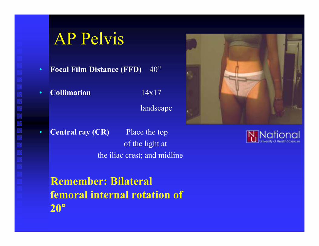
AP Pelvis
• Focal Film Distance (FFD) 40”
• Collimation 14x17
landscape
• Central ray (CR) Place the top
of the light at
the iliac crest; and midline
Remember: Bilateral
femoral internal rotation of
20°

AP PELVIS
Structures Visualized
•SI Joints
•Hip Joints
•Pubic Symphysis
•Greater and Lesser
Trochanters
•Obturator Foramen
•Femoral Heads
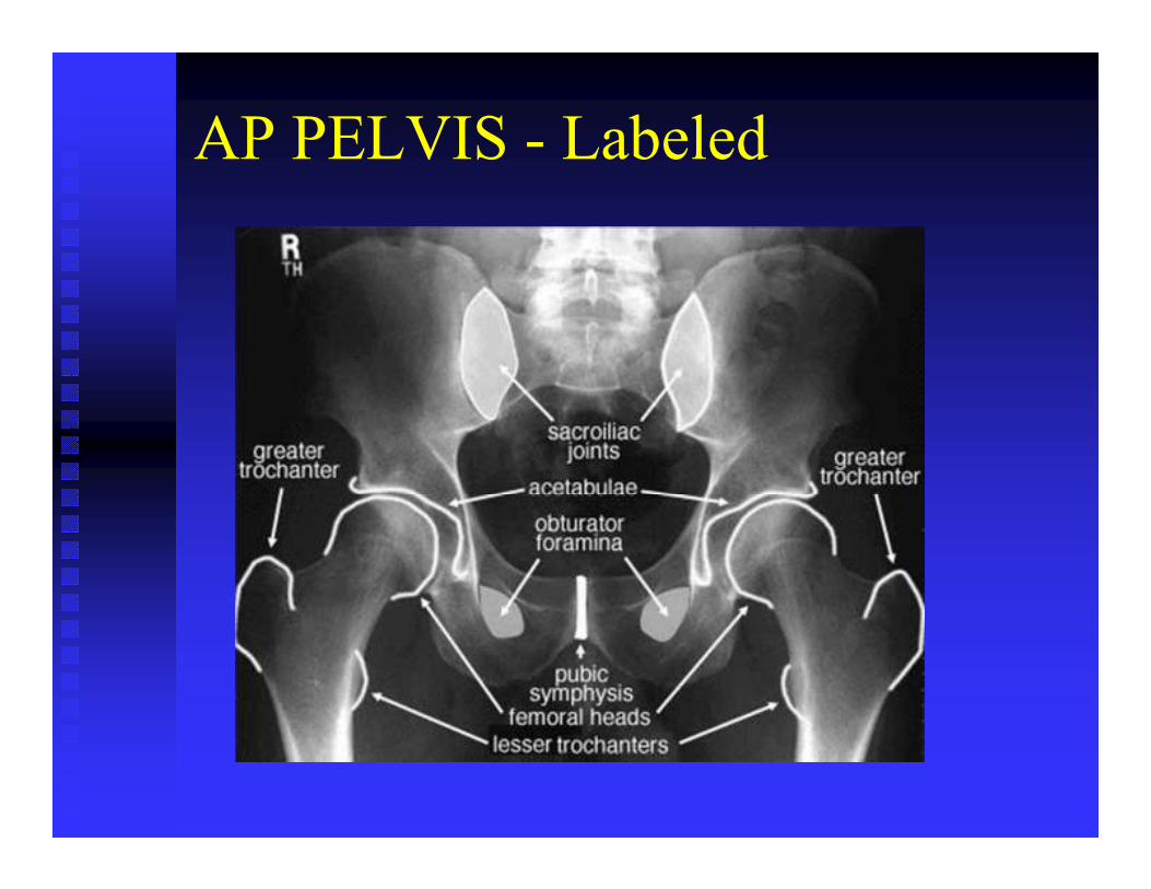
AP PELVIS - Labeled

AP Spot View of the Hip
• FFD 40”
• Collimation 10x12
• Central ray (CR) Femoral
pulse, mid groin
• Remember: Femur
internally rotated 20°

AP SPOT HIP
Structures Visualized
•Femoral Head
•Femoral Neck
•Greater and Lesser
Trochanter
•Femoral Shaft
•Kohler’s Teardrop
•Pubic Rami
•Iliac Fossa

LATERAL FROG-LEG VIEW
• FFD 40”
• Collimation 10x12
landscape
• CR Femoral pulse
• Remember to flex, abduct and external rotate the femur

FROG-LEG (HIP)
Structures Visualized
•Femoral head
•Femoral Neck
•Hip Joint space
•Kohler’s Teardrop
•Pubic Rami
•Obturator Foramen
•Femoral Shaft

Alignment Evaluation
• Iliofemoral, Klein’s, and Shenton’s line
• Iliopectineal line and ilioischial line

• Red= Iliofemoral
Line should be a
smooth arc
• Blue= Shenton’s
Line should be a
smooth arc
• If not a smooth arc
transition, the femur
has been displaced.
www.raddaily.com

• Green= Klein’s Line
should intersect the
femoral epiphysis. If
the line does not
intersect the femoral
epiphysis then the
epiphysis has
migrated medially.
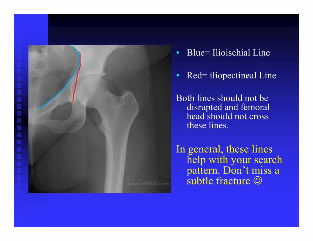
• Blue= Ilioischial Line
• Red= iliopectineal Line
Both lines should not be disrupted and femoral head should not cross these lines.
In general, these lines help with your search pattern. Don’t miss a subtle fracture ☺

Trabecular Pattern of Femoral Neck
• Trabecular pattern within the femoral neck should be intact. If disrupted, the trabecular pattern typically demonstrates linear area of sclerosis traversing the femoral neck. Disrupted trabecular pattern= FRACTURE
Journal of Biomedical & Pharmaceutical Engineering 1:1 (2007) 45-51

Case
• Hx: Hip and groin pain

AP Pelvis and Right Frogleg Lateral-
Where is the abnormality?

Osseous bump at the
lateral aspect of the
head-neck junction
of the right femur
with secondary
degenerative
changes. This
‘bump’ was not
caused by acute
trauma.

Femoroacetabular Impingement
Syndrome (FAI)
• FAI, cam type• Osseous bump at the lateral aspect of the
head-neck junction of the femur
• FAI was not caused by the acute trauma.
The patient had these bony prominences.
FAI is associated with labral tear.

Femoroacetabular Impingement
• Lateral osseous bump along the femoral head-neck junction=
cam
• “Pistol grip” deformity
• Osseous extension of the lateral aspect of the acetabulum resulting in overcoverage of the femoral
head= pincer
www.hipfai.com
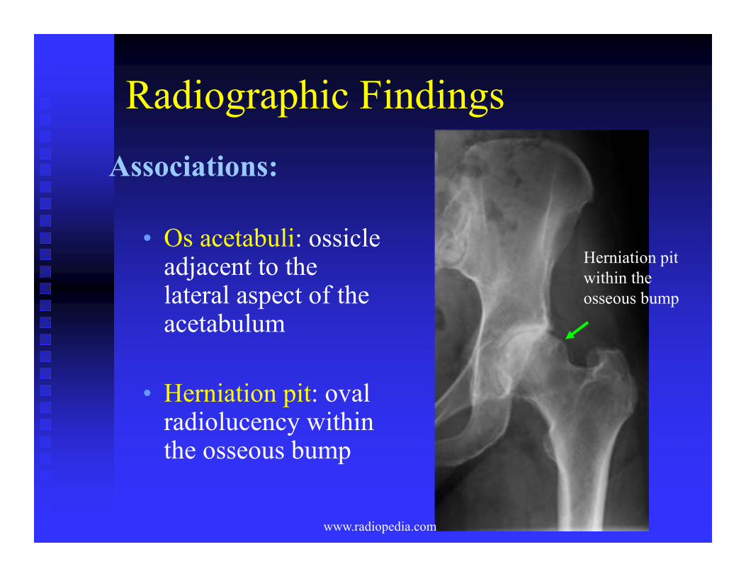
Radiographic Findings
Associations:
• Os acetabuli: ossicle adjacent to the lateral aspect of the acetabulum
• Herniation pit: oval radiolucency within the osseous bump
www.radiopedia.com
Herniation pit
within the
osseous bump

• Herniation pit is from wearing of the lateral
aspect of the femur due to contacting with
the acetabulum creating a synovial-filled
‘divot’ or cavity.

Clinical Findings
• Chief Complaint: groin pain with hip rotation in
the sitting position or during/after sports; or
trochanteric pain radiating to the lateral thigh.
• Decreased range of motion & pain
with flexion, adduction and
internal rotation of the femur

FYI on FAI
• Think of sport specific
limitations: patient with FAI
would not be a candidate as a
hockey goalie since it requires
hip flexion and femoral
internal rotation.

Complications of FAI
• Decreased joint clearance between femoral
neck and acetabulum
• Premature degeneration, and tears in
the labrum and adjacent articular
cartilage
Filigenzi F and Bredella M. MR imaging of Femoroacetabular Impingement. Applied Radiology, April 2008, 12-19.
Tannast M, et al. Femoroacetabular Impingment: Radiographic Diagnosis-What The Radiologist Should Know.
AJR:188;1540-1552, June 2007.

Follow-up
• MRI with arthrography: evaluate for
labral tears, and articular cartilage damage
• Orthopedic surgeon consultation
• Osseous resection
• Labral repair/refixation with suture
anchors or labral debridement
Larson C and Giveans M. Arthroscopic Management of Femoroacetabular Impingement: Early Outcome Measures. J of Arthroscopic and Related Surgery, May 2008, 24 (5); 540-546.

Case
Hx: 16 year-old female (yof) sprinter with
bilateral hip pain.

AP Pelvis- Where is the abnormality?


Bilateral Avulsion of ASIS
• Attachment of the Sartorius and Tensor
fascia latae tendon/muscle
• Normal open growth plates of the iliac crest
• Subtle bilateral femoroacetabular
impingement, cam type.

Anterior Superior Iliac Spine (ASIS)
Avulsion
Mechanism of injury (MOI): forceful extension of hip
Treatment: rest>>>rehab; healing 4-6 weeks to 6 months
Surgical: displacement greater than 2 cm; complication is nerve entrapment with displacement.

Case
Hx: 13 yom complains of knee pain
What other regions should be evaluated
beside the knee joint, particularly in a
young individual?
Other regions or joints need to be
evaluated:
• Ankle/foot
• Knee
• Hip
• And Lumbar

AP Pelvis- Where is the abnormality?

Recumbent Bilateral Lateral
Frog-leg View


Radiographic Findings
• Medial migration of the right femoral epiphysis
• Positive Klein’s Line- not intersecting the femoral epiphysis.
• Decreased femoral epiphyseal height and size
• Decreased bone density, subtle
• Varus deformity of the femoral neck (‘bending appearance’)

Slipped Capital Femoral Epiphysis
• Age: 10-17 yoa of boys; 8-15 yoa of girls
• Causes (separate or combination of)
• Overweight
• New activity- strenuous exercise
• Growth spurt
• Trauma
Resnick D. Diagnosis of Bone and Joint Disorders, 4th ed. 2002; 2729-34.

SCFE
Complications
• Severe varus deformity and foreshortening
• Osteonecrosis
• Premature degenerative joint disease
Follow-up
• Orthopedic surgeon consultation
• Reduction
• Severe cases: Pin the femoral epiphysis at the current location

Case
Hx: 18 yof athlete complains of hip pain

AP Pelvis- Can you find the abnormality?

• X-ray demonstrated no findings. The patient
continued to have pain.
• Further History: The 18 yof patient is a long
distance runner, and is running 60 miles per
week
• What is your differential diagnosis?
What advanced imaging would be best
for this patient?

MRI: Coronal T1 & T2 Weighted
Images

Diffuse, fanned
appearance is noted
within the femoral neck
with high signal on T2
and low signal intensity
on T1 weighted images.
Also, a linear low signal
is noted within the
medial aspect of the
femoral neck.

Stress fracture of the left femoral
neck
• Normal density with abnormal stress
• Bone scan and MRI would be positive
• Treatment: Non-weight bearing activity, for
example swimming.

Case
• 28 year-old soccer player was hit from
behind falling forward; inducing hip flexion
and internal rotation

www.bjsmcom

Hip Fracture/Dislocation
• Posterior wall fracture of the acetabulum
fracture and dislocation
• Treatment (Tx): Reduction; surgical
• Remember: Incidence of avascular necrosis
of the femoral head greatly increases if the
time to reduction is greater than six hours
• Br J Sports Med 2004

3D CT & Post-surgical
www.bjsmcom

Knee

Knee Views
• AP
• Lateral
•Tunnel
•Tangential (Sunrise)

AP Knee
• Collimation 8x10
• CR patellar apex
• Tube tilt 5 ° cephalad
• If standing & PA, 15 degree cephalad tube tilt.

AP KNEE
Structures Visualized
•Patella
•Femur
•Tibia
•Fibula
•Condyles of the Tibia and Femur
•Adductor tubercle
•Joint Space

AP KNEE - Labeled

LATERAL KNEE
• FFD 40”
• Collimation 8x10
• CR Joint line
• Knee flexion of 90-120 degrees

LATERAL KNEE
Structures Visualized
•Patella
•Femur
•Tibia
•Fibula
•Infra & Supra Patellar Fat
Pads

LATERAL
KNEE - Labeled

Tunnel View
• FFD 31
(corrected 40-9)
• Collimation 8x10
• CR Joint line
• Tilt 45 ° caudad
• Measure from mid-hamstring to anterior knee (not just through popliteal fossa)

TUNNEL VIEW (KNEE)
Structures Visualized
Intercondylar notch
Femoral condyles
Tibial eminences
Tibia
Fibula

Sunrise/ Tangential View
• FFD 40”
• Collimation 8x10
• CR Patellofemoral
joint

TANGENTIAL (SUNRISE) VIEW
Structures Visualized
•Medial and Lateral
Patellofemoral Joint
•Patella
•Patellar facets
•Trochlear groove
•Femoral Condyles

Case
Hx: 15 yom twisted his knee during basketball

AP View-Where is the abnormality?

Magnified

Findings
• Avulsion along the proximal lateral tibia.
• Segond fracture at the insertion site of the lateral (fibular) collateral ligament and fibers of distal iliotibial band.
•Associated with 75-100% ACL tear due to mechanism of injury
• Avulsion of the Tibial eminences
• Associated with anterior cruciate ligament tear

Avulsion: Segond & Avulsion of
Tibial Eminences
• Mechanism of Injury (MOI): Excessive internal
rotation and varus stress of tibia
• MRI
• Anterior cruciate ligament, Lateral collateral
ligament & distal Iliotibial band
• Most commonly tear of the medial mensicus.
Case
• Boy fell off his bike and has knee pain

AP and Sunrise View- Abnormal?
www.wikiradiography.com

Bipartite Patella
• Normal variant, superolateral aspect of the patella
• Smooth margins= not a fracture
• Clinically correlate for symptoms of this region
• Most commonly bilateral with smooth margins
• Conservative treatment
Rajinder Singh Gaheer, MS, MRCS, MRCPS, MCh(Orth); Sandeep Kapoor, FRCS(Tr&Orth);
Martin Rysavy, MD, PhD Orthopedics; November 2009 - Volume 32 · Issue 11

Bipartite Patella
• If symptoms of this region: MRI for
further evaluation
• Trauma to bipartite patella can cause
symptoms with effusion and bone
marrow edema on MRI.

Case
• 15 yof with knee pain

AP and PA Tunnel View

Sunrise (patellofemoral) View

Patellar Fracture-Dislocation
• Laterally displaced patella with heterotopic ossification medially or avulsion site of the medial patellar retinaculum
• MOI- twisting; direct blow; anomalous
• Treatment- conservative; possible surgical
• What advanced imaging would be best to evaluate this region?

MRI for Patellar dislocation
• MRI would demonstrate:
• Medial patellar retinaculum tear
• Possible bone contusion/ bone marrow edema
along medial patella and lateral femoral
condyle (kissing contusion).

Case
Hx: Twisted knee

AP and Lateral View-Where is the
abnormality?

Findings
• Radiolucency along the lateral aspect of the medial femoral condyle
• What advanced imaging should be performed for further evaluation?

MRI: Coronal T2 Weighted

Osteochondral Dissecans
• Age: 10-20
• Define: Necrosis of bone followed by reossification and healing
• Flap fragment with defect of the articular cartilage and fluid on MRI>>>unstable
• surgical

OCD
• Mechanism of Injury (MOI): shearing and rotary forces
• Tx:
• Intact cartilage: Walking with crutches, beneficial for the reconstitution of cartilage.
• Fragment/Defect in Cartilage: surgical• Joint locking

Case
• 12 yom limping with knee pain


Findings
• Soft tissue effusion of Hoffa’s (infrapatellar
region) and suprapatellar bursa
• Thickening of the patellar tendon
• Fragmentation and displacement tibial
tuberosity

Osgood-Schlatter’s Disease
• Traction apophysitis: Overuse injury age 9-14
with a male predominance.
• Repetitive strain from running, basketball, or other
repetitive sports leads to chronic avulsion of the
apophysis of the tibial tubercle.
• Callous formation with prominent tender tibial
tuberosity

Osgood-Schlatter’s
• S/S: Tenderness; tight quads; patella alta
• 2-6 months of conservative treatment;
eliminate stressful activity
• No jumping, running during
rehab
• If chronic, possible surgical excision of
ossicle.

Case
• Chronic knee pain; soccer player


Arcuate Sign
• Avulsion of the styloid process of the fibula at the insertion site of the fibular collateral ligament and bicep femoris.
• Associated with posterior cruciate ligament tears
• MOI: external rotation of tibia with varus stress; hyerperextension.

Case
• Chronic knee pain; previous trauma


•Malunion of the
proximal fibula
•Heterotopic
ossification along
the medial aspect of
the medial femoral
condyle. What
attaches to this
region?
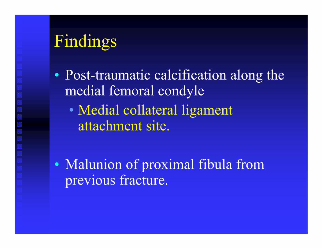
Findings
• Post-traumatic calcification along the medial femoral condyle
• Medial collateral ligament attachment site.
• Malunion of proximal fibula from previous fracture.

Pellegrini Stieda Syndrome
• Chronic avulsion of the medial collateral ligament
• Associated with anterior cruciate ligament and meniscal tears
• MRI is the next step if clinically indicated.

Case
• Long jumper with knee pain and
cannot extend the knee

www.feinberg.northwestern.edu

Findings
• Soft tissue swelling surrounding the patella
• Cephalad migration of patella
• Calcific densities within the infrapatellar region may represent avulsion fragments

Avulsion of the Patellar Tendon
• Rupture of the patellar tendon from the tibial tuberosity
• MRI to evaluation the patellar tendon and retraction distance, and for other ligamentous or meniscal tears
• Orthopedic referral

Ankle

Ankle Views
• AP
• Medial Oblique•Optional: Lateral oblique
•Lateral

AP View
• FFD 40”
• CR between the
malleoli

AP ANKLE
Structures Visualized
•Talar Dome
•Navicular
•Medial, Lateral and Posterior
Malleoli
•Tibial shaft
•Fibular Shaft
•Tibial Plafond

AP ANKLE - Labeled

MEDIAL OBLIQUE ANKLE
• FFD 40”
• CR between malleoli

MEDIAL OBLIQUE ANKLE
Structures Visualized
•Talar Dome
•Medial, Lateral and Posterior
Malleoli
•Tibial Plafond
•Navicular
•Calcaneus
•Tibia and Fibula
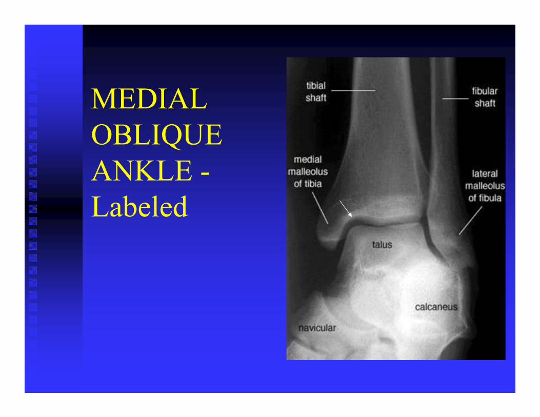
MEDIAL
OBLIQUE
ANKLE -
Labeled

LATERAL ANKLE
• FFD 40”
• CR medial
malleolus

LATERAL ANKLE
Structures Visualized
•Tibia
•Fibula
•Talus
•Calcaneus
•Navicular
•Cuboid

LATERAL
ANKLE -
Labeled

Case
• Long distance runner with foot and ankle
pain

Lateral Ankle

Linear sclerosis of the calcaneus.
What advanced imaging would be best
for further evaluation?

Stress Fracture of the Calcaneus
• X-ray: Linear sclerosis or no x-ray findings
• MRI- Bone marrow edema with high signal intensity and linear fracture line
Stress Fracture
• Healing time: weeks to months
• Treatment: conservative care
• First 6 weeks, reduced weight bearing
followed by gradual weight bearing
activities.

Case
• Football player twisted ankle

AP View

Findings
• Radiolucency along the medial aspect of the
talar dome
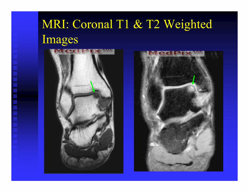
MRI: Coronal T1 & T2 Weighted
Images

Osteochondral Lesion (OCL) of
the Medial Talus
• MOI: Inversion+plantar flexion+external rotation of tibia on talus (medial OCL)
• Shearing and rotary forces
• 25 yoa; 5-6th decade
• MRI necessary for further evaluation
• Conservative Treatment
• If flap or loose fragment, surgical.

Case
Hx: 15 yom twisted ankle during soccer game

Medial Oblique and AP Views


Findings
• Salter Harris type III, Fracture of epiphysis extending to the growth plate
• MOI:Abduction and external rotation
• Complications- early closure; partial closure results in angular deformity

Salter Harris Fractures-growth plates
www.radiologyassistant.nl

Case
Hx: 23 yom, soccer player twisted his ankle
with plantar flexion + inversion (high ankle
sprain).
What other region should be
evaluated?

AP and Lateral Ankle

AP and Lateral Tib/Fib

Masseoneuve Fracture
• Fracture of the medial malleolus extending posteriorly AND Fracture of proximal fibula.
• Associated with disruption of interosseous membrane & tibiofibular syndesmosis; deltoid ligament (medial); joint widening
• Treatment- closed reduction & cast

Case
Hx: 32 yom playing tennis and felt a sharp
pain in the back of the lower leg.

Lateral View

Findings
• Atherosclerosis of the Posterior tibial artery
• Indistinct Achilles tendon margins= soft
tissue swelling

Achilles tendon rupture
• S/S: Indentation of tendon, weakness or loss
of motion
• MRI and/or diagnostic ultrasound for
further evaluation

Normal MRI vs Abnormal
www.musculoskeletalmri.blogspot.com
www.faoj.org
Complete tear of Achilles

Rupture of Achilles Tendon
• Tx: Surgical
• Conservative care if no retraction: non-weight
bearing with cast for 6 weeks; followed by
short walking cast for 2 weeks
• Rehab for 6 months; heel lift
• Surgical: Complete tear and retraction

Foot

Foot Views
• DP
•Medial Oblique
•Lateral

DORSOPLANTAR FOOT (DP)
• FFD 40-2
• Collimation 8 X 10
• CR 3rd MT base
• Tube Tilt 10 ° cephalad

DORSOPLANTAR FOOT (DP)
Structures Visualized
•Cuboid
•Calcaneus
•Talus
•Navicular
•1st-3rd Cuneiform
•1st-5th metatarsals
•phalangeal bones

DORSOPLANTAR
FOOT - Labeled

MEDIAL OBLIQUE FOOT
• FFD 40-2
• CR 3rd Metatarsalbase
• Tube tilt 10 ° cephalad
tube tilt

MEDIAL OBLIQUE FOOT
Structures Visualized
•Styloid process of 5th Metatarsal
bone>>> best projection to
evaluate this region
•Calcaneus
•Talus
•Navicular
•Cuboid
•1st-3rd Cuneiforms
•Metatarsals and sesamoids
•Phalanges

MEDIAL
OBLIQUE FOOT -
Labeled
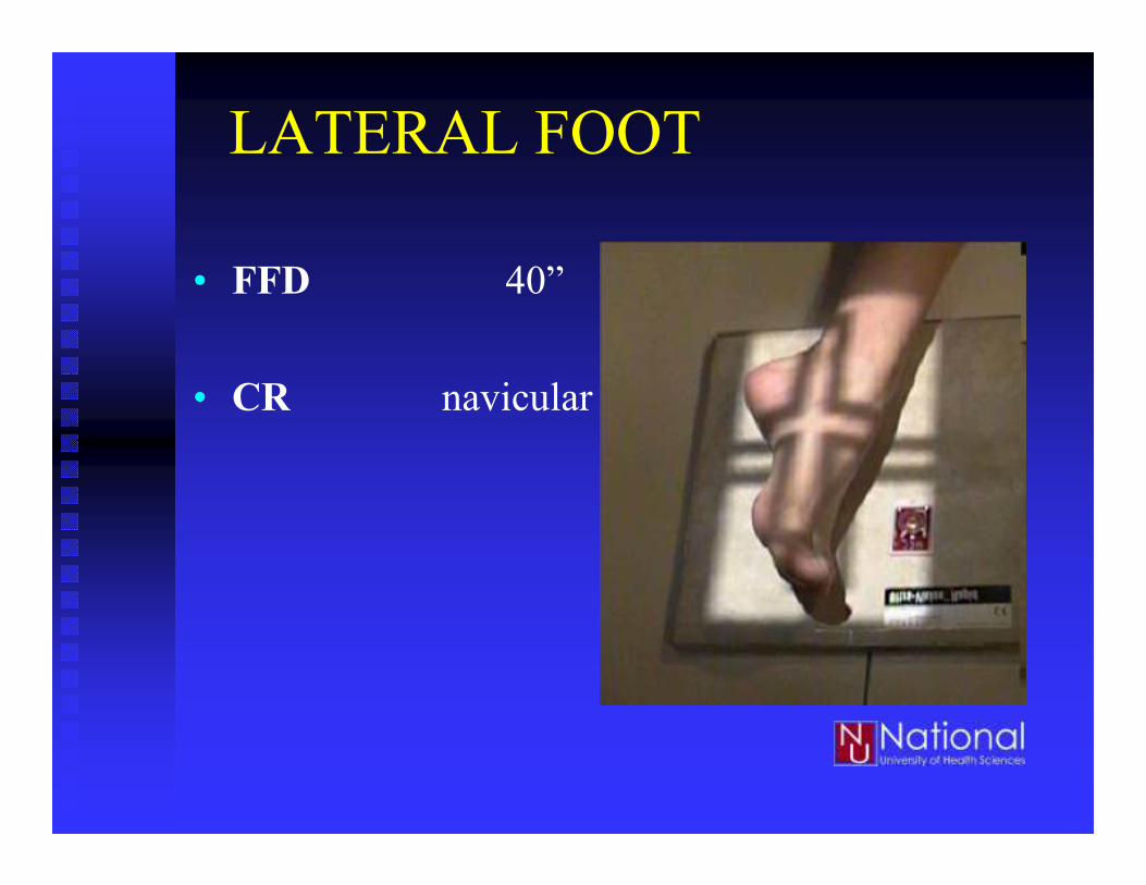
LATERAL FOOT
• FFD 40”
• CR navicular

LATERAL FOOT
Structures Visualized
•Calcaneus
•Talus
•Navicular
•Cuboid
•Metatarsals

LATERAL FOOT - Labeled

Remember: X-ray Study of the Foot
• DP, medial oblique and lateral views of the
foot should include the bony structures from
the distal tufts or toes to the distal
tibia.
• Whereas the ankle x-ray studies do not
include the toes.

Case
• Wrestling barefoot
• Foot pain

DP Foot
Donated by Dr. L. Nicholson

Magnified- 4th toe
Donated by Dr. L. Nicholson

Fracture of Proximal Phalanx
• Fracture of 4th proximal phalanx
• Bipartite sesamoid, normal variant
• Tx: immobilize/splint
Case
• Twisted ankle with inversion + plantar
flexion


Medial Oblique

Avulsion of the Styloid Process
• 5th metatarsal, styloid
process, base of the 5th
metatarsal.
www.radiopaedia.org

Versus Normal Apophysis
www.atpracticeinsights.com
Normal
apophysis:
parallel to
the long
axis of the
proximal
metatarsal.

Styloid Avulsion
• MOI: inversion with plantar flexion
• Avulsion from the lateral cord of the plantar
aponeurosis, and/or peroneus brevis tendon

Treatment
• Non-displaced: Immobilize for 4-6 or 8
weeks; conservative.
• Surgical: Comminuted fracture;
Displacement; and fracture involving greater
than 30% of cubometatarsal articulation
(articular surface)

Case
• Dull, achey foot pain for several weeks


Stress Fracture with Callous
Formation
• Callous formation at the diaphysis of the 2nd
metatarsal
• Aka March fracture
• Osteopenia, disuse
• Tx: Conservative care; reduce weight bearing activities

Case
• Athlete twisted his/her foot

www.mypacs.net
Which foot and
joint are affected?

Right Foot Pain and Swelling

Lisfranc Joint or Tarsometatarsal
Diastasis (widening)
• Severe sprain or possible dislocation of the tarsometatarsal joint
• Widening of the tarsometatarsal joint; swelling
• unable or difficult to bear weight; dropped arch
• May have associated fracture (this patient does not have a fracture)
• MOI: Twisting with plantar flexion of forefoot or direct blow.

X-ray Findings of Lisfranc Joint
Fracture or Diastasis/Dislocation
• Evaluation of a Normal DP foot x-ray study
• Alignment of 1st metatarsal to 1st
cuneiform; and 2nd metatarsal to 2nd
cuneiform
• Widening of 1st-2nd metatarsal interspace;
Lateral dislocation of the metatarsals

Stress Radiograph for Lisfranc
Injury
• Special Radiograph: Bilateral or
unilateral weight bearing DP
view of the feet, 10 cephalad
tube tilt .

Lisfranc Joint Injury
• Lisfranc ligament (tarsometatarsal
ligament) is a major stabilizer of the
Tarsometatarsal (TMT) joint; torn
ligament results in midfoot instability.
• Origin: first cuneiform
• Insertion: medial aspect of the base
of the second metatarsal.

Lisfranc Injury
• Remember: Disruption causes midfoot instability
•This can end an athlete’s career!!

Lisfranc Joint Injury: Follow-up
• Rest; boot>>>6-8 weeks or more
• Advanced Imaging
• MRI- ligament and bone marrow edema
• CT-fracture fragments
• If unstable, Surgical
• Percutaneous wire or plate & screw fixation

MRI of Torn Tarsometatarsal
ligament
www.radsource.us

If unstable=Surgery
www.lisfranc.org
www.jbjs.org
www.jaaos.org

References
• Cornuelle A; Gronefeld D. Radiographic Anatomy & Positioning, Integrated Approach. 1998
• Yochum T, Rowe L. Essentials of Skeletal Radiology, 3rd ed. Baltimore: Williams & Wilkins, 2005.
• Resnick D. Diagnosis of Bone and Joint Disorders, 4th ed. 2002.
• Prentice W. Arnheim’s Principles of Athletic Training, 13 ed. 2009
• Hyde T; Gengenbach M. Conservative Management of Sports Injuries. 2007
• Stoller D, et al. Diagnostic Imaging, Orthopedics. 2004
• Juhl J, et al. Essentials of Radiologic Imaging, 6th ed. 1993.
• Stoller D. MRI in Orthopedics & Sports Medicine, 3rd ed; 2007
• Filigenzi F and Bredella M. MR imaging of Femoroacetabular Impingement. Applied Radiology, April 2008, 12-19.
• Tannast M, et al. Femoroacetabular Impingement: Radiographic Diagnosis-What The Radiologist Should Know. AJR:188;1540-1552, June 2007.
• Campos J, et al. Pathogenesis of the Segond Fracture: Anatomic and MR Imaing Evidence of an Iliotibial Tract or Anterior Oblique Band Avulsion. Radiology May 2001; 219, 381-386.
• Arnaiz J, et al. Imaging Findings of Lower Limb Apophysitis. AJR March 2011; 196, W316-25.
• Strub W. Signs in Imaging: The Arcuate Sign. Radiology 2007; 244: 620-621.
• Theodorou, et al. Radiology. March 2003; 226, 857-865.
• AAFP.org. Fractures of Proximal 5thMetatarsal. 59 (9),May 1, 1999.
• Timpone V, et al. Intermetatarsal Fat Pad Sign: Radiographic Aid to Diagnosis of Occult Tarsometatarsal Joint Injuries. AJR January 2009; 192 (1), W36-37.
• Rajinder Singh Gaheer, MS, MRCS, MRCPS, MCh(Orth); Sandeep Kapoor, FRCS(Tr&Orth); Martin Rysavy, MD, PhD Orthopedics; November 2009 - Volume 32 · Issue 11
