back and upper limb
TRANSCRIPT

Back and upper limb

General division of the back
musclesSuperficial extrinsic back muscles
(posterior thoracoappendicular muscles)
Intermediate extrinsic back muscles
(spinocostal muscles)
Extrinsic back muscles
Superficial layer
(Splenius muscles)
Intermediate layer
(Erector spinae)
Deep layer
(Transverospinal muscles)
Minor deep layer
(Short back muscles)
Intrinsic back muscles
Back muscles
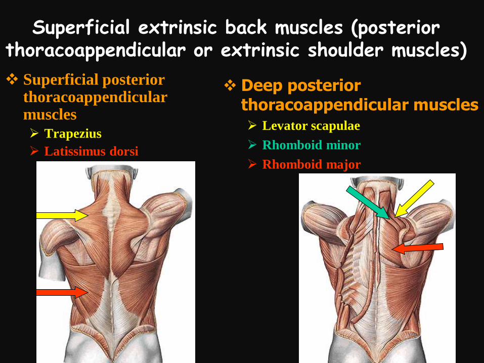
Superficial extrinsic back muscles (posterior thoracoappendicular or extrinsic shoulder muscles)
Superficial posterior thoracoappendicular muscles
Trapezius
Latissimus dorsi
Deep posterior thoracoappendicular muscles Levator scapulae
Rhomboid minor
Rhomboid major

Superficial extrinsic back muscles (posterior thoracoappendicular or extrinsic shoulder
muscles)Trapezius
Attachments
Origin (proximal
attachment)
• Occipital bone,
nuchal ligament and
spinous processes of
C7 to T12 vertebrae
Insertion (distal
attachment)
• Clavicle, acromion
and spine of scapula

Superficial extrinsic back muscles (posterior thoracoappendicular or extrinsic shoulder muscles)
Trapezius
Innervation
Spinal roots of
accessory nerve
(CN XI)
Cervical plexus

Superficial extrinsic back muscles (posterior thoracoappendicular or extrinsic shoulder muscles)
Trapezius
Function- movements of scapula:
Superior part- elevation
Middle part- retraction
Inferior part- depression
Common action of superior and
inferior parts results in rotation
of scapula which enables
elevation of the arm above the
level of the shoulder.
Trapezius cooperates then with
serratus anterior muscle
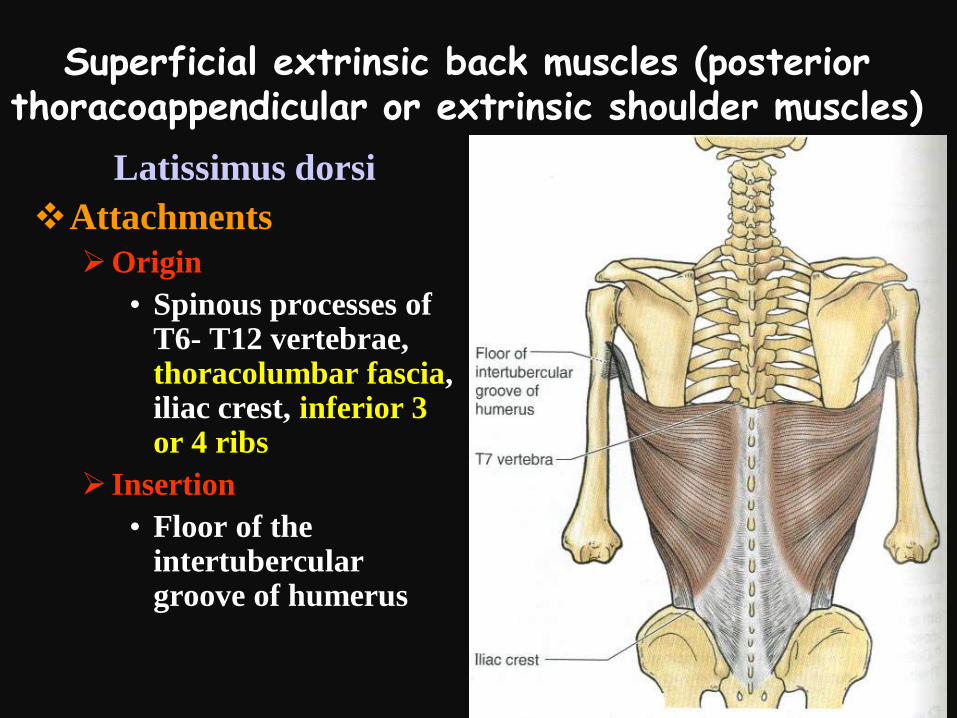
Superficial extrinsic back muscles (posterior thoracoappendicular or extrinsic shoulder muscles)
Latissimus dorsi
Attachments
Origin
• Spinous processes of T6- T12 vertebrae, thoracolumbar fascia, iliac crest, inferior 3 or 4 ribs
Insertion
• Floor of the intertubercular groove of humerus

Superficial extrinsic back muscles (posterior thoracoappendicular or extrinsic shoulder muscles)
Latissimus dorsi
Innervation
Thoracodorsal nerve-
branch of the brachial
plexus
Thoracodorsal nerve
is situated on the
posterior wall of the
axilla and may be
injured during
surgery in this region

Superficial extrinsic back muscles (posterior thoracoappendicular or extrinsic shoulder muscles)
Functions of latissimus dorsi
Action on the glenohumeral joint:
Extension, adduction and medial rotation (folding arms behind the back)
Together with pectoralis major
Depresses elevated upper limb (for example during chopping wood)
Raises body towards arms during climbing

Latissimus dorsi
Raising the body

Superficial extrinsic back muscles (posterior thoracoappendicular or extrinsic shoulder muscles)
Levator scapulae
Attachments
Origin
• Transverse
processes of C1 to
C4 vertebrae
Insertion
• Superior part of
medial border of
scapula
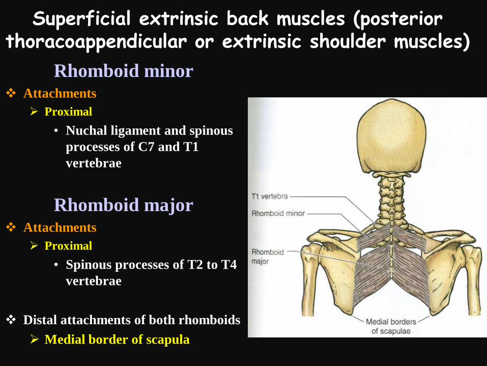
Superficial extrinsic back muscles (posterior thoracoappendicular or extrinsic shoulder muscles)
Rhomboid minor Attachments
Proximal
• Nuchal ligament and spinous
processes of C7 and T1
vertebrae
Rhomboid major Attachments
Proximal
• Spinous processes of T2 to T4
vertebrae
Distal attachments of both rhomboids
Medial border of scapula

Superficial extrinsic back muscles (posterior thoracoappendicular or extrinsic shoulder muscles)
Levator scapulae and rhomboids
Function- movements of scapula:
Elevation of scapula and tilting the glenoid cavity inferiorly by rotating the scapula
Retraction of scapula (rhomboids)
Fixing scapula to the thoracic wall

Superficial extrinsic back muscles (posterior thoracoappendicular or extrinsic shoulder muscles)
Levator scapulae and rhomboids
Innervation
Dorsal scapular nerve-branch of the brachial plexus (its supraclavicular part-root C4- C5)
Testing the rhomboids or dorsal scapular nerve or C4- C5 roots
Patient has its hands on the hips and pushes elbows posteriorly against the resistence provided by examiner
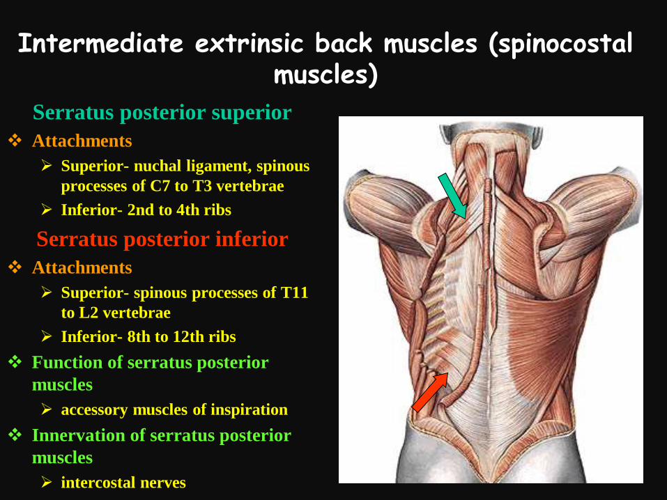
Intermediate extrinsic back muscles (spinocostal muscles)
Serratus posterior superior
Attachments
Superior- nuchal ligament, spinous
processes of C7 to T3 vertebrae
Inferior- 2nd to 4th ribs
Serratus posterior inferior
Attachments
Superior- spinous processes of T11
to L2 vertebrae
Inferior- 8th to 12th ribs
Function of serratus posterior
muscles
accessory muscles of inspiration
Innervation of serratus posterior
muscles
intercostal nerves

Intrinsic (deep) back muscles
Innervation- dorsal rami of the
spinal nerves
Superficial extrinsic back muscles
(posterior thoracoappendicular muscles)
Intermediate extrinsic back muscles
(spinocostal muscles)
Extrinsic back muscles
Superficial layer
(Splenius muscles)
Intermediate layer
(Erector spinae)
Deep layer
(Transverospinal muscles)
Minor deep layer
(Short back muscles)
Intrinsic back muscles
Back muscles

Intrinsic (deep) back musclesAction
•Acting bilaterally (together)-
extend trunk and head
•Acting unilaterally (alone)- flex
laterally and rotate trunk and
head

Superficial layer of the intrinsic back muscles
Splenius capitis
Splenius cervicis
Location
Lateral and posterior side of neck
Extended between midline and cervical vertebrae(transverse processes-splenius cervicis) and skull (mastoid process of temporal bone- splenius capitis)
Main action
Acting alone- bend and rotate head to side of active muscle
Acting together- extend head and neck

Erector spinaeErector spinae

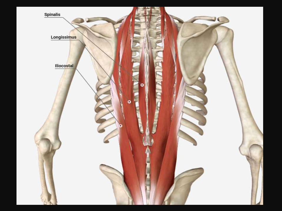

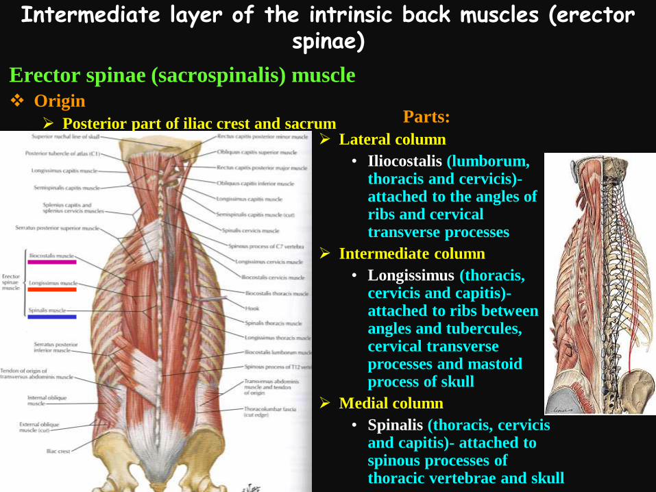
Intermediate layer of the intrinsic back muscles (erector spinae)
Erector spinae (sacrospinalis) muscle
Origin
Posterior part of iliac crest and sacrum Parts:
Lateral column
• Iliocostalis (lumborum, thoracis and cervicis)-attached to the angles of ribs and cervical transverse processes
Intermediate column
• Longissimus (thoracis, cervicis and capitis)-attached to ribs between angles and tubercules, cervical transverse processes and mastoid process of skull
Medial column
• Spinalis (thoracis, cervicis and capitis)- attached to spinous processes of thoracic vertebrae and skull

Deep layer of the intrinsic back muscles (transversospinal muscles)
Transversospinal
muscles
Attachments
Extended between
transverse and
spinous processes of
vertebrae
Parts (have different
length):
Semispinalis
(thoracis, cervicis
and capitis)- spanns
4-6 segments
Multifidi- spann 2- 4
segments
Rotatores muscles-
spann 1-2 segments

Minor deep layer of the intrinsic back muscles
Interspinales muscles
Intertransversarii
muscles
Attachments
• Extended between
spinous or transverse
processes of adjacent
vertebrae
Levatores costarum
muscles
Extended between
transverse processes of
thoracic vertebrae and rib
situated inferolaterally

Suboccipital region
Situated in the upper
part of the posterior
surface of the neck,
below the occipital bone,
at the level of C1 and C2
vertebrae
Covered by the trapezius,
splenius capitis and
semispinalis capitis
muscles

Suboccipital muscles (deep neck muscles)
Rectus capitis posterior minor
Rectus capitis posterior major
Rectus capitis lateralis
Superior oblique of the head
Inferior oblique of the head
Suboccipital muscles are
extended between C1 or C2
vertebrae and occipital bone.
The only exception is the inferior
oblique of the head extended
between C1 and C2 vertebrae

Suboccipital muscles (deep neck muscles)
Rectus capitis posterior minor
Rectus capitis posterior major
Rectus capitis lateral
Superior oblique of the head
Inferior oblique of the head
Suboccipital muscles are
innervated by the suboccipital
nerve (motor nerve being the
posterior ramus of C1 spinal
nerve)
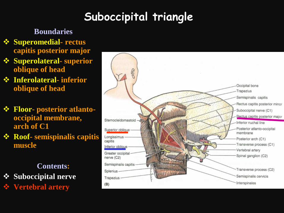
Suboccipital triangleBoundaries
Superomedial- rectus capitis posterior major
Superolateral- superior oblique of head
Inferolateral- inferior oblique of head
Floor- posterior atlanto-occipital membrane, arch of C1
Roof- semispinalis capitis muscle
Contents:
Suboccipital nerve
Vertebral artery

Innervation of the skin in the occipital region
In the innervation of the skin in the occipital region participate:
Greater occipital nerve- posterior branch of the 2nd cervical spinal nerve (C2)
Lesser occipital nerve- branch of the cervical plexus (contains the anterior branch of the 3rd cervical spinal nerve C3)
1st cervical spinal nerve C1 (and suboccipital nerve) has no sensory fibers and it results in lack of the C1 dermatome of the skin

Greater occipital nerve Exits the vertebral canal
between the vertebrae C1 i C2
Runs superiorly under the trapezius and splenius capitis muscles
Pierces lateral border of the aponeurosis of the trapezius, just under the inferior nuchal line of the occipital bone
In the occipital region runs together with occipital artery

Lesser occipital nerve
Is a branch of the
cervical plexus
Arises in the
midpoint of the
posterior edge of the
sternocleidomastoid
muscle

Blocks of the lesser and greater occipital nerves
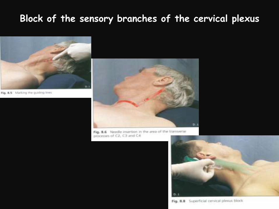
Block of the sensory branches of the cervical plexus

Superficial muscles of the thorax (anterior thoracoappendicular muscles)
Pectoralis major
Extended between clavicle, sternum and ribs and lateral lip of the intertubercular groove of humerus
Main action- adduction, medial rotation and flexion of humerus, muscle of climbing
Innervation- medial and lateral pectoral nerves
Pectoralis minor
Extended between ribs and coracoid process of scapula
Main action- protraction anddepression of scapula
Innervation- medial pectoral nerve

Boxer’s muscle
Serratus anterior


Superficial muscles of the thorax (anterior thoracoappendicular muscles)
Serratus anterior
Extended between ribs and medial border of scapula
Innervation- long thoracic nerve
Main action- protraction and rotation of scapula, participation in elevation of arm above the level of shoulder
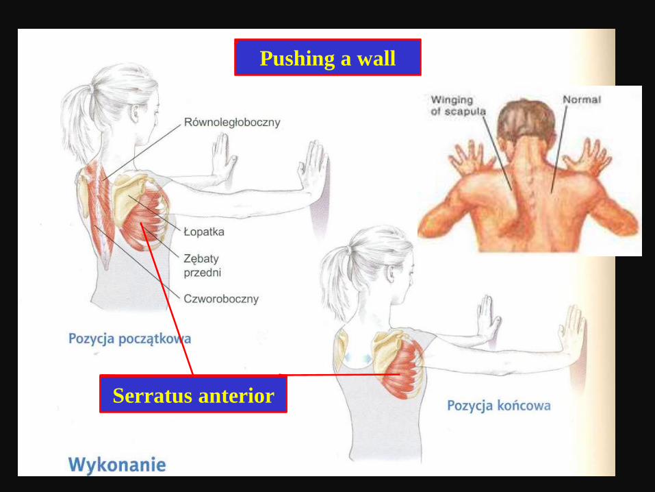
Erector spinaeSerratus anterior
Pushing a wall

Scapulohumeral (intrinsic shoulder) muscles
Deltoid muscle
Rotator cuff muscles
Supraspinatus
Infraspinatus
Teres minor
Subscapularis
Teres major muscle

Scapulohumeral (intrinsic shoulder) muscles
Deltoid muscle
Attachments
Proximal
• Posterior part: spine of
scapula
• Middle part- acromion
• Anterior part- clavicle
Distal
Deltoid tuberosity of humerus
Innervation
Axillary nerve (branch of the
posterior cord of brachial
plexus)

Scapulohumeral (intrinsic shoulder) muscles
Deltoid muscle
Main action
Posterior part:
extension and lateral
rotation of arm
Middle part-
abduction of arm
(cooperates with
supraspinatus)
Anterior part-
flexion and medial
rotation of arm

Scapulohumeral (intrinsic shoulder) musclesMuscles of rotator cuff
Rotator cuff muscles form a
musculotendinous cuff around
the glenohumeral joint
Rotator cuff muscles reinforce
and stabilize the glenohumeral
joint blending with its articular
capsule
Supraspinatus muscle
Infraspinatus muscle
Teres minor
Subscapularis muscle

Scapulohumeral (intrinsic shoulder) musclesMuscles of rotator cuff
Supraspinatus muscle
Attachments
Proximal
• Supraspinous fossa of scapula
Distal
• Superior facet on greater tubercle of humerus
Innervation
Suprascapular nerve(branch of the superior trunk of brachial plexus)
Action
Abduction of arm

Between the acromion and the tendon of
supraspinatus is located the subacromial bursa.
Subacromial bursa, filled with synovial fluid, diminish
friction of supraspinatus during its contraction.

Scapulohumeral (intrinsic shoulder) musclesMuscles of rotator cuff
Infraspinatus muscle
Attachments
Proximal
• Infraspinous fossaof scapula
Distal
• Middle facet on greater tubercle of humerus
Innervation
Suprascapular nerve (branch of the superior trunk of brachial plexus)

Scapulohumeral (intrinsic shoulder) musclesMuscles of rotator cuff
Teres minor
Attachments
Proximal
• Lateral border of scapula
Distal
• Inferior facet on greater tubercle of humerus
Innervation
Axillary nerve (branch of the posterior cord of
brachial plexus)
Action of
infraspinatus and teres minor
Lateral rotation of arm

Scapulohumeral (intrinsic shoulder) musclesMuscles of rotator cuff
Subscapularis muscle
Attachments
Proximal
• Subscapular fossa of scapula
Distal
• Lesser tubercle of humerus
Innervation
Subscapular nerves (branches of
the posterior cord of brachial
plexus)
Action
Medial rotation of arm

Tearing of the rotator cuff tendons

Scapulohumeral (intrinsic shoulder) muscles
Teres major
Attachments
Proximal
• Inferior angle of scapula
Distal
• Medial lip of intertubercular groove of humerus
Innervation
Subscapular nerves(branches of the posterior cord of brachial plexus)
Action
Adduction and medial rotation of arm

Axilla (armpit)

Axilla (armpit)
Pyramidal space between
thorax and upper limb
Axilla provides a
passageway from neck for
vessels and nerves supplying
upper limb

Boundaries of axilla
Apex- entrance from neck
Situated between 1st rib, clavicle and
subscapularis
Base of axilla
Skin, subcutaneous tissue and axillary
fascia

Boundaries of axilla
Anterior wall- formed by pectoralis
major and pectoralis minor muscles
Posterior wall- formed by scapula
and subscapularis muscle and
latissimus dorsi and teres major
muscles
Medial wall- formed by wall of
thorax and covering it serratus
anterior
Lateral wall (or narrow lateral
border)- formed by intertubercular
groove of humerus

Contents of axilla
Brachial plexus (its cords
and branches)
Axillary blood vessels
Axillary artery and its
branches
Axillary vein and its
tributaries
Axillary lymph nodes

Openings in the posterior wall of axilla
Quadrangular space
Boundaries
Superior- teres minor
Inferior- teres major
Medial- long head of triceps brachii muscle
Lateral- surgical neck of humerus
Contents
Axillary nerve
Posterior circumflex humeral artery

Openings in the posterior wall of axilla
Triangular space
Boundaries
Superior- teres minor
Inferior- teres major
Lateral- long head of triceps brachii muscle
Contents
Circumflex scapular artery
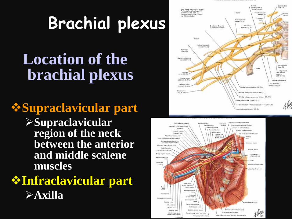
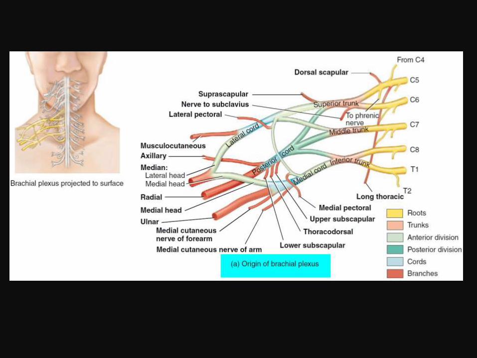
Brachial plexus
Location of the brachial plexus
Supraclavicular partSupraclavicular
region of the neck between the anterior and middle scalene muscles
Infraclavicular partAxilla


Brachial plexus (supraclavicular part)
Roots of the brachial
plexus
Ventral rami of last four
cervical spinal nerves and
first thoracic nerve
(C5 to T1 nerves)
Situated in the neck
(posterior triangle) between
anterior and middle scalene
muscles
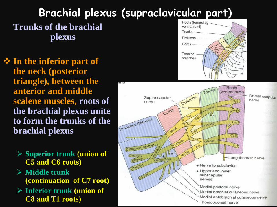
Brachial plexus (supraclavicular part)Trunks of the brachial
plexus
In the inferior part of the neck (posterior triangle), between the anterior and middle scalene muscles, roots of the brachial plexus unite to form the trunks of the brachial plexus
Superior trunk (union of C5 and C6 roots)
Middle trunk (continuation of C7 root)
Inferior trunk (union of C8 and T1 roots)
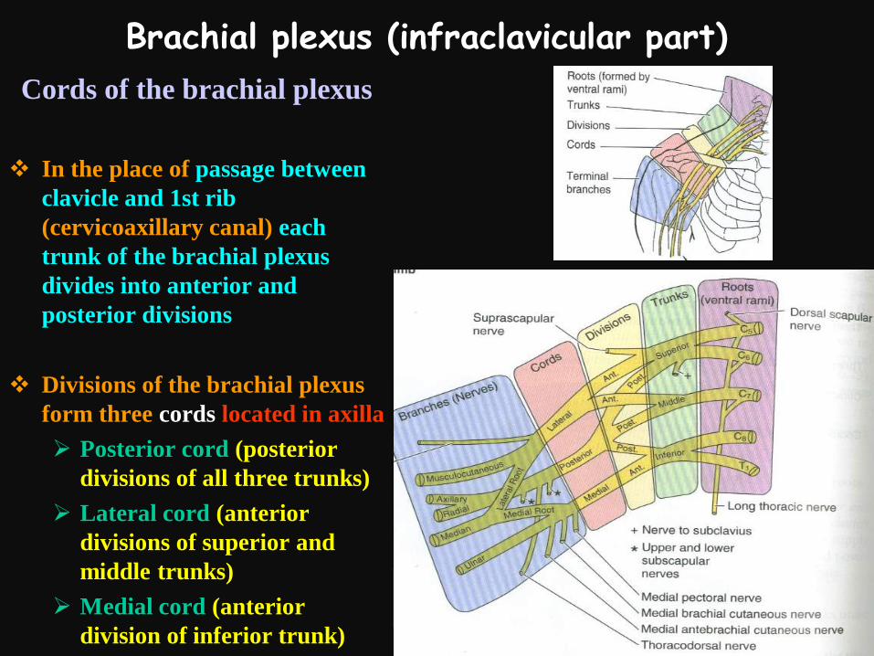
Brachial plexus (infraclavicular part)
Cords of the brachial plexus
In the place of passage between
clavicle and 1st rib
(cervicoaxillary canal) each
trunk of the brachial plexus
divides into anterior and
posterior divisions
Divisions of the brachial plexus
form three cords located in axilla
Posterior cord (posterior
divisions of all three trunks)
Lateral cord (anterior
divisions of superior and
middle trunks)
Medial cord (anterior
division of inferior trunk)
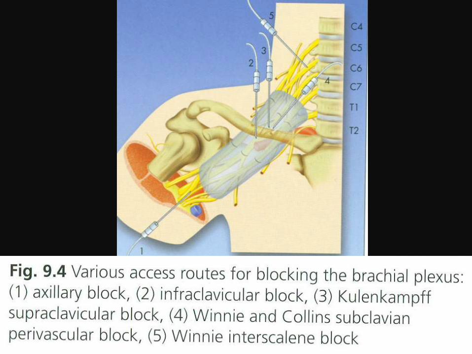

Interscalene block of the brachial plexus

Supraclavicular block of the brachial plexus

Infraclavicular block of the brachial plexus

Axillary block of the brachial plexus

Supraclavicular nerves of brachial plexus
Dorsal scapular nerve
Arises from C4 and
C5 roots
Runs along medial
border of scapula
Innervates
Levator scapulae
Rhomboid minor
Rhomboid major

Supraclavicular nerves of brachial plexus
Long thoracic nerve
Arises from C5, C6 and C7
roots
Runs on the lateral wall of
thorax, on the surface of
serratus anterior muscle
Innervates
Serratus anterior muscle
Symptoms of injury
Winged scapula

Supraclavicular nerves of brachial plexus
Suprascapular nerve
Arises from superior trunk of the
brachial plexus
Passes across the posterior
triangle of the neck, and via the
scapular notch reaches the
supraspinous fossa of scapula
Innervates
Supraspinatus muscle
Infraspinatus muscle
Symptoms of injury
Difficulties with abduction of arm

Supraclavicular nerves of brachial plexusNerve to subclavius
Arises from superior trunk of the brachial plexus
Runs posteriorly to clavicle
Innervates
Subclavius muscle

Short (side) nerves of the infraclavicular part of brachial plexus
Lateral (from lateral
cord) and medial (from
medial cord) pectoral
nerves
Pierce the clavipectoral
fascia and reach deep
surfaces of the
pectoralis major and
minor muscles
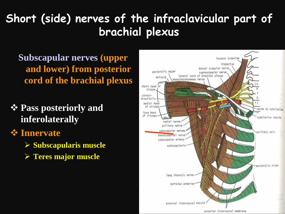
Short (side) nerves of the infraclavicular part of brachial plexus
Subscapular nerves (upper
and lower) from posterior
cord of the brachial plexus
Pass posteriorly and
inferolaterally
Innervate
Subscapularis muscle
Teres major muscle

Short (side) nerves of the infraclavicular part of brachial plexus
Thoracodorsal nerve
from posterior cord
of the brachial
plexus
Runs inferolaterally
along the posterior
axillary wall to
latissimus dorsi
Innervate
Latissimus dorsi
muscle

Short nerves of the infraclavicular part of brachial plexus
Axillary nerve from posterior cord of the
brachial plexus
Leaves the axilla via the quadrangular space, winds around the surgical neck of humerus, reaches deltoid and teres minor, gives rise to lateral brachial cutaneous nerve
Innervate
Deltoid muscle
Teres minor muscle
Shoulder joint
Skin over inferior part of deltoid
Injury
May be caused by fracture of the surgical neck of humerus or dislocation of the head of humerus from the shoulder joint
Symptoms
Difficulties with abduction of shoulder joint
Area of anesthesia on the lateral surface of the shoulder
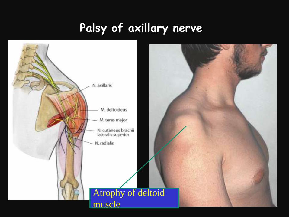
Palsy of axillary nerve
Atrophy of deltoid
muscle

Long nerves of the infraclavicular part of brachial plexus
Posterior cord
Radial nerve
Lateral cord
Musculocutaneous nerve
Lateral root of the median nerve
Medial cord
Ulnar nerve
Medial root of the median nerve
Medial brachial cutaneous nerve
Medial antebrachial cutaneous nerve