b cell development and activation in healthy people, there are mature b cells with the capacity to...
TRANSCRIPT
B CELL DEVELOPMENT AND ACTIVATION
• In healthy people, there are mature B cells with the capacity to make antibodies to virtually any antigen.
• Bone marrow is the primary lymphoid organ in which B cell development occurs.

• Bone marrow is the primary lymphoid organ in which B cell development occurs.
• Following initial development in bone marrow, mature B cells migrate to various secondary lymphoid tissues, including lymph nodes, spleen, gut-associated lymphoid tissue and blood.
• There, mature B cells can interact with antigen, become activated, and further differentiate into antibody-secreting cells

• B and T cells undergo distinct differentiation pathways.
• B cells are generated in the bone marrow, with mature B cells, which are ready to respond to antigen, then exiting and migrating to lymph nodes and spleen. T cells are generated in the thymus.

• The development of B cells, starting from hematopoietic stem cells and ending with cells that produce antibodies, can be divided into four phases:

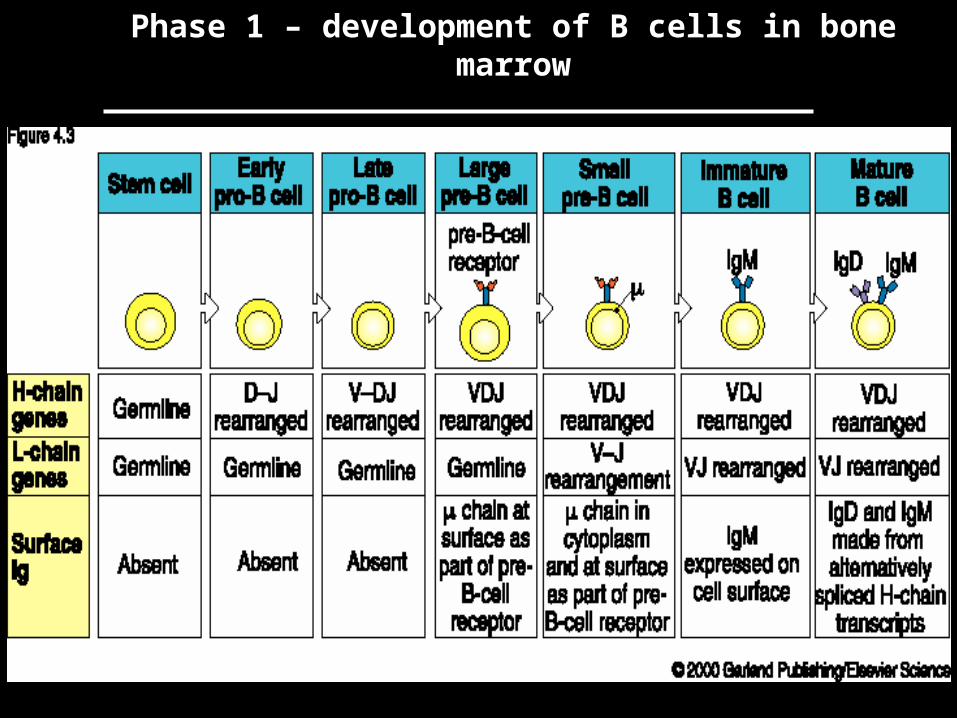
Phase 1 – development of B cells in bone marrow
• This first phase of B cell development is the generation of B cells in bone marrow.
• There, stem cells develop into pro-B cells, then pre-B cells, and finally mature B cells, which exit the bone marrow and migrate to secondary lymphoid organs.
• This phase of B cell development is not driven by contact with antigen: antigen independent.
• The DNA rearrangements that result in a functional cell-surface immunoglobulin molecule occur during this phase.
Phase 1 – development of B cells in bone marrow
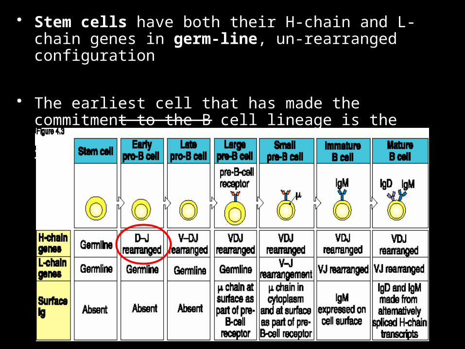
• Stem cells have both their H-chain and L-chain genes in germ-line, un-rearranged configuration
• The earliest cell that has made the commitment to the B cell lineage is the pro-B cell: pro-B cells have begun to rearrange their H-chain gene.

• Once a B lineage cell expresses cell surface H-chain (m) it is defined as a pre-B cell.
• However, the early pre-B cell receptor is not the final form of surface immunoglobulin: H-chain + surrogate light chain (molecule that mimics L-chain)
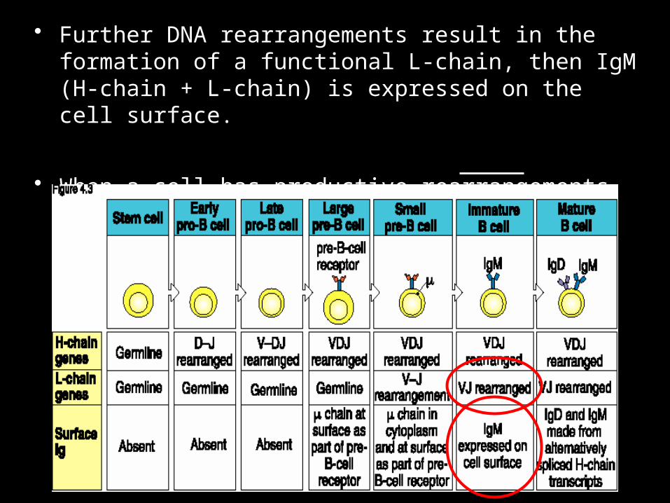
• Further DNA rearrangements result in the formation of a functional L-chain, then IgM (H-chain + L-chain) is expressed on the cell surface.
• When a cell has productive rearrangements of both H- and L-chains) it becomes an immature B cell:

• This first phase of B cell development in bone marrow is dependent on association with stromal cells.
• Stromal cells are non-lymphoid cells that provide an appropriate microenvironment for B cell development.
• Bone marrow stromal cells produce both cell surface-stimulatory molecules, as well as growth factors and cytokines, which help drive B cell development.

• For a B cell to survive this phase of development, it must have productive rearrangements of both H-chain and L-chain.
• Failure to do this results in cell death - cells that have unproductive rearrangements (such as rearrangements that are not in a correct reading frame) are eliminated.
• A given B cell can undergo repeated rearrangements.
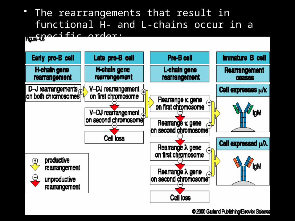
• The rearrangements that result in functional H- and L-chains occur in a specific order:
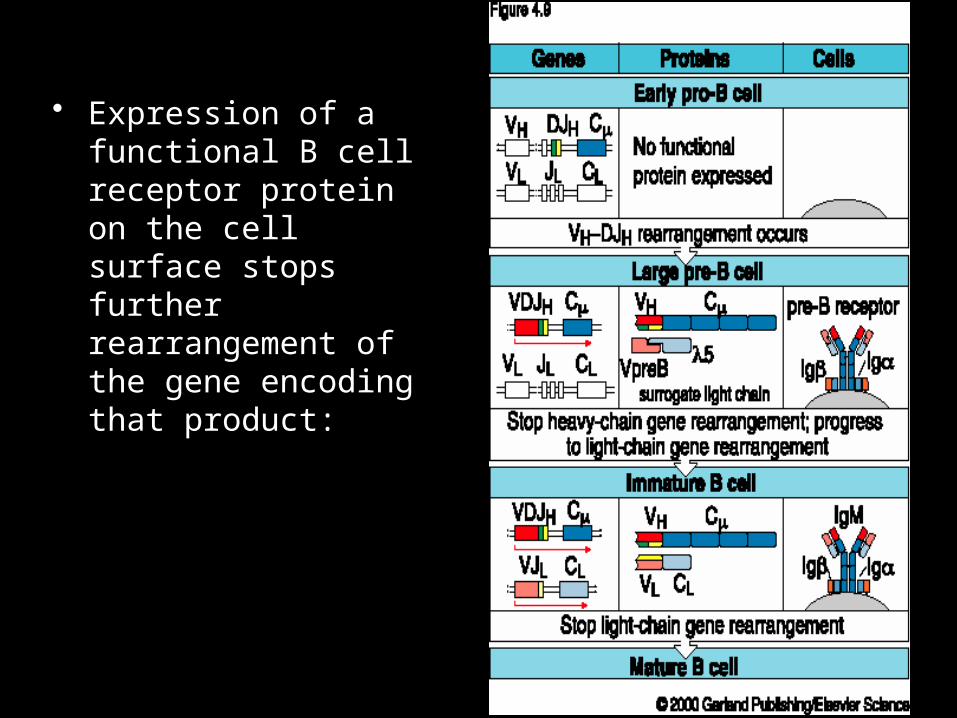
• Expression of a functional B cell receptor protein on the cell surface stops further rearrangement of the gene encoding that product:
Phase 2 – elimination of self-reactive cells
• Once B cells express a functional cell surface receptor for antigen (immature/mature B cell stage) they have the potential to be stimulated by contact with antigen, and to become antibody-secreting cells.
• Since the DNA rearrangements that result in functional H-chain and L-chain are not antigen-driven, a fraction of immature B cells will have a BCR that, by chance, reacts with some component of self - self antigen reactive cells
• These immature B cells are removed by clonal deletion, either in the bone marrow, or shortly after leaving the bone marrow.
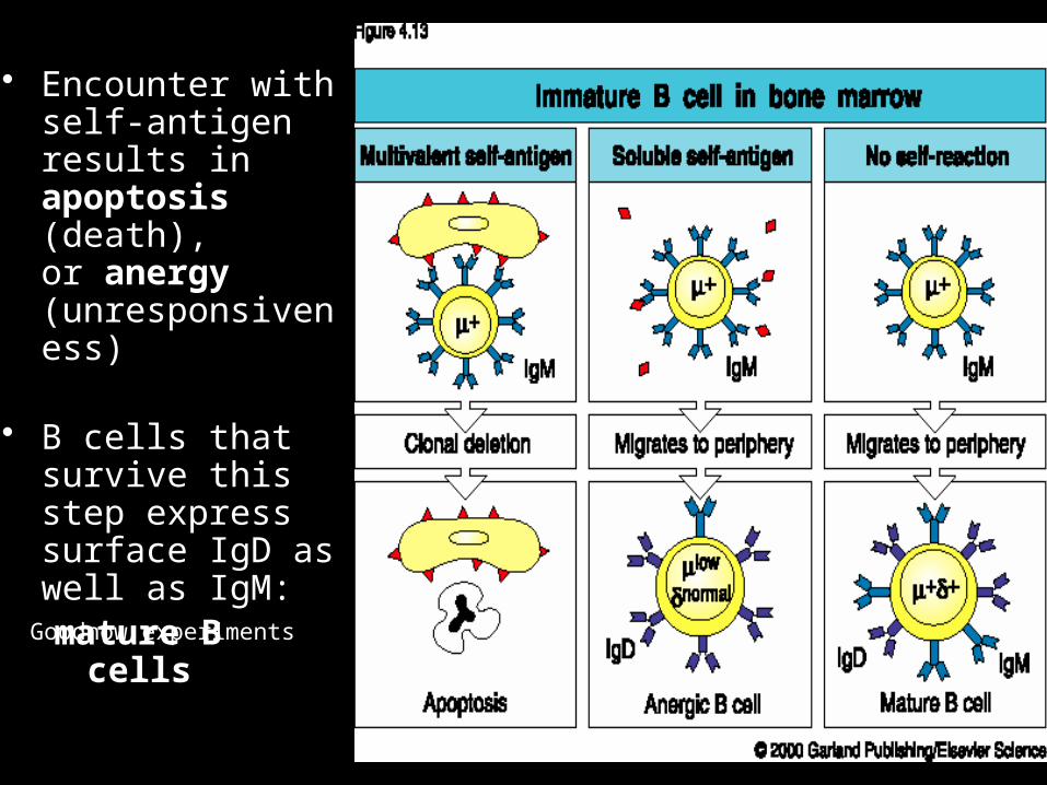
• Encounter with self-antigen results in apoptosis (death), or anergy (unresponsiveness)
• B cells that survive this step express surface IgD as well as IgM: mature B cells
Goodnow experiments
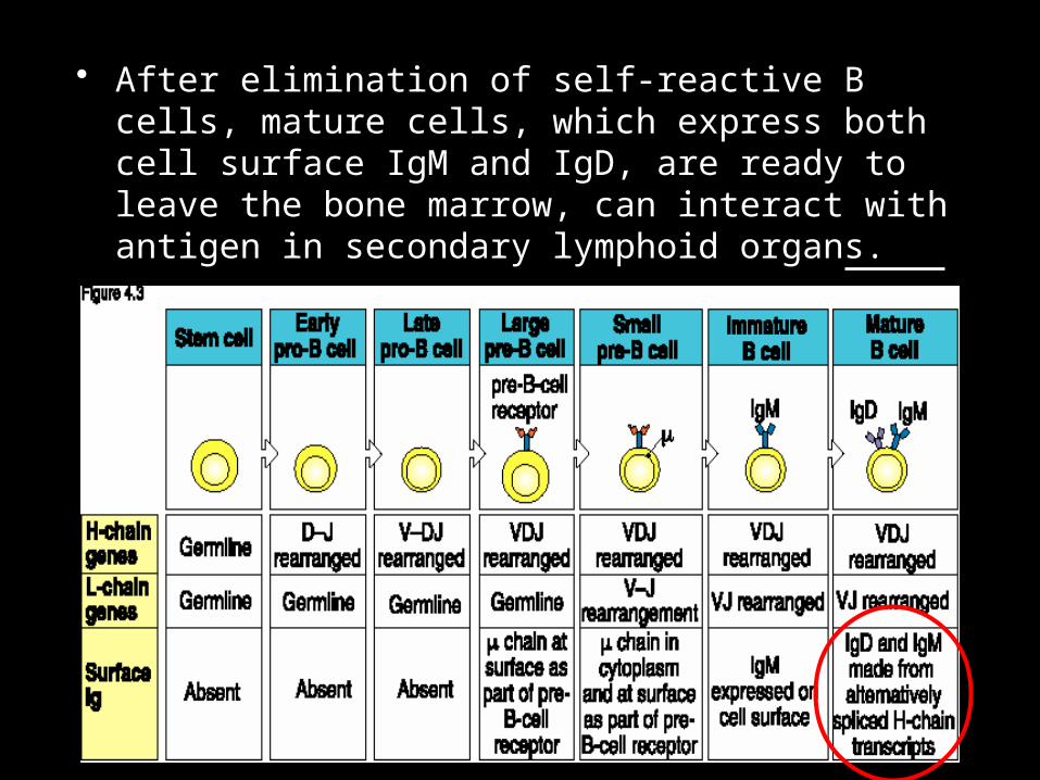
• After elimination of self-reactive B cells, mature cells, which express both cell surface IgM and IgD, are ready to leave the bone marrow, can interact with antigen in secondary lymphoid organs.

Phase 2 – elimination of self-reactive B cells, generation of mature IgM+, IgD+ cells:

Phase 3 – activation of B cells on contact with antigen
• Following the generation of a functional B cell receptor for Ag, and the removal of self-reactive cells, mature Ag-responsive B cells (IgM+, IgD+) emigrate from bone marrow.
• These mature B cells go to secondary lymphoid organs.

• In the lymph node, B cells gather in primary lymphoid follicles, where they receive viability-promoting signals, interacting with follicular dendritic cells, and wait for antigen
• B cells enter lymph nodes via high endothelial venules (HEV) to reach these primary follicles.
• B cells can recirculate out via the lymphatic circulation, and back into blood.

What is meant by ‘B cell activation’?
• Go -> G1 of cell cycle (increase in size)• upregulate
– MHC class II– costimulatory molecules (B7-2)– adhesion molecules (ICAM1)– cytokine receptors (IL-2R)
• migrate to outer T zone– altered response to chemokines
• become receptive to T cell help– protected from fas
• enter mitosis if provided with submitogenic doses of other stimuli (LPS, CD40L, IL-4)
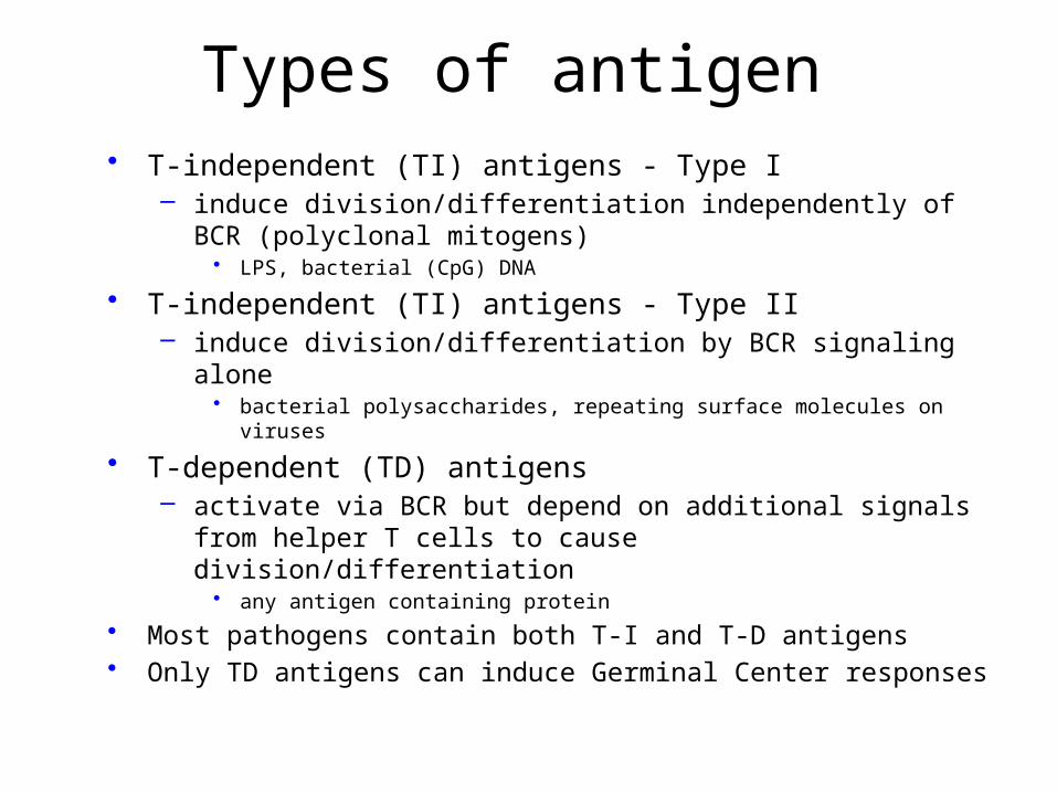
Types of antigen• T-independent (TI) antigens - Type I
– induce division/differentiation independently of BCR (polyclonal mitogens)• LPS, bacterial (CpG) DNA
• T-independent (TI) antigens - Type II– induce division/differentiation by BCR signaling alone
• bacterial polysaccharides, repeating surface molecules on viruses
• T-dependent (TD) antigens– activate via BCR but depend on additional signals from helper T cells
to cause division/differentiation• any antigen containing protein
• Most pathogens contain both T-I and T-D antigens• Only TD antigens can induce Germinal Center responses

T cell
mitogenic BcR signal
'activation' signalbut not mitogenic
mitogenesisdifferentiation
presentAg
T-independent (TI) T-cell dependent (TD)
Types of B cell Antigens
-> most pathogens contain both T-independent and T-dependent antigens

Innate features of pathogens act as B cell costimulators
• pathogen multivalency– provides a level of BCR crosslinking optimal for activation
• many pathogens activate TLRs – TLR signaling synergizes with BCR signal
• many pathogens activate the complement cascade and become C3d coated– complement receptor (CR) crosslinking synergizes with BCR
signal
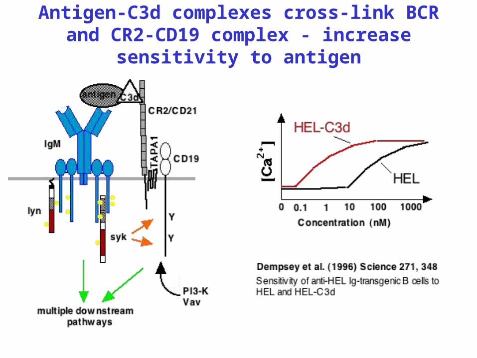
Antigen-C3d complexes cross-link BCR and CR2-CD19 complex - increase
sensitivity to antigen

Current Paradigm :
B cell plasma cellsB cell
B cell plasma cells
DC
Emerging Model:
DC
B cell
T-Independent (type II) Responses
Multivalent Antigen
B cell plasma cells
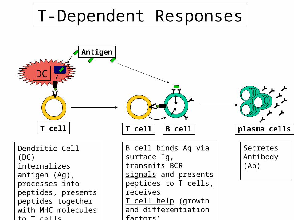
T-Dependent Responses
DC
Antigen
T cell T cell
Dendritic Cell (DC)internalizes antigen (Ag), processes into peptides, presents peptides together with MHC molecules to T cells
B cell binds Ag via surface Ig, transmits BCR signals and presents peptides to T cells, receives T cell help (growth and differentiation factors)
Secretes Antibody (Ab)

• Antigen-specific B cells are detained in the T cell-areas, where they interact with antigen, and with antigen-specific activated helper T cells. Stimulated antigen-specific B cells then proliferate and differentiate, eventually forming plasma cells and germinal centers:
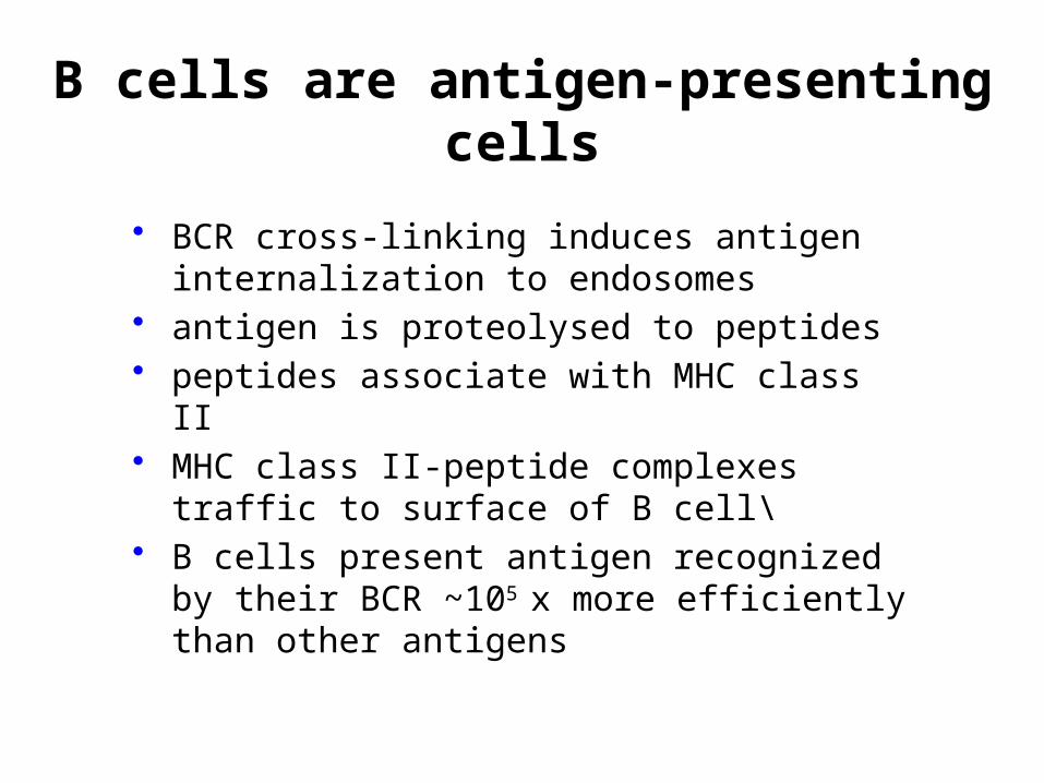
B cells are antigen-presenting cells
• BCR cross-linking induces antigen internalization to endosomes
• antigen is proteolysed to peptides• peptides associate with MHC class II• MHC class II-peptide complexes traffic to
surface of B cell\• B cells present antigen recognized by their BCR
~105 x more efficiently than other antigens

B cell antigen presentation and the concept of linked
help protein
sugar
T
Sugar SpecificB cell
Protein SpecificT cellAntigen internalization, proteolysis
-> presentation of peptides
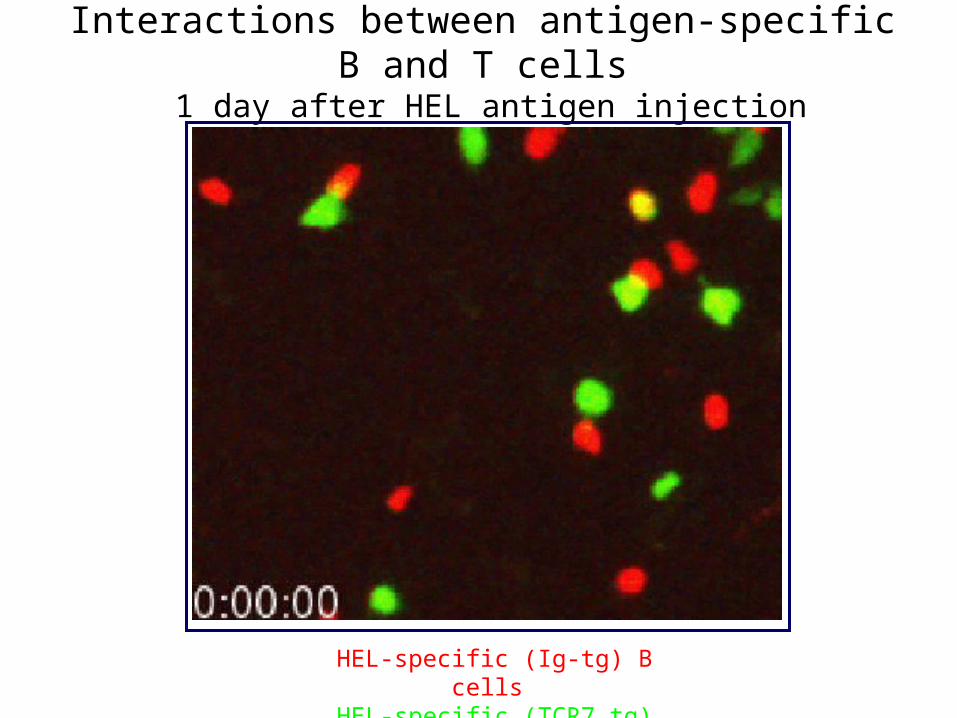
HEL-specific (Ig-tg) B cells HEL-specific (TCR7 tg) T cells
Interactions between antigen-specific B and T cells 1 day after HEL antigen injection

Onset of B-T interaction
HEL-specific B cells HEL-specific T cells
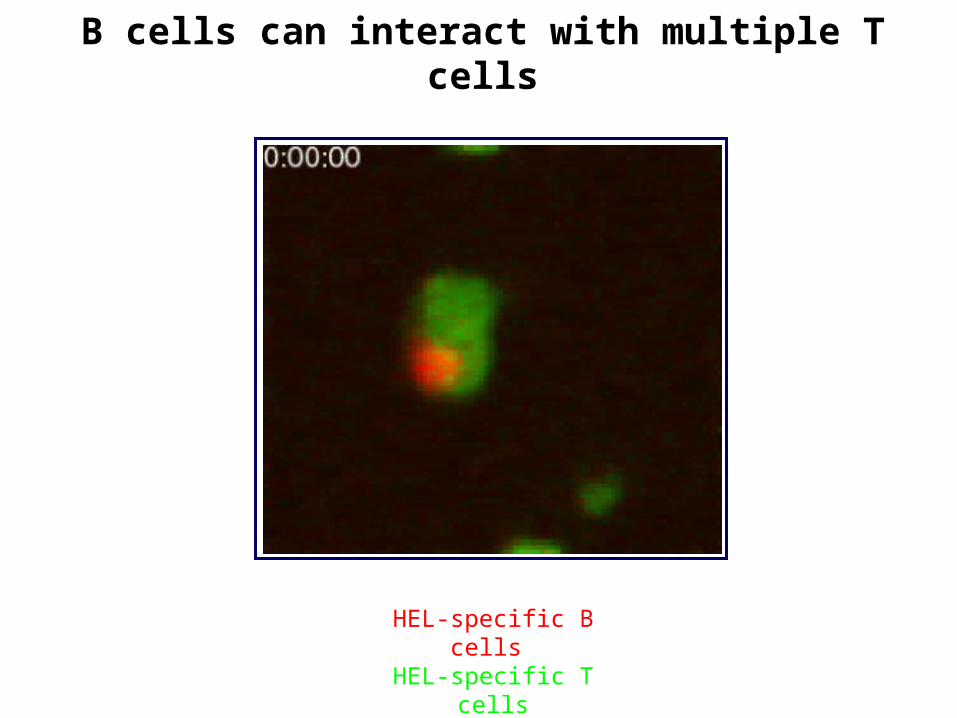
B cells can interact with multiple T cells
HEL-specific B cells HEL-specific T cells
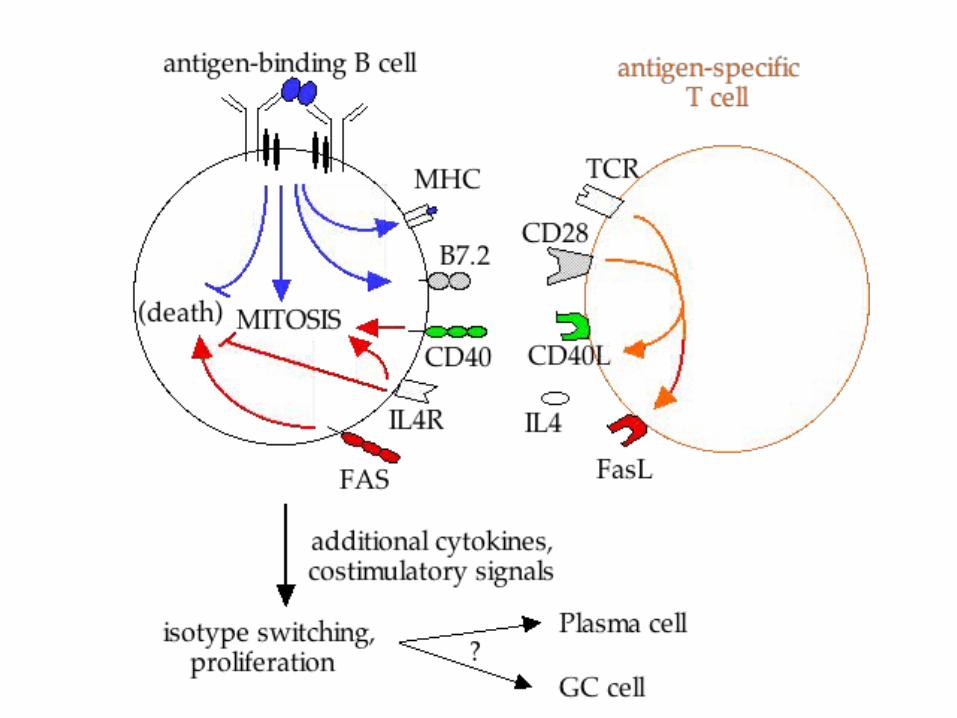
Cardinal features of B - T interaction
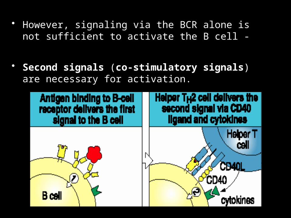
• However, signaling via the BCR alone is not sufficient to activate the B cell -
• Second signals (co-stimulatory signals) are necessary for activation.

-or-deficient mice and human do not undergo isotype switching
They only have IgM: hyper IgM syndrom

Phase 4 – differentiation to antibody-secreting cells
• Some of the progeny of these antigen-activated B cells differentiate into IgM-secreting plasma cells (antibody-secreting cells).
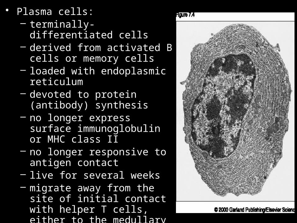
• Plasma cells:– terminally-differentiated cells– derived from activated B cells or
memory cells– loaded with endoplasmic
reticulum– devoted to protein (antibody)
synthesis– no longer express surface
immunoglobulin or MHC class II– no longer responsive to antigen
contact– live for several weeks– migrate away from the site of
initial contact with helper T cells, either to the medullary cords of the lymph nodes or to the bone marrow

Plasma cells
Surface Surface High rate Growth Somatic Isotype Ig MHC II Ig secretion hypermut’n switch
B
BMature B cell
Plasma cell
High Yes No Yes Yes Yes
Low No Yes No No No

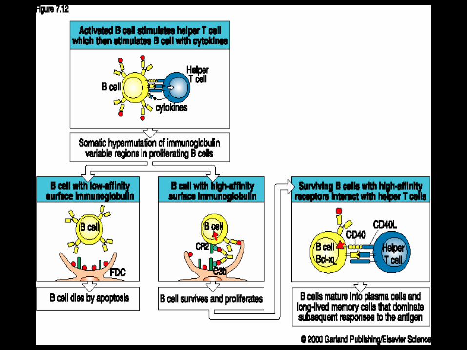
• Other antigen activated B cells give rise to germinal centers (GC), zones of proliferating activated B cells:

• These germinal centers (GC) contains:– proliferating (D - centroblasts) B cells express low
levels of Ig, especially IgD – differentiating (L - centrocytes) B cells express high
levels of Ig

• B cells can interact with an antigen that is bound to the surface of follicular dendritic cells in the lymph nodes.
• These cells trap and concentrate antigen, maximizing the interaction of antigen with B cells

Follicular Dendritic cells (stained blue)in the Germinal Centre

Retention of Antigens on Follicular Dendritic Cells
Radiolabelled antigen localises on the surface of Follicular Dendritic cellsand persists there, without internalisation, for very long periods
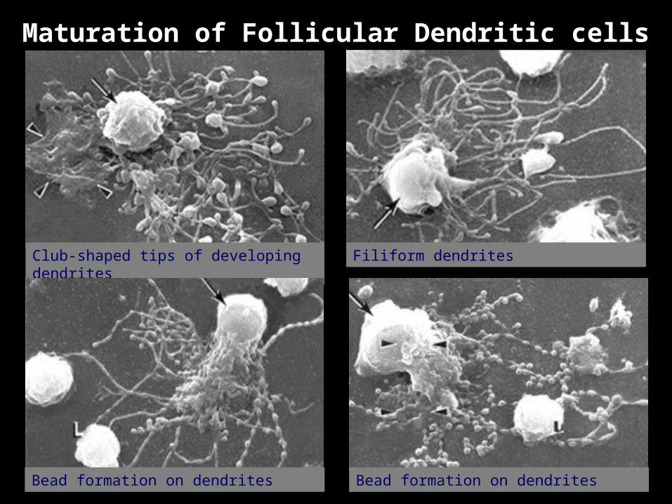
Maturation of Follicular Dendritic cells
Club-shaped tips of developing dendrites Filiform dendrites
Bead formation on dendrites Bead formation on dendrites

The veils of antigen-bearing dendritic cell surround the beads and the layer of immune complexes is thickened by transfer from the dendritic cell. These beads are then released and are then called ICCOSOMES
Iccosome formation and release
DC veils
Iccosomes (black coated particles) bind to and are taken up by B cell
surface immunoglobulin
Y YY
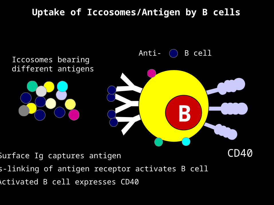
Iccosomes bearing different antigens
B
Uptake of Iccosomes/Antigen by B cells
Anti- B cell
Surface Ig captures antigen
Cross-linking of antigen receptor activates B cell
Activated B cell expresses CD40
CD40
B
B
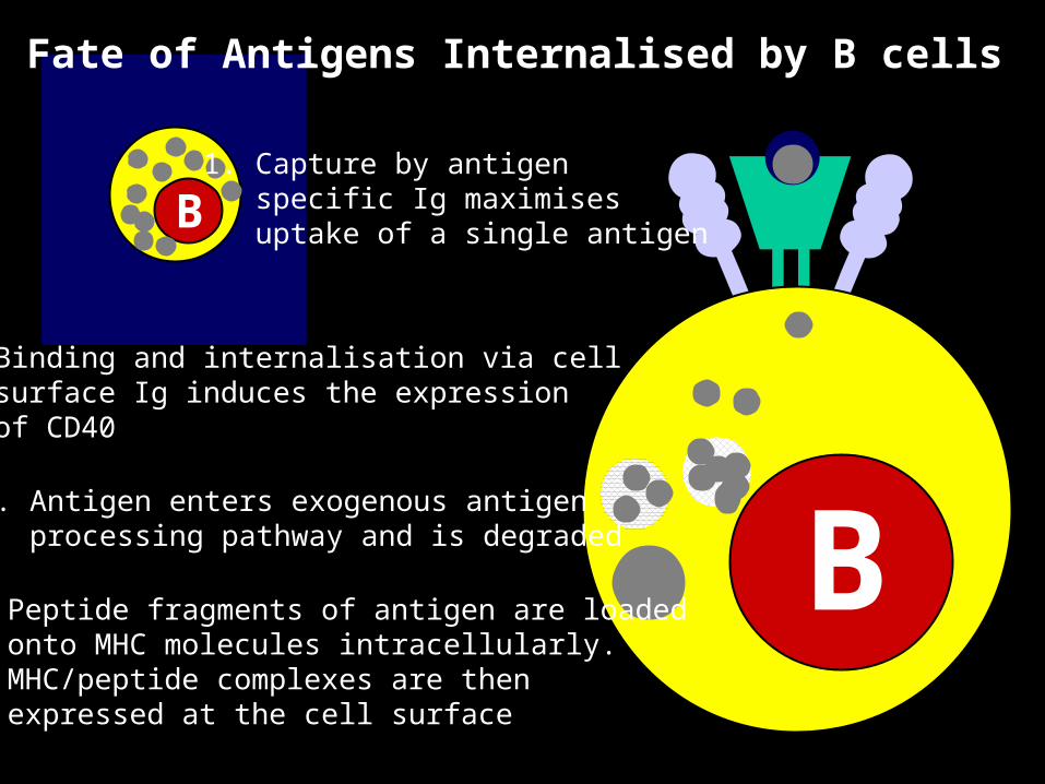
2. Binding and internalisation via cellsurface Ig induces the expressionof CD40
3. Antigen enters exogenous antigenprocessing pathway and is degraded
4. Peptide fragments of antigen are loadedonto MHC molecules intracellularly.MHC/peptide complexes are thenexpressed at the cell surface
Fate of Antigens Internalised by B cells
1. Capture by antigenspecific Ig maximisesuptake of a single antigen
• This selection process involves competition for both antigen and for helper T cells.
• Antigen is trapped on the surface of follicular dendritic cells in the form of immune complexes (antigen + antibody complexes).
• B cells that bind to Ag with high affinity live, others die by apoptosis.
• Centrocytes interact with T cells by presenting processed antigen to them via their MHC class II molecules.
• Centrocytes that receive co-signaling (via CD40, MHC class II and cytokines), as well as signaling via their antigen receptor survive
• Centrocytes that do not bind antigen and T cells with sufficient affinity die by apoptosis.

• Germinal centers are where isotype switching and somatic hypermutation occur.
• Somatic hypermutation:
– rapid mutation (hypermutation) of immunoglobulin genes
– results in antigen-binding affinity that is higher, or lower, than its original binding affinity
– selection by antigen results in the generation of BCR with increased affinity for antigen
• Only those B cells that have enhanced their antigen receptor’s binding affinity survive.
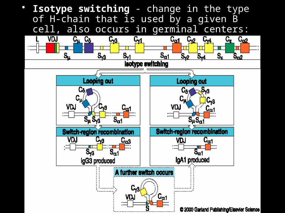
• Isotype switching - change in the type of H-chain that is used by a given B cell, also occurs in germinal centers:

• Isotype switching involves DNA rearrangements - replacement of one H-chain class gene with another
• Isotype switching also occurs in germinal centers
• Isotype switching is guided by the pattern of cytokines that are produced by helper T cells:
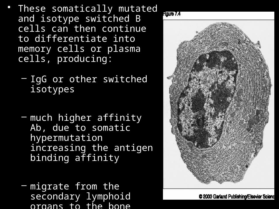
• These somatically mutated and isotype switched B cells can then continue to differentiate into memory cells or plasma cells, producing:
– IgG or other switched isotypes
– much higher affinity Ab, due to somatic hypermutation increasing the antigen binding affinity
– migrate from the secondary lymphoid organs to the bone marrow, where cytokines (IL-6, IL-11) produced by bone marrow stromal cells keep these cells viable and producing Abs

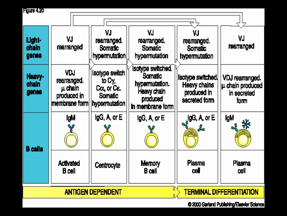
Together, these processes result in a 1-2 log increase in the number of antigen-specific B cells (clonal selection and expansion), an increase in antibody binding affinity for antigen (somatic hypermutation), and the expression of new Ig subclasses (Ig isotype switching):
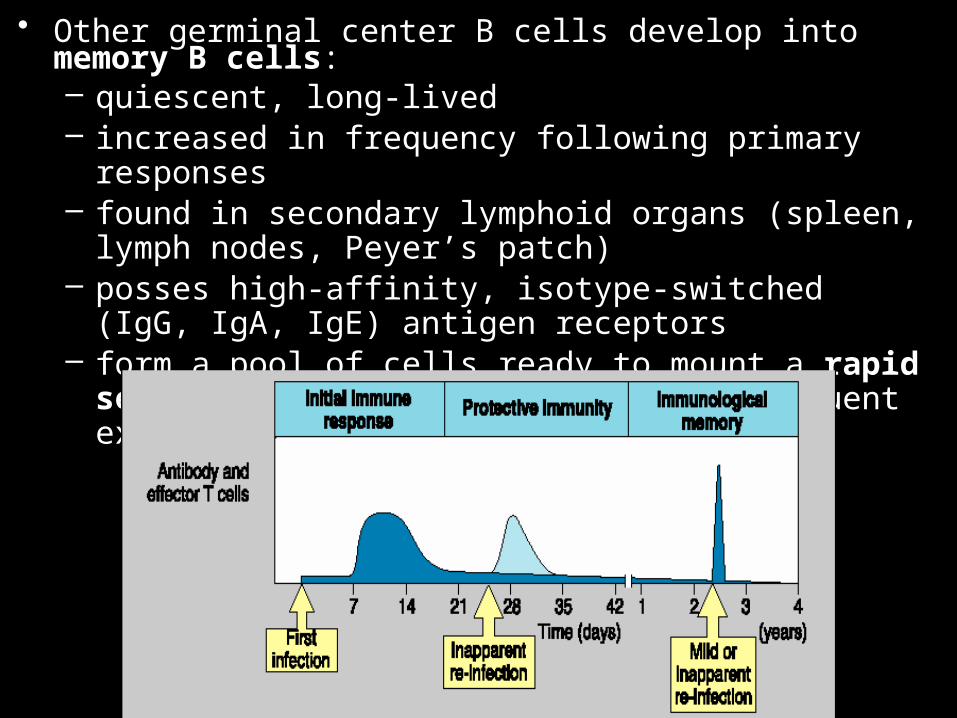
• Other germinal center B cells develop into memory B cells:– quiescent, long-lived– increased in frequency following primary responses– found in secondary lymphoid organs (spleen, lymph nodes,
Peyer’s patch)– posses high-affinity, isotype-switched (IgG, IgA, IgE)
antigen receptors– form a pool of cells ready to mount a rapid secondary
antibody response, on subsequent exposure to antigen:

• The combined result of clonal selection (expansion of pool size of Ag-reactive cells), somatic hypermutation, isotype switching, and the generation of memory cells, is the creation of a pool of cells that can respond rapidly and vigorously to subsequent contact with antigen, with high-affinity, IgG and IgA antibodies.

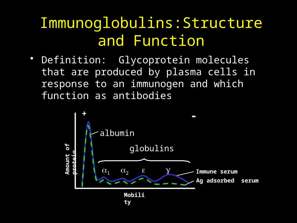
Immunoglobulins:Structure and Function
• Definition: Glycoprotein molecules that are produced by plasma cells in response to an immunogen and which function as antibodies
Immune serum
Ag adsorbed serum
a1 a2 e
+ -
albumin
globulins
Mobility
Am
oun
t of
pro
tein
g
General Functions of Immunoglobulins
• Effector functions – Fixation of complement– Binding to various cells
(Usually require Ag binding)
• Ag binding– Can result in protection

Immunoglobulin Structure
• Heavy & Light Chains
• Disulfide bonds– Inter-chain– Intra-chain
CH1
VL
CL
VH
CH2 CH3
Hinge Region
Carbohydrate
Disulfide bond

Immunoglobulin Structure
• Variable & Constant Regions– VL & CL
– VH & CH
• Hinge RegionCH1
VL
CL
VH
CH2 CH3
Hinge Region
Carbohydrate
Disulfide bond
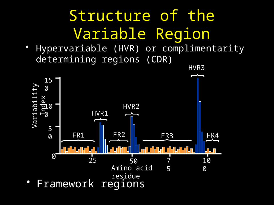
Structure of the Variable Region• Hypervariable (HVR) or complimentarity determining
regions (CDR)HVR3
FR1 FR2 FR3 FR4
HVR1HVR2
Var
iabi
lity
Ind
ex
25 7550 100Amino acid residue
150
100
50
0
• Framework regions

Immunoglobulin Fragments: Structure/Function Relationships
• Fab– Ag binding– Valence = 1– Specificty
determined by VH and VL
Papain
Fc
Fab
• Fc– Effector functions
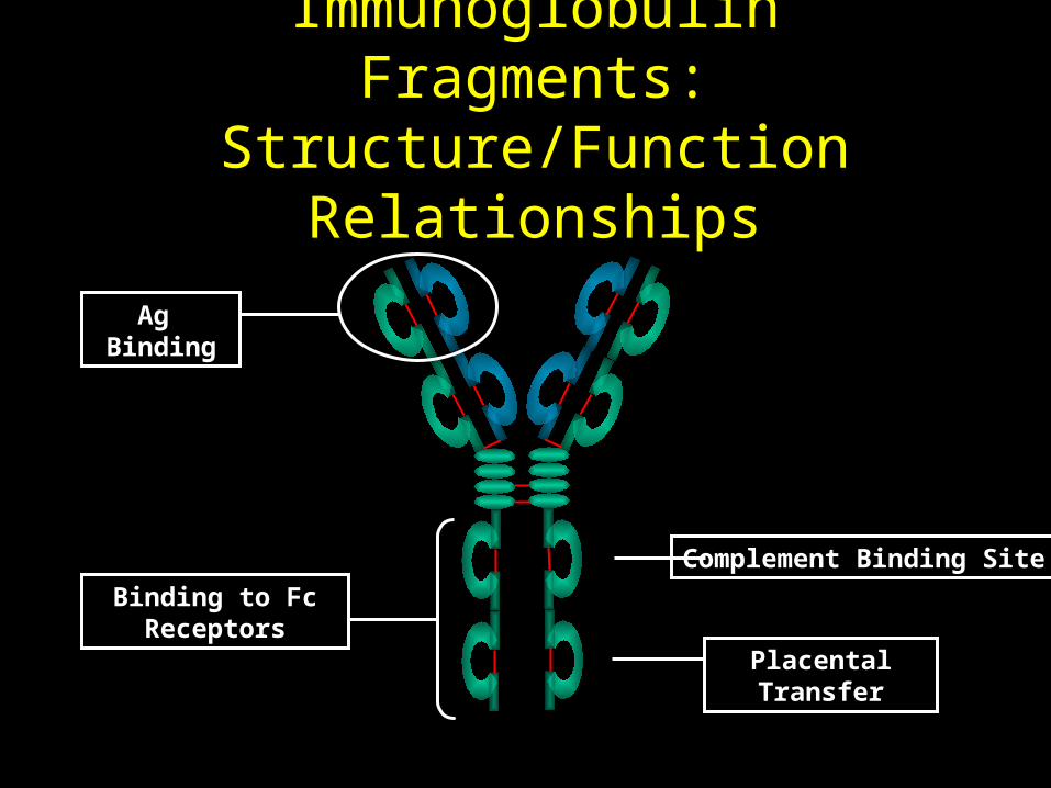
Immunoglobulin Fragments: Structure/Function Relationships
Ag Binding
Complement Binding Site
Placental Transfer
Binding to Fc Receptors
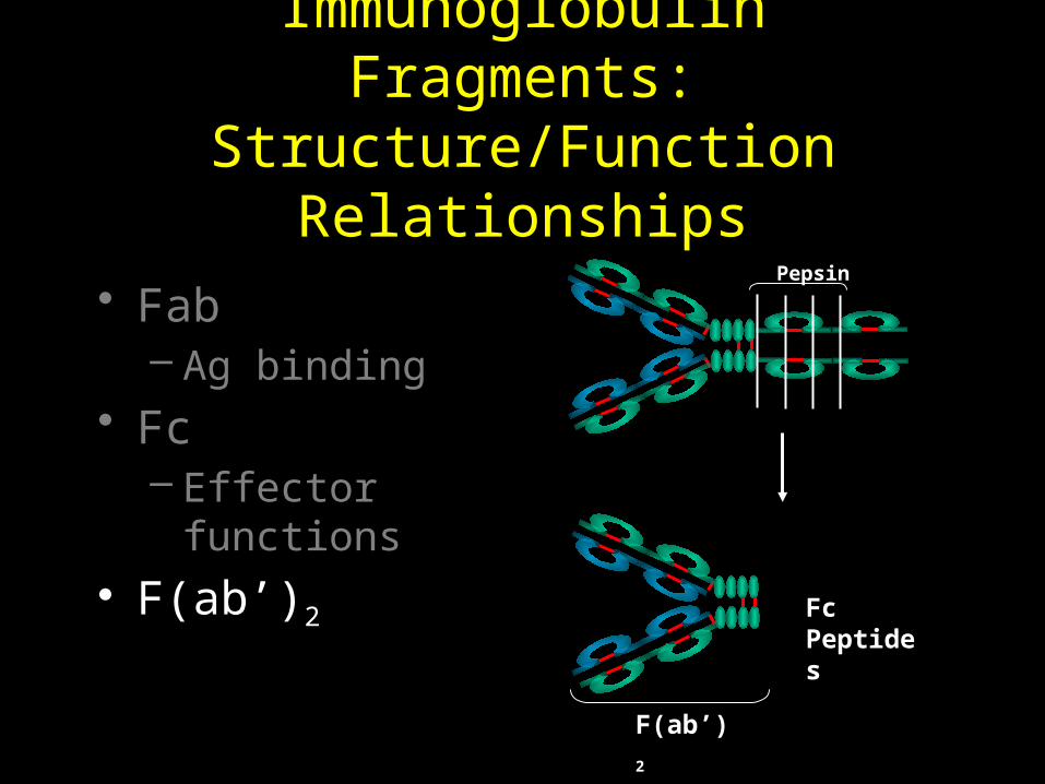
Immunoglobulin Fragments: Structure/Function Relationships
• Fab– Ag binding
• Fc– Effector functions
• F(ab’)2
Pepsin
Fc Peptides
F(ab’)2

Human Immunoglobulin Classes
• IgG - Gamma heavy chains• IgM - Mu heavy chains• IgA - Alpha heavy chains• IgD - Delta heavy chains• IgE - Epsilon heavy chains
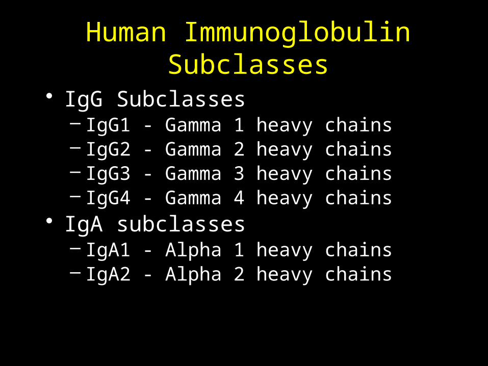
Human Immunoglobulin Subclasses
• IgG Subclasses– IgG1 - Gamma 1 heavy chains– IgG2 - Gamma 2 heavy chains– IgG3 - Gamma 3 heavy chains– IgG4 - Gamma 4 heavy chains
• IgA subclasses– IgA1 - Alpha 1 heavy chains– IgA2 - Alpha 2 heavy chains
Human ImmunoglobulinLight Chain Types
• Kappa ()• Lambda ()

Human ImmunoglobulinLight Chain Subtypes
• Lambda light chains– Lambda 1 (1)– Lambda 2 (2)– Lambda 3 (3) – Lambda 4 (4)
Immunoglobulins
• Nomenclature– IgM (kappa)– IgA1(lambda 2)– IgG
• Heterogeneity

Monomeric IgM
IgM only exists as a monomer on the surface of B cells
Cm4 contains the transmembrane and cytoplasmic regions. These are
removed by RNA splicing to produce secreted IgM
Monomeric IgM has a very low affinity for antigen
Cm4
Cm3Cm2 Cm1
N.B. Only constant heavy chain
domains are shown
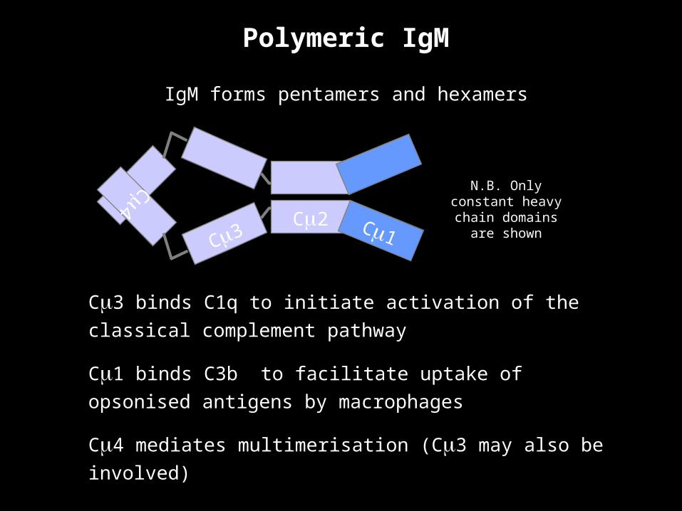
Cm3 binds C1q to initiate activation of the classical
complement pathway
Cm1 binds C3b to facilitate uptake of opsonised antigens
by macrophages
Cm4 mediates multimerisation (Cm3 may also be involved)
Cm4
Cm3Cm2 Cm1
N.B. Only constant heavy chain
domains are shown
Polymeric IgM
IgM forms pentamers and hexamers

CC
C
C
C C
Multimerisation of IgM
Cm
4
Cm
3
Cm2
C
C
Cm4
Cm
3Cm
2
C C
Cm4
Cm3
Cm2
C
C
Cm4
Cm3Cm2
C
C
Cm
4Cm3
Cm2
C
C
s s
ss
ss
C
C
ss
1. Two IgM monomers in the ER(Fc regions only shown)
2. Cysteines in the J chain form disulphide bonds with cysteines from each monomer to form a dimer
3. A J chain detaches leaving the dimer disulphide bonded.
4. A J chain captures another IgM monomer and joins it to the dimer.
5. The cycle is repeated twice more
6. The J chain remains attached to the IgM pentamer.

Antigen-induced conformational changes in IgM
Planar or ‘Starfish’ conformation found in
solution.
Does not fix complement
Staple or ‘crab’ conformation of IgM
Conformation change induced by
binding to antigen.
Efficient at fixing complement
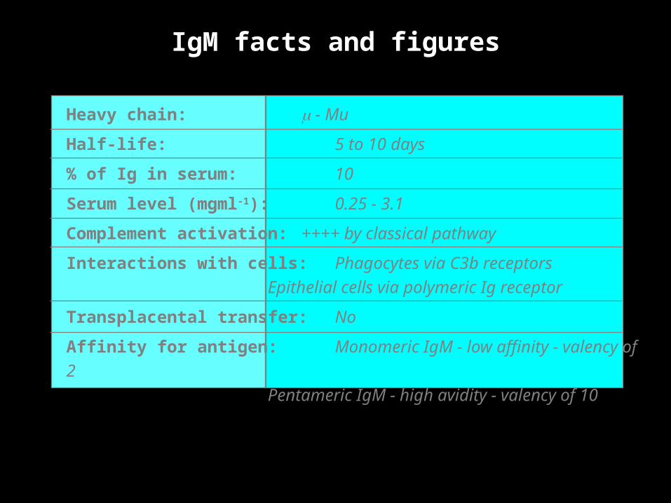
IgM facts and figures
Heavy chain: m - Mu
Half-life: 5 to 10 days
% of Ig in serum: 10
Serum level (mgml-1): 0.25 - 3.1
Complement activation: ++++ by classical pathway
Interactions with cells: Phagocytes via C3b receptors
Epithelial cells via
polymeric Ig receptor
Transplacental transfer: No
Affinity for antigen: Monomeric IgM - low affinity - valency of
2
Pentameric IgM - high
avidity - valency of 10
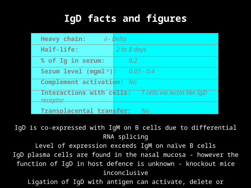
IgD facts and figures
IgD is co-expressed with IgM on B cells due to differential RNA splicing
Level of expression exceeds IgM on naïve B cells
IgD plasma cells are found in the nasal mucosa - however the function of IgD in
host defence is unknown - knockout mice inconclusive
Ligation of IgD with antigen can activate, delete or anergise B cells
Extended hinge region confers susceptibility to proteolytic degradation
Heavy chain: d - Delta
Half-life: 2 to 8 days
% of Ig in serum: 0.2
Serum level (mgml-1): 0.03 - 0.4
Complement activation: No
Interactions with cells: T cells via lectin like IgD receptor
Transplacental transfer: No
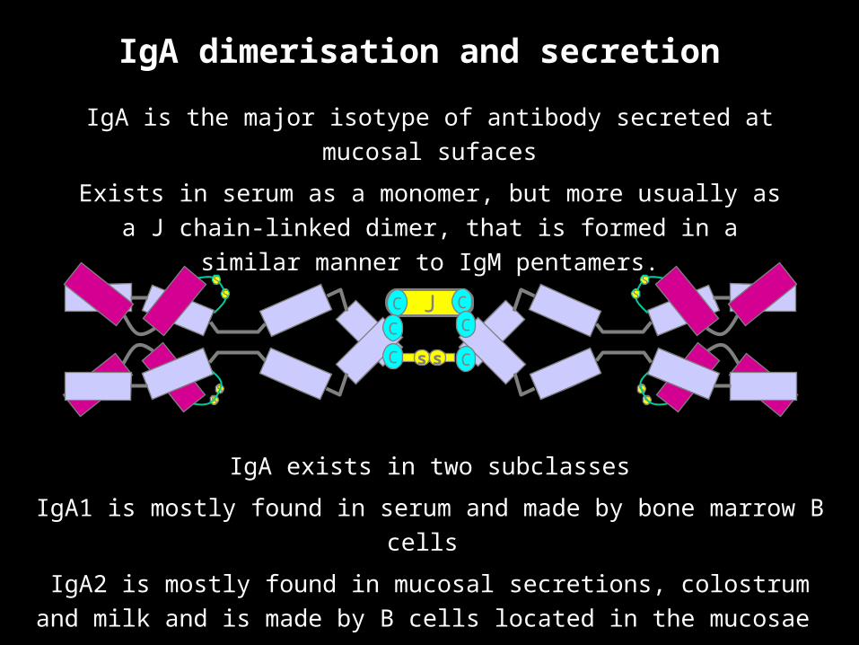
IgA dimerisation and secretion
IgA is the major isotype of antibody secreted at mucosal sufaces
Exists in serum as a monomer, but more usually as a J chain-
linked dimer, that is formed in a similar manner to IgM pentamers.
JC C
SS
SS
C
C
SS
SS
C
C
s s
IgA exists in two subclasses
IgA1 is mostly found in serum and made by bone marrow B cells
IgA2 is mostly found in mucosal secretions, colostrum and milk and is made
by B cells located in the mucosae

Epithelialcell
JC C
SS
SS
C
C
SS
SS
CC
ss
Secretory IgA and transcytosis
B
JC C
SS
SS
CC
SS
SS
CCss
JC C
SS
SS
C
C
SS
SS
CC
ss
JC C
SS
SS
C
C
SS
SS
CC
ss
pIgR & IgA areinternalised
‘Stalk’ of the pIgR is degraded to release IgA containing part of the pIgR - the secretory component
JC C
SS
SS
C
C
SS
SS
CC
ss
IgA and pIgR are transported to the apical surface in vesicles
B cells located in the submucosaproduce dimeric IgA
Polymeric Ig receptors are expressed on the basolateral surface of epithelial cells to capture IgA produced in the mucosa
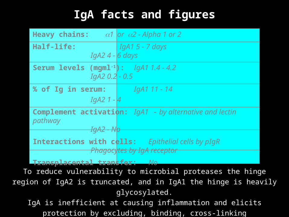
IgA facts and figures
Heavy chains: a1 or a2 - Alpha 1 or 2
Half-life: IgA1 5 - 7 daysIgA2 4 - 6 days
Serum levels (mgml-1): IgA1 1.4 - 4.2IgA2 0.2 - 0.5
% of Ig in serum: IgA1 11 - 14
IgA2 1 - 4
Complement activation: IgA1 - by alternative and lectin pathwayIgA2 - No
Interactions with cells: Epithelial cells by pIgRPhagocytes by IgA receptor
Transplacental transfer: No
To reduce vulnerability to microbial proteases the hinge region of IgA2 is truncated,
and in IgA1 the hinge is heavily glycosylated.
IgA is inefficient at causing inflammation and elicits protection by excluding, binding,
cross-linking microorganisms and facilitating phagocytosis
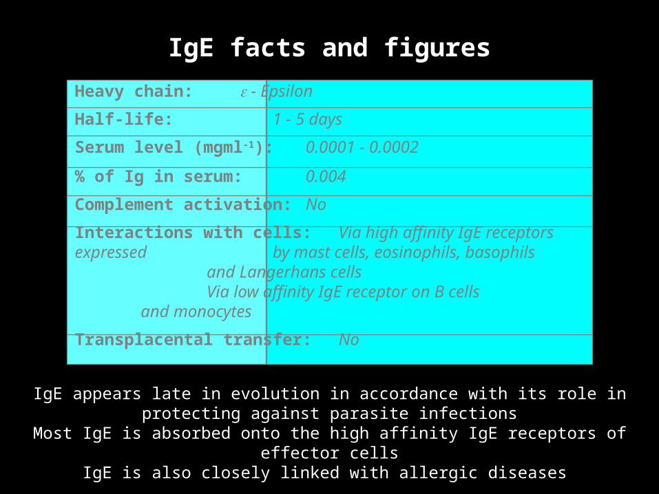
IgE facts and figures
IgE appears late in evolution in accordance with its role in protecting against parasite infections
Most IgE is absorbed onto the high affinity IgE receptors of effector cellsIgE is also closely linked with allergic diseases
Heavy chain: e - Epsilon
Half-life: 1 - 5 days
Serum level (mgml-1): 0.0001 - 0.0002
% of Ig in serum: 0.004
Complement activation: No
Interactions with cells: Via high affinity IgE receptors expressed by mast cells, eosinophils,
basophils and Langerhans cells
Via low affinity IgE receptor on B cells and monocytes
Transplacental transfer: No
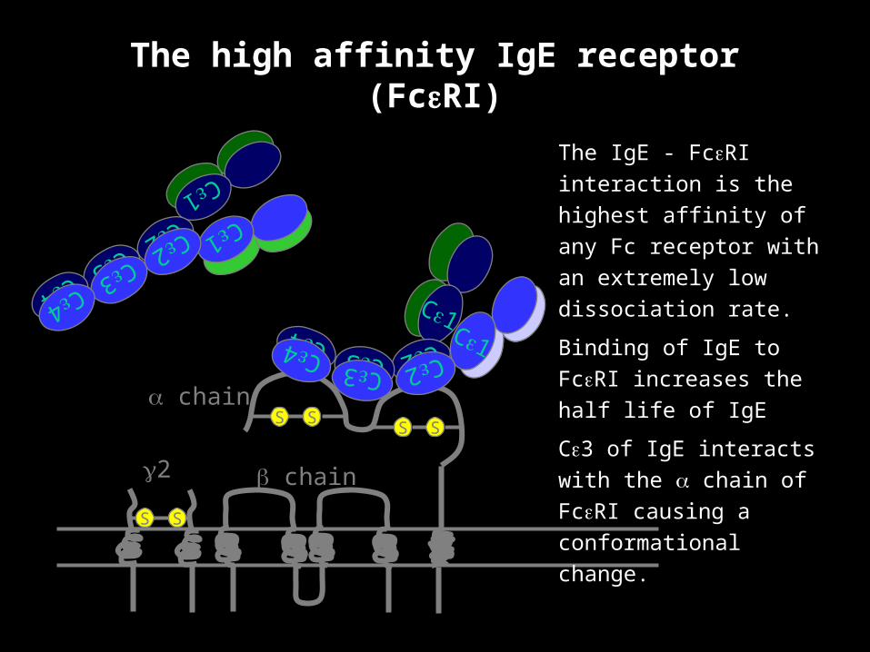
The high affinity IgE receptor (FceRI)
a chain
b chaing2
S SS S
S S
Ce1Ce1Ce2Ce2Ce3Ce3
Ce4Ce4
Ce1
Ce1Ce2Ce2
Ce3Ce3
Ce4Ce4
The IgE - FceRI interaction
is the highest affinity of any
Fc receptor with an
extremely low dissociation
rate.
Binding of IgE to FceRI
increases the half life of IgE
Ce3 of IgE interacts with the
a chain of FceRI causing a
conformational change.

IgG facts and figures
Heavy chains: g 1 g 2 g3 g4 - Gamma 1 - 4
Half-life: IgG1 21 - 24 days IgG2 21 - 24 days
IgG3 7 - 8 days IgG4 21 - 24 days
Serum level (mgml-1): IgG1 5 - 12 IgG2 2 - 6
IgG3 0.5 - 1IgG4 0.2 - 1
% of Ig in serum: IgG1 45 - 53IgG2 11 - 15
IgG3 3 - 6IgG4 1 - 4
Complement activation: IgG1 +++ IgG2 +
IgG3 ++++ IgG4 No
Interactions with cells: All subclasses via IgG receptors on macrophages and phagocytes
Transplacental transfer: IgG1 ++IgG2 +
IgG3 ++IgG4 ++

Carbohydrate is essential for complement activation
Subltly different hinge regions between subclasses accounts for differing abilities to activate complement
C1q binding motif is located on the Cg2 domain

Fcg receptors
Receptor Cell type Effect of ligation
FcgRI Macrophages Neutrophils,
Eosinophils, Dendritic cells Uptake, Respiratory burst
FcgRIIA Macrophages Neutrophils,
Eosinophils, Platelets
Langerhans cells Uptake, Granule release
FcgRIIB1 B cells, Mast Cells No Uptake, Inhibition of
stimulation
FcgRIIB2 Macrophages Neutrophils,
Eosinophils Uptake, Inhibition of
stimulation
FcgRIII NK cells, Eosinophils,
Macrophages, Neutrophils
Mast cells Induction of killing (NK cells)
High affinity Fcg receptors from the Ig superfamily: