award number: w81xwh-14-1-0111 - apps.dtic.mil · (sap and pdc), hr (faculty and staff) and other...
TRANSCRIPT
AWARD NUMBER: W81XWH-14-1-0111
TITLE: A Molecular Framework for Understanding DCIS
PRINCIPAL INVESTIGATOR: Herbert Lyerly
CONTRACTING ORGANIZATION: Duke University
Durham, NC 27705
REPORT DATE: October 2016
TYPE OF REPORT: Annual
PREPARED FOR: U.S. Army Medical Research and Materiel Command
Fort Detrick, Maryland 21702-5012
DISTRIBUTION STATEMENT: Approved for Public Release;
Distribution Unlimited
The views, opinions and/or findings contained in this report are those of the author(s) and should
not be construed as an official Department of the Army position, policy or decision unless so
designated by other documentation.

REPORT DOCUMENTATION PAGE Form Approved
OMB No. 0704-0188 Public reporting burden for this collection of information is estimated to average 1 hour per response, including the time for reviewing instructions, searching existing data sources, gathering and maintaining the data needed, and completing and reviewing this collection of information. Send comments regarding this burden estimate or any other aspect of this collection of information, including suggestions for reducing this burden to Department of Defense, Washington Headquarters Services, Directorate for Information Operations and Reports (0704-0188), 1215 Jefferson Davis Highway, Suite 1204, Arlington, VA 22202-4302. Respondents should be aware that notwithstanding any other provision of law, no person shall be subject to any penalty for failing to comply with a collection of information if it does not display a currently valid OMB control number. PLEASE DO NOT RETURN YOUR FORM TO THE ABOVE ADDRESS.
1. REPORT DATE
October 20162. REPORT TYPE
Annual 3. DATES COVERED
30 Sep 2015 - 29 Sep 2016
A Molecular Framework for Understanding DCIS
5a. CONTRACT NUMBER
5b. GRANT NUMBER
W81XWH-14-1-0111 5c. PROGRAM ELEMENT NUMBER
6. AUTHOR(S)
H. Kim Lyerly, M.D. 5d. PROJECT NUMBER
5e. TASK NUMBER
E-Mail: [email protected] 5f. WORK UNIT NUMBER
7. PERFORMING ORGANIZATION NAME(S) AND ADDRESS(ES)
AND ADDRESS(ES)
8. PERFORMING ORGANIZATION REPORTNUMBER
Duke University
2200 W. Main St., STE 700
Durham, NC 27705
9. SPONSORING / MONITORING AGENCY NAME(S) AND ADDRESS(ES) 10. SPONSOR/MONITOR’S ACRONYM(S)
U.S. Army Medical Research and Materiel Command
Fort Detrick, Maryland 21702-5012 11. SPONSOR/MONITOR’S REPORT
12. DISTRIBUTION / AVAILABILITY STATEMENT
Approved for Public Release; Distribution Unlimited
13. SUPPLEMENTARY NOTES
14. ABSTRACT
DCIS is proposed to be a precursor to invasive breast cancer. Improvements in early diagnosis have led to increased numbers of DCIS
cases, however, we presently have no way to predict which DCIS lesions are at risk for progression to invasive cancer. We also lack the
extensive molecular profiling necessary to place DCIS within the framework used to classify and guide treatment for invasive disease. We
propose to take advantage of a unique set of specimens, comprising DCIS and early invasive disease, and using next-generation
sequencing, understand the nature of DCIS and the events that determine and promote its progression. We have already developed the
approaches necessary for obtaining profiles from the tumor and stromal compartments of DCIS, lesions from which only limited numbers
of cells can be obtained. The availability of our datasets will transform the understanding of early disease and may ultimately alter the
course of treatment for women with a diagnosis of DCIS.
15. SUBJECT TERMS
Breast cancer, DCIS
16. SECURITY CLASSIFICATION OF:
U 17. LIMITATION
OF ABSTRACT
18. NUMBER
OF PAGES
19a. NAME OF RESPONSIBLE PERSON
USAMRMC
a. REPORT
Unclassified
b. ABSTRACT
Unclassified
c. THIS PAGE
Unclassified Unclassified 28
19b. TELEPHONE NUMBER (include area
code)
Standard Form 298 (Rev. 8-98) Prescribed by ANSI Std. Z39.18

Table of Contents
Page
1. Introduction ............................................................................................4
2. Approach ...............................................................................................4
3. Key Research Accomplishments ...........................................................5
4. Reportable Outcomes .......................................................................... 17
5. Participants & Other Collaborating Organizations ............................... 18
6. Conclusion ........................................................................................... 19
7. Appendices .......................................................................................... 19

4
A Molecular Framework for Understanding DCIS
Award No. W81XWH-14-1-0111 Annual Report Year 2
1. Introduction
This project centers on creating a molecular framework of DCIS (ductal carcinoma in situ). DCIS is considered to be the precursor to Invasive Ductal Carcinoma (IDC), the most common form of breast cancer. IDC accounts for 80% of all breast cancers, predominantly affecting women aged 55 and older; however, at least a third of women with IDC are diagnosed before they reach 55.
Utilizing a unique bank of frozen mammary biopsies, containing samples with DCIS alone, and a combination of DCIS and IDC, we aim to profile both DCIS and related tissue components. It is our aim to sample the ~300 biopsies, and compare both by RNA seq, and whole genome amplification, DCIS lesions, within, and between patients, and see how these may be correlated with IDC lesions. We also intend to look for changes in the stroma between those patients that present with IDC and those that do not. This work aims to identify characteristics that may be suggestive of a patients’ likelihood of progressing from DCIS to IDC, with the purpose of reducing the need for over treatment for this disease.
2. Approach
We first applied and received appropriate regulatory approval from the Duke SPORE tissue use committee, the Duke Cancer Center Protocol Review Committee, the Duke IRB, and the DoD. An MTA was established with the University of Cambridge.
We have created a DCIS Clinical Annotation database which was created and is being managed by Cedars-Sinai Medical Center in Los Angeles. A remote, web based case report from was also created to enable data abstraction for Duke medical records.
All Duke breast cancer research specimens were identified and prioritized based on pathologic diagnosis. Clinical data was abstracted from Duke clinical data by prioritizing pure DCIS, mixed DCIS and invasive BC samples, and invasive BC samples. Normal breast and atypical will be annotated if needed as controls.
Data was gathered from 1) electronic medical records at Duke University Medical Center and 2) extracted from DCIS patient charts (which hold the specific clinical annotation including patient outcome).
After identifying breast cancer research specimens with pathologic and clinical data consistent with DCIS and/or DCIS and invasive breast cancer, shipping to Dr. Greg Hannon for further processing was performed.

5
3. Key Research Accomplishments
Major goals of the project (as stated in SOW)
1. Regulatory approval and MTA (Duke/Cambridge)2. Database creation and remote web based CRF created (Duke)3. Sample collection/annotation, shipping to Cambridge (Duke)4. Laser capture of frozen material (Duke pathologist working remotely or at
CSHL/Cambridge)5. Exome capture and DNA sequencing (CSHL/Cambridge)6. RNAseq library construction (CSHL/Cambridge)7. Analysis DNA data (CSHL/NYGC)8. Analyze RNA differential expression (NYGC)9. Analyze stroma compartments (CSHL/Cambridge)10. Technical validation of potential markers (Cambridge)11. Validate potential markers in FFPE cohort (Duke/Cambridge)12. Validate in longitudinal cohort (Duke /Cambridge)13. Nominate candidates for clinical validation (Duke /Cambridge)
Dr. Greg Hannon of Cancer Research UK Cambridge Institute will provide his research accomplishments via a separate report.
What was accomplished under these goals:
Tissue Use protocol
A Tissue Use protocol, “Molecular Framework of Early Breast Cancer” (IRB# Pro00059726) was approved by the Duke IRB, Cancer Protocol Committee (CPC), and DOD to allow for the use of banked tissue samples for the proposed studies (see Appendix for protocol). The 2016 continuing review by the Duke IRB is included in the Appendix as well.
Pathologic and Clinical Annotation Database
A clinical annotation database titled the Breast Oncology Database has been established to complement the procured SPORE sample characteristics and annotated pathology data. This Breast Oncology Database is an offsite clinical annotation database created in collaboration with Cedar-Sinai Medical Center. This database is built on the REDCap platform and includes the full functionality of REDCap. This database is housed on a server at Cedars-Sinai Medical Center (CSMC) and is accessible on the web via https (no VPN required). This database adheres to CSMC Enterprise Information Services (EIS) research database security standards. The Breast Oncology Database consists of: 9 Baseline forms and 4 Follow-up forms with a total of 779 possible entry fields. The clinical data elements collected include: demographics, radiographic findings (mammography, U/S, MRI, CT, PET), pathologic diagnosis (histology, IHC, and FISH), surgical and radiation treatment received, systemic treatment received (dates and dosing regimens), and survival data.
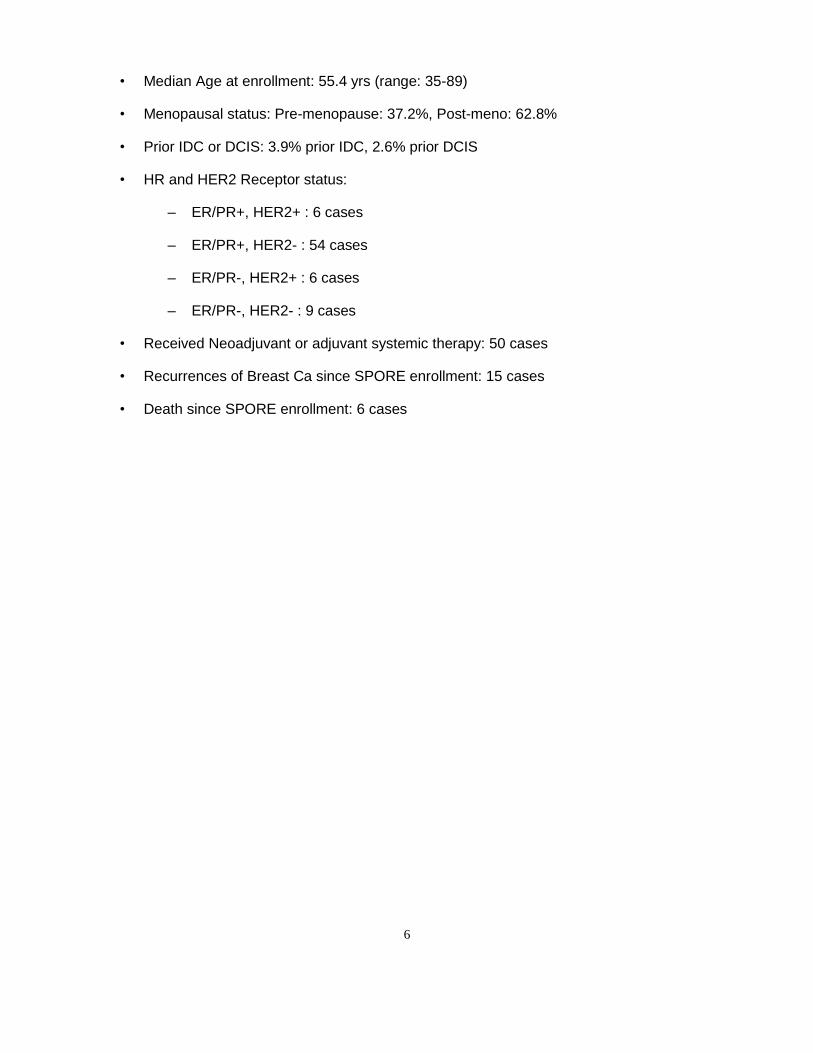
To date we have annotated 197 cases of DCIS and DCIS/IDC contained in the SPORE tissue bank, this includes the 78 cases sent to Dr. Hannon’s lab for genomic evaluation. Below is a clinical summary of the clinical cases related to the 78 tumor samples sent to Dr. Hannon’s lab:
6
• Median Age at enrollment: 55.4 yrs (range: 35-89)
• Menopausal status: Pre-menopause: 37.2%, Post-meno: 62.8%
• Prior IDC or DCIS: 3.9% prior IDC, 2.6% prior DCIS
• HR and HER2 Receptor status:
– ER/PR+, HER2+ : 6 cases
– ER/PR+, HER2- : 54 cases
– ER/PR-, HER2+ : 6 cases
– ER/PR-, HER2- : 9 cases
• Received Neoadjuvant or adjuvant systemic therapy: 50 cases
• Recurrences of Breast Ca since SPORE enrollment: 15 cases
• Death since SPORE enrollment: 6 cases

7
Demographics

8
Radiology

9
Pathology
• Include: ER/PR/HER2, T N M- Stage, pathologic and clinical staging options
• Research Pathology description and concurrence
• Related genomic mutations (BRCA1/2, etc.) detected in clinical work-up

10
Searching of the Database: Predefined Queries

11
Searching of the Database: Predefined Queries Results

12
Searching of the Database: New Database Queries – SPORE cases with neoadjuvant
therapy

13
Searching of the Database: SPORE cases with neoadjuvant therapy Query – Results

Database Statistical Analysis Options

15
Database Statistical Analysis – Disease Free Survival and Overall Survival Curves of
Annotated Cases
Kaplan-Meier Disease Free Survival curves from Breast Oncology Database annotation of tissues sent to Dr.
Hannon’s Lab:
Kaplan-Meier Overall Survival curves from Breast Oncology Database annotation of tissues sent to Dr.
Hannon’s Lab:

16
Duke Pathology Archives
Between 600 and 700 breast surgical procedures are performed at Duke University Medical
Center each year, and even though the number of surgeries has been relatively steady, a much
smaller percentage of cancers have been cryopreserved and banked in recent years.
Fortunately, we currently have over 50,000 samples have been archived following formalin
fixation and paraffin embedding.
Utilizing Duke’s Breast Data Mart
We have identified the Duke Breast Data Mart as a valuable resource. The Breast Research
Data Mart is a component of the Research Management Data Mart (RMDM) which allows:
Data integration from MaestroCare (EPIC), IRB (eIRB), CTMS (eResearch), Finance
(SAP and PDC), HR (Faculty and Staff) and other base operational systems to present a
complete view of research activities
Reports and Dashboards (such as Enrollment, Protocol, Finance etc.) to be created to
assist with monitoring of individual projects.
A semantic layer of the Business Objects Universe has been created to provide end users the
ability to intuitively query and retrieve data consistently. Schematic below shows the current
state and long term vision for RMDM.
Utilizing the Breast Data Mart, we have identified nearly 1,000 additional patients with clinical
and pathologically confirmed diagnosis of ductal carcinoma in situ (DCIS).
We have drafted a retrospective data analysis protocol entitled “The Natural History of Ductal
Carcinoma In Situ and the Identification of Clinical Outliers” and plan to submit this to the Duke
IRB in September 2016. This protocol would allow us access to clinically annotate the medical
records of the previously identified nearly 1,000 DCIS patients seen at Duke from 2004 to 2013.
Clinical annotation of DCIS patients may allow us to observe trends in molecular subtypes of
DCIS (i.e., hormone sensitive, HER2 over-expression, etc.), treatment modalities, progression
of disease, and survival outcomes. In addition, we plan to file a waiver of consent with the Duke
IRB in order to identify patients treated at Duke with DCIS. Once patients with a history of DCIS
are identified, we may approach them to request consent to use excess tissue, previously
collected for clinical pathology, for research purposes.
Dr. Greg Hannon of Cancer Research UK Cambridge Institute will provide his research accomplishments creating libraries and sequencing these samples via a separate report.

17
Opportunities for training and professional development
Nothing to report (not intended for training).
Results disseminated to communities of interest
Nothing to report.
Plan for next reporting period
We plan to continue to clinically annotate the Duke frozen tissue breast bank. We will then extend our clinical annotation to the archival breast samples from Duke pathology, consisting of formaldehyde fixed paraffin embedded tissue after identifying these from the Duke pathology reports.
These samples will be identified and initial attempts to assemble them into a validation cohort for subsequent markers.
4. Reportable outcomes
Dr. Joe Geradts continues to perform the tissue microdissection and accomplished the following during this past year:
1. Confirmed the presence of DCIS and related pathologic lesions in frozen sections preparedin the Hannon lab from biopsies provided by the Duke group.
2. Annotated a large number of images from the breast core biopsies sent to the Hannon labfor microdissection. The exact number of cases and the range of annotated regions (up to~60 per case) should be included in the Hannon report. The different regions of interestwere numerically coded as follows:
1 = invasive carcinoma 2 = DCIS 3 = benign epithelium 4 = normal epithelium 5 = stroma adjacent to invasive carcinoma 6 = stroma adjacent to DCIS 7 = stroma away from DCIS/invasive ca 8 = inflammatory focus 9 = other
For each type of annotation region, a/b/c/d indicate individual foci within that region.
3. Confirmed that a large number of annotated regions were correctly microdissected. Theexact number may be included in the Hannon report.

18
4. Several face-to-face meetings, telephone conversations, and extensive e-mailcorrespondence with the Hannon and Duke groups, given separate physical locations.
Changes in use or care of human subjects
Nothing to report.
Products
Nothing to report.
5. Participants & Other Collaborating Organizations
Individuals worked on the project
Name: H. Kim Lyerly
Project Role: Partnering PI – provides strategic insight into DCIS biology and clinical
outcomes, and continues to engage thought leaders to support ongoing activities interrogating
DCIS biology. Coordinates efforts to identify appropriate tumor samples for analysis from the
Duke SPORE tissue bank, the clinical annotation of the samples in both the SPORE tissue bank,
and the general Duke breast tissue bank, and administration of the the general tissue
microdissection activities that will be directly supervised by Dr. Geradts and his team. Nearest person month worked: 2.05 CM
Name: Joseph Geradts Project Role: Co-investigator – microdissection of tissues and interpretation of stained tissue
sections Nearest person month worked: 1.2 CM
Name: Katherine Kalinowski Project Role: Associate in Research – clinical data abstraction and annotation into secure
DCIS clinical annotation database Nearest person month worked: 4.8 CM
Name: Amy Hobeika Project Role: Regulatory Administrator – ensures all necessary regulatory documentation is
submitted and maintained Nearest person month worked: 1.32 CM
Name: Qing Cheng Project Role: Co-investigator – provide support for RNAseq analysis and bioinformatics
coordination of genomics data with the clinical outcomes data with the clinical database
Nearest person month worked: 3.18 CM

19
Name: Kimberly Egler Project Role: Research Project Manager – oversees administrative and financial activities Nearest person month worked: 1.92 CM
Name: Delila Serra Project Role: Laboratory Research Analyst – performs immunohistochemical assays on breast tumor tissue Nearest person month worked: 6 CM
Name: Karrie Comatas Project Role: Laboratory Research Analyst – performs immunohistochemical assays on breast tumor tissue Nearest person month worked: 6 CM
Change in active support since last report
Dr. Lyerly’s effort changed from 2.52 to 2.05 CM; as the grant moved into year 2, research staff were trained on their tasks and less time was needed to oversee efforts and provide guidance. Similarly, Kimberly Egler’s effort went from 2.4 to 1.92 CM.
Other organizations involved as partners
Cancer Research UK Cambridge Institute – collaboration to perform the RNA sequencing and the DNA sequencing and have laser microdissected 35 patients, as detailed in the grant application.
Cedars Sinai Medical Center (CSMC) – storage and management of secure DCIS database; as detailed in the grant application.
6. Conclusions
Annotation will continue and identification and annotation of the validation cohort from the Duke pathology archives will continue.
7. Appendices
Tissue Use Protocol: Molecular Framework of Early Breast Cancer Duke IRB Continuing Review Approval

PI: H. Kim Lyerly, MD Version Date: 1/16/15 IRB# Pro00059726
1
Tissue Use Protocol
Molecular Framework of Early Breast Cancer
Study Investigators
Principal Investigator: H. Kim Lyerly, MD Duke University Medical Center Department of Surgery MSRB1 Research Dr Box 2606 Durham, NC 27710
Sub-Investigators: Michael A. Morse, MD Rajesh Dash, MD Shannon McCall, MD William Gwin III, MD Duke University Medical Center Greg Hannon, PhD Cold Spring Harbor
Statistician: Steven Piantadosi, MD, PhD Cedars Sinai
Regulatory Coordinator: Amy Hobeika, PhD Duke University Medical Center
Study Location: Duke University Medical Center FWA FWA00009025

PI: H. Kim Lyerly, MD Version Date: 1/16/15 IRB# Pro00059726
2
Research Summary
1. Protocol Title: Molecular Framework of Early Breast Cancer
2. Purpose of the Study: The objective of the study is to perform molecular profiling ofearly breast cancer using human breast cancer specimens that have been banked and will be banked in the future in the Breast SPORE tissue bank (Protocol # Pro00014678), the DUHS Biospecimen Repository and Processing Core (BRPC) Facility (Protocol #Pro00035974), the DOD TVA tissue bank (Protocol #Pro00045965), and the DOD CTRA tissue bank (Protocol #Pro00044981).
3. Background & Significance: Breast cancer remains a leading cause of deathamongst women worldwide. There has been a dramatic rise in the incidence of ductal carcinoma in situ (DCIS) breast cancer in the US which is at least partially due to the use of screening mammography [1]. Currently, therapy for DCIS involves surgical resection with or without radiation and sometimes adjuvant hormone therapy [2-4]. Although Page et al. [5] have shown that overall there is a nine-fold increased likelihood for women with DCIS to develop invasive breast carcinoma, there are few biomarkers to predict behavior in individual patients with DCIS [6,7].
Early discovery of a DCIS lesion should be a lifesaver, it should not be the beginning of needless treatment and suffering for half the people diagnosed. Our objective is to undertake extensive molecular profiling of DCIS and early invasive disease, identifying signatures and patterns that can be used to predict long term, and short term, outcomes and help guide choices among treatment options for early breast cancer.
Exome Capture
The common perception of DCIS is that it is the precursor to invasive disease. This however, has never been shown, nor is there any understanding of how DCIS lesions within the same patient and across patients are related to each other. We will use exome capture to establish copy number variations and somatic variations of DCIS lesions to determine if each lesion is a result of an independent event or a founder effect, and how evolutionary connected different pathological grades of DCIS are. We will also identify shared variations between invasive cells and DCIS, to finally establish if invasive disease is a result of DCIS progression. Shared variations between patients will provide possible biomarkers for predictive power over patient outcome.
Transcriptional analysis of early breast cancer
Using the latest in laser capture and high-throughput technology, we will isolate DCIS lesions and neighboring cells from biopsies. Making use of RNAseq methods specific for low input samples we will provide a transcriptional multilayered landscape. We will look for clustered, and unique, expression changes across hundreds of patient biopsies, covering a range of DCIS and early breast cancer subtypes, and where possible associated normal tissue. This will allow us to generate hypotheses for molecular markers.
The role of stroma in DCIS outcomes

PI: H. Kim Lyerly, MD Version Date: 1/16/15 IRB# Pro00059726
3
Current literature suggests that the stromal compartment surrounding invasive breast cancers, and even DCIS, could play a role in the progression of the disease. The break from DCIS to invasive carcinoma could be a result not of the cancer cells them selves changing, but of the stromal compartment shifting to accommodate the expansion of tumor cells. It is not yet know what stromal cell types, if any, play a role in the development of early invasive breast cancer. We will carry out histology of a subset of samples. Using cell type specific markers, we will visualize the cellular community surrounding both the DCIS and the early invasive carcinoma. 4. Design & Procedures: We plan to evaluate the biology of human DCIS through genomic analysis including tissue laser capture, transcriptome sequencing (RNA seq), exome sequencing, and immunohistochemistry. We plan to obtain the breast and associated normal tissues either from biopsies banked by the DUHS Biospecimen Repository, Processing Core (BRPC) Facility or the Breast SPORE Tumor Bank, the DOD TVA tissue bank, and/or the DOD CTRA tissue bank. We are interested in studying paraffin embedded tissue, fresh tissue, and/or frozen early breast cancer specimens and benign breast tissue. The Duke SPORE Breast tumor bank contains 7,000 core needle samples from 1,700 patients. These samples are pathologically confirmed and annotated. The DUHS Biospecimen Repository and Processing Core (BRPC) Facility is a centralized biospecimen collection and processing infrastructure with standardized protocols with a robust quality assurance program to assure sample integrity, data security and patient safety. The facility is responsible for maintenance of the Breast SPORE tumor bank collection and also prospectively collects breast tumor specimens for the Breast site-based group at DUMC. The DOD TVA tissue bank ) archives core biopsies and blood from subjects with mammographically detected breast abnormalities that are undergoing breast biopsy. These samples are pathologically confirmed and annotated. The DOD CTRA tissue bank archives tumor tissue and blood from subjects with a history of breast cancer that are undergoing medically indicated surgical or biopsy procedures related to their breast cancer. These samples are pathologically confirmed and annotated. The tissue provided for this use protocol will contain study ID numbers based on the tissue bank from which they are received. Study investigators from Duke will have access to review clinical data related to tissues used in this protocol, including access to patient PHI, including name and MRN. Brief description of assays to be prioritized. ‘ Genomic Analysis (Exome sequencing) We will perform tissue laser capture on breast tumor samples followed by in depth profiling methods to define the genetic alterations associated with early breast cancer

PI: H. Kim Lyerly, MD Version Date: 1/16/15 IRB# Pro00059726
4
and answer critical questions on how lesions from the same person are associated with each other, and how clonal evolution contributes to the progression to invasive cancer. Exome sequencing provides an effective means of obtaining both copy number and somatic mutation information for studies with a large number of patients (and many lesions per patient) compared to whole genome sequencing. We plan to use the ExomeCNV algorithm [8] to call copy number aberrations in breast samples as this algorithm was designed for tumor-normal comparison data. We plan to identify variants using the GATK toolkit, which shows the highest precision and recall for identifying single nucleotides variants (SNVs) and small indels. Copy number variation (CNV) and SNV data from multiple DCIS and IDC lesions will be used to determine the relatedness between samples. Transcriptome Sequencing (RNA Sequencing) We will isolate DCIS lesions and neighboring cells from the same patient biopsies using high-throughput technology methods including the laser capture. Making use of RNAseq methods specific for low input samples we will look for clustered, and unique, expression changes in patient biopsies, covering a range of DCIS and early breast cancer subtypes, and associated normal tissue to identify transcriptional changes that lead to DCIS and early invasive cancers. This data will be used to generate hypotheses for molecular markers. We will also use RNAseq to determine the role of stroma in early breast cancer outcomes. For this analysis, high quality RNA seq libraries will be made using small sample inputs captured by laser microscopy. The RNAseq libraries will be mapped to the human genome and RNA-seQC used to evaluate the transcript coverage rates, duplication rates, GC content bias, and exonic/intronic alignment fractions. Expression profiles will be analyzed to see if the pathological grades of DCIS cluster together and if samples from the same patient cluster together. Validating biomarkers of DCIS progression (Immunohistochemistry) Information gathered from genomic analysis will be used to generate hypotheses regarding potential prognosis makers. Assays will be optimized and applied to paraffin sections from DCIS lesions. Immunostains will be manually scored by a pathologist. Evaluation of the IHC stains will take into account subcellular localization, percentage of staining cells, staining pattern (diffuse, mosaic, patchy, focal) and staining intensity. 5. Selection of Subjects: We will identify up to 400 individual specimens (multiple may be from the same donor) in total to complete the proposed studies from the following biospecimen repositories: DUHS Biospecimen Repository, Processing Core (BRPC) biobank or the Breast SPORE Tumor Bank, the DOD TVA tissue bank, and/or the DOD CTRA tissue bank. We will request treatment refractory breast cancer specimens of each subtype (ER+, HER2+, triple negative). 6. Subject Recruitment & Compensation: We are obtaining archived tumor tissue samples and tissues which may be collected in the future from the Breast SPORE Tissue Bank and from the DUHS Biospecimen Repository and Processing Core (BRPC) Facility (Director Shannon McCall). Samples collected under the DOD TVA and DOD CTRA tissue banks may also be used. There will not be compensation to the subjects.

PI: H. Kim Lyerly, MD Version Date: 1/16/15 IRB# Pro00059726
5
7. Consent Process: Subjects have been consented or will be consented for tissue banking and study under the Duke Breast SPORE grant (Pro00014678),the DUHS Biospecimen Repository and Processing Core (BRPC) Facility protocol, the DOD TVA tissue bank (Protocol #Pro00045965), or the DOD CTRA tissue bank (Protocol #Pro00044981). They will not be re-consented in this study. 8. Subject’s Capacity to Give Legally Effective Consent: Subjects will not be re-consented in this study. 9. Study Interventions: There is no study intervention. 10. Risk/Benefit Assessment: Because the tissue has already been obtained, we do not expect any risks to the patients from whom the tissue specimens were obtained other than a small risk that patient confidentiality could be compromised by review of clinical data. There are no immediate benefits to patients. Results of these studies may benefit future patients with breast cancer. 11. Costs to the Subject: There is no cost to the subject. 12. Data Analysis & Statistical Considerations: Sample Size and Power: The power of RNA sequencing for detecting transcriptional changes that leads to DCIC and earlier invasive cancers is estimated with RNASeqPower package in R [9]. With the depth of sequencing of 20, the coefficient of variation of 0.75, the significance level of 0.05, and the effect size of 1.5, the minimal sample sizes of 59 and 79 for each group are required to achieve the statistical power of 80% and 90% respectively. The power to detect CNVs is estimated with the Online Exome Power Calculator (exomepower.ssg.uab.edu). With the number of mutations m = 300, total number of genes M = 20,000, sensitivity of detecting mutations Ps = 0.8, the mutation probability equals the genome-wide average w = 1, and genetic heterogeneities of 0.05, the minimal sample size of 211 is required to achieve the 80% power for detecting mutations. There will be enough specimens for each group, since we have up to 400 individual specimens in this study. Data Analysis: While ExomeCNV software will be used for CNV detection, we plan to use our own transcriptome analysis pipeline for RNA-seq, which uses the Tophat (http://tophat.cbcb.umd.edu/) software package for performing gapped alignments against the reference genome, DESeq for detecting differential gene expression, and Cuffsuite[10] for detecting differential isoform expression. These tools provide a list of differentially expressed genes and isoforms and provide p-values, FDR, q-value, and normalized expressions values (normalized read counts in the case of Cuffdiff). We typically use a combination of FDR (<0.05), log fold change, and a noise filter with a minimum number of reads or FPKM for a gene or isoform in all the samples to identify differentially expressed genes and isoforms. After we have both the CNV, somatic mutation, and the differentiated expressed genes and isoforms, we will construct eQTL mapping with linear regression to dissect the genetic basis of gene expression. PPI networks in BioGRID and other online functional databases (e.g. Gene Ontology (GO), KEGG, and Enzyme Commission (EC) will be used to explore the biological mechanisms of the identified genes. Prediction models such as support vector machines and logistic regression will be used to identify the potential biomarkers and evaluate their

PI: H. Kim Lyerly, MD Version Date: 1/16/15 IRB# Pro00059726
6
predict powers. 13. Data & Safety Monitoring: There will be no data and safety monitoring of the study. The protocol will be conducted in accordance with the protocol submitted to and approved by the USAMRMC ORP HRPO and will not be initiated until written notification of approval of the research project is issued by the USAMRMC ORP HRPO. 14. Privacy, Data Storage & Confidentiality: All records pertaining to the identity of samples obtained from the tissue repository will be maintained as private and confidential. Patient information accessed for this study will be kept in a password-protected database on a server managed by the Duke Department of Surgery IT support team that will be accessible only to Dr. Lyerly and the study key personnel within Duke. All records pertaining to the identity of participants in the tissue repository will be maintained as private and confidential. PHI will not be provided to personnel outside of Duke. RNA sequencing data will be generated at Cold Spring Harbor Laboratories by Dr. Greg Hannon. Deidentified tissue will be sent to Dr. Hannon at Cold Spring Harbor for RNA sequencing. Dr. Hannon will not receive any PHI and will not seek to obtain PHI. Once RNA sequencing is completed by Dr. Hannon, data will be sent to Dr. Lyerly. Key Personnel PI: H. Kim Lyerly, MD Sub-Investigators: Qing Cheng, PhD, Rajesh Dash, MD, Greg Hannon, PhD, William Gwin III, MD References 1. Ernster VL, Barclay J, Kerlikowske K, Grady D, Henderson C. Incidence of and treatment for ductal carcinoma in situ of the breast. JAMA. 1996;275(12):913–918. 2. Czerniecki BJ, Koski GK, Koldovsky U, Xu S, Cohen PA, Mick R, Nisenbaum H, Pasha T, Xu M, Fox KR, Weinstein S, Orel SG, Vonderheide R, Coukos G, DeMichele A, Araujo L, Spitz FR, Rosen M, Levine BL, June C, Zhang PJ. Targeting HER-2/neu in early breast cancer development using dendritic cells with staged interleukin-12 burst secretion. Cancer Res. 2007;67(4):1842–1852. 3. Gonzalez RJ, Buzdar AU, Fraser Symmans W, Yen TW, Broglio KR, Lucci A, Esteva FJ, Yin G, Kuerer HM. Novel clinical trial designs for treatment of ductal carcinoma in situ of the breast with trastuzumab (herceptin) Breast J. 2007;13(1):72–75. 4. Yen TW, Kuerer HM, Ottesen RA, Rouse L, Niland JC, Edge SB, Theriault RL, Weeks JC. Impact of randomized clinical trial results in the national comprehensive cancer network on the use of tamoxifen after breast surgery for ductal carcinoma in situ. J Clin Oncol. 2007;25(22):3251–3258. 5. Page DL, Dupont WD, Rogers LW, Jensen RA, Schuyler PA. Continued local recurrence of carcinoma 15–25 years after a diagnosis of low grade ductal carcinoma in situ of the breast treated only by biopsy. Cancer. 1995;76(7):1197–1200. 6. Gauthier ML, Berman HK, Miller C, Kozakeiwicz K, Chew K, Moore D, Rabban J, Chen YY, Kerlikowske K, Tlsty TD. Abrogated response to cellular stress identifies DCIS associated with subsequent tumor events and defines basal-like breast tumors. Cancer Cell. 2007;12(5):479–491. 7. Hu M, Peluffo G, Chen H, Gelman R, Schnitt S, Polyak K. Role of COX-2 in epithelial-stromal cell interactions and progression of ductal carcinoma in situ of the breast. Proc Natl Acad Sci USA. 2009;106(9):3372–3377. 8. Sathirapongsasuti JF1, Lee H, Horst BA, Brunner G, Cochran AJ, Binder S, Quackenbush J, Nelson SF. Exome sequencing-based copy-number variation and loss

PI: H. Kim Lyerly, MD Version Date: 1/16/15 IRB# Pro00059726
7
of heterozygosity detection: ExomeCNV. Bioinformatics. 2011;27(19):2648-54. 9. Hart SN1, Therneau TM, Zhang Y, Poland GA, Kocher JP., Calculating sample size estimates for RNA sequencing data. J Comput Biol. 2013 Dec;20(12):970-8. doi: 10.1089/cmb.2012.0283. 10. Trapnell C, Roberts A, Goff L, Pertea G, Kim D, Kelley DR, Pimentel H, Salzberg SL, Rinn JL, Pachter L (2012), Differential gene and transcript expression analysis of RNA-seq experiments with TopHat and Cufflinks. Nat Protoc. 2012 Mar 1;7(3):562-78. doi: 10.1038/nprot.2012.016.

From: [email protected]: Amy Hobeika, Ph.D.Subject: eIRB: Continuing review approvedDate: Monday, February 15, 2016 12:29:02 PM
IRB NOTIFICATION OF CONTINUING REVIEW APPROVAL
Continuing Review ID: CR001_Pro00059726
Principal Investigator: Herbert Lyerly
Protocol Title: Molecular Framework of Early Breast Cancer
Sponsor/Funding Source(s):
Duke Cancer Center
US Department of Defense
Federal Funding Agency ID: W81XWH-14-0111
Date of Declared Concordance with federally funded grant, if applicable: N/A
The Duke University Health System Institutional Review Board for Clinical Investigations has conducted
the following activity on the study cited above:
Activity: Continuing Review Review Type: Expedited
Review Date: 2/15/2016 Issue Date: 2/15/2016 Anniversary Date: 3/9/2016 Expiration Date: 3/9/2017
DUHS IRB approval encompasses the following specific components of the study:

Protocol, version/date: --
Summary, version/date: --3/5/2015
Consent form reference date: --
Investigator Brochure, version/date: --
Pediatric Risk Category: --
Other: --
The DUHS IRB has determined the specific components above to be in compliance with all applicable
Health Insurance Portability and Accountability Act ("HIPAA") regulations.
This study expires at 12 AM on the Expiration Date cited above. At that time, all study activity must
cease. If you wish to continue specific study activities directly related to subject safety, you must
immediately email Jody Power at [email protected] or call the IRB Office at 668-5111 and follow
the instructions to reach the IRB Chair on call. Continuing review submissions (renewals) must be
received by the DUHS IRB office 60 to 45 days prior to the Expiration Date.
No change to the protocol, consent form or other approved document may be implemented without
first obtaining IRB approval for the change. Any proposed change must be submitted as an
amendment. If necessary in a life-threatening situation, where time does not permit your prior
consultation with the IRB, you may act contrary to the protocol if the action is in the best interest of
the subject. You must notify the IRB of your action within five (5) working days of the event.
The Duke University Health System Institutional Review Board for Clinical Investigations (DUHS IRB),
is duly constituted, fulfilling all requirements for diversity, and has written procedures for initial and
continuing review of human research protocols. The DUHS IRB complies with all U.S. regulatory
requirements related to the protection of human research participants. Specifically, the DUHS IRB
complies with 45CFR46, 21CFR50, 21CFR56, 21CFR312, 21CFR812, and 45CFR164.508-514. In
addition, the DUHS IRB complies with the Guidelines of the International Conference on
Harmonization to the extent required by the U. S. Food and Drug Administration.
DUHS Institutional Review Board
2424 Erwin Rd | Suite 405 | Durham, NC | 919.668.5111
Federalwide Assurance No: FWA 00009025