award number: w81xwh-04-1-0734 do ebv encoded small rnas ... · w81xwh-04-1-0734 / final report /...
TRANSCRIPT

AD_________________
Award Number: W81XWH-04-1-0734 TITLE: Do EBV Encoded Small RNAs Interfere with Tumor Suppressor APC in EBV Associated Breast Cancers PRINCIPAL INVESTIGATOR: Sajal K. Ghosh CONTRACTING ORGANIZATION: Boston University Boston, MA 02118 REPORT DATE: August 2006 TYPE OF REPORT: Final PREPARED FOR: U.S. Army Medical Research and Materiel Command Fort Detrick, Maryland 21702-5012 DISTRIBUTION STATEMENT: Approved for Public Release; Distribution Unlimited The views, opinions and/or findings contained in this report are those of the author(s) and should not be construed as an official Department of the Army position, policy or decision unless so designated by other documentation.

REPORT DOCUMENTATION PAGE Form Approved
OMB No. 0704-0188 Public reporting burden for this collection of information is estimated to average 1 hour per response, including the time for reviewing instructions, searching existing data sources, gathering and maintaining the data needed, and completing and reviewing this collection of information. Send comments regarding this burden estimate or any other aspect of this collection of information, including suggestions for reducing this burden to Department of Defense, Washington Headquarters Services, Directorate for Information Operations and Reports (0704-0188), 1215 Jefferson Davis Highway, Suite 1204, Arlington, VA 22202-4302. Respondents should be aware that notwithstanding any other provision of law, no person shall be subject to any penalty for failing to comply with a collection of information if it does not display a currently valid OMB control number. PLEASE DO NOT RETURN YOUR FORM TO THE ABOVE ADDRESS. 1. REPORT DATE 01-08-2006
2. REPORT TYPEFinal
3. DATES COVERED 15 Jul 2004 – 14 Jul 2006
4. TITLE AND SUBTITLE
5a. CONTRACT NUMBER
Do EBV Encoded Small RNAs Interfere with Tumor Suppressor APC in EBV Associated Breast Cancers
5b. GRANT NUMBER W81XWH-04-1-0734
5c. PROGRAM ELEMENT NUMBER
6. AUTHOR(S)
5d. PROJECT NUMBER
Sajal K. Ghosh
5e. TASK NUMBER
5f. WORK UNIT NUMBER
7. PERFORMING ORGANIZATION NAME(S) AND ADDRESS(ES)
8. PERFORMING ORGANIZATION REPORT NUMBER
Boston University Boston, MA 02118
9. SPONSORING / MONITORING AGENCY NAME(S) AND ADDRESS(ES) 10. SPONSOR/MONITOR’S ACRONYM(S) U.S. Army Medical Research and Materiel Command
Fort Detrick, Maryland 21702-5012 11. SPONSOR/MONITOR’S REPORT NUMBER(S) 12. DISTRIBUTION / AVAILABILITY STATEMENT Approved for Public Release; Distribution Unlimited
13. SUPPLEMENTARY NOTES Original contains colored plates: ALL DTIC reproductions will be in black and white.
14. ABSTRACT Epstein Barr virus (EBV) infection in human is associated with variety of malignant diseases including Burkitt’s Lymphoma (BL),nasopharyngeal carcinoma, Hodgkin’s disease and lymphoproliferative disorders and significant portions of breast cancers. EBV infectioncauses acute infectious mononucleosis but ultimately establishes persistent lifetime latent infection. In all latently infected cells EBVexpresses two small non-polyadenylated RNAs (EBERs). Recent studies have shown that EBERs alone provide tumorigenic potential. Wehave identified that EBERs (which possess extensive secondary structure) has strong nucleotide sequence homology to the coding exon ofkinesin superfamily of motor protein Kif3C. Kinesin is an essential member of the multiprotein ß-catenin degradation complex whichincludes tumor suppressor adenomatos poliposis coli (APC) and GSK3ß. ß-catenin, an activator of Wnt signaling pathway is activated inmany breast cancers. Here we demonstrate that EBER expression in mammary epithelial cell line BT549 induce elevated level of ß-cateninprotein and upregulation of its dependent genes. We also show that in epithelial cells EBER RNA is processed into 19 base small RNA,which is homologous to a 3’-noncoding region of Kif3C mRNA. Further, we demonstrate that Kif3C mRNA is also down-regulated inepithelial cells following EBER expression. Our pilot study thus provides intriguing data that suggests EBER mediated ß-cateninderegulation in epithelial malignancies which possibly takes place via tiny-RNA production from EBERs.
15. SUBJECT TERMS EBV, non-coding RNA, kinesin, ß-catenin, RNA-interference, epithelial cell growth
16. SECURITY CLASSIFICATION OF:
18. NUMBER OF PAGES
19a. NAME OF RESPONSIBLE PERSON USAMRMC
a. REPORT U
b. ABSTRACT U
c. THIS PAGE U
UU
16
19b. TELEPHONE NUMBER (include area code)
Standard Form 298 (Rev. 8-98) Prescribed by ANSI Std. Z39.18

W81XWH-04-1-0734 / Final Report / Sajal K. Ghosh
Table of Contents
Cover…………………………………………………………………………………… 1
SF 298……………………………………………………………………………..…… 2
Table of Contents…………………………………………………………………… 3
Introduction…………………………………………………………….………….... 4
Body……………………………………………………………………………………. 4
Key Research Accomplishments………………………………………….……… 11
Reportable Outcomes………………………………………………………………. 12
Conclusions………………………………………………………………………….. 13
References…………………………………………………………………………… 14
Appendices…………………………………………………………………………… 16
Supporting Data………………………………………………..…………………… 16
List of Personnel……………………………………………………………………. 16

W81XWH-04-1-0734 / Final Report / Sajal K. Ghosh
4
INTRODUCTION
Epstein Barr Virus (EBV) has been detected in significant portion of estrogen negative invasivebreast cancers and in large numbers of rapidly growing fibroadenomas of the breast inimmunocompromised patients.1-3 One recent study also detected EBV from estrogen positive breastcancer patients.4 Because of close association of EBV with various epithelial and lymphoid cancers ithas been suggested that EBV plays important role in the genesis of this subset of breast cancers. EpsteinBarr virus (EBV) infection in human initially causes acute infectious mononucleosis and laterestablishes persistent lifetime latent infection. In all latently EBV-infected cells, only a restricted set ofEBV genes is expressed. Two EBV-encoded small non-polyadenylated RNAs (EBERs) are constantmember of this set. Recent studies have shown that EBERs alone provide tumorigenic potential in EBVnegative BL cell lines.5-8 In recent years several studies, including our own, reported that RNAmolecules act as a coactivator or corepressor of gene transcription. Also, small interfering RNAs andmicro RNAs are increasingly being identified as important regulator of gene expression. ThroughBLAST search we have identified that EBERs (which possess extensive secondary structure) has strongsequence homology to kinesin superfamily of motor proteins. Recently, it has been reported thatassociation of kinesin protein KIF3 is required for the transport of tumor suppressor proteinadenomatous polyposis coli (APC) along cytoplasmic microtubules.9 Recent studies have shown thatAPC, whose mutation was originally identified in colorectal cancers, is also often mutated in breastcancer patients (as high as 18%).10 APC is known to facilitate proteasomal degradation of ß-catenin, anactivator of Wnt signaling pathway that is activated in many breast cancers. Suppression of kinesin hasbeen recently shown to disrupt APC transport and facilitate nuclear accumulation of ß-catenin.11 Wehypothesize that EBERs down regulate KIF3 protein by RNA interference mechanism and interfere withthe functionality and antitumor activity of APC. The specific aims of this proof of principle study is toanalyze if EBERs indeed alter expression of KIF3 protein via RNA interference and to analyze if suchalteration provide growth advantages in mammary epithelial cells. Analysis of EBER’s role as a tumorsuppressor antagonist is highly significant as tumorigenesis is a multistep process, which involves manyfactors including activation of oncogenes and inactivation of tumor suppressor genes or their products. Ifour hypothesis becomes true, inhibition of EBERs’ expression or their inactivation could be an importtherapeutic avenue to treat this subset of breast cancers and the concept may even be used for other EBVassociated cancers.
BODYTo fulfill the specific aims of this study we set out six individual tasks as follows:
Task 1. Development of EBER expression system.
Task 2. Analysis of kinesin mRNA and protein expression in cells that are expressing EBV EBER.
Task 3. Analysis of activity of tumor suppressor APC in cells expressing EBER RNA.
Task 4. Identification of small interfering RNA molecules complementary to various kinesins.
Task 5. Analysis of growth properties of cells that are expressing EBER.
Task 6. Data integration and evaluation of the concept hypothesis and pilot experiments to carry theconcept ahead.

W81XWH-04-1-0734 / Final Report / Sajal K. Ghosh
5
In this section data obtained so far for individual tasks are presented.
Task 1: Development of EBER expression system
To develop a system for EBER expression in vitro we cloned the EBV genome that contain both theEBER1 and EBER2 coding frames from genomic DNA of EBV positive lymphoid cell line P3HR1. Wealso PCR amplified individual genes EBER1 and EBER 2.
Primers used for this purpose were (bold bases were enzyme sites and small letters are deviations fromoriginal virus sequence):
C: CGGGGTCTCGGAtCcCCTAGGTCAD: TGCCGTTTAATGATAGATctCCAGGAG1B: CCCGTTTAGGTaGAtCTGCGGGATAA2A: GGGCTTAACGTTGgATCCCAGAAGATG
These primers were used according to the figure below (Figure 1) to amplify both EBERs (EBER12using CD) or individual EBER1 (C-1B) or EBER2 (2A-D) in PCR amplification reaction using highfidelity Taq polymerase. Amplification products were purified, digested with BamHI and BglII (as
appropriate), gel purified and cloned into the BamHI site of pDNA3.1 (Figure 2). The inserts ofrepresentative clones were completely sequenced to confirm that there was no PCR introduce error.
To test whether these clones could express EBER RNA, they were transfected in various epithelialcell lines. Total RNA was extracted from transfected cells by Trizol extraction and analyzed by northernblot analysis. EBER1 specific sequence was PCR amplified from pCDNA-EBER1 clone and used astemplate for the 32P-labeled probe for northern blot analysis. Total RNA preparations from EBVpositive and negative Burkitt’s lymphoma cell lines (Akata and Ramos, respectively) were used as
C
D2A1B
EBER1 EBER2
Figure 1. Schematic diagram of the PCR amplification strategy for the EBER1 and EBER2 genes.
C-1B 2A-D C-D Mar
ker
EBER
12
EBER
2
EBER
1M
arke
r
Mar
ker
Figure 2. A. Analysis of the PCR amplificationproduct from EBER genes. 100 bp ladder fromInvitrogen was used as molecular weightmarker. B. Restriction digestion analysis of thepCDNA3.1 clones. 100 bp ladder from Bio-Radwas the marker in this experiment.

W81XWH-04-1-0734 / Final Report / Sajal K. Ghosh
6
positive and negative controls, respectively. As shown in the Figure 3 below, epithelial cells HEK 293,HeLa and MCF-7, all expressed EBER abundantly demonstrating that epithelial cells support EBERproduction.
Production of EBER in mammary epithelial cell line MCF-10A has not been tested although thatin MCF-10F has been successfully performed with the EBER12 plasmid construct. It may be noted thatMCF-10A and MCF-10F both comes from same original cell stock (adherent or floating cells) and invitro they both behave similarly. Therefore, use of MCF-10F in place of MCF-10A is not a deviationfrom original approved SOW.
Task 2: Analysis of kinesin mRNA and protein expression in cells that are expressing EBV EBER
To determine if Kinesin Kif3 expression level is altered in cells that are expressing EBV EBER,we analyzed Kinesis mRNA level. To get a better assessment of the effect of EBER, we decided togenerate cell lines that would stably express EBER and then to test Kif3 mRNA level.
We transfected MCF-7 and HeLa cells with pCDNA-EBER12 clones and maintained them inpresence of G418 drug. Several drug resistant colonies were selected, cloned and EBER expression wasanalyzed by northern blot analysis. As shown in Figure 4, we isolated several G418 resistant colonies
Cyclo-philin
EBER1/2
Ram
os
Aka
ta
pCD
NA
3.0
EBER
1/2
HEK 293 MCF-7EBV+BL cells
EBV -
pCD
NA
3.0
EBER
1/2
pCD
NA
3.0
EBER
1/2
HeLa
Figure 3. Northern Blot analysis of total RNA fromHEK293, MCF-7 and HeLa cells transientlytransfected with EBER12 expression plasmid.Total RNA from untransfected BL cells Ramos(EBV-) and AKATA (EBV+) was used as control.Probe for housekeeping gene Cyclophilin wasused as a loading control.
Figure 4. Generation of cell lines that stably express EBER RNA. Total RNA from either MCF-7 or HeLacells were analyzed for EBER RNA by northern blot analysis. Prefix “ME” denote MCF-7 cells transfectedwith EBER plasmid and “MP” transfected with vector only. Similarly on the right side panel prefix “HE”denote HeLa cells transfected with EBER plasmid. Position of the EBER and house-keeping genecyclophilin are indicated on the left. Although cyclophilin probe was not included in northern blot of HeLacell lines, equal loading was confirmed by ethidium bromide staining of the gel (data not shown).
HE.
1
HE.
2
HE.
3
HE.
4
HE.
5
HE.
6
ME.
1
MP.
2
ME.
2
ME.
3
ME.
4
ME.
5
ME.
6
ME.
7
MP3
MP.
1
Cyclophilin
EBER1/2
MCF-7 HeLa
W81XWH-04-1-0734 / Final Report / Sajal K. Ghosh
7
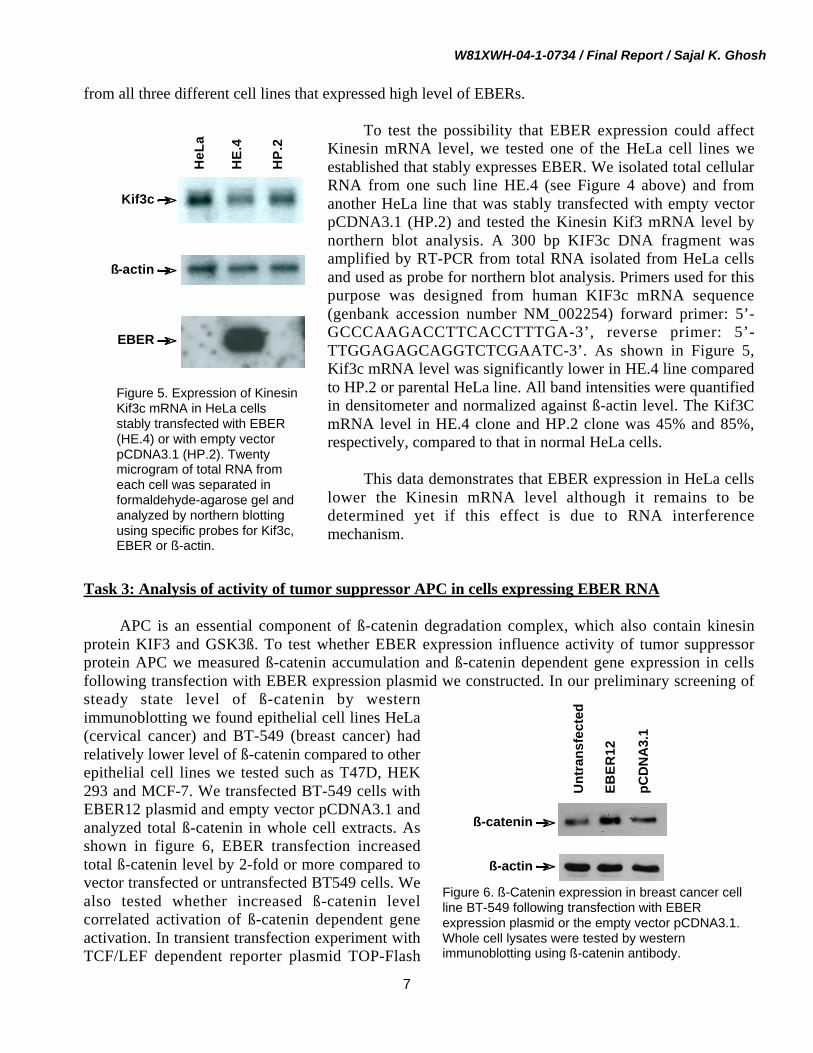
from all three different cell lines that expressed high level of EBERs.
To test the possibility that EBER expression could affectKinesin mRNA level, we tested one of the HeLa cell lines weestablished that stably expresses EBER. We isolated total cellularRNA from one such line HE.4 (see Figure 4 above) and fromanother HeLa line that was stably transfected with empty vectorpCDNA3.1 (HP.2) and tested the Kinesin Kif3 mRNA level bynorthern blot analysis. A 300 bp KIF3c DNA fragment wasamplified by RT-PCR from total RNA isolated from HeLa cellsand used as probe for northern blot analysis. Primers used for thispurpose was designed from human KIF3c mRNA sequence(genbank accession number NM_002254) forward primer: 5’-GCCCAAGACCTTCACCTTTGA-3’, reverse primer: 5’-TTGGAGAGCAGGTCTCGAATC-3’. As shown in Figure 5,Kif3c mRNA level was significantly lower in HE.4 line comparedto HP.2 or parental HeLa line. All band intensities were quantifiedin densitometer and normalized against ß-actin level. The Kif3CmRNA level in HE.4 clone and HP.2 clone was 45% and 85%,respectively, compared to that in normal HeLa cells.
This data demonstrates that EBER expression in HeLa cellslower the Kinesin mRNA level although it remains to bedetermined yet if this effect is due to RNA interferencemechanism.
Task 3: Analysis of activity of tumor suppressor APC in cells expressing EBER RNA
APC is an essential component of ß-catenin degradation complex, which also contain kinesinprotein KIF3 and GSK3ß. To test whether EBER expression influence activity of tumor suppressorprotein APC we measured ß-catenin accumulation and ß-catenin dependent gene expression in cellsfollowing transfection with EBER expression plasmid we constructed. In our preliminary screening ofsteady state level of ß-catenin by westernimmunoblotting we found epithelial cell lines HeLa(cervical cancer) and BT-549 (breast cancer) hadrelatively lower level of ß-catenin compared to otherepithelial cell lines we tested such as T47D, HEK293 and MCF-7. We transfected BT-549 cells withEBER12 plasmid and empty vector pCDNA3.1 andanalyzed total ß-catenin in whole cell extracts. Asshown in figure 6, EBER transfection increasedtotal ß-catenin level by 2-fold or more compared tovector transfected or untransfected BT549 cells. Wealso tested whether increased ß-catenin levelcorrelated activation of ß-catenin dependent geneactivation. In transient transfection experiment withTCF/LEF dependent reporter plasmid TOP-Flash
Unt
rans
fect
ed
EBER
12
pCD
NA
3.1
ß-actin
ß-catenin
Figure 6. ß-Catenin expression in breast cancer cellline BT-549 following transfection with EBERexpression plasmid or the empty vector pCDNA3.1.Whole cell lysates were tested by westernimmunoblotting using ß-catenin antibody.
Figure 5. Expression of KinesinKif3c mRNA in HeLa cellsstably transfected with EBER(HE.4) or with empty vectorpCDNA3.1 (HP.2). Twentymicrogram of total RNA fromeach cell was separated informaldehyde-agarose gel andanalyzed by northern blottingusing specific probes for Kif3c,EBER or ß-actin.
HE.
4
HP.
2
HeL
aKif3c
ß-actin
EBER

W81XWH-04-1-0734 / Final Report / Sajal K. Ghosh
8
which contain 4 TCF/LEF binding sites in the promoter(Upstate Cell Signaling) or a mutant variety FOP-Flash(where all TCF/LEF sites are mutated) we found thatEBER expression significantly enhances reporter geneexpression in BT549 cells (Figure 7). This data suggestedthat increase in ß-catenin level due to EBER expressionalso leads to increase in ß-catenin dependent geneexpression in BT-549 cells.
In unstimulated cells, ß-catenin is rapidly degradedin the cytoplasm following its phosphorylation by thedegradation complex (which consists of APC,axin/kinesin and GSK3ß) and subsequent ubiquitinationand proteasomal degradation. We postulated that EBERexpression could degrade Kinesin/Kif3C and render ß-catenin degradation complex non-functional. In such ascenario there will be reduced degradation of ß-catenin.To test this possibility we analyzed the stability of ß-
catenin in EBER-expressing cells. BT-549 cells were transfected with vector or EBER expressionplasmid and stable-expression was selected using G418 drug resistance. Cells were treated with 20 µMcycloheximide to inhibit new protein synthesis and ß-catenin level in the cells were determined over aperiod of 4 hrs. Figure 8 shows that the rate of degradation of ß-catenin was appreciably less in EBER-expressing cells compared to vector control cells, as by two hours following cyclohemide treatment, theß-catenin level in vector transfected cell line was 3-times lower than that of EBER transfected cell line .
Task 4: Identification of small interfering RNA molecules complementary to various kinesins.
The main hypothesis of this Concept Grant application was that EBERs are probably modified intoshort inhibitory RNA to exert its effect in EBV infected cells. The rationale for this proposition camefrom the fact that through BLAST sequence search we identified strong sequence homology between
ß-Cateninß-Actin
0 11/2 2 4
ß-Cateninß-Actin EBER
Vector
Figure 8. BT-549 cells stably transfectedwith EBER expression plasmid or thevector (pCDNA3.1) were treated with 20µM cycloheximide for 0-4 hrs, at whichtime whole cell lysates were preparedand analyzed for ß-catenin. ß-actincontent of the lysates was alsomeasured to confirm equal loading.
Homo sapiens kinesin family member 3CGenbank Accession: BC092406
Macaca mulatta kinesin family member 3CGenbank Accession: XM_001084502
Figure 9. Sequence homology of EBER and mammalian Kif3C as identified through BLAST search.
Rel
ativ
e Lu
cife
rase
Act
ivity
01234567
FOP TOP
pCDNA3.1EBER12
Figure 7. Analysis of ß-Catenin dependentgene expression in breast cancer cell lineBT-549 following EBER expression. BT-549cells were cotransfected with TCF/LEFdependent luciferase reporter plasmid TOP-Flash or a mutant variety FOP-Flash andEBER expression plasmid or the emptyvector pCDNA3.1.
W81XWH-04-1-0734 / Final Report / Sajal K. Ghosh
9
EBER and mammalian kinesin family protein Kif3C (Figure 9). Homology of EBER sequence 106-128with 3’-noncoding mRNA sequence of human and monkey Kif3C are shown.
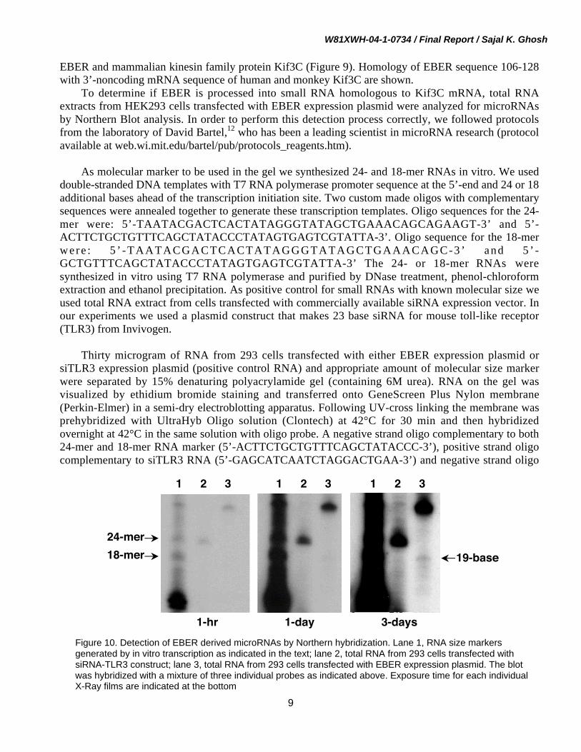
To determine if EBER is processed into small RNA homologous to Kif3C mRNA, total RNAextracts from HEK293 cells transfected with EBER expression plasmid were analyzed for microRNAsby Northern Blot analysis. In order to perform this detection process correctly, we followed protocolsfrom the laboratory of David Bartel,12 who has been a leading scientist in microRNA research (protocolavailable at web.wi.mit.edu/bartel/pub/protocols_reagents.htm).
As molecular marker to be used in the gel we synthesized 24- and 18-mer RNAs in vitro. We useddouble-stranded DNA templates with T7 RNA polymerase promoter sequence at the 5’-end and 24 or 18additional bases ahead of the transcription initiation site. Two custom made oligos with complementarysequences were annealed together to generate these transcription templates. Oligo sequences for the 24-mer were: 5’-TAATACGACTCACTATAGGGTATAGCTGAAACAGCAGAAGT-3’ and 5’-ACTTCTGCTGTTTCAGCTATACCCTATAGTGAGTCGTATTA-3’. Oligo sequence for the 18-merwere : 5 ’ -TAATACGACTCACTATAGGGTATAGCTGAAACAGC-3’ and 5 ’ -GCTGTTTCAGCTATACCCTATAGTGAGTCGTATTA-3’ The 24- or 18-mer RNAs weresynthesized in vitro using T7 RNA polymerase and purified by DNase treatment, phenol-chloroformextraction and ethanol precipitation. As positive control for small RNAs with known molecular size weused total RNA extract from cells transfected with commercially available siRNA expression vector. Inour experiments we used a plasmid construct that makes 23 base siRNA for mouse toll-like receptor(TLR3) from Invivogen.
Thirty microgram of RNA from 293 cells transfected with either EBER expression plasmid orsiTLR3 expression plasmid (positive control RNA) and appropriate amount of molecular size markerwere separated by 15% denaturing polyacrylamide gel (containing 6M urea). RNA on the gel wasvisualized by ethidium bromide staining and transferred onto GeneScreen Plus Nylon membrane(Perkin-Elmer) in a semi-dry electroblotting apparatus. Following UV-cross linking the membrane wasprehybridized with UltraHyb Oligo solution (Clontech) at 42°C for 30 min and then hybridizedovernight at 42°C in the same solution with oligo probe. A negative strand oligo complementary to both24-mer and 18-mer RNA marker (5’-ACTTCTGCTGTTTCAGCTATACCC-3’), positive strand oligocomplementary to siTLR3 RNA (5’-GAGCATCAATCTAGGACTGAA-3’) and negative strand oligo
Figure 10. Detection of EBER derived microRNAs by Northern hybridization. Lane 1, RNA size markersgenerated by in vitro transcription as indicated in the text; lane 2, total RNA from 293 cells transfected withsiRNA-TLR3 construct; lane 3, total RNA from 293 cells transfected with EBER expression plasmid. The blotwas hybridized with a mixture of three individual probes as indicated above. Exposure time for each individualX-Ray films are indicated at the bottom
24-mer18-mer 19-base
1 2 3 1 2 3 1 2 3
3-days1-hr 1-day

W81XWH-04-1-0734 / Final Report / Sajal K. Ghosh
10
complementary to EBER sequence 106-128 (5’-GGCAGAAAGCAGAGTCTGGGAAG-3’) were endlabeled with 32P-γATP. Each of these three probes was mixed in the same hybridization mixture.Following hybridization, the membrane was washed in non-stringent wash buffer (3% SSC, 5% SDS)for 20 min (twice, 10 min each) and stringent wash buffer (1% SSC, 1% SDS) for 5 min and exposed toKodak BioMax films for appropriate duration of time.
As shown in Figure 10, we were able to clearly identify a 23-base RNA in total RNA from 293 cellstransfected with siRNA-TLR3 construct (lane 2). Interestingly, a band corresponding to approximately19 bases was detected in total RNA from EBER-transfeced 293 cells. However, the intensity of thissignal was significantly lower than that observed for siRNA-TLR3 and was visible only in longerexposure of the blot. In other experiments we demonstrated that positive strand siTLR3 RNA probe orthe negative strand 24-mer probe does not hybridize with EBER RNA or the corresponding micro RNA(data not shown). This experiment thus demonstrated that a ~19 base EBER-specific microRNA isindeed made in cells those express full-length EBER RNA.
Task 5: Analysis of growth properties of cells that are expressing EBER.
We hypothesized that expression of EBER would inhibit kinesin expression by RNA interferenceand consequently block degradation of ß-catenin. Increased amount of ß-catenin would then lead toenhanced expression of ß-catenin dependent genes and ultimately provide proliferative signals to thecell. To test this hypothesis, in our original statement of work we proposed to perform cell proliferationassay in mammary epithelial cell line MCF-10A (or MCF-10F) and in breast cancer cell line MCF-7 thatare stably transfected with EBER. Unfortunately, our repeated attempt to establish MCF-10F line stablyexpressing EBER was unsuccessful. Transfection efficiency was always too low and cells did notsurvive G418 drug selection.
However, we performed cell proliferation assay with breast cancer cell line MCF-7. We used
Figure 11. EBER expression doesnot provide cell proliferative signalto MCF-7 cells. MCF-7 cells stablytransfected with either EBER orbackbone vector pCDNA3.1 wereplated at a low density (1000 cellsin 100 mm plates) and allowed togrow for two weeks. Cells wereGiemsa stained, colonies countedand photograph taken againstfluorescent light.
ME.6 (EBER/MCF-7)
MP.1 (pCDNA3.1/MCF-7)

W81XWH-04-1-0734 / Final Report / Sajal K. Ghosh
11
MCF-7 line stably transfected with EBER (please see figure 2. ME.6) and with vector pCDNA3.1(MP.1, figure 2). We plated 1000 actively growing cells of both lines in 100 mm tissue culture dishes (intriplicate) and allowed to grow and form colonies over two weeks time. At the end of this 2-weekperiod, number of colonies and their size for each clone were compared. As shown in Figure 11, nostatistically significant difference in the size or the number of colonies between cells expressing EBERor the plasmid vector was observed. This data suggested that at least in MCF-7 breast cancer cell lineEBER expression does not provide any growth advantage.
Task 6: Data integration and evaluation of the concept hypothesis and pilot experiments to carrythe concept ahead.
In this concept grant application our objective was to test whether EBERs interfere with thefunction of tumor suppressor APC in epithelial cells. The rationale for such a study was that improperfunction of APC is associated with various cancers. If EBERs indeed interfere with normal functions ofAPC, this could be a mechanism by which EBERs influence epithelial malignancy. The other aspect ofthis application was to test the idea that small interfering RNA is possibly made from EBERs thatdegrade a protein (Kif3C) required for proper function of APC.
We have been able to demonstrate that the function of APC (as measured by ß-catenin regulatedgene transcription) is indeed deregulated in epithelial breast cancer cell line BT549 (Task 3). Inefficientfunction of APC cannot degrade cytoplasmic ß-catenin properly and allows ß-catenin to migrate tonucleus and upregulate specific gene expression.
Our study on expression of small RNAs in cells expressing EBER in Task 4 demonstrated thatRNA species of approximately 19 bases are made in epithelia cells such as HEK293. We alsodemonstrated that EBER expression leads to decrease in Kif3C mRNA level (Task 2). We however, didnot get a clear answer to whether EBERs helps directly in epithelial cell proliferation (Task 5).
In our view the work from this concept grant provided evidence that EBER makes small RNA inepithelial cells, which is similar in sequence to Kif3C, and EBER expression leads to diminution ofKif3C mRNA level. This decrease is possibly associated with failure of APC/Kinesin/GSK3ß mediatedß-catenin degradation. This information is significant both with respect to the mechanism of EBERfunction as well as role of EBV in EBV-associated epithelial cancers. However, there are several areasas elucidated in the “conclusion” section that needs in-depth study.
KEY RESEARCH ACCOMPLISHMENTS
The non-polyadenylated small RNAs from Epstein-Barr virus (EBER) is readily expressed in allepithelial cells without the need of any external promoter. This was observed in transientlytransfected cells as well as in stable-expression cell lines.
EBER expression increased the stability of ß-catenin and its steady state level in breast cancercell line BT549. EBERs also upregulated the expression of ß-catenin dependent genes in thesecells.
Expression of mRNA of kinesin protein Kif3C was found to be significantly lower in epithelialcell line HeLa that was stably transfected with EBER.

W81XWH-04-1-0734 / Final Report / Sajal K. Ghosh
12
Small, approximately 19-base RNA processed from EBER, with matching sequence to humanKif3C was identified in epithelial cells HEK293 transfected with EBER.
We were unsuccessful in establishing stable EBER-expressing mammary epithelial cell line(MCF-10F) and could not test the role of EBER in the growth of those cells. Cell proliferationassay in breast cancer cell line MCF-7 however, showed that EBER expression did not providethem with any added growth advantage.
REPORTABLE OUTCOMES
A.Data obtained from this work was presented as a Poster in the “Era of Hope” Breast Cancer Meetingorganized by Department of Defense, which was held in Pennsylvania Convention Center, Philadelphia,June 8-11, 2005.
Title: ROLE OF EPSTEIN-BARR VIRUS ENCODED SMALL RNAS IN BETA-CATENINDEGRADATIONAuthors: Sajal K. Ghosh and Idowu AkinsheyeAbstract:Significant improvement of breast cancer survival rate in recent years has been possible because of ourbetter knowledge of the risk factors for the disease and molecular mechanism of the abonormalities aswell as, early detection. In recent years, Epstein Barr Virus (EBV) has been detected in significantportion of estrogen receptor negative invasive breast cancers. EBV has also been detected in largenumbers of rapidly growing fibroadenomas of the breast in immuno-compromised patients. EBV isassociated with variety of malignant diseases including Burkitt’s Lymphoma (BL), nasopharyngealcarcinoma, Hodgkin’s disease and lymphoproliferative disorders of immunosuppressed patients.Because of this close association it has been suggested that EBV plays important role in the genesis ofEBV associated breast cancers. Although most of the EBV genes are not expressed in EBV associatedtumors, two non-coding RNA molecules (EBERs) are expressed consistently. Recent studies havesuggested that these EBERs may have oncogenic potential. We have identified that EBERs (whichpossess extensive secondary structure) has strong sequence homology to kinesin superfamily of motorproteins. Suppression of kinesin protein (KIF3C) has been recently shown to disrupt adenomatouspolyposis coli (APC) transport and facilitate nuclear accumulation of beta-catenin. ß-Catenin is animportant activator of Wnt signaling pathway that is activated in many breast cancers. We hypothesizethat EBERs down regulate KIF3 protein by RNA interference mechanism and interfere with thefunctionality and antitumor activity of APC.In order to test our hypothesis, we have cloned the EBER genes in pCDNA 3.1 vector. In transienttransfection experiments in epithelial cells such as HeLa, human embryonic kidney cells 293 and also inhuman breast cancer cell line MCF-7, followed by northern blot analysis, we confirmed that theseconstructs make large amount of EBERs. Next, we analyzed KIF3 gene expression in EBER expressingcells by northern blotting. A 350 base pair cDNA segment of KIF3 mRNA was amplified from totalcellular RNA from HeLa cells and was used as probe for this purpose. Our preliminary data indicate thatKIF3C mRNA level was significantly lower in HeLa cells that were expressing EBER but not in cellsthat were transfected with empty pCDNA vector. We are currently testing how this apparent reduction inKIF3C level affects ß-catenin degradation in a transient ß-catenin dependent reporter assay system.Using commercially available antibodies, we are also analyzing KIF3 protein expression in cells that

W81XWH-04-1-0734 / Final Report / Sajal K. Ghosh
13
are expressing EBER. Analysis of EBER’s role as a tumor suppressor antagonist is highly significant astumorigenesis is a multistep process which involve many factors including activation of oncogenes andinactivation of tumor suppressor genes or their products. Detail results of these studies will be presentedin the meeting.
B.A NIH small grant application (R03) entitled “ß-CATENIN ACTIVATION IN EBV LATENCY” hasbeen made by the PI based on the finding of this concept grant.
C.A manuscript with tentative title “Epstein Barr virus encoded small RNAs facilitate ß-cateninstabilization” is in preparation.
CONCLUSIONS
This concept grant provided us with nice opportunity to test our hypothesis that small interferingRNAs are possibly made from EBV encoded RNAs (EBER) and may be responsible for EBER’s role inEBV associated mammary epithelial tumors. We hypothesized because of sequence similarity of EBERwith kinesin Kif3C, interfering RNAs against Kif3C is probably made in cells that express EBER. Asoutlined above, we clearly demonstrated that EBER expression reduces Kif3C mRNA level in HeLacells. We also demonstrated that ß-catenin is relatively stable in cells that express EBER over EBER-negative cells and there is a consequent upregulation of ß-catenin dependent gene expression.
The Wnt/ß-catenin signaling pathway plays an important role in tissue development and as such itis strictly regulated. Any mutation that constitutively activates this pathway could therefore involve ininitiation and progression of cancer. In fact, multiple in vivo and in vitro studies already implicatedaberrant ß-catenin signaling as the determining factor in variety of cancers13-15. Mutation in the ß-cateningene (ctnnb1) has been documented in the cancers of colon16,17, prostate18, ovary19, liver20, lung21, skin22,uterine endometrium23,24 and urinary bladder25 among others. Studies based on these presumptive linksindeed show that ß-catenin is activated in latent EBV infection. In one study EBV latent protein LMP2Adirectly activated ß-catenin signaling in telomerase-immortalized human foreskin keratinocytes26. Ourdata shows that EBV latent gene product EBERs (the non-coding RNAs) also activates ß-catenindependent gene expression. It is noteworthy that usurpation of ß-catenin pathway by several otherhuman pathogenic viruses has also been documented in recent years27-31. As ß-catenin stabilization hasbeen demonstrated in BL cells, which displays type 1 latency and expresses only EBNA1 and EBERs32,it is possible that EBV gene products other than LMPs may also have a role in ß-catenin activation. Ourdata that EBERs independently activate ß-catenin signaling is thus highly significant and deserves to betested thoroughly.
We have demonstrated EBER-mediated inhibition of ß-catenin degradation and subsequent geneupregulation in only BT549 cells which is a breast cancer cell line. It will be necessary in futureexperiments to test whether other breast cancer cell lines and mammary epithelial line behave similarlyin response to EBER expression. Since the level of ß-catenin is often elevated in cancer cells, it ispossible that many of the breast cancer cell line may have already elevated ß-catenin level. In choosingother breast cancer lines to test EBER function we have to consider that. Further, it will be very useful totest whether those cells support production of additional amount of ß-catenin and associated geneactivation by introducing ß-catenin protein expression vector into these cells. BT-549 cells do have

W81XWH-04-1-0734 / Final Report / Sajal K. Ghosh
14
lower level of ß-catenin compared to other breast cancer lines such as MCF-7 and T47D. Ourobservation that EBER enhances ß-catenin mediated gene transcription is thus a genuine effect.
Analysis of RNA preparations from cells expressing EBER provided evidence that EBERs areprocessed into small RNAs which potentially could interfere with expression of cellular mRNAs such asthat of Kif3C. We used a 23 base negative strand EBER probe that had sequence homology to Kif3CmRNA. Because we found a distinct band of approximately 19 bases, it is unlikely that this was part ofrandomly broken down piece of the 172-base EBER RNA. However, it is noteworthy that the amount ofthis product was very low compared to EBER expression level and was detected in 293 cells, which ishighly transfectable. We will need to test whether this small RNA can also be detected in other epithelialcells, in particular, those cells where EBERs downregulate ß-catenin degradation or where Kif3CmRNA level are down because of EBER expression. It may be noted that the sequence of EBER that ishomologous to Kif3C is in positive orientation. However, this stretch of EBER sequence also formshairpin structure in RNA folding prediction analysis (by M-fold program). Thus the other strand of thehairpin can actually act as the interfering antisense RNA for the Kif3C mRNA. This idea need to betested in future experiments.
We found reduction of Kif3C mRNA level due to EBER expression in one stably transfected HeLacell line. In future studies we must test other EBER stable lines to establish clearly the effect of EBERon Kif3C mRNA degradation. We have been unsuccessful in determining the role of EBER inproliferation mammary epithelial cells during the course of this study. We are continuing our attempt totest mammary epithelial cell line MCF-10F for this purpose with better transfection technique.
In summary, data generated from this concept grant clearly suggest that EBERs are processed intosmall RNAs, which potentially could upregulate ß-catenin mediated gene regulation via downregulationof ß-catenin degradation. A recent study published in May 2006 demonstrated that decrease in ß-cateninexpression in breast cancer patients is associated with poor prognosis.33 Our results are thus areintriguing preliminary data to launch a more comprehensive study on the role of EBER in ß-cateninregulation which has clear potential to shed light on EBV’s role in not only breast cancers but also otherepithelial malignancies.
REFERENCES
1. Labrecque LG, Barnes DM, Fentiman IS, Griffin BE. Epstein-Barr virus in epithelial cell tumors: abreast cancer study. Cancer Res. 1995;55:39-45.
2. Kleer CG, Tseng MD, Gutsch DE, et al. Detection of Epstein-Barr virus in rapidly growingfibroadenomas of the breast in immunosuppressed hosts. Modern Pathology. 2002;15:759-764.
3. Fina F, Romain S, Ouafik L, et al. Frequency and genome load of Epstein-Barr virus in 509 breastcancers from different geographical areas. Br J Cancer. 2001;84:783-790.
4. Arbach H, Viglasky V, Lefeu F, et al. Epstein-Barr virus (EBV) genome and expression in breastcancer tissue: effect of EBV infection of breast cancer cells on resistance to paclitaxel (Taxol). JVirol. 2006;80:845-853.
5. Ruf IK, Rhyne PW, Yang H, et al. EBV regulates c-MYC, apoptosis, and tumorigenicity inBurkitt's lymphoma. Curr Top Microbiol Immunol. 2001;258:153-160.
6. Ruf IK, Rhyne PW, Yang C, Cleveland JL, Sample JT. Epstein-Barr virus small RNAs potentiatetumorigenicity of Burkitt lymphoma cells independently of an effect on apoptosis. J Virol.2000;74:10223-10228.

W81XWH-04-1-0734 / Final Report / Sajal K. Ghosh
15
7. Komano J, Sugiura M, Takada K. Epstein-Barr virus contributes to the malignant phenotype and toapoptosis resistance in Burkitt's lymphoma cell line Akata. J Virol. 1998;72:9150-9156.
8. Komano J, Maruo S, Kurozumi K, Oda T, Takada K. Oncogenic role of Epstein-Barr virus-encoded RNAs in Burkitt's lymphoma cell line Akata. J Virol. 1999;73:9827-9831.
9. Jimbo T, Kawasaki Y, Koyama R, et al. Identification of a link between the tumour suppressorAPC and the kinesin superfamily. Nat Cell Biol. 2002;4:323-327.
10. Furuuchi K, Tada M, Yamada H, et al. Somatic mutations of the APC gene in primary breastcancers. Am J Pathol. 2000;156:1997-2005.
11. Cui H, Dong M, Sadhu DN, Rosenberg DW. Suppression of kinesin expression disruptsadenomatous polyposis coli (APC) localization and affects beta-catenin turnover in young adultmouse colon (YAMC) epithelial cells. Exp Cell Res. 2002;280:12-23.
12. Bartel DP. MicroRNAs: genomics, biogenesis, mechanism, and function. Cell. 2004;116:281-297.13. Kikuchi A. Tumor formation by genetic mutations in the components of the Wnt signaling
pathway. Cancer Sci. 2003;94:225-229.14. Taketo MM. Shutting down Wnt signal-activated cancer. Nat Genet. 2004;36:320-322.15. Morin PJ, Weeraratna AT. Wnt signaling in human cancer. Cancer Treat Res. 2003;115:169-187.16. Sparks AB, Morin PJ, Vogelstein B, Kinzler KW. Mutational analysis of the APC/beta-catenin/Tcf
pathway in colorectal cancer. Cancer Res. 1998;58:1130-1134.17. Samowitz WS, Powers MD, Spirio LN, Nollet F, van Roy F, Slattery ML. Beta-catenin mutations
are more frequent in small colorectal adenomas than in larger adenomas and invasive carcinomas.Cancer Res. 1999;59:1442-1444.
18. Voeller HJ, Truica CI, Gelmann EP. Beta-catenin mutations in human prostate cancer. Cancer Res.1998;58:2520-2523.
19. Palacios J, Gamallo C. Mutations in the beta-catenin gene (CTNNB1) in endometrioid ovariancarcinomas. Cancer Res. 1998;58:1344-1347.
20. de La Coste A, Romagnolo B, Billuart P, et al. Somatic mutations of the beta-catenin gene arefrequent in mouse and human hepatocellular carcinomas. Proc Natl Acad Sci U S A.1998;95:8847-8851.
21. Sunaga N, Kohno T, Kolligs FT, Fearon ER, Saito R, Yokota J. Constitutive activation of the Wntsignaling pathway by CTNNB1 (beta-catenin) mutations in a subset of human lungadenocarcinoma. Genes Chromosomes Cancer. 2001;30:316-321.
22. Rubinfeld B, Robbins P, El-Gamil M, Albert I, Porfiri E, Polakis P. Stabilization of beta-cateninby genetic defects in melanoma cell lines. Science. 1997;275:1790-1792.
23. Ikeda T, Yoshinaga K, Semba S, Kondo E, Ohmori H, Horii A. Mutational analysis of theCTNNB1 (beta-catenin) gene in human endometrial cancer: frequent mutations at codon 34 thatcause nuclear accumulation. Oncol Rep. 2000;7:323-326.
24. Fukuchi T, Sakamoto M, Tsuda H, Maruyama K, Nozawa S, Hirohashi S. Beta-catenin mutation incarcinoma of the uterine endometrium. Cancer Res. 1998;58:3526-3528.
25. Shiina H, Igawa M, Shigeno K, et al. Beta-catenin mutations correlate with over expression of C-myc and cyclin D1 Genes in bladder cancer. J Urol. 2002;168:2220-2226.
26. Morrison JA, Klingelhutz AJ, Raab-Traub N. Epstein-Barr virus latent membrane protein 2Aactivates beta-catenin signaling in epithelial cells. J Virol. 2003;77:12276-12284.
27. Huang H, Fujii H, Sankila A, et al. Beta-catenin mutations are frequent in human hepatocellularcarcinomas associated with hepatitis C virus infection. Am J Pathol. 1999;155:1795-1801.
28. Schroeder JA, Adriance MC, McConnell EJ, Thompson MC, Pockaj B, Gendler SJ. ErbB-beta-catenin complexes are associated with human infiltrating ductal breast and murine mammarytumor virus (MMTV)-Wnt-1 and MMTV-c-Neu transgenic carcinomas. J Biol Chem.2002;277:22692-22698.

W81XWH-04-1-0734 / Final Report / Sajal K. Ghosh
16
29. Cha MY, Kim CM, Park YM, Ryu WS. Hepatitis B virus X protein is essential for the activationof Wnt/beta-catenin signaling in hepatoma cells. Hepatology. 2004;39:1683-1693.
30. Bose S, Banerjee AK. Beta-catenin associates with human parainfluenza virus type 3ribonucleoprotein complex and activates transcription of viral genome RNA in vitro. Gene Expr.2004;11:241-249.
31. Street A, Macdonald A, McCormick C, Harris M. Hepatitis C virus NS5A-mediated activation ofphosphoinositide 3-kinase results in stabilization of cellular beta-catenin and stimulation of beta-catenin-responsive transcription. J Virol. 2005;79:5006-5016.
32. Everly DN, Jr., Kusano S, Raab-Traub N. Accumulation of cytoplasmic beta-catenin and nuclearglycogen synthase kinase 3beta in Epstein-Barr virus-infected cells. J Virol. 2004;78:11648-11655.
33. Dolled-Filhart M, McCabe A, Giltnane J, Cregger M, Camp RL, Rimm DL. Quantitative in situanalysis of beta-catenin expression in breast cancer shows decreased expression is associated withpoor outcome. Cancer Res. 2006;66:5487-5494.
APPENDICES
None
SUPPORTING DATA
None
LIST OF PERSONNEL RECEIVING PAY FROM THE RESEARCH EFFORT
Sajal K. Ghosh