award number: w81xwh-04-1-0222 title: analysis of ... of morphogenic ... such alternations will...
TRANSCRIPT

AD_________________
Award Number: W81XWH-04-1-0222 TITLE: Analysis of Morphogenic Effect of hDAB2IP on Prostate Cancer and its Disease Correlation PRINCIPAL INVESTIGATOR: Jer-Tsong Hsieh, Ph.D. CONTRACTING ORGANIZATION: University of Texas, Southwestern Medical Center Dallas, TX 75390-9110 REPORT DATE: February 2007 TYPE OF REPORT: Annual PREPARED FOR: U.S. Army Medical Research and Materiel Command Fort Detrick, Maryland 21702-5012 DISTRIBUTION STATEMENT: Approved for Public Release; Distribution Unlimited The views, opinions and/or findings contained in this report are those of the author(s) and should not be construed as an official Department of the Army position, policy or decision unless so designated by other documentation.

REPORT DOCUMENTATION PAGE Form Approved
OMB No. 0704-0188 Public reporting burden for this collection of information is estimated to average 1 hour per response, including the time for reviewing instructions, searching existing data sources, gathering and maintaining the data needed, and completing and reviewing this collection of information. Send comments regarding this burden estimate or any other aspect of this collection of information, including suggestions for reducing this burden to Department of Defense, Washington Headquarters Services, Directorate for Information Operations and Reports (0704-0188), 1215 Jefferson Davis Highway, Suite 1204, Arlington, VA 22202-4302. Respondents should be aware that notwithstanding any other provision of law, no person shall be subject to any penalty for failing to comply with a collection of information if it does not display a currently valid OMB control number. PLEASE DO NOT RETURN YOUR FORM TO THE ABOVE ADDRESS. 1. REPORT DATE (DD-MM-YYYY)
01-02-2007 2. REPORT TYPE
Annual 3. DATES COVERED (From - To)
1 Feb 2006 - 31 Jan 20074. TITLE AND SUBTITLE
Analysis of Morphogenic Effect of hDAB2IP on Prostate Cancer and its Disease 5a. CONTRACT NUMBER
Correlation
5b. GRANT NUMBER
W81XWH-04-1-0222 5c. PROGRAM ELEMENT NUMBER
6. AUTHOR(S)
Jer-Tsong Hsieh, Ph.D. 5d. PROJECT NUMBER
5e. TASK NUMBER
E-Mail: [email protected]
5f. WORK UNIT NUMBER
7. PERFORMING ORGANIZATION NAME(S) AND ADDRESS(ES)
8. PERFORMING ORGANIZATION REPORT
NUMBER
University of Texas, Southwestern Medical Center Dallas, TX 75390-9110
9. SPONSORING / MONITORING AGENCY NAME(S) AND ADDRESS(ES) 10. SPONSOR/MONITOR’S ACRONYM(S)
U.S. Army Medical Research and Materiel Command
Fort Detrick, Maryland 21702-5012 11. SPONSOR/MONITOR’S REPORT
NUMBER(S)
12. DISTRIBUTION / AVAILABILITY STATEMENT
Approved for Public Release; Distribution Unlimited
13. SUPPLEMENTARY NOTES
14. ABSTRACT: Imbalance of apoptotic and/or survival signaling cascade is a hallmark of malignant cell. In prostate cancer (PCa), constitutive activation of phosphatidylinositol 3-kinase (PI3K)-Akt/PKB and inactivation of apoptosis-stimulated kinase (ASK1)-JNK pathway signaling are often detected in metastatic cell. Understanding the underlying mechanism leading to such alternations will provide a better treatment strategy to control the terminal stage of this disease. In this project, we have proposed that DAB2IP protein, a novel RASGAP, is a part of homeostatic machinery and plays an important in modulating signal pathways elicited by exogenous survival/death stimuli. Our data clearly demonstrated that DAB2IP is a novel scaffold protein that complexes with key proteins involved in cell growth or death. The outcome of this study has led us to unveil a new mechanism of DAB2IP, which provides a better understanding how PCa cells switch from survival to death under the stimuli of exogenous signals.
15. SUBJECT TERMS
Cell differentiation, GTPase activating protein, tumor suppressor, Epigenetic regulation, signal transduction, prostate cancer
16. SECURITY CLASSIFICATION OF:
17. LIMITATION
OF ABSTRACT
18. NUMBER
OF PAGES
19a. NAME OF RESPONSIBLE PERSON
USAMRMC a. REPORT
U b. ABSTRACT
U c. THIS PAGE
U
UU
2219b. TELEPHONE NUMBER (include area
code)
Standard Form 298 (Rev. 8-98)
Prescribed by ANSI Std. Z39.18

- 3 -
Table of Contents
Introduction…………………………………………………………….………….. 4
Body…………..…………………………………………………………………… 4
Key Research Accomplishments………………………………………….……… 7
Reportable Outcomes………………………………………………………………. 7
Conclusions………………………………………………………………………….. 8
References…………………………………………………………………………… 8
Appendices…………………………………………………………………………… 9

- 4 -
INTRODUCTION
Expression of Ras protein has also been assessed in primary and metastatic prostate
cancer (PCa) tumors. Most tumors expressed higher Ras protein than normal prostate tissues (1).
Also, an increased c-H-Ras mRNA expression in PCa is associated with the progression of PCa
to androgen independence (2). Surprisingly perhaps, PCa specimens from American men have
an extremely low rate of mutation in the Ras gene (3). This implies that other effectors may be
involved in increasing RAS protein levels in PCa. DAB2IP appears to be a good candidate
because it contains an amino acid sequence that is homologous to the GTPase activating protein
(GAP) domain of other RasGAPs and it is functional active based on biochemical and molecular
biologic studies (4). In addition to the GAP domain, DAB2IP contains several other functional
motifs such as a pleckstrin homology domain (aa 20-70) with a high affinity to certain
phosphoinositides, a C2 domain (aa 90-120) involved in binding phospholipids in a calcium-
dependent or -independent manner, a proline-rich (PR) domain (aa 796-805) involved in
interacting with proteins that contain a SH3 domain, and a leucine zipper (aa 842-861), which
mediates dimerization. For example, the C2 domain of DAB2IP can bind and then activate
apoptosis-stimulated kinase (ASK1) involved in the TNF- -mediated apoptosis (5).
In addition to the interaction of C2 domain with ASK1, DAB2IP mutants defective in
GAP activity failed to increase ASK1 activity, suggesting that the GAP activity of DAB2IP is
required for TNF- -elicited ASK1 activity (5). Also, the GAP activity of DAB2IP is known to
be a negative regulator for Ras-Raf-ERK activation critical for cell growth. Thus, DAB2IP
appears as a unique factor in modulating both cell growth and death by inhibiting Ras-Raf-ERK
proliferative pathway (6) and activating ASK1-JNK/p38 apoptotic signaling. Very likely, other
functional domains in DAB2IP have different roles in maintaining homeostasis of prostate
epithelium.
RECENT PROGRESS Overall, Task 1 and 2 are completed and Task 3 is completed 60%. In summary, we made
several key progresses in this project: (1) Loss of DAB2IP expression is associated with the
recurrent of androgen-independent (AI) PCa; (2) Loss of DAB2IP expression is mainly due to
epigenetic regulation; (3) DAB2IP is a key homeostatic factor in balancing cell growth and
aoptosis. Thus, I have summarized these new results as follows:
Task 3. To delineate signal cascade mediated by hDAB2IP protein complex in prostatic epithelium. The role of DAB2IP in modulating cell survival and death Cell homeostasis is a balance between cell proliferation, apoptosis and differentiation in basal
and luminal epithelia. It is believed that loss of homeostatic control in these cells renders the
onset of neoplasm in prostatic epithelium. Until now, a little is known about homeostatic
machinery in normal prostatic epithelium. Recently, we identified DAB2IP as a new member of
GTPase activating protein (GAP) family as a potent growth inhibitor in metastatic PCa cell lines
(7) by inducing G0 cell cycle arrest and promoting apoptosis under stress factor (Fig. 1). It is
known that the C2 domain can interact with ASK1 to facilitate TNF- -mediated apoptosis by
activating ASK1. Our preliminary data further indicated that the PR domain in DAB2IP is

- 5 -
critical for binding PI3K regulatory subunit (p85) and then suppresses the activation of Akt (Fig.
2); the first six amino acids of PR domain appear to be key binding site (Fig. 3). It appears that
DAB2IP-mediated Akt inactivation can concomitantly enhance ASK1 activation leading to cell
apoptosis. In contrast, DAB2IP siRNA can restore Akt activity, suppress ASK1 activation and
reduce apoptosis (Fig. 4), indicating that DAB2IP is critical factor for interaction between Akt
and ASK1. Taken together, we believe that DAB2IP is a novel scaffold protein for both survival
and death signal molecules, by which DAB2IP complex is a key machinery to maintain
homeostasis in normal cell.
Figure 1 The role of DAB2IP in PI3K inhibitor (LY294002)-induced cell apoptosis. DAB2IP-transfected PCa lines (C4-2) were determined for DAB2IP expression (a), then the total
cell number (b), the percentage of apoptotic cell (c) and apoptotic hallmark-PARP cleavage (d)
were determined after 24 hrs of treatment.
Figure 2 The level of phosphorylated Akt and total Akt in DAB2IP-transfected C4-2. Cells
were treated LY294002 (10 μM) (a) or TNF- (100 ng/ml) (b) for 30 min and total cell lysate
was subjected to western blot analysis.
Figure 3 Determination of interactive domain in DAB2IP with the regulatory domain PI3K (p85). Using pull down (a) or immunoprecipitation (b), the first six amino acids in PR domain
of DAB2IP is a critical binding domain with p85. The mutant can diminish the activity of
DAB2IP in LY294002- or TNF- -elicited cell apoptosis.

- 6 -
Figure 4 Reduction of LY294002- or TNF- -mediated cell apoptosis by DAB2IP siRNA. Endogenous DAB2IP levels in PZ-HPV7 was decreased by DAB2IPsiRNA (a), which resulted
in elevated activated Akt (b), decreased activated ASK1 (c) and decreased cell apoptosis (d) after
treating with LY294002- or TNF- .

- 7 -
KEY RESEARCH ACCOMPLISHMENT
• Clone mouse DAB2IP gene, map the promoter region and characterize epigenetic regulation
(This work has been published on the cover page of DNA and Cell Biology journal).
• Generate a series of DAB2IP mutant constructs.
• Generate a DAB2IP knock out ES cell.
• Map new functional domain in DAB2IP for controlling cell survival.
• Determine the role of DAB2IP in cell survival/ apoptosis of PCa cells by various exogenous
stimuli.
REPORTABLE OUTCOMES
1. Chen, H, Karam. J.A., Schultz, R., Zhang, Z., Ducan, C., and Hsieh, J.T. (2006) Cloning of mDAB2IP gene, a novel member of the RasGTPase-activating protein family and characterization of its regulatory region. DNA and Cell Biol., 25: 232-245.
2. Hsieh, J.T., Karam, J.A., and Min, W. (2007) The Link of Genetic and Biologic Evidence from DAB2IP gene with Aggressive Prostate Cancer. JNCI, (submiited).
CONCLUSIONS
Prostate homeostasis relies on a balance between cell proliferation, apoptosis and
differentiation. We initially identified DAB2IP, a novel Ras-GTPase activating protein (Ras-
GAP), as a DOC-2/DAB2-interacting protein from a yeast two-hybrid system. A blast search
indicated that DAB2IP consists of several conserved structural domains –the pleckstrin
homology (PH), the protein kinase C conserved region 2 (C2) and RasGAP at the N-terminal
half, and at the C-terminal half a proline-rich sequence (PR) and a leucine-zipper motif (LZ). We
have extensively characterized the structural and functional relationship of AIP1 involved in the
TNF signaling pathways. Interestingly, DAB2IP mutants defective in GAP activity failed to
increase ASK1 activity, suggesting that the GAP activity of AIP1 is required for AIP1-enhanced
ASK1 activity. The GAP activity of Ras-GAP is known to be critical for regulation of Ras-Raf-
ERK activation. The requirement for AIP1 GAP activity in ASK1-JNK activation is supported
by observation that ERK exerts an inhibitory effect on ASK1-JNK signaling. Crosstalk among
MAPKs, i.e. one MAPK (e.g. ERK) may inhibit or oppose the activation of another MAPK (e.g,
JNK), has been well documented. Many stimuli reciprocally regulate ERK and JNK activation
Conversely, ERK inhibition by DAB2IP (via GAP activity) may require for DAB2IP-enhanced
ASK1-JNK activation. Now, our data have further established a linkage of PI3K, Ras-GAP to
stress-activated ASK1-JNK signaling pathway. Further study of the role of DAB2IP in signal
transduction will provide more understanding of recurrent AIPCa.

- 8 -
REFERENCES
1. Bushman EC, Nayak RN, Bushman W. Immunohistochemical staining of ras p21:
staining in benign and malignant prostate tissue. J Urol. 1995; 153:233-7.
2. Bakin RE, Gioeli D, Bissonette EA, Weber MJ. Attenuation of Ras signaling restores
androgen sensitivity to hormone-refractory C4-2 prostate cancer cells. Cancer Res 2003;
63:1975-80.
3. Konishi N, Hiasa Y, Tsuzuki T, Tao M, Enomoto T, Miller GJ. Comparison of ras
activation in prostate carcinoma in Japanese and American men. Prostate. 1997; 30:53-7.
4. Wang Z, Tseng CP, Pong RC, Chen H, McConnell JD, Navone N, et al. A Novel
RasGTPase activating protein that interacts with DOC-2/DAB2: A downstream effector
leading to the suppression of prostate cancer. J Biol Chem 2002; 277: 12622-31.
5. Zhang R, He X, Liu W, Lu M, Hsieh JT, Min W. AIP1 mediated TNF-induced ASK1
activation by facilitating dissociation of ASK1 from inhibitor 14-3-3. J. Clin. Invest
2003; 11:1933-43.
6. Zhou J, Scholes J, Hsieh JT. Signal transduction targets in androgen independent prostate
cancer. Cancer and Metastasis Review, 2001; 20:351-362.
7. Wang Z, Tseng CP, Pong RC, Chen H, McConnell JD, Navone N, et al. A Novel
RasGTPase activating protein that interacts with DOC-2/DAB2: A downstream effector
leading to the suppression of prostate cancer. J Biol Chem 2002; 277: 12622-31.

DNA AND CELL BIOLOGYVolume 25, Number 4, 2006© Mary Ann Liebert, Inc.Pp. 232–245
Cloning of Mouse Dab2ip Gene, a Novel Member of theRasGTPase-Activating Protein Family and Characterization of
Its Regulatory Region in Prostate
HONG CHEN,1 JOSE A. KARAM,1 ROGER SCHULTZ,2 ZHENGWANG ZHANG,1 CHRISTINE DUNCAN,2
and JER-TSONG HSIEH1
ABSTRACT
Disabled homolog 2 (Drosophila) interacting protein (DAB2IP/Dab2IP) is a member of the GTPase-activat-ing protein for downregulating the Ras-mediated signal pathway and TNF-mediated apoptosis. The down-regulation of human DAB2IP mRNA levels was detected in prostate cancer cells due to the epigenetic regu-lation. Here, we isolated a mouse Dab2ip gene with a highly homologous sequence to that of the human andrat gene and mapped it at chromosome 2B. The mDab2ip gene contains 14 exons and 13 introns and spansapproximately 65 kb. Exon1 contains at least three splicing variants (Ia, Ib, and Ic). The deduced amino acidsequence of mouse Dab2IP encompasses 1065 residues containing several unique protein interaction motifsas well as a Ras-like GAP-related domain, which shares a high homology with both humans and rats. Datafrom real-time RT-PCR analysis revealed a diverse expression pattern of the mDab2ip gene in various organs,implying differential regulation of this gene from various tissues. We have mapped a 1.3-kb segment con-taining a 5�-upstream region from exon Ia as a promoter region (�147/�545) in prostatic epithelial cell lines(TRAMP-C); this region is highly GC-rich, and mDab2ip appears to be a TATA-less promoter. It appearsthat epigenetic regulation, particularly histone acetylation of the Dab2ip gene promoter, plays an importantrole in modulating its gene expression in the mouse prostate cancer cell.
232
INTRODUCTION
DISABLED HOMOLOG 2 (Drosophila) interacting protein(DAB2IP/Dab2IP) is a novel member of the Ras GTPase-
activity family protein (Chen et al., 2002; Wang et al., 2002)DAB2IP is able to interact with DOC-2/DAB2 (Fulop et al.,1998; Tseng et al., 1999; Zhou and Hsieh, 2001; Zhou et al.,2003), and this protein complex modulates the Ras-mediatedsignal pathway, then causes the growth inhibition in prostatecancer cells (Wang et al., 2002). Also, DAB2IP (also namedAIP1: ASK interacting protein 1) is involved in TNF-�-medi-ated cell apoptosis by facilitating dissociation of ASK1 fromits inhibitor 14-3-3 (Zhang et al., 2003, 2004)
The higher hDAB2IP mRNA levels are detected in normalhuman prostatic epithelium than in prostate cancer cells, whichis due to the epigenetic regulation (Wolffe and Matzke, 1999;Jones and Takai, 2001) such as DNA methylation and histoneacetylation of the human DAB2IP (hDAB2IP) gene promoter
(Chen et al., 2002, 2003). In breast cancer, DNA hypermethy-lation of hDAB2IP is also found in both breast cancer cell linesand specimens with lymph node metastasis, and hDAB2IP geneexpression can be restored in methylated cell lines treated with5-aza-2�-deoxycytidine (Dote et al., 2004). Also, hDAB2IP(alias for AFQ34) is identified as a novel MLL fusion partnerfrom an acute myeloid leukemia (AML) patient with a t(9;11)(q34;q23); the intron 9 of the MLL gene is translocated into theexon 2 of DAB2IP, and causes the disruption of the pleckstrinhomology (PH) domain in the DAB2IP protein, implying thatthis fusion protein may alter RAS activity as a part of leukemiatransformation process (von Bergh et al., 2004). Thus, DAB2IPshould be involved in carcinogenesis of various tissues.
To further unveil the physiological functions of mouseDab2ip, we decided to clone and map its chromosomal loca-tion. With analyzing the structure of the mouse Dab2ip(mDab2ip) gene, we have assembled the entire gene sequenceand its full-length mRNA with an open reading frame. We also
Departments of 1Urology and 2Pathology, University of Texas Southwestern Medical Center, Dallas, Texas.

profiled the mDab2ip expression pattern from a variety of or-gans and cell lines. By determining the promoter sequence fromthe 5’- flanking region of the mDab2ip gene in mouse prosta-tic epithelial cell lines provided clues for the transcriptional reg-ulation of the mDab2ip gene.
MATERIALS AND METHODS
Tissue culture, treatment, and RNA isolation
Three mouse transgenic prostate adenocarcinoma cell lines(TRAMP-C1, TRAMP-C2, and TRAMP-C3) (Greenberg et al.,1995; Foster et al., 1997; Gingrich et al., 1997) were main-tained in DMEM supplemented with 5% FBS (HyClone, Lo-gan, UT) plus 5% Nu-serum™ IV (BD Bioscience, Bedford,MA) and 50 ng/ml insulin (Sigma St. Louis, MO). The NIH-3T3 cell line was maintained in DMEM with 10% fetal bovineserum (FBS), and the PC3 cell line was maintained in T mediumsupplemented with 5% FBS. Total RNA from variant organsand cell lines were isolated using the RNAzol B (Tel-Test Inc.,Friendswood, TX) according to the manufacturer’s instructions.
For tissue RNA isolation, various organs (approximate 50mg) were harvested from nude mice after euthanasia and weresnap-frozen in liquid nitrogen until analysis was performed. Tis-
sues were submerged with RNAzol B, quickly homogenized,then subjected to the same isolation procedure.
To study the effect of hisone acteylation, different concen-trations (20, 40, and 100 nM) of Trichostatin (TSA), a histonedeacetylase (i.e., HDAC) inhibitor, was changed every 24 h for48 h. For studying the effect of DNA methylation, different con-centrations (1, 2, and 5 �M) of 5-aza-2�-deoxycytidine (5�-Aza),a DNA methyltransferase (DNMT) inhibitor, was changed every48 h for 96 h. For the combination, 5�-Aza was first added andchanged every 48 h; then TSA was added 72 h after treatment.Cells were collected at 96 h after treatment.
Cloning mouse Dab2ip gene and sequence analysis
To obtain the entire coding region of mouse Dab2ip cDNA,we performed RT-PCR from total cellular RNA from the mousebrain. Based on the high homology sequences between humanand rat DAB2IP cDNA (Chen et al., 2002; Wang et al., 2002),two sets of primer were synthesized (Table 1). PCR products werecloned into pCR2.1-TOPO vector (Invitroge, Carlsbad, CA) andsequenced, then used as probes for screening the mDab2ip gene.
A mouse bacterial artificial chromosomal (BAC, RPCI.22) li-brary (ResGen Invitrogen Corp., Huntsville, AL) was screened.The positive clones were subjected to Southern blot analysis andDNA sequencing for confirmation.
CLONING OF MOUSE Dab2ip GENE 233
TABLE 1. PCR PRIMERS USED ON 5� RACE, REAL-TIME RT-PCR, PROBE AND PROMOTER CONSTRUCTS
Application Primer name Primer 5�–3� sequence
5� RACE Sp1 (outer) ATACAGCACATCGTCCAGGSp2 (inner) GTTCTCCATCCACTTATCGCGC
Real-time RT-PCR F-mDab2ip CGATAAGTGGATGGAGAACCTGAGR-mDab2ip AGATGCTGACGGTCTGGTAGCGTGCF-Actin TGTGTGGATTGGTGGCTCTATCR-Actin CTGCTTGCTGATCCACATCTG
Probe 1 (800 bp) F1-DAB2IP TCGTGGAAGGACTCATGACCR1-DAB2IP TCCACCACCCTGTTGCTGTA
Probe 2 (300 bp) F2-DAB2IP TGGACGATGTGCTCTATGCCR2-DAB2IP GGATGGTGATGGTTTGGTAG
Promoters F1 GCTCCTCACCTGCCTCTTCATTAGF6 CCTGCTCTCCCAGCCTTAGTTTCF8 CACCAAGAGCCAGCCCAAACF10 ACCCTTCGTTGCTTTCACCGF12 GAGTCCCTCGCTGTCCGATACR2 TGCTCCTCCCCTCCAGATGTTC
Mutagenesis F-�Sp1 GGCGGGGGCGACGGGCCGCGAGGGR-�Sp1 CCCTCGCGGCCCGTCGCCCCCGCCF-�AP2 CCTTCCCCCTCTTGCAGGGCTTCCTCAGR-�AP2 CTGAGGAAGCCCTGCAAGAGGGGGAAG
Bisulfite-treated Fm ATTAAGAGTTAGTTTAAATTGGATDNA Rm ACCTCCAACACCCTCCTAAATAC
Immunoprecipitated F (outer) CCTGCTCTCCCAGCCTTAGTTTCDNA (ChIP) R (outer) TGTGGTTGCGGTCCAGTTTG
F (inner) ATGGCAACGGCGGCTTAGGR (inner) TTTGGGCTGGCTCTTGGTG

Identification of transcriptional starting site (TSS) by 5� RACE
To determine the transcriptional starting site of the mDab2ipgene, the total cellular RNA (10 �g) from two mouse organs (thebrain and kidney) and two TRAMP-C lines (TRAMP-C1 andTRAMP-C2) was subjected to 5� RACE using the FirstChoice™RLM-RACE Kit (Ambion Inc., Austin, TX) according to themanufacturer’s manual. A random-primed reverse-transcriptionreaction and nested PCR (Fig. 2B and Table 1) were performedto amplify the 5�end of the mDab2ip mRNA transcript. We an-alyzed the PCR product in a 2% NuSieve® 3:1 agarose gel (Cam-brex BioScience, Rockland, MA) and then cloned it into pCR2.1-TOPO for sequence identification.
Determination of mDAB2IP mRNA levels by real-timequantitative RT-PCR (qRT-PCR) assay
Two micrograms of total cellular RNA were reversely tran-scribed into cDNA and amplified using either the mDab2ipprimer set (2 ng/�l) or Actin primer set (6 ng/�l) (Table 1) ina 40-�l reaction mixture containing 20-�l IQ™ SYBR® Green
Supermix (Bio-Rad, Hercules, CA). The PCR was performedusing an iCycler machine (Bio-Rad) and the reaction condi-tion was as follow: 95°C (3 min) and 40 cycles amplificationcycle (95°C [30 sec], 55°C [30 sec], and 72°C [1 min]). Toassure the quality of each reaction, melting curves analysiswas performed using 95°C (1 min), 55°C (1 min), and 80 cy-cles of 0.5°C increment beginning at 55°C. Each sample wasperformed in duplicate. The level of mDab2ip mRNA fromeach organ was calculated as follows: �Ct (threshold cycle)of each sample � mean of Ct(Dab2ip) � mean of Ct(Actin). Therelative expression of mDab2ip in each organ was calculatedas 1/2�Ct(sample tissue) � �Ct(spleen), since the spleen had the low-est Dab2ip mRNA level. Meanwhile, the levels of themDab2ip mRNA from each cell line were calculated as1/2�Ct(sample cell) � �Ct(TRAMP-C3), since TRAMP-C3 had thelowest expression of mDab2ip.
Fluorescence in situ hybridization (FISH) analysis
To determine the chromosomal localization of the mDab2ipgene, cells were sterilely isolated from the spleens of 6-week-
CHEN ET AL.234
FIG. 1. Structure of mDab2ip gene. (A) Map of the mDab2ipgene dispersed over approximately 65 kb. The exon 1 (blackbox) contains at least three variants (Ia, Ib, and Ic), and the rel-ative position of BAC clones is displayed. (B) Schematic dis-play of two specific primers (Sp1 [outer] and Sp2 [inner]), andcorresponding two universal primers for 5�RACE. (C) Alter-native slicing of mDab2ip mRNA detected from the brain,TRAMP-C1/TRAMP-C2 and kidney.

old mice and set up in RPMI 1640 with glutamine, 20% FBS, and50 �g lipopolysaccharide at 37°C for 42 h. At 42 h 0.75 �l of 10�g/ml colcemid was added and incubated for 10 min. Cells werepelletted, resuspended in 1 ml of hypotonic KCl (prewarmed to37°C), and incubated at room temperature for 15 min. The cellswere then fixed in methanol:acetic acid (3:1) and spread on slidesusing heat treatment. DNA probes from BAC clones 22N15 and301E21 were fluorescently labeled by nick translation using stan-dard conditions. The probe was hybridized to mouse metaphaseslides overnight in a HYBrite (Vysis, Inc., Downers Grove, IL)hybridization chamber and washed. The hybridization signal wasviewed and analyzed on an Olympus AX70 fluorescence micro-scope and images captured using MacProbe software (version 4.4,Applied Imaging, San Jose, CA).
Construction of luciferase reporter plasmid containing the 5�-upstream regulatory sequence of the mDab2ip gene
To analyze the 5�-upstream regulatory sequence of themDab2ip gene, a 1.3-kb fragment from �730 to �545 (tran-scription initial site as �1 predicted by 5�RACE data) con-taining the upstream region, exon Ia and partial intron 1a wasamplified by PCR from clone 22N15. To further define the pro-moter region in the mDab2ip gene, a series of deletion mutantswere generated by PCR (Table 1). The PCR products were sub-cloned into the pCR(-Blunt II TOPO vector (Invitrogen, Carls-bad, CA). After sequencing confirmation, they were furthercloned a into pGL3 basic vector (Promega, Madison, WI) us-ing KpnI/XhoI sites to generate pGL3–F1/R2 (from �730 to�545), pGL3–F6/R2 (from �421 to �545), pGL3–F8/R2
(from �6 to �545), pGL3–F10/R2 (from �249 to �545),pGL3–F12/R2 (from �445 to �545), The pGL3–F7/R2 con-tains a NcoI fragment (from �147 to �545) of mDab2ip. ThepGL3–F6/NcoI contains a 0.3-kb insert from �421 to �157and pGL3-F6/SanDI contains a 0.4-kb insert from-421 to �97.
For mutagenesis studies, we used pGL3–F7/R2 as a templateto generate deletion mutants: pGL3–�Sp1 (deletion of poten-tial Sp1 site from �124 to �114) and pGL3–�AP2 (deletionof potential AP2 site from �47 to �26) using a Quikchange®
II XL Site-Directed Mutagenesis Kit (Stratagene, La Jolla, CA)with the primer set (Table 1).
Measurement of mDab2ip putative promoter activity using reporter gene assay
Both TRAMP-C1 and TRAMP-C3 were plated at a densityof 0.6 � 105 cells per well in a six-well plate. After 16 h, cellswere transfected with both 0.8 �g of reporter vectors and the0.2 �g �-galactosidase (�-gal) vector (pCH110) using Lipo-fectamine Plus transfection reagent (Invitrogen). Twenty-fourhours after incubation, the transfected cells were treated withTSA for 24 h, 5�-Aza for 48 h, or a combination of both drugsby incubating 5�-Aza for 24 h then adding TSA for an addi-tional 24 h. Cells were washed twice with cold phosphate-buffered saline (PBS) and harvested them in lysis buffer(Promega); then cell lysate was subjected to luciferase and �-gal assays as described previously (Chen et al., 2003). Therelative luciferase activity (RLA) from each sample was deter-mined by normalizing the luciferase activity with its �-gal ac-tivity. All experiments were repeated at least three times in trip-licate.
CLONING OF MOUSE Dab2ip GENE 235
FIG. 2. Chromosomal localization of the mDab2ip gene by FISH analysis. (A) A DAPI stained chromosomes (blue) with hy-bridized probe (red), and (B) a reversed image of the chromosomal staining.

Chromatin immunoprecipitation (ChIP) assay
Chromatin Immunoprecipitation (ChIP) assay was per-formed as described previously (Chen et al., 2003) with a spe-cific PCR primer set (Table 1). Precleared chromatin from 2 �106 cells was used for each ChIP sample. The 5-�g anti-AcetylHistone H3 (Upstate, Lake Placid, NY) antibodies were usedin the per ChIP assays. The immunoprecipitated DNA was am-plified by genomic-PCR, and the PCR products were subjectedto gel electrophoresis.
Bisulfite genomic sequencing
High molecular weight genomic DNA was obtained from NIH-3T3, TRAMP-C1, TRAMP-C2, and TRAMP-C3 cell lines andsubjected to bisulfite modification as previously described (Chenet al., 2003). Bisulfite-modified DNA was amplified by PCR withspecific primer set (Table 1). The PCR products were further sub-cloned into a TA cloning vector pCR2.1-TOPO, and at least fiveindividual clones were sequenced using a reverse M13 primer.
RESULTS
Characterization of the mDab2ip gene
A mouse bacterial artificial chromosomal (BAC) library wasscreened with two partial mDab2ip cDNA probes. Three posi-tive clones were identified (22N15, 301E21, and 538A16). Twoof them (22N15 and 301E21) were chosen for further study be-cause both contained the mDab2ip gene confirmed by PCR andSouthern blot analysis using a 800-bp partial mDab2ip cDNAprobe (data not shown). We performed sequencing analysis us-ing Sp6 and T7 primers to analyze both clones. With the BLASTprogram (National Center for Biotechnology Information[NCBI], http://www.ncbi.nlm.nih.gov), we matched these twoclones with the Mus musculus chromosome 2 genomic contig se-quence (accession no. NT_039206). The sequence data showed
the 3’end sequencing (T7 primer) of clone 22N15 aligned withthe middle portion of the mDab2ip gene and the 5� end sequence(Sp6 primer) aligned with 5� upstream of NT_039206. The se-quence of clone 301E 21 spans the entire mDab2ip gene exceptthe 5� upstream regulation region (Fig. 1A). Furthermore, we per-formed flourescence in situ hybridization (FISH) analysis using22N15 and 301E21 as probes; we were able to locate themDab2ip gene at chromosome 2B (Fig. 2A and B).
We deduced the exon–intron junction of mDab2ip by align-ing its cDNA sequence with NT_039206. Furthermore, we con-firmed all predicted exon–intron boundaries by PCR and DNAsequencing, which junction coincides with the GT . . . AG rule.It appears that the mDab2ip gene contains 14 exons and 13 in-trons (Fig. 1A and Tables 2 and 3). Noticeably, exon 1 is a non-coding exon that was separated from exon 2 by a large intron(�10 kb). The translation initiation site (ATG) is mapped atthe 11-bp downstream from the 5�-end of exon 2 and the pro-tein termination site (TAA) is located at exon 14 followed witha large untranslated sequence.
To further determine the transcription starting site(s) of themDab2ip gene, we designed two mDab2ip-specific primers(Sp1, Sp2) to combine with universal outer and inner adapterprimers for nested PCR (Fig. 1B). As shown in Figure 1C, twoPCR transcripts (300 and 600 bp) were detected from RNA iso-lated from the mouse brain and two mouse prostatic epithelialcell lines (TRAMP-C1 and -C2). In contrast, only one singletranscript (300 bp) was detected from kidney RNA. DNA se-quencing data revealed at least three variants form the exon 1.The mDab2ip mRNA from the mouse brain and TRAMP-Ccontains both exon Ia and exon Ib; the mDab2ip mRNA fromthe mouse kidney contains exon Ic.
A high homology of deduced protein sequence amongmouse Dab2IP, and human, and rat DAB2IP
In our recent study (Chen et al., 2002), we show that the de-duced Dab2IP protein sequence is remarkably similar between
CHEN ET AL.236
TABLE 2. DIFFERENT SPLICING VARIANTS OF MOUSE DAB2IP
mDab2ip mRNA a mDab2ip mRNA b mDab2ip mRNA c Amino acidExon (6109bp) (6392bp) (6051bp) (1065aa)
Ia 1–134Ib 1–417Ic 1–76II 135–288 (154) 418–571 (154) 77–230 (154) 1–48 (48)
*ATG (145–147) *ATG (428–440) *ATG (87–89)III 289–387 (99) 572–670 (99) 231–329 (99) 49–81 (33)IV 388–942 (555) 671–1225 (555) 330–884 (555) 82–266 (185)V 943–1087 (145) 1226–1370 (145) 885–1029 (145) 267–314 (48)VI 1088–1232 (145) 1371–1515 (145) 1030–1174 (145) 315–363 (49)VII 1233–1469 (237) 1516–1752 (237) 1175–1411 (237) 364–442 (79)VIII 1470–1671 (202) 1753–1954 (202) 1412–1613 (202) 443–509 (67)IX 1672–1850 (179) 1955–2133 (179) 1614–1792 (179) 510–569 (60)X 1851–2739 (889) 2134–3022 (889) 1793–2681 (889) 570–865 (296)XI 2740–2892 (153) 3023–3175 (153) 2682–2834 (153) 866–915 (50)XII 2893–3086 (194) 3176–3369 (194) 2835–3028 (194) 916–981 (66)XIII 3087–3174 (88) 3370–3457 (88) 3029–3116 (88) 982–1010 (29)XIV 3175–6109 (2935) 3458–6392 (2935) 3117–6051 (2935) 1011–1065 (55)
*TAA (3340–3342) *TAA (3623–3625) *TAA (3282–3284)

rats and humans (94% homology). To obtain the entire openreading frame of mDab2ip, we performed RT-PCR from RNAisolated from the mouse brain using primer sets based on therat DAB2IP cDNA sequence (Chen et al., 2002; Wang et al.,2002). The deduced 1065 amino acids of mDab2ip revealed ahigh homology sequence with human and rat counterparts (Fig.3A). Using the program “NCBI conserved domain database”(http://www.ncbi.nih.gov/structure/cdd/wrpsb.cgi), ScanPrositeprogram (http://au.expasy.org/cgi-bin/scanprosite), and MotifScan Graphic program (http://scansite.mit.edu), there are fiveconserved protein domains predicted: PH (pleckstrin homologydomain 30–79), C2 (protein kinase C conserved region2 [CalB,amino acid 90–189), RasGAP (GTPase-activating protein,amino acid 212–539), proline-rich domain (amino acid796–805), and a leucine zipper domain (amino acid 911–932)(Fig. 3B). These data indicate that mDab2ip appears to be anew member of the RasGAP family protein.
Expression profile of mDab2ip mRNA in differentmouse tissues and cell lines
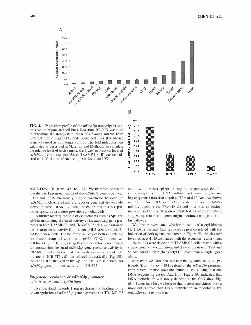
To determine the tissue distribution of mDab2ip mRNA, weperformed a real-time RT-PCR analyses (Fig. 4A). Results indi-cated a unique expression pattern in certain organs. For example,mDab2ip was most abundant in the brain (72.5-fold), salivarygland (38.7-fold), and testis (21.3-fold); moderate expression inthe kidney (15.0-fold) and heart (11.3-fold); low expression in thelung (7.4-fold), seminal vesicle (7.1-fold), ventral prostate (6.5-fold), epididymis (6.1-fold), liver (5.9-fold), and bladder (5.6-fold); quite low expression in the coagulation gland (3.6-fold) andskeleton muscles (2.0-fold) compared with the spleen, which hasthe lowest expression level (�1.0) among all the organs tested.This pattern of expression is consistent with our previous report(Wang et al., 2002) and gene card expression pattern (http://bioin-formatics.weizann.ac.il/cards-bin/arddisp?DAB2IP) except theexpression level of Dab2ip detected from the heart.
In several established mouse cell lines, we also observed a dif-ferential expression level of the mDab2ip transcript. In general, thehighest level of mDab2ip mRNA was detected in the NIH 3T3
cell line (�8.5-fold higher than TRAMP-C3); TRAMP-C1 and -C2 lines (�3-fold) had moderate expressions, and the TRAMP-C3 line had the lowest expression (Fig. 4B). PC-3, a human prostatecancer cell line, was used as a negative control (Chen et al., 2003).
Analysis of the 5�-upstream sequence of the mDab2ip gene
To analyze the promoter region of the mDab2ip gene, a 1.3-kb fragment from position �730 to �545 (transcription initial siteas �1 predicted by 5�RACE) containing a 5� upstream region ofthe exon Ia, exon Ia, and partial intron1a region, was amplifiedby PCR from clone 22N15 using primer set F1/R2 (Table 1). Sequencing analysis indicated that this region is very GC-rich.Using the TFSEARCH program (http://www.cbrc.jp/research/db/TFSEARCH.html); Promoter Scan II program (http://thr.cit.nih.gov/molbio/proscan/), and MacVector 7.0 program, we identifiedseveral potential trans-factor binding sites (Fig. 5A) includingSp1, AP-1 AP-2, SREBP, and p300, GATA-1/2, PEA2, AML-1a, and MalT_box. Neither the TATA-box nor the CAAT-boxwas identified. The similar trans-factor binding sites were alsodetected in the hDAB2IP gene (Chen et al., 2002). It indicates thatthe Dab2ip gene is a typical TATA-less promoter.
To define the potential promoter region in the mDab2ip gene,we examined the reporter gene activities of a series of deletionconstructs (Fig. 5B) generated from the clone 22N15. Consis-tent with Figure 4B, the luciferase activity of the mDab2ip pro-moter constructs was much higher in NIH-3T3 cell than twoother mouse prostatic epithelial cells (Fig. 5C).
In both TRAMP-C1 and TRAMP-C3 cells (Fig. 5D), we ob-served two constructs (i.e., pGL3–F6/R2 [from �421 to �545]and pGL3–F7/R2 [from �147 to �545]) expressed higher lu-ciferase reporter gene activity than that of pGL3–F1/R2 (from�730 to �545), suggesting the presence of a negative cis-ele-ment between �730 and �421. Moreover, the reporter geneactivity decreased significantly in the rest of the deletion con-structs (i.e., pGL3–F8/R2, pGL3–F10/R2, and pGL3– F12/R2).Also, very little reporter gene activity was observed in cellstransfected with either pGL3–F6/NcoI (from �421 to �157) or
CLONING OF MOUSE Dab2ip GENE 237
TABLE 3. EXON–INTRON BOUNDARIES OF THE MOUSE DAB2IP
3� intron/5� exon 3� exon/5� intronExon Size (bp) boundary boundary Intron Size (kb)
Ia 134 TGAGAG/gtaggccc 1a 29.3Ib 417 cctcctcag/GTCCCA GCCAAG/gtctgtgac 1b 4.3Ic 76 cctcctcag/GTCCCA CACTTG/gtgagtggc 01c 10.0II 154 cctcctcag/GTCCCA TTCGAG/gtgggtgtc 2b 1.0III 99 gtttgacag/GTGACG AACAAG/gtacctgta 3b 0.9IV 555 cttatgcag/GACAAC GTGAAG/gtgagtgtg 4b 2.7V 145 cctccctag/GACTTT CACTAG/gtagtgggg 5b 0.1VI 145 gcccacag/GTGAGT CTACTG/gttagtgcca 6b 2.7VII 237 cctatgcag/TGCTTC TGCCAA/gtgagtgtt 7b 1.6VIII 202 ttctgtccag/GTTTGG GATCAG/gtgcctgtt 8b 1.4IX 179 attcttgtag/AGCGTT CTCTGG/gtaagagc 9b 0.9X 889 tccctgcag/TCTGAT AAGCAG/gtcagcacc 10b 0.5XI 153 ttcatcgtag/GGCCCT GAAAAG/gtaaaactg 11b 1.6XII 194 tgctggcag/GATCTG CAGCAG/gtgagcagg 12b 4.2XIII 88 ctgttcacag/GTTGAT GCCCAG/gttggggctc 13b 0.5XIV 2935 gcccacag/GAAAAG

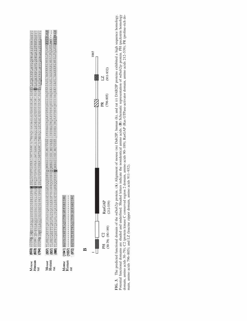
FIG
. 3.
The
pre
dict
ed f
unct
iona
l do
mai
n of
the
mD
ab2i
ppr
otei
n. (
A)
Alig
nmen
t of
mou
se (
m)
Dab
2IP,
hum
an (
h),
and
rat
(r)
DA
B2I
P pr
otei
ns e
xhib
ited
a hi
gh s
eque
nce
hom
olog
y.Po
tent
ial
func
tiona
l do
mai
ns a
re s
hade
d an
d un
derl
ined
. Sh
aded
let
ters
ind
icat
e th
e no
nide
ntic
al a
min
o ac
ids.
(B
) Sc
hem
atic
rep
rese
ntat
ion
of m
Dab
2ip
prot
ein.
PH
(pe
ckst
rin
hom
olog
ydo
mai
n, a
min
o ac
ids
30–7
9); C
2 (p
rote
in k
inas
e C
con
serv
ed r
egio
n 2
dom
ain,
am
ino
acid
s 90
–189
); R
asG
AP
(Ras
GT
Pase
-act
ivat
or d
omai
n, a
min
o ac
ids
212–
539)
; PR
(pr
olin
e-ri
ch d
o-m
ain,
am
ino
acid
s 79
6–80
5);
and
LZ
(le
ucin
e zi
pper
dom
ain,
am
ino
acid
s 91
1–93
2).
B
pGL3–F6/SanDI (from�421 to �97). We therefore concludethat the basal promoter region of the mDab2ip gene is between�147 and �545. Noticeably, a good correlation between themDab2ip mRNA level and the reporter gene activity was ob-served in these TRAMP-C cells, indicating that this is a pro-moter operative in mouse prostatic epithelial cells.
To further identify the role of cis-elements such as Sp1 andAP2 in modulating the basal activity of the mDab2ip gene pro-moter in both TRAMP-C1 and TRAMP-C3 cells, we examinedthe reporter gene activity from either pGL3–�Sp1, or pGL3–�AP2 in these cells. The luciferase activity of both mutants didnot change compared with that of pGL3–F7/R2 in these twocell lines (Fig. 5D), suggesting that either factor is not criticalfor maintaining the basal mDab2ip gene promoter activity inTRAMP-C cells. In contrast, the luciferase activities of bothmutants in NIH-3T3 cell line reduced dramatically (Fig. 5E),indicating that that either the Sp1 or AP2 site is critical formDab2ip gene promoter activity in NIH-3T3.
Epigenetic regulation of mDab2ip promoter activity in prostatic epithelium
To understand the underlying mechanism(s) leading to thedownregulation of mDab2ip gene expression in TRAMP-C3
cells, two common epigenetic regulatory pathways (ie., hi-stone acetylation and DNA methylation) were analyzed us-ing epigenetic modifiers such as TSA and 5�-Aza. As shownin Figure 6A, TSA or 5�-Aza could increase mDab2ipmRNA levels in the TRAMP-C3 cell in a dose-dependentmanner, and the combination exhibited an additive effect,suggesting that both agents might mediate through a simi-lar pathway.
We further investigated whether the status of acetyl histoneH3 (H3) in the mDab2ip promoter region correlated with theinduction of both agents. As shown in Figure 6B, the elevatedlevels of acetyl H3 associated with the promoter region (from�154 to �7) were detected in TRAMP-C3 cells treated with asingle agent or a combination, and the combination of TSA and5�-Aza could elicit higher acetyl H3 levels than a single agentalone.
Moreover, we examined the DNA methylation status of CpGislands (from �8 to �294 region) of the mDab2ip promoterfrom several mouse prostatic epithelial cells using bisulfiteDNA sequencing assay. Data from Figure 6C indicated thatDNA methylation was rarely detected at the CpG sites (Fig.6C). Taken together, we believe that histone acetylation play amore critical role than DNA methylation in modulating themDab2ip gene expression.
CHEN ET AL.240
A
B
FIG. 4. Expression profile of the mDab2ip transcript in var-ious mouse organs and cell lines. Real-time RT-PCR was usedto determine the steady-state levels of mDab2ip mRNA fromdifferent mouse organs (A) and mouse cell lines (B). Mouseactin was used as an internal control. The fold induction wascalculated as described in Materials and Methods. To calculatethe relative level of each sample, the lowest expression level ofmDab2ip from the spleen (A), or TRAMP-C3 (B) was consid-ered as 1. Variation of each sample is less than 10%.

(con
tinu
ed)
FIG
. 5.
Cha
ract
eriz
atio
n of
the
mD
ab2i
pge
ne p
rom
oter
s. (
A)
The
pre
dict
ed r
egul
ator
y se
quen
ces
of t
he m
Dab
2ip
gene
. Exo
n Ia
seq
uenc
e of
the
mD
ab2i
pis
und
erlin
ed. T
hepu
tativ
e ci
s-ac
ting
elem
ents
are
box
ed. P
rim
er s
eque
nces
(bo
ld l
ette
rs)
and
rest
rict
ion
endo
nucl
ease
s si
tes
(bol
d le
tters
and
und
erlin
ed)
wer
e us
ed i
n su
bseq
uent
clo
ning
for
re-
port
er c
onst
ruct
s. T
he t
rans
crip
tion
star
t si
te (
TSS
) as
�1
was
pre
dict
ed b
y 5�
RA
CE
.

CHEN ET AL.242
FIG. 5. Continued. (B) Schematic representation of the mDab2ip promoter construct. A series of reporter gene constructs weregenerated by series deletion or removing the potential cis-element of the mDab2ip promoter sequence using pGL3 as a backbonevector described in Materials and Methods. (C) Characterization of mDab2ip promoter activity in various mouse cell lines.
DISCUSSION
The mDab2ip gene spans approximately 65 kb, containing14 exons and 13 introns with at least three variants: exon Ia,exon Ib, and exon Ic found from different sources of RNA us-ing the 5�RACE assay. All three splicing sequences of mDab2ipP cDNA have been submitted to the GenBank™ (AY305656[mDab2ip a]; AY 305657 [mDab2ip b]; AY305658 [mDab2ipc]). Using FISH analysis, mDab2ip was localized at chromo-some band 2B (Fig. 2), which is consistent with the LocusLinkprogram analysis (http://www.ncbi.nih.gov/ LocusLink).
In this study, we performed a real-time RT-PCR to demon-
strate the mDab2ip mRNA levels in different organs (Fig. 4A).Very abundant mDab2ip mRNA levels were found in the brain,salivary gland, and testis, and the moderate levels were foundin the kidney and heart. In addition, organs such as the lung,seminal vesicle, ventral prostate, epididymis, liver, and bladderonly express low levels of Dab2ip mRNA. Also, the lowest levelof Dab2ip mRNA was detected in the coagulation gland, skele-tal muscles, and spleen. Such a diverse expression pattern of theDab2ip gene implies that mDab2ip may have a unique physio-logical function in a specific organ. To gain the insight of themechanisms of mDab2ip transcriptional regulation, we isolatedan �1.3 kb (Fig. 5A) fragment containing a 5�-upstream region

from exon Ia, and we found very rich GC-rich sequences andno canonical TATA boxes in this region. Nevertheless, we haveshown that the 5�-flanking region from positions �730/�545could enhance the reporter gene activity, and it contained thebasal promoter (�147/�545) and a negative regulatory element(�730/�421) (Fig. 5). In this region, several putative cis-ele-ments could underlie the differential Dab2ip gene expressionsin various organs or cells, so we have employed a series of mu-tants (Fig. 5D and E) to examine the role of two common cis-elements such as Sp1 and AP2 in maintaining the basal promoteractivity of the mDab2ip gene. In NIH-3T3, both cis-elementsare critical for maintaining the basal promoter activity of the
mDab2ip gene (Fig. 5E). However, in prostatic epithelium,either cis-element may not play a critical role in modulating thebasal promoter activity of the mDab2ip gene (Fig. 5D). Instead,the status of histone aceylation but not DNA methylation asso-ciated with the mDab2ip promoter region correlates with its geneinduction in the TRAMP-C3 (Fig. 6). It also appears that 5�-Azais able to increase the acetylated histone levels associated withthe mDab2ip promoter region. A similar observation wasdemonstrated in our previous studies (Chen et al., 2003), sinceit is known that the hypomethylation agent can cause the disso-ciation of the transcription repressor complex containing bothDNMT and HDAC (Jones and Baylin, 2002).
CLONING OF MOUSE Dab2ip GENE 243
FIG. 5. Continued. (D) Mapping core of the mDab2ip pro-moter region in TRAMP-C1 and TRAMP-C3. (E) Evaluating therole of two cis-elements (Sp1 and AP2) in modulating mDab2ippromoter activity in NIH-3T3 cells. Bars, SD.

In our previous publications (Chen et al., 2002; Wang et al.,2002), we found the translation initiation site (ATG) of DAB2IPat 63-bp from the 5�-end of exon 3 and predicted a putative openreading frame encoding the 967-amino acid for hDAB2IP or the996- amino acid for rat DAB2IP (rDAB2IP). However, the up-dated sequence data from NCBI (accession no. NP_619723) in-dicate that an additional ATG site is mapped at 11-bp from the5�-end of exon 2 that was also detected in mDab2ip. Thus, pre-dicted mDab2ip protein encodes the 1065-amino acid contain-ing an additional 69-amino acid with a PH domain. The PH do-main is a short motif that mediates membrane localization, andis found in many proteins involved in signal transduction, in-cluding GAPs for Ras (Shaw, 1996; Rebecchi and Scarlata,1998). The predicted protein sequence alignment between mouse and human DAB2IP is remarkably conserved. Von Berghet al. (2004) reported that the hDAB2IP is the alias for the
AF9Q34gene (accession no. AY032952) as a novel fusion part-ner of MLL in the AML patient with (9:11) translocation. Thejuxaposition of MLL intron 9 into exon 2 of AF9Q34 will re-sult in the loss of the exon 2 splicing donor site. Consequently,the hDAB2IP/AF9Q34 exon 2 sequences will be spliced out andresult in an MLL-exon 9/AF9Q34-exon 3 fusion product. In thiscase, the AF9Q34-MLL fusion protein does not contain the PHdomain, implying that the normal function of the AF9Q34 genemay be altered due to the chromosomal translocation.
Sequence analysis of mDab2ip revealed the presence of ahighly conserved GAP-related domains (GRD), the catalyticunit to stimulate the GTPase activity of Ras proteins, in the N-terminus of mDab2ip. GRD is a characteristic domain in the allRasGAPs such as human neurofiibromin (NF1), rat SynGAP,p120GAP, and human nGAP (Bernards et al., 1992; Davis etal., 1993; Li et al., 1996; Kim et al., 1998; Noto et al., 1998;
CHEN ET AL.244
FIG. 6. Epigenetic regulation of the mDab2ip gene promoter in mouse prostatic epithlium. (A) The effect of TSA and/or 5�-Aza on mDab2ip mRNA expression in TRAMP-C3 cells. *Statistically significant (p�). (B) The effect of TSA and/or 5�-Azaon acetyl histone H3 levels associated with the mDab2ip promoter in TRAMP-C3 cells. (C) Profiling DNA methylation statuson the CpG site in the mDab2ip gene promoter region (from �8 to �294) in several mouse cell lines. The position of the CpGdinucleotide site (vertical tick) was indicated. �, unmethylated CpG; �, methylated CpG.

Glanzer et al., 2002). Homayouni et al. (2003) suggest thatDab2IP may function as a downstream effector in the Reelin-signaling pathway that influences Ras signaling during braindevelopment. With the cloning of this gene from the mouse,studying the function al role of this gene in brain developmentas well as prostate carcinogeneis can be feasible.
ACKNOWLEDGMENTS
We thank Dr. Norman Greenberg (Baylor College of Medi-cine) for providing TRAMP-C cell lines, and Kenneth S. Koen-eman (UT Southwestern Medical Center) for reading this man-uscript. This work is supported in part by United States ArmyGrants W81XWH-04-1-0222 and DAMD17-03-2-0033. Se-quence data from this article have been deposited into theEMBL/GenBank Data Libraries under accession numberAY305656, AY305657, and AY305658.
REFERENCES
BERNARDS, A., HAASE, V.H., MURTHY, A.E., MENON, A., HAN-NIGAN, G.E., and GUSELLA, J.F. (1992). Complete human NF1cDNA sequence: two alternatively spliced mRNAs and absence ofexpression in a meuroblastoma line. DNA Cell Biol. 11, 727–734.
CHEN, H., PONG, R.C., WANG, Z., and HSIEH, J.T. (2002). Differ-ential regulation of the human gene DAB2IP in normal and malig-nant prostatic epithelia: Cloning and characterization. Genomics 79,573–581.
CHEN, H., TOYOOKA, S., GAZDAR, A. F., and HSIEH, J.T. (2003).Epigenetic regulation of a novel tumor suppressor gene (hDAB2IP)in prostate cancer cell lines. J. Biol. Chem. 278, 3121–3130.
DAVIS, M. M., CATINO, J.J., SATOH, T., KAZIRO, Y., andPERKINS, L.M. (1993). Sequence of the cDNA encoding Ras GT-Pase-activating protein from rat. Gene 134, 305–306.
DOTE, H., TOYOOKA, S., TSUKUDA, K., YANO, M., OUCHIDA,M., DOIHARA, H., SUZUKI, M., CHEN, H., HSIEH, J.T., GAZ-DAR, A.F., et al. (2004). Aberrant promoter methylation in humanDAB2 interactive protein (hDAB2IP) gene in breast cancer. Clin.Cancer Res. 10, 2082–2089.
FOSTER, B.A., GINGRICH, J.R., KWON, E.D., MADIAS, C., andGREENBERG, N.M. (1997). Characterization of prostatic epithelialcell lines derived from transgenic adenocarcinoma of the mouseprostate (TRAMP) model. Cancer Res. 57, 3325–3330.
FULOP, V., COLITTI, C.V., GENEST, D., BERKOWITZ, R.S., YIU,G.K., NG, S.W., SZEPESI, J., and MOK, S.C. (1998). DOC-2/hDAB2, a candidate tumor suppressor gene involved in the devel-opment of gestational trophoblastic diseases. Oncogene 17, 419–424.
GLANZER, J.G., LIAO, L., BAKER, T., MCMULLEN, M.H., LAN-GAN, A.S., CRANDALL, L.Z., and VORCE, R.L. (2002). Organi-zation and regulation of the human rasGAP gene. Gene 285, 149–156.
GINGRICH, J.R., BARRIOS, R.J., KATTAN, M.W., NAHM, H.S.,FINEGOLD, M.J., and GREENBERG, N.M. (1997). Androgen-in-dependent prostate cancer progression in the TRAMP model. Can-cer Res. 57, 4687–4691.
GREENBERG, N.M., DEMAYO, F., FINEGOLD, M.J., MEDINA, D.,TILLEY, W.D., ASPINALL, J.O., CUNHA, G.R., DONJACOUR,A.A., MATUSIK, R.J., and ROSEN, J.M. (1995). Prostate cancer ina transgenic mouse. Proc. Natl. Acad. Sci. USA 92, 3439–3443.
HOMAYOUNI, R., MAGDALENO, S., KESHVARA, L., RICE, D.S., and CURRAN, T. (2003). Interaction of Disabled-1 and the GT-Pase activating protein Dab2IP in mouse brain. Mol. Brain Res. 115,121–129.
JONES P.A., and BAYLIN, S.B. (2002). The fundamental role of epi-genetic events in cancer. Nat. Rev. 3, 415–428.
JONES, P.A., and TAKAI, D. (2001). The role of DNA methylation inmammalian epigenetics. Science 293, 1068–1070.
KIM, J.H., LIAO, D., LAU, L.F., and HUGANIR, R. (1998). SynGAP:A synaptic RasGAP that associates with the PSD-95/SAP90 proteinfamily. Neuron 20, 683–691.
LI, S., SATOH, H., WATANABE, T., NAKAMURA, S., and HAT-TORI, S. (1996). cDNA cloning and chromosomal mapping of anovel human GAP (GAP1M), a GTPase-activating protein of Ras.Genomics 35, 625–627.
NOTO, S., MAEDA, T., HATTORI, S., INAZAWA, J., IMAMURA,M., ASAKA, M., and HATAKEYAMA, M. (1998). A novel humanRasGAP-like gene that maps within the prostate cancer susceptibil-ity locus at chromosome 1q25. FEBS Lett. 441, 127–131.
REBECCHI, M.J., and SCARLATA, S. (1998). Pleckstrin homologydomains: A common fold with diverse functions. Annu. Rev. Bio-phys. Biomol. Struct. 27, 503–528.
SHAW, G. (1996). The pleckstrin homology domain: An intriguingmultifunctional protein module. Bioessays 18, 35–46.
TSENG, C.P., ELY, B.D., PONG, R.C., WANG, Z., ZHOU, J., andHSIEH, J.T. (1999). The role of DOC-2/DAB2 protein phosphory-lation in the inhibition of AP-1 activity. An underlying mechanismof its tumor-suppressive function in prostate cancer. J. Biol. Chem.274, 31981–31986.
VON BERGH, A.R., WIJERS, P.M., GROOT, A.J., VANZELDEREN-BHOLA, S., FALKENBURG, J.H., KLUIN, P.M., andSCHUURING, E. (2004). Identification of a novel RAS GTPase-ac-tivating protein (RASGAP) gene at 9q34 as an MLL fusion partnerin a patient with de novo acute myeloid leukemia. Genes Chromo-somes Cancer 39, 324–334.
WANG, Z., TSENG, C.P., PONG, R.C., CHEN, H., MCCONNELL,J.D., NAVONE, N., and HSIEH, J.T. (2002). The mechanism ofgrowth-inhibitory effect of DOC-2/DAB2 in prostate cancer. Char-acterization of a novel GTPase-activating protein associated with N-terminal domain of DOC-2/DAB2. J. Biol. Chem. 277, 12622–12631.
WOLFFE, A.P., and MATZKE, M.A. (1999). Epigenetics: Regulationthrough repression. Science 286, 481–486.
ZHANG, H., ZHANG, R., LUO, Y., D’ALESSIO, A., POBER, J. S.,and MIN, W. (2004). AIP1/DAB2IP, a novel member of the Ras-GAP family, transduces TRAF2-induced ASK1-JNK Activation. J.Biol. Chem. 279, 44955–44965.
ZHANG, R., HE, X., LIU, W., LU, M., HSIEH, J.T., and MIN, W.(2003). AIP1 mediates TNF-�-induced ASK1 activation by facili-tating dissociation of ASK1 from its inhibitor 14-3-3. J. Clin. Invest.111, 1933–1943.
ZHOU, J., and HSIEH J.T. (2001). The inhibitory role of DOC-2/DAB2in growth factor receptor-mediated signal cascade. DOC-2/DAB2-mediated inhibition of ERK phosphorylation via binding to Grb2. J.Biol. Chem. 276, 27793–27798.
ZHOU, J., SCHOLES, J., and HSIEH, J.T. (2003). Characterization ofa novel negative regulator (DOC-2/DAB2) of c-Src in normal pro-static epithelium and cancer. J. Biol. Chem. 278, 6936–6941.
Address reprint requests to:J.-T. Hsieh, Ph.D.
Department of UrologyUniversity of Texas Southwestern Medical Center
5323 Harry Hines Blvd.Dallas, TX 75390-9110
E-mail: [email protected]
Received for publication June 12, 2005; received in revisedform August 15, 2005; accepted January 17, 2006.
CLONING OF MOUSE Dab2ip GENE 245