autonomic neuropathy in diabetes dr.j.kanakamani
TRANSCRIPT

AUTONOMIC NEUROPATHY IN
DIABETESDr.J.Kanakamani

Plan of presentation
• Introduction• Epidemiology• Pathogenesis• Systems involved in DAN
– Clinical manifestations– Evaluation– Management
• Summary

INTRODUCTION


The ANSSNS• activate body• thoracolumbar (T1-
L2)• short
preganglionic/long postganglionic fibers
• global responses• postganglionic
transmitter: NE (except
sweat glands – ACh)
PSNS• prepare body for
rest/digest• craniosacral (CN
III, VII,IX, X & S2-4)
• long preganglionics/ short postganglionic fibers
• discrete/local responses
• postganglionic transmitter: ACh

The ANS
SNS• “Fight -Flight” system
– Activation– increases heart rate– increases sweating– dilates pupil– inhibits GI movement– closes sphincters– diverts blood from skin
and GI tract to skeletal muscles
PSNS• “Rest-digest” system
– promotes digestion, GI peristalsis
– slows heart rate– constricts pupil– empties bladder– relaxes sphincters– mediates genital
erection

EPIDEMIOLOGY

Why recognise DAN?
• 25-50% die within 5-10 years of diagnosis
• 5-year mortality rate is 3-5 times higher
• Marker of adverse cardiovascular, renal and cerebrovascular outcomes

Is it common?
• Varyingly reported from 5-35%• Symptomatic autonomic neuropathy
- long after the onset of diabetes. • Subclinical autonomic dysfunction -
common

Association with peripheral neuropathy
• tthough there is an association parasympathetic dysfunction may appear independant
• Hence, tests for sensory and motor nerve functions (eg. monofilament, quantitative sensory testing, nerve conduction studies, muscle strength testing) may not be effective in detecting DAN that cardiovascular autonomic function testing can detect at early stage of emergence.
• Thus, tests for other forms of diabetic peripheral nerve dysfunction should not substitute for the tests for cardiovascular autonomic dysfunction.

Epidemiology
• Risk factors– poor glycemic control– long duration of diabetes– increasing age– female sex– higher body mass index– ? smoking and elevated triglycerides

PATHOGENESIS

Pathogenesis
Diabetes
Neurohumoral growth factor
and EFA deficiency
Autoimmune damage
Neurovascular insufficiency
Hyperglycemia
Free radical injury
Sorbitol, AGE, PKC

SYSTEMS INVOLVED IN DAN
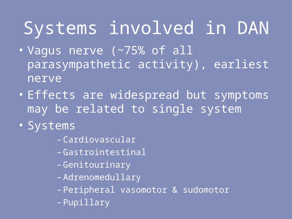
Systems involved in DAN• Vagus nerve (~75% of all
parasympathetic activity), earliest nerve• Effects are widespread but symptoms
may be related to single system• Systems
– Cardiovascular– Gastrointestinal– Genitourinary– Adrenomedullary– Peripheral vasomotor & sudomotor– Pupillary
CARDIOVASCULAR AUTONOMIC
NEUROPATHY

Clinical manifestations• Heart Rate changes
– Impaired Heart rate variability – Resting tachycardia and fixed HR
• BP changes– Nocturnal hypertension– Orthostatic hypotension– postprandial hypotension
• Limited exercise tolerance

Other clinical implications
• QT prolongation, altered repolarisation, nocturnal arrhytmogenesis and death
• Silent myocardial ischemia• Diabetic cardiomyopathy• Intraoperative cardiovascular liability
(vasopressor support, severe intraop hypothermia)
• Stroke
Evaluation
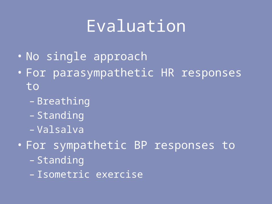
• No single approach• For parasympathetic HR responses to
– Breathing– Standing– Valsalva
• For sympathetic BP responses to– Standing– Isometric exercise

EvaluationTest Technique InterpretationHR reponse to deep breathing
HR response tostanding
HR response to Valsalva
Patient lies quietly and breathes deeply at a rate of six breaths per minute and ECG is recorded. The difference between the maximum and minimum heart rate and Expiration to Inspiration (E:I) R-R interval ratio are calculated.
ECG is recorded in lying followed by full upright position. The R-R interval is measured at beats 15 and 30 after the patient stands.
The patient forcibly exhales into the mouthpiece of a manometer, exerting a pressure of 40 mm Hg for 15 seconds. There are 4 phases during this maneuver. The longest and shortest R-R intervals are measured. The ratio is called valsalva ratio.
A difference in HR of < 10 bpm and E:I ratio is >1.17 are abnormal.
A 30:15 ratio of < 1.03 is abnormal.
Valsalva ratio of < 1.2 is abnormal.

EvaluationTest Technique InterpretationBP response to standing
BP response to isometric exercise
BP is measured when the patient is lying down and 2 minutes after the patient stands
The patient squeezes a handgrip dynamometer to establish his or her maximum. The patient then maintains the grip at 30% maximum for 5 minutes. BP is measured in the contralateral arm.
Systolic BP fall of ≥ 20 mm Hg or diastolic BP fall of ≥ 10 mm Hg is abnormal *.
A diastolic BP rise of < 16 mm Hg is abnormal.

AFT LAB

Stages
• Early stage: abnormality of heart rate response during deep breathing alone
• Intermediate stage: an abnormality of Valsalva response
• Severe stage: the presence of postural hypotension

Safety
• High value-to-risk ratio.• Some adverse effects. Valsalva
maneuver - transient increase in intracranial, intrathoracic and intraabdominal pressures - theoretical possibility of intraocular hemorrhage and lens dislocation.
• Children, mentally disabled and aged – difficult to perform

Evaluation
• Newer noninvasive tests– Power spectral analysis– MIBG SPECT– 11-C-hydroxyephedrine scintigraphy

Treatment of impaired HRV
• Prolonged QT– Acute mgt. (Mg, temp pacing,
isoproteronol)– Chronic mgt. ( avoid ppt. factors,
electrolytes, pem pacing.
• SCD– ICD

Treatment of OH
• General measures– Gravity suits and stockings– Changes in posture to be made slowly in "stages”– Tensing the legs, dorsiflexing the feet, or doing
handgrip exercise before standing– High salt diet, increasing water consumption– Treat anemia, avoid drugs aggrevating OH
• Pharmacological measures– Glycemic control and multifactorial risk reduction– Alpha lipoic acid, ACEi

Drugs for OH• Specific Drugs
• Midodrine 2.5 – 10 mg tid• Fludrocortisone 0.05 mg hs – 0.4
mg/day– blockers (pindolol) – not clear– Clonidine – severe side effects– desmopressin
• Octreotide esp. for postprandial hypotension 25 – 200 mcg/day

GASTROINTESTINAL AUTONOMIC
NEUROPATHY

Clinical manifestations
• Esophageal dysmotility– GERD common, dysphagia is uncommon
• Gastroparesis diabeticorum• Enteropathy
– Nocturnal watery painless diarrhea
• Constipation• Fecal incontinence• Gall bladder atony and enlargement
Gastroparesis
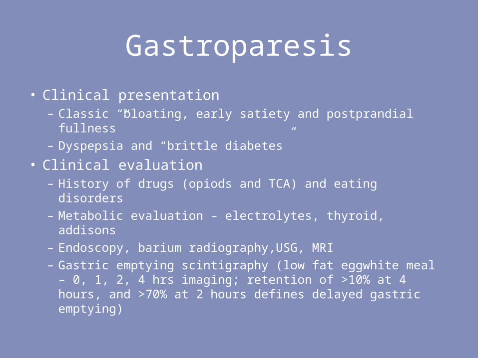
• Clinical presentation– Classic “bloating, early satiety and postprandial
fullness”– Dyspepsia and “brittle diabetes”
• Clinical evaluation– History of drugs (opiods and TCA) and eating disorders– Metabolic evaluation – electrolytes, thyroid, addisons– Endoscopy, barium radiography,USG, MRI– Gastric emptying scintigraphy (low fat eggwhite meal –
0, 1, 2, 4 hrs imaging; retention of >10% at 4 hours, and >70% at 2 hours defines delayed gastric emptying)

Treatment of Gastroparesis
Cisapride not more than 1 mg/kg/d

Pathogenesis of diabetic diarrhea
Autonomic dysfunction
Anorectal dysfunction
Bacterial overgrowth
Incresed secretion
Altered motility
? Exocrine pancreatic
insufficiencyAssociated
factors
Diabetic diarrhea

Pathogenesis of diabetic diarrhea
Autonomic dysfunction
Associated factors
Diabetic diarrhea
? bile acid malabsorptio
n
Concurrent diseases
Dietitic foods - sorbitol

Evaluation for diarrheaLevel of invesigation
Tests
First line(a) Blood biochemistry; Stool – weight, 72 hour fecal fat, elastase,
chymotrypsin, leucocytes, parasites, occult blood; Upper GI
Barium studies with dedicated small bowel follow through - for
gastric retention, pattern of malabsorption, small intestinal and
colonic wall thickness
(b) D-Xylose test for small intestinal malabsorptionSecond line (a) Upper GI endoscopy with duodenal biopsy for histology and
bacteriology
(b) Colonoscopy and biopsy for histology
(c) Glucose hydrogen breath test for bacterial overgrowthThird line (a) Ambulatory small intestinal manometry for intestinal
pseudoobstruction
(b) Empiric cholestryamine for possible bile acid malabsorption
(c) Enteroscopy with biopsy and enteroclysis
(d) Secretin-pancreozymin test for pancreatic exocrine insufficiency
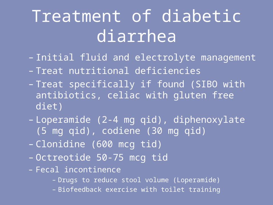
Treatment of diabetic diarrhea
– Initial fluid and electrolyte management– Treat nutritional deficiencies– Treat specifically if found (SIBO with
antibiotics, celiac with gluten free diet)– Loperamide (2-4 mg qid), diphenoxylate
(5 mg qid), codiene (30 mg qid)– Clonidine (600 mcg tid) – Octreotide 50-75 mcg tid– Fecal incontinence
– Drugs to reduce stool volume (Loperamide)– Biofeedback exercise with toilet training
GENITOURINARY AUTONOMIC
NEUROPATHY

Clinical manifestations
• Neurogenic bladder– Decreased bladder sensation, hesitancy, later
incomplete evacuation and frequent UTI
• Erectile dysfunction– Neuropathy, vascular disease, metabolic
control, nutrition, endocrine disorders, psychogenic factors, and anti-diabetes drugs.
• Female sexual dysfunction– Decreased libido and vaginal lubrication
causing dypareunia

Evaluation• Bladder
– Urine culture, postvoidal residue, renal function tests, cystometry and voiding cystometrogram
• Erectile dysfunction– History, physical examination,
biochemistry, hormones, penile doppler, therapeutic trial with sildanefil, intracavernosal injections of vasodilator
• Retrograde ejaculation– Azoospermia with spermaturia in
postcoital urine specimen


Treatment• Bladder
– CBD initially– later timed voiding often with Crede’s
maneuvre– bethenechol at the time of voiding – external sphincter relaxation with doxazosin– Severe cases, clean intermittent
catheterisation– Rarely, bladder neck resection
• ED– 5-PDE inhibitors (> 60% patients respond)– Intracavernosal papaverine– Transurethral alprodostil

MISCELLANEOUS

Others
• Metabolic– Hypoglycemia unawareness
• Sudomotor– Peripheral dry skin and paradoxical excess
sweating in trunk– Gustatory sweating
• Peripheral vasomotor– changes in the texture of skin, loss of nails,
anhidrosis, callus formation and the development of fissures and cracks

– Peripheral edema and venous prominences– The loss of sympathetic vascular innervation
results in high peripheral blood flow through arteriovenous (AV) shunts and abnormal local reflex vascular control - increased osteoclastic activity resulting in reduced bone density, proneness to fractures - ?pathogenesis of Charcot’s neuroarthropathy
• Pupillary involvement– AR pupil, diminished hippus, reduced dark
adaptation

GUIDELINES FOR DIAGNOSIS

San Antonio conference, 1988
• Symptoms not to be considered as markers of its presence.
• Noninvasive validated autonomic function tests should be used taking into account confounding factors like concomitant drug use, concurrent illness, age, etc.
• Abnormality in more than one test on more than one occasion is desirable.
• Both sympathetic and parasympathetic functions should be tested independently.

San Antonio conference, 1988
• For the assessment of CAN, the panel recognized three tests of heart rate control and two tests of BP control
• These tests were judged suitable for both routine screening and monitoring the progress of autonomic neuropathy.
• No other tests including those for GI, genitourinary, sudomotor, microvascular skin blood flow and pupillary function were considered to be sufficiently well standardized for routine clinical use.

Thanks
