automated detection of aortic annulus sizing...
TRANSCRIPT

AUTOMATED DETECTION OF AORTIC ANNULUS SIZING BASED ON
DECISION LEVEL FUSION
NORHASMIRA BINTI MOHAMMAD
A thesis submitted in fulfilment of the
requirements for the award of the degree of
Master of Philosophy
Faculty of Biosciences and Medical Engineering
Universiti Teknologi Malaysia
FEBRUARY 2018

vi
Specially dedicated to husband, M ak and Abah.
Thank you so much for your prayers.
I love all o f you.

vii
ACKNOW LEDGEM ENT
Firstly, I would like to give a token of my gratitude to all who have contributed
through their wide range of support to the success of this work. Special thanks to my
supervisor Dr. Zaid bin Omar. He has enriched me with his dedicated care and constant
willingness to discuss with a wide range o f information for thought and assisted in
completing this research.
I would also like to express gratitude to Dr. Dyah Ekashanti Octorina Dewi as
my co-supervisor who has helped in stimulating suggestions and encouragement. Last
but not least, I would like to thank my beloved parents, family and friends dearly for
the daily support and assistance throughout my research period at Universiti Teknologi
Malaysia.
viii
ABSTRACT
Aortic valve disease occurs due to calcification on the area of leaflets and it is
progressive over time. Surgical Aortic Valve Replacement (SAVR) can be performed
to treat the patient. However, due to invasive procedure of SAVR, a new method
known as Transcatheter Aortic Valve Implantation (TAVI) has been introduced,
where a synthetic catheter is placed within the patient’s heart valve. Traditionally,
aortic annulus sizing procedure requires manual measurement of scanned images
acquired from different imaging modalities which are Computed Tomographic (CT)
and echocardiogram where both of the modalities produce inconsistency in measuring
the aortic annulus yet able to produce different parameters which lead to accurate
measurement. In this research, the image processing techniques of CT scan and
echocardiogram images are done separately in order to obtain the aortic annulus size.
Intensity adjustment and median filter are applied to CT scan image pre-processing,
Watershed Transformation associated with the morphological operation has been used
to perform the aortic annulus segmentation while image resizing and wavelet
denoising method have been performed in echocardiogram image pre-processing
followed by the implementation of Otsu N-clustering and morphological operation
method for object segmentation. Then, Euclidean distance formula is applied to
measure the distance between two points that indicates the diameter of the aortic
annulus. Finally, a decision fusion technique based on the mathematical statistic
approach has been applied to fuse the measured annulus size obtained from both
modalities. Results affirmed the approach’s ability to achieve accurate annulus
measurements when the final results are compared with the ground truth. In addition,
the application o f non-probabilistic estimation on the decision level fusion approach
which does not required the dataset training produces fast computational time and
helps in determining the optimal size of new aortic valve to be implemented in human
heart.

ix
ABSTRAK
Penyakit injap aortik biasanya terjadi disebabkan oleh kalsium yang
menempel di bahagian pembuka injap aorta dan ianya akan menjadi semakin kritikal
yang akan mengganggu mekanisma pengaliran darah dalam jantung manusia.
Pembedahan boleh dilakukan untuk merawat pesakit dimana prosedur ini memerlukan
bukaan pada dada pesakit untuk menukar injap yang rosak. Walaubagaimanapun,
prosedur ini berisiko tinggi dan terdapat cara yang lebih selamat untuk menjalankan
rawatan iaitu dengan menjalankan implantasi menggunakan kateter. Tradisionalnya,
saiz injap diukur secara manual dan pengukuran boleh dibuat pada imej yang diperoleh
daripada pengimbas CT dan mesin ultrasound dimana kedua alat pengimejan ini
menghasilkan ukuran yang tidak konsisten namun masing-masing mampu
menghasilkan parameter yang berbeza yang dapat menyumbang kepada penghasilan
ukuran yang tepat apabila digabungkan. Oleh itu, penggunaan kaedah pemprosesan
imej daripada kedua-dua alatan pengimejan dilakukan secara berasingan. Kaedah
pelarasan intensiti dan penapis median digunakan dalam pra-pemprosesan imej
pengimbas CT, transformasi Watershed dibantu oleh operasi morfologi digunakan
untuk melakukan objek segmentasi manakala kaedah pengecilan imej dan wavelet
telah digunakan dalam pra-pemprosesan ultrasound imej diikuti pelaksanaan kaedah
Otsu dan morfologi untuk segmentasi objek. Seterusnya, jarak antara dua titik yang
mewakili diameter injap diukur menggunakan formula jarak Euclidean. Akhirnya,
teknik gabungan keputusan yang berasaskan statistik digunakan untuk menyatukan
diameter injap yang diperolehi daripada kedua alatan pengimejan tersebut.
Berdasarkan keputusan yang diperolehi, teknik yang digunakan dalam kajian ini
mampu menghasilkan keputusan yang tepat apabila perbandingan keputusan akhir
dengan pengukuran manual dilakukan. Disamping itu, penggunan statistik yang bukan
berasaskan probabiliti pada peringkat gabungan membantu mempercepatkan masa
operasi dan membantu dalam menentukan saiz injap aorta palsu.

x
CHAPTER TITLE PAGE
DECLARATION v
DEDICATION vi
ACKNOW LEDGEM ENT vii
ABSTRACT viii
ABSTRAK ix
TABLE OF CONTENTS x
LIST OF TABLES xii
LIST OF FIGURES xiii
LIST OF ABBREVIATIONS xx
1 INTRODUCTION
1.1 Background of Research 1
1.2 Problem Statement 6
1.3 Objectives of the Study 10
1.4 Scope of the Study 10
1.5 Significance of Study 11
2 LITERATURE REVIEW
2.1 Introduction to Heart Disease 12
2.2 Devices for TAVI 14
2.3 Imaging Modalities for TAVI Assessment 16
2.3.1 CT Scan Modality 18
2.3.2 Echocardiogram Modality 19
2.4 CT scan Image Processing Algorithm 22
TABLE OF CONTENTS
xi
2.5 Echocardiogram Image Processing Algorithm 26
2.6 Data Fusion 29
3 M ETHODOLOGY
3.1 Introduction 33
3.2 Data Collection 35
3.3 CT Scan Image 36
3.3.1 Preprocessing Algorithm 36
3.3.2 Marker-Controlled Watershed Transform40
for CT scan Image Segmentation
3.3.3 Morphological Operation o f Binary49
Image
3.4 Echocardiogram Image Processing 50
3.4.1 Image Resizing 50
3.4.2 Otsu N-Clustering 56
3.4.3 Morphological Operation o f Binary58
Image
3.5 Minimax Method for Decision Fusion 60
4 RESULTS ANALYSIS
4.1 Analysis on CT scan Image Processing 64
4.2 Analysis on Echocardiogram image processing 75
4.3 Decision level stage 82
5 CONCLUSION AND FUTURE W ORKS 87
5.1 Conclusion 87
5.2 Recommendation and Future Work 89
REFERENCES 91
APPENDIX 103

xii
LIST OF TABLES
TABLE NO. TITLE
2.1 (a) Available size o f Edward Sapien prosthetic valve.
2.1 (b) Available size o f CoreValve prosthetic valve.
3.1 Available size of Edward Sapien prosthetic valve.
3.2 Available size of CoreValve prosthetic valve.
4.1 Comparison o f annulus sizing between manual marking
and other methods for CT scan images.
4.2 Comparison o f annulus sizing between manual marking
and other methods for Echocardiogram images.
4.3 Error representing the difference between the values o f
automated measurement and available size o f Edward
SAPIEN prosthetic valve.
4.4 MaxMax, MaxMin and Minimax values o f statistical
test of data.
4.5 Error representing the difference between the values of
automated measurement and available size o f
CoreValve prosthetic valve.
4.6 MaxMax, MaxMin and Minimax values o f statistical
test of data.
PAGE
15
15
63
63
74
82
84
84
86
86

xiii
FIGURE NO. TITLE PAGE
1.1 Prevalence of valvular heart disease by age. 2
1.2 Progression of aortic stenosis. 3
1.3 Percentage of patient survival. 4
1.4 Reasons of why AVR is non-referral. 5
1.5 Normal and abnormal aortic valve. 8
1.6 (a) Normal and (b) Abnormal aortic valve in 8
Echocardiogram image.
1.7 (a) Normal and (b) abnormal aortic valve in CT scan 9
image.
2.1 Anatomy of human heart. 13
2.2 Anatomy of aortic annulus. 17
2.3 Aortic Valve imaging in CT 19
2.4 Positions of ultrasonic transducer using 21
echocardiogram.
2.5 Aortic annulus dimension in PLAX view. 21
2.6 Manual measurement of annulus on Echocardiogram 22
image.
3.1 Flowchart of methodology. 34
3.2 Area of ascending aorta (AA), left ventricular outflow 36
tract (LVOT) and the diameter of aortic valve marked on
(a) CT scan and (b) Echocardiogram image.
3.3 CT scan image of (a) Original data and (b) image after 37
performing the contrast enhancement.
3.4 Histogram of (a) original CT scan image and (b) image 38
after performing contrast enhancement.
LIST OF FIGURES

3.5
3.6
3.7
3.8
3.9
3.10
3.11
3.12
3.13
3.14
3.15
3.16
3.17
3.18
3.19
3.20
3.21
3.22
4.1
4.2
4.3
xiv
Image (a) before and (b) after performing the median 40
filtering.
Steps to implement Watershed Transformation. 41
Structuring element of 4-neighborhood. 42
Reconstruction of opening image. 44
Reconstruction of opening-closing image. 44
Marker image. (a) Regional maxima of opening-closing 46
by reconstruction image and (b) superimpose of regional
maxima with original image.
Topographic relief o f watershed tool. 47
Application of watershed transform in remote sensing 47
research area on a signal with (a) three markers and (b)
two markers.
Image (a) before and (b) after watershed transform. 48
(a) Outline of segmented object and (b) intersection 49
points of skeleton image.
Echocardiographic of raw image. 51
Features along ascending aorta including aortic annulus 51
(AV ann), sinuses of Valsalva (Sinus Val)
andsinotubular junction (ST Jxn).
Example of three-level wavelet decomposition. 52
Four level of wavelet decomposition. 55
Denoised image. 55
Otsu clustering, n=3. 58
(a) Segmented ROI and (b) skeletonization of segmented 59
image.
(a) Thinning object of skeleton image and (b) 60
measurement of annulus using MATLAB tool.
Sequential output of enhanced image where (a) original 65
image, (b) contrast adjustment and (c) Median filtering.
Effects of segmentation output after applying median 66
filter with kernel size of 5x5, 7x7 and 9x9.
Sobel convolution kernels. 67

xv
4.4 Gradient magnitude. 68
4.5 Computation of marker image. 69
4.6 Superimpose of gradient magnitude and marker image. 71
4.7 Watershed segmentation output after conversion of (a) 71
label to RGB image and (b) RGB to greyscale image.
4.8 Outline of segmented region. 72
4.9 Morphological operation of ‘branchpoints’, (a) without 72
and (b) with image skeleton operation.
4.10 Manual measurement of aortic annulus. 73
4.11 Steps of implementing the algorithm for benchmarking 74
purpose for CT image.
4.12 Images after annulus segmentation o f (a) region 75
growing, (b) deformable models and (c) proposed
method.
4.13 Preprocessing output; (a) Original, (b) resized, (c) 77
wavelet decomposition and (d) denoised image.
4.14 Sequential output of segmentation stage; (a) Clustered 78
image, n = 5 (b) dilation of greyscale image, (c)
conversion o f greyscale to binary image with fill holes,
and (d) skeletonized image.
4.15 Aortic annulus detection steps; (a) thinning operation of 80
‘skel’ image, (b) intersection points of thinning object
and (c) superimpose of intersection points with the
resized image.
4.16 Steps of implementing the algorithm for benchmarking 81
purpose on CT image.
4.17 Segmented ROI based on (a) region growing, (b) K- 82
means clustering and (c) proposed method.
4.18 Annulus size from CT scan and Echocardiogram images 84
for Edward SAPIEN prosthetic valve.
4.19 Annulus size from CT scan and Echocardiogram images 86
for CoreValve prosthetic valve.

xx
AS - Aortic Stenosis
SAVR - Surgical Aortic Valve Replacement
LV - Left Ventricular
TAVI - Transcatheter Aortic Valve Implantation
ESC - European Society of Cardiology
CT - Computed Tomography
MRI - Magnetic Resonance Imaging
IJN - Institut Jantung Negara
PVL - Paravalvular Leak
BAV - Balloon Aortic Valvuloplasty
CE - Conformite Europeene
LVOT - Left Ventricle Outflow Tract
AA - Ascending Aorta
TTE - Transthoracic Echocardiography
TEE - Transesophageal Echocardiography
PLAX - Parasternal Long Axis
PSAX - Parasternal Short Axis
IWT - Interactive Watershed Transform
PNN - Probabilistic Neural Network
SVM - Support Vector Machine
CBP - Complex Back-Propagation
FCM - Fuzzy C-Mean
DIP - Digital Image Processing
SAR - Synthetic Aperture Radar
SBGFRLS - Selective Binary and Gaussian Filtering Regularized
Level Set
PET - Positron Emission Tomography
LIST OF ABBREVIATION

xxi
ROI - Region of Interest
PSNR - Power Signal to Noise Ratio
AV Ann - Aortic Valve Annulus
Sinus Val - Sinuses of Valsalva
ST Jxn - Sino Tubular Junction
CWT - Continuous Wavelet Transform

xxii
LIST OF APPENDICES
APPENDIX TITLE PAGE
A CT scan Image Processing of Patient A 103
B Echocardiogram Image Processing of Patient A 106
C Annulus sizing of Patient A 109
D CT scan Image Processing of Patient B 112
E Echocardiogram Image Processing of Patient B 115
F Annulus sizing of Patient B 118
G CT scan Image Processing of Patient C 120
H Echocardiogram Image Processing of Patient C 123
I Annulus sizing of Patient C 126
J CT scan Image Processing of Patient D 128
K Echocardiogram Image Processing of Patient D 131
L Annulus sizing of Patient D 134
CHAPTER 1
INTRODUCTION
1.1 Background of the Research
Aortic Stenosis (AS) is the most common heart valve disease in western countries.
The growth in the number of AS patients is strongly linked to the phenomenon of
population ageing [1] as elder people are usually affected by this disease where typically
it presents in the age of 70 years and above. The prevalence percentage of adults aged
around 50 to 59 years who are likely to be affected by AS is only 0.2%, approximately.
However, the number increased to 1.3% in a group of patient aged from 60 to 69 years
and inclined up to 9.8% in the group of patients aged from 80 to 89 years [2]. Generally,
the prevalence percentage of AS among adults over 75 years is 3% [3]. In addition, one
out o f eight people over 75 years are reported to have moderate or severe AS [3]. Surgical
aortic valve replacement (SAVR) is the therapeutic procedure to treat patients with severe
symptoms AS such as left ventricular (LV) dysfunction [4-7]. Figure 1.1 shows the
percentage of adults affected by the heart valve disease according to the groups of age
where the statistics can be categorized into two which are the percentage of patients who

2
undergo the surgery and the percentage of total prevalence of AS including patients who
have gone through the surgery.
Age 50-59 Age 60-69 Age 70-79 Age 80-89 Age cohort
Figure 1.1: Prevalence of valvular heart disease by age [112].
AS is a high risk disease as it perceives no symptoms in the early stage of the
appearance, however, once the symptoms present, it becomes progressive [4]. A study has
reported that many symptomatic patients with severe AS decided not to refer themselves
to the heart team for diagnosis and treatment [8]. Once the symptoms appear, untreated
patients have a poor prognosis where the worsening symptoms eventually leads to death.
The initial stage of AS happens due to calcification and thickening of the aortic valve
leaflets that disrupt the blood flow where this condition is known as aortic sclerosis. The
aortic sclerosis can be confirmed using an Echocardiogram where the changes in systemic
system are noticed where the early-peaking and systolic ejection murmur are presence.
About 25% of people with age more than 65 years will have aortic sclerosis where almost
3
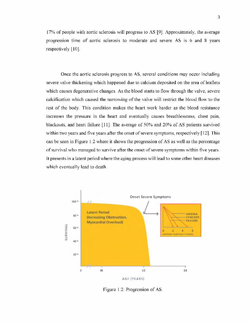
17% of people with aortic sclerosis will progress to AS [9]. Approximately, the average
progression time o f aortic sclerosis to moderate and severe AS is 6 and 8 years
respectively [10].
Once the aortic sclerosis progress to AS, several conditions may occur including
severe valve thickening which happened due to calcium deposited on the area of leaflets
which causes degenerative changes. As the blood starts to flow through the valve, severe
calcification which caused the narrowing of the valve will restrict the blood flow to the
rest of the body. This condition makes the heart work harder as the blood resistance
increases the pressure in the heart and eventually causes breathlessness, chest pain,
blackouts, and heart failure [11]. The average of 50% and 20% of AS patients survived
within two years and five years after the onset of severe symptoms, respectively [12]. This
can be seen in Figure 1.2 where it shows the progression of AS as well as the percentage
of survival who managed to survive after the onset of severe symptoms within five years.
It presents in a latent period where the aging process will lead to some other heart diseases
which eventually lead to death.
Onset Severe Symptoms
0 40 60 80
A G E ( YE A R S )
Figure 1.2: Progression of AS.

4
Traditionally, AS patients need to undergo the open heart surgery to replace the
faulty valve with the new valve as there is no medication to slow down the progression of
AS. The standard procedure to treat the AS patient is by performing SAVR. However, it
is an invasive procedure that requires an open heart surgery and this procedure should be
performed right after the onset of symptoms [5, 12]. Figure 1.3 shows the percentage of
asymptomatic and symptomatic survival who have performed the AVR as well as the
percentage of survival who have not gone through the AVR. Almost 50% of
asymptomatic and symptomatic patients survived after 15 years undergone the AVR. In
contrast, almost 50% of untreated patients with AS symptoms are reported to survive less
than 7 years after having the AS. This proves that SAVR can improve the quality o f life
if the patient immediately seek for treatment [13-14].
oo 2 3 4 5 6 7 8 9 10 11 12 13 14 15
YEARS
AVR, Asymptomatic
AVR, symptomatic
No AVR, Asymptomatic
N o AVR, symptomatic
Figure 1.3: Percentage of patient survival.
5
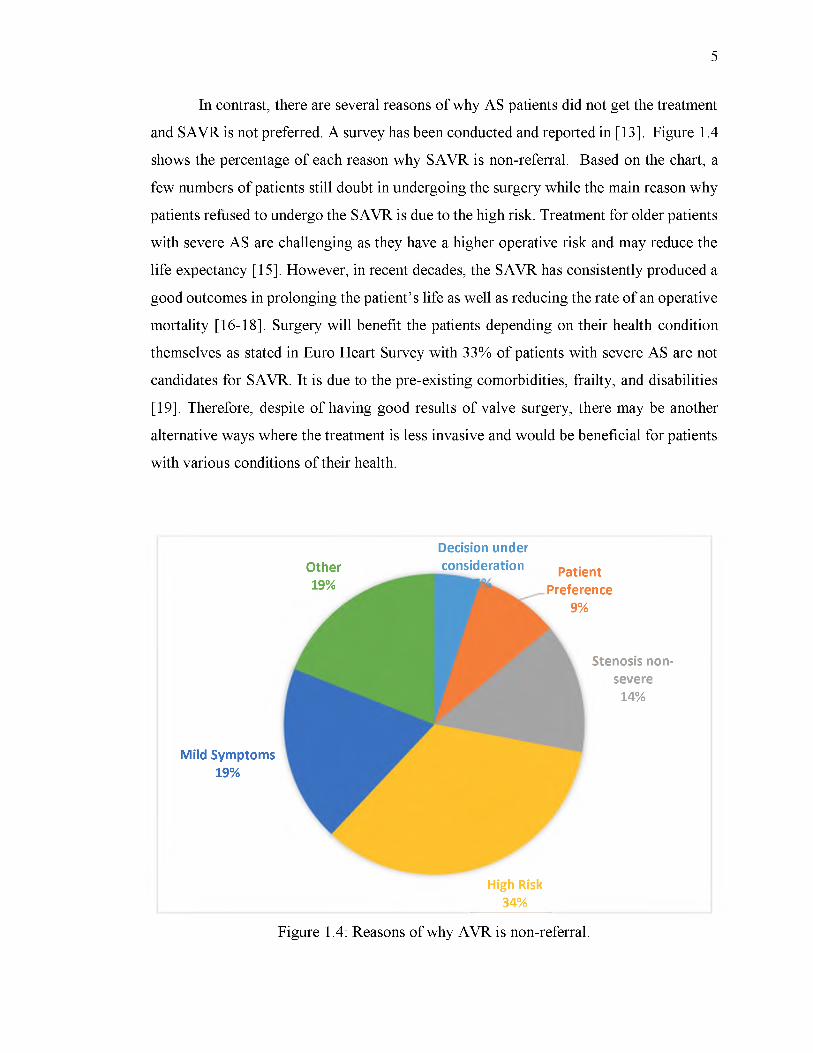
In contrast, there are several reasons of why AS patients did not get the treatment
and SAVR is not preferred. A survey has been conducted and reported in [13]. Figure 1.4
shows the percentage of each reason why SAVR is non-referral. Based on the chart, a
few numbers of patients still doubt in undergoing the surgery while the main reason why
patients refused to undergo the SAVR is due to the high risk. Treatment for older patients
with severe AS are challenging as they have a higher operative risk and may reduce the
life expectancy [15]. However, in recent decades, the SAVR has consistently produced a
good outcomes in prolonging the patient’s life as well as reducing the rate of an operative
mortality [16-18]. Surgery will benefit the patients depending on their health condition
themselves as stated in Euro Heart Survey with 33% of patients with severe AS are not
candidates for SAVR. It is due to the pre-existing comorbidities, frailty, and disabilities
[19]. Therefore, despite of having good results of valve surgery, there may be another
alternative ways where the treatment is less invasive and would be beneficial for patients
with various conditions of their health.
Other19%
Decision under consideration Patient
Preference9%
Mild Symptoms 19%
Stenosis non- severe
14%
High Risk 34%
Figure 1.4: Reasons of why AVR is non-referral.

6
Transcatheter Aortic Valve Implantation (TAVI) has been developed as a
promising alternative to conventional valve replacement for patients with severe,
symptomatic AS who are otherwise left untreated due to the perceived high risk of
operative mortality [20]. TAVI is less invasive compared to SAVR as it does not require
an open heart surgery in the procedure. The strategy of new valve insertion can be divided
into two common routes; transfemoral where the new valve is inserted through the femoral
artery and transapical which goes through a small cut on the left side of patient’s chest to
reach the apex of the heart.
In order to determine whether the TAVI procedure would benefit the patients,
several diagnoses or tests can be performed by referring to the guidelines proposed by the
European Society o f Cardiology (ESC) [13]. Once the decision has been made, TAVI
candidates need to be examined using several medical imaging modalities such as
computed tomography (CT) scan, angiogram and magnetic resonance imaging (MRI) in
order to measure the anatomy of valve structure. In addition, these kinds of modalities
may help in determining the selection of the suitable prosthesis valve to be implanted.
1.2 Problem Statem ent
Without appropriate aortic valve replacement, AS patients with severe symptoms
will have high mortality where the percentage of mortality is 3% to 4% soon after
symptoms have appeared and 7% among patients who are waiting for the SAVR [21]. In
addition, a significant number of patients with some other pre-comorbidities are not
recommended to undergo the SAVR as it will not benefit them. Thus, this will cause the
AS patients with severe symptoms to be left untreated. As TAVI has been proposed as an
alternatives to SAVR, AS patients still have a chance to survive and have a good quality
of life.
7
In regards of the matter, the evaluation of the anatomic characteristics o f the aortic
valve is critical in determining the TAVI’s success. Annulus size and shape vary for every
patient. Figure 1.5 illustrates the different structures of normal and abnormal aortic valve.
Basically, the normal aortic valve will have a proper opening and closing o f the leaflets
while valves that has been severely calcified yield an improper opening and closing of the
leaflets. Currently, the procedure to determine the size of the prosthetic valve is done
manually by experts. Common diagnosis includes the use o f Echocardiogram and CT scan
imaging modalities. Severe AS patients can be assured by referring to the characteristics
provided by the Echocardiogram modality such as the aortic valve area, indexed valve
area, mean transvalvular pressure and velocity ratio.
In addition, all those information are obtained in real time measurement which
provides high accuracy results. However, when it comes to measuring the size in space
which is in real time 2-D Echocardiogram image, it needs to be done manually and still
requires experts. Besides, Echocardiogram modality is highly operator-dependent. It
usually has limitations on its resolution and always suffer from speckle noises as it deals
with real time measurement. This will affect the measurement accuracy as well as lead to
deployment of wrong size o f prosthetic valve. Figure 1.6 shows the normal and abnormal
aortic valves in Echocardiogram images where the red circle inside images represents the
area of aortic annulus.

8
Figure 1.5: Normal and abnormal aortic valve.
(a) (b)
Figure 1.6: (a) Normal and (b) Abnormal aortic valve in Echocardiogram image.
Meanwhile, CT scan modality is often used to further evaluate an abnormality
seen on other tests such as an X-ray or an Echocardiogram. CT scan produces cross
sectional images that appear to open the body up, allowing the doctor to look at it from
the inside. It is able to create a detailed picture of heart and its blood vessels. However,
CT scan is prone to calcification. A soft thrombus can be partially covered and masked
by contrast. Then, errors are possible. Figure 1.7 shows the normal and abnormal aortic
valve in CT scan modality where the red circle in image (a) shows the area of valve that

9
has no calcium deposited on it while image (b) shows the AS that has been severely
calcified.
(a) (b)
Figure 1.7: (a) Normal and (b) abnormal aortic valve in CT scan image.
Based on the characteristics from both CT scan and Echocardiogram image, each
of them produces important features which are useful in order to measure the diameter of
aortic annulus as well as choosing the appropriate size of prosthetic valve before
performing the TAVI procedure. Currently, pre-TAVI procedures are performed
manually by the specialists and require a lot of work and procedures as well as time
consuming to ensure the success of TAVI. Therefore, in this research, the applications of
digital image processing associated with the decision fusion technique has been used in
order to reduce the workloads and human error.
Basically, the framework includes four stages of processing which are image
enhancement, object segmentation, annulus measurement and decision fusion. The
proposed algorithms are applied to both CT scan and Echocardiogram image separately.
Final features refer to the measured size of aortic annulus from CT scan and
Echocardiogram image obtained after performing the object segmentation. Decision
fusion approach is chosen as the best fusion technique to fuse this two features in order to
obtain the exact size of prosthetics valve that should be implanted into human heart. This

10
is due to other fusion approaches such as data/image and features fusion which are not
well-matched with the obtained final data.
1.3 Objectives of the Study
The following are the objectives of this study: -
i. To perform automated segmentation of aortic annulus using cardiac CT scan and
Echocardiogram image separately.
ii. To predict the diameter of segmented aortic annulus.
iii. To perform decision fusion of annulus size obtained from CT scan and
Echocardiogram in order to attain a more precise dimension of aortic annulus.
1.4 Scope of the Study
The scope of this research includes the measurement of the aortic annulus size of
human heart in 2D image obtained from both CT scan (Dual-source Somatom Definition,
Siemens, Germany) and Ultrasound (Philips) imaging modalities. The original file type
of DICOM is converted to JPEG with size of 550 x 550 for CT and 600 x 800 for
Echocardiogram image. Datasets of four patients which have been obtained from Institut
Jantung Negara (IJN) are tested using the proposed approach where all of the
Echocardiogram images are captured from the parasternal long axis (PLAX) view. In
addition, all o f the datasets are from the patients who will undergo TAVI procedure and
the size of aortic annulus obtained from their measurement will constitute a ground truth
in this experiment. Therefore, a validation can be made by comparing the proposed
approach to the ground truth. The 64-Bit MATLAB software version o f R2016a and an

11
operating system o f Window 8.1 with Intel core i5 processor are used to perform the
analysis.
1.5 Significance of Study
TAVI is regarded as an advanced technology in the medical field to treat patients
with severe aortic valve disease through blood vessel. Presently, the size o f the prosthetic
valve is chosen based on the manual measurement on CT scanned image performed by
the experts, and the verification o f the selected size may also be done using
Echocardiogram. False determination of annulus size may cause serious complications
for TAVI where deployment of a large valve may cause incomplete opening and closing
of leaflets, and excessive oversizing can lead to annular rupture, while deployment of a
small valve can cause paravalvular leak (PVL). Therefore, in this research, a new
framework is introduced to perform an accurate measurement as well as to overcome the
disparity or wrong aortic annulus measurement. TAVI procedure is less invasive as it does
not require an open heart surgery. AS patient with severe symptoms will still have a
chance to live in a good quality as this procedure has been proven to produce promising
results to the TAVI patients. Besides, this research also helps the clinicians in solving
uncertainty in a short time as the approach is efficient and produces a faster results in
determining the exact size o f prosthetic valve that should be implemented in human heart.
Thus, hopefully this will help in alleviating the constraints before performing the TAVI
procedure in Malaysia.

92
REFERENCES
[1] A. Martinsson, X. Li, C. Andersson, J. Nilsson, J. Smith and K. Sundquist,
"Temporal Trends in the Incidence and Prognosis of Aortic Stenosis: A
Nationwide Study of the Swedish Population", Circulation, vol. 131, no. 11, pp.
988-994, 2015.
[2] G. Eveborn, H. Schirmer, G. Heggelund, P. Lunde and K. Rasmussen, "The
evolving epidemiology of valvular aortic stenosis. The Troms0 Study", Heart, vol.
99, no. 6, pp. 396-400, 2012.
[3] V. Nkomo, J. Gardin, T. Skelton, J. Gottdiener, C. Scott and M. Enriquez-Sarano,
"Burden of valvular heart diseases: a population-based study", The Lancet, vol.
368, no. 9540, pp. 1005-1011, 2006.
[4] Martin B. Leon, M.D., Craig R. Smith, M.D., Michael Mack, M.D., D. Craig
Miller, M.D., Jeffrey W. Moses, M.D., Lars G. Svensson, M.D., Ph.D., E. Murat
Tuzcu, M.D., John G. Webb, M.D., Gregory P. Fontana, M.D., Raj R. Makkar,
M.D., David L. Brown, M.D., Peter C. Block, M.D., Robert A. Guyton, M.D.,
Augusto D. Pichard, M.D., Joseph E. Bavaria, M.D., Howard C. Herrmann, M.D.,
Pamela S. Douglas, M.D., John L. Petersen, M.D., Jodi J. Akin, M.S., William N.
Anderson, Ph.D., Duolao Wang, Ph.D., and Stuart Pocock, Ph.D, “Transcatheter
Aortic-Valve Implantation for Aortic Stenosis in Patients Who Cannot Undergo
Surgery”, N Engl J Med, 2010.
[5] R. Bonow, B. Carabello, K. Chatterjee, A. de Leon, D. Faxon, M. Freed, W.
Gaasch, B. Lytle, R. Nishimura, P. O'Gara, R. O'Rourke, C. Otto, P. Shah and J.
Shanewise, "ACC/AHA 2006 Guidelines for the Management of Patients With
Valvular Heart Disease: A Report of the American College of
Cardiology/American Heart Association Task Force on Practice Guidelines
(Writing Committee to Revise the 1998 Guidelines for the Management of
Patients With Valvular Heart Disease): Developed in Collaboration With the
Society of Cardiovascular Anesthesiologists: Endorsed by the Society for

93
Cardiovascular Angiography and Interventions and the Society of Thoracic
Surgeons", Circulation, vol. 114, no. 5, pp. e84-e231, 2006.
[6] A. Vahanian, H. Baumgartner, J. Bax, E. Butchart, R. Dion, G. Filippatos, F.
Flachskampf, R. Hall, B. Iung, J. Kasprzak, P. Nataf, P. Tornos, L. Torracca, A.
Wenink, S. Priori, J. Blanc, A. Budaj, J. Camm, V. Dean, J. Deckers, K. Dickstein,
J. Lekakis, K. McGregor, M. Metra, J. Morais, A. Osterspey, J. Tamargo, J.
Zamorano, J. Zamorano, A. Angelini, M. Antunes, M. Fernandez, C. Gohlke-
Baerwolf, G. Habib, J. McMurray, C. Otto, L. Pierard, J. Pomar, B. Prendergast,
R. Rosenhek, M. Uva and J. Tamargo, "Guidelines on the management of valvular
heart disease: The Task Force on the Management of Valvular Heart Disease of
the European Society o f Cardiology", European Heart Journal, vol. 28, no. 2, pp.
230-268, 2006.
[7] Popma, J.J., Adams, D.H., Reardon, M.J., Yakubov, S.J., Kleiman, N.S.,
Heimansohn, D., Hermiller, J., Hughes, G.C., Harrison, J.K., Coselli, J. and Diez,
J., “Transcatheter aortic valve replacement using a self-expanding bioprosthesis
in patients with severe aortic stenosis at extreme risk for surgery”, Journal of the
American College of Cardiology, vol. 63, no 19, pp.1972-1981, 2014.
[8] B. Iung, G. Baron, P. Tornos, C. Gohlke-Barwolf, E. Butchart and A. Vahanian,
"Valvular Heart Disease in the Community: A European Experience", Current
Problems in Cardiology, vol. 32, no. 11, pp. 609-661, 2007.
[9] C. Otto, "Why is aortic sclerosis associated with adverse clinical outcomes?”,
Journal of the American College o f Cardiology, vol. 43, no. 2, pp. 176-178,
2004.
[10] J. Cosmi, S. Kort, P. Tunick, B. Rosenzweig, R. Freedberg, E. Katz, R.
Applebaum and I. Kronzon, "The Risk of the Development of Aortic Stenosis in
Patients With "Benign" Aortic Valve Thickening", Archives of Internal Medicine,
vol. 162, no. 20, p. 2345, 2002.
[11] Maganti, Kameswari, Vera H. Rigolin, Maurice Enriquez Sarano, and Robert O.
Bonow, "Valvular heart disease: diagnosis and management”, In Mayo Clinic
Proceedings, vol. 85, no. 5, pp. 483-500, 2010.

94
[12] "Aortic Stenosis Prevalence", Edwards.com, 2017. [Online]. Available:
http://www.edwards.com/eu/procedures/aorticstenosis/Pages/prevalence.aspx.
[Accessed: 30- Jul- 2017].
[13] A. Vahanian, et al., "Guidelines on the management of valvular heart disease
(version 2012): The Joint Task Force on the Management of Valvular Heart
Disease o f the European Society of Cardiology (ESC) and the European
Association for Cardio-Thoracic Surgery (EACTS)", European Journal of Cardio-
Thoracic Surgery, vol. 42, no. 4, pp. S1-S44, 2012.
[14] M. Brown, P. Pellikka, H. Schaff, C. Scott, C. Mullany, T. Sundt, J. Dearani, R.
Daly and T. Orszulak, "The benefits of early valve replacement in asymptomatic
patients with severe aortic stenosis", The Journal of Thoracic and Cardiovascular
Surgery, vol. 135, no. 2, pp. 308-315, 2008.
[15] M.B. Leon, C.R. Smith, M. Mack, et al., “Transcatheter aortic-valve implantation
for aortic stenosis in patients who cannot undergo surgery”, N Engl J Med, pp.
1597-1607, 2010.
[16] P. Kolh, A. Kerzmann, C. Honore, L. Comte and R. Limet, "Aortic valve surgery
n octogenarians: predictive factors for operative and long-term results", European
Journal of Cardio-Thoracic Surgery, vol. 31, no. 4, pp. 600-606, 2007.
[17] S. Melby, A. Zierer, S. Kaiser, T. Guthrie, J. Keune, R. Schuessler, M. Pasque, J.
Lawton, N. Moazami, M. Moon and R. Damiano, "Aortic Valve Replacement in
Octogenarians: Risk Factors for Early and Late Mortality", The Annals of
Thoracic Surgery, vol. 83, no. 5, pp. 1651-1657, 2007.
[18] A. Vahanian, O. Alfieri, N. Al-Attar, M. Antunes, J. Bax, B. Cormier, A. Cribier,
P. De Jaegere, G. Fournial, A. Kappetein, J. Kovac, S. Ludgate, F. Maisano, N.
Moat, F. Mohr, P. Nataf, L. Pierard, J. Pomar, J. Schofer, P. Tornos, M. Tuzcu, B.
van Hout, L. Von Segesser and T. Walther, "Transcatheter valve implantation for
patients with aortic stenosis: a position statement from the European Association
of Cardio-Thoracic Surgery (EACTS) and the European Society of Cardiology
(ESC), in collaboration with the European Association of Percutaneous
Cardiovascular Interventions (EAPCI)", European Heart Journal, vol. 29, no. 11,
pp. 1463-1470, 2008.

95
[19] B. Iung, A. Cachier, G. Baron, et al., “Decision-making in elderly patients with
severe aortic stenosis: why are so many denied surgery?” Eur Heart J, pp. 2714
2720, 2005.
[20] C. Tamburino, D. Capodanno, A. Ramondo, A. Petronio, F. Ettori, G. Santoro, S.
Klugmann, F. Bedogni, F. Maisano, A. Marzocchi, A. Poli, D. Antoniucci, M.
Napodano, M. De Carlo, C. Fiorina and G. Ussia, "Incidence and Predictors of
Early and Late Mortality after Transcatheter Aortic Valve Implantation in 663
Patients with Severe Aortic Stenosis", Circulation, vol. 123, no. 3, pp. 299-308,
2011.
[21] Chambers JB., “Aortic stenosis”, European Journal of Echocardiography, pp. i l l —
i19, 2009.
[22] M. Leon et al, "Transcatheter Aortic-Valve Implantation for Aortic Stenosis in
Patients Who Cannot Undergo Surgery", New England Journal o f Medicine, vol.
363, no. 17, pp. 1597-1607, 2010.
[23] Ross J Jr, Braunwald E, “Aortic stenosis”, Circulation 1968;38:61-7.
[24] Cheitlin MD, Gertz EW, Brundage BH, Carlson CJ, Quash JA, Bode RS Jr. ,“Rate
of progression of severity o f valvular aortic stenosis in the adult”,^m Heart
1979;98:689-700.
[25] Otto CM, Pearlman AS, Gardner CL. “Hemodynamic progression of aortic
stenosis in adults assessed by Doppler echocardiography”.
[26] Davies SW, Gershlick AH, Balcon R. “Progression of valvular aortic stenosis: a
long-term retrospective study”,.Eur Heart J,1991.
[27] Peter M, Hoffmann A, Parker C, Luscher T, Burckhardt D. , “Progression of aortic
stenosis: role of age and concomitant coronary artery disease”, Chest 1993;103:
1715-9
[28] "Aortic Stenosis: Practice Essentials, Background, Pathophysiology",
Emedicine.medscape.com, 2016. [Online]. Available:
http://emedicine.medscape.com/article/150638-overview. [Accessed: 14- Apr-
2016].
[29] Bergler-Klein J., “Natriuretic peptides in the management of aortic stenosis”,
CurrCardiol Rep, 2009.
96
[30] Schwarz F, Baumann P, Manthey J, et al., “The effect of aortic valve replacement
on survival”, Circulation, 1982.
[31] Murphy ES, Lawson RM, Starr A, Rahimtoola SH.,“Severe aortic stenosis in
patients 60 years o f age or older: left ventricular function and 10-year survival
after valve replacement”, Circulation, 1981.
[32] Lund O,“Preoperative risk evaluation and stratification of long-term survival after
valve replacement for aortic stenosis: reasons for earlier operative
intervention”, Circulation, 1990.
[33] Bouma BJ, van den Brink RBA, van der Meulen JHP, et al.,“To operate or not on
elderly patients with aortic stenosis: the decisio n and its consequences”,Heart,
1999.
[34] Iung B, Cachier A, Baron G, et al.,“Decision-making in elderly patients with
severe aortic stenosis: why are so many denied surgery?”, Eur Heart J, 2005.
[35] Varadarajan P, Kapoor N, Bansal RC, Pai RG,“Clinical profile and natural history
of 453 nonsurgically managed patients with severe aortic stenosis”,^nn
ThoracSurg, 2006.
[36] Bach DS, Siao D, Girard SE, Duvernoy C, McCallister BD Jr, Gualano
SK,“Evaluation of patients with severe symptomatic aortic stenosis who do not
undergo aortic valve replacement”,CircCardiovascQual Outcomes, 2009.
[37] A. Cribier, H. Eltchaninoff and A. Bash, "Percutaneous transcatheter implantation
of an aortic valve prosthesis for calcific aortic stenosis", ACC Current Journal
Review, vol. 12, no. 2, p. 95, 2003.
[38] Cribier A, Eltchaninoff H, Bash A, Borenstein N, Tron C, Bauer F, et al.
Percutaneous transcatheter implantation of an aortic valve prosthesis for calcific
aortic stenosis: first human case description. Circulation 2002; 106: 3006-8.
[39] Lardizabal JA, O’Neill BP, Desai HV, Macon CJ, Rodriguez AP, Martinez CA, et
al. The transaortic approach for transcatheter aortic valve replacement: initial
clinical experience in the United States. J Am Coll Cardiol 2013; 61: 2341-5. doi:
10.1016/j.jacc.2013.02.076.

97
[40] B. Clayton, G. Morgan-Hughes and C. Roobottom, "Transcatheter aortic valve
insertion (TAVI): a review", The British Journal of Radiology, vol. 87, no. 1033,
p. 20130595, 2014.
[41] Sinning JM, Werner N, Nickenig G, Grube E. Next-generation transcatheter heart
valves: current trials in Europe and the USA. Methodist Debakey Cardiovasc J
2012; 8: 9-12.
[42] Webb J, Cribier A. Percutaneous transarterial aortic valve implantation: what do
we know? Eur Heart J 2011; 32: 140-7. doi: 10.1093/ eurheartj/ehq453
[43] Bloomfield GS, Gillam LD, Hahn RT, Kapadia S, Leipsic J, Lerakis S, et al. A
practical guide to multimodality imaging of transcatheter aortic valve replacement.
JACC Cardiovasc Imaging 2012; 5: 441-55.
[44] Messika-Zeitoun D, Serfaty JM, Brochet E, Ducrocq G, Lepage L, Detaint D, et
al. Multimodal assessment of the aortic annulus diameter. J Am Coll Cardiol 2010;
55:186-94
[45] Leipsic J, Gurvitch R, Labounty TM, Min JK, Wood D, Johnson M, et al.
Multidetector computed tomography in transcatheter aortic valve implantation.
JACC Cardiovasc Imaging 2011; 4: 416-29.
[46] Piazza N, de Jaegere P, Schultz C, Becker AE, Serruys PW, Anderson RH.
Anatomy o f the aortic valvar complex and its implications for transcatheter
implantation of the aortic valve. Circ Cardiovasc Interv 2008; 1: 74-81. doi:
10.1161/CIRCINTERVENTION S. 108.780858
[47] C. Bennett, J. Maleszewski and P. Araoz, "CT and MR Imaging o f the Aortic
Valve: Radiologic-Pathologic Correlation", RadioGraphics, vol. 32, no. 5, pp.
1399-1420, 2012.
[48] Tanaka R, Yoshioka K, Niinuma H, Ohsawa S, Okabayashi H, Ehara
S.,“Diagnostic value of cardiac CT in the evaluation o f bicuspid
aortic stenosis: comparison with echocardiography and operative
findings”,AJR Am J Roentgenol, 2010;195(4):895-899.
[49] Altiok E, Koos R, Schroder J, et al.,“Comparison of two-dimensional and three
dimensional imaging techniques for measurement of aortic annulus diameters
before transcatheter aortic v alve implantation”, Heart, 2011;97(19):1578-1584.

98
[50] Dashkevich A, Blanke P, Siepe M, et al.,“Preoperative assessment of aortic
annulus dimensions: comparison of noninvasive and intraoperative
measurement”,Ann ThoracSurg, 2011;91(3):709-714.
[51] Li X, Tang L, Zhou L, et al., “Aortic valves stenosis and regurgitation: assessment
with dual source computed tomography”,/nt J Cardiovasc Imaging, 2009.
[52] Dashkevich A, Blanke P, Siepe M, et al.,“Preoperative assessment of aortic
annulus dimensions: comparison of noninvasive and intraoperative
measurement”,Ann ThoracSurg, 2011.
[53] Haralick, R. M., and Shapiro, L. G.,“Survey : image segmentation techniques.
Comp. Vis. Graph”,Im. Proc., 1985.
[54] 2016. [Online]. Available: http://www.bioss.ac.uk/people/chris/ch4.pdf.
[Accessed: 28- Aug- 2016].
[55] Lecture 4: Thresholding", Webcache.googleusercontent.com, 2016. [Online].
Available:http://webcache.googleusercontent.com/search?q=cache:haMMH9iNA
J:homepags.inf.ed.ac.uk/rbf/CVonline/LOCAL COPIES/MORSE/threshold.pdf
+&cd =1&hl=en&ct=clnk&gl=my. [Accessed: 28- Aug- 2016].
[56] 2016. [Online].Available:
http://www.uio. no/studier/emner/matnat/ifi/INF43 00/h 11/undervisningsmateriale
/IN F4300- 2011-f04-segmentation.pdf. [Accessed: 28- Aug- 2016].
[57] 2016. [Online]. Available: http://www.bioss.ac.uk/people/chris/ch4.pdf.
[Accessed: 28- Aug- 2016].
[58] Jia-xin, C., & Sen, L., “A medical image segmentation method based on watershed
transform”, The Fifth International Conference on Computer and Information
Technology, 2005.
[59] Roerdink, Jos BTM, and Arnold Meijster. "The watershed transform: Definitions,
algorithms and parallelization strategies." Fundamenta informaticae, pp 187-228,
2000.
[60] Beucher, S., L. C., “Use o f watersheds in contour detection.” , in Proceeding o f
International Workshop on Image Processing, Real-Time Edge and Motion
Detection/Estimation, pp. 17-21, 1979.

99
[61] Roerdink, J. B., &Meijster, A.,“The watershed transform: Definitions, algorithms
and parallelization strategies”, Fundamentainformaticae, 2000.
[62] Straka, M., La Cruz, A., Kochl, A., Sramek, M.,Groller, E., & Fleischmann,
D.,“3D watershed transform combined with a probabilistic atlas for
medical image segmentation”, Journal o f Medical Informatics &
Technologies, 2003.
[63] Grau, V., Mewes, A. U. J., Alcaniz, M., Kikinis, R., & Warfield, S. K., “Improved
watershed transform for medical image segmentation using prior
information”,IEEE transactions on medical imaging, 2004.
[64] Hahn, H. K., &Peitgen, H. O., “IWT-interactive watershed transform: a
hierarchical method for efficient interactive and automated segmentation of
multidimensional gray-scale images”, In Medical Imaging 2003.
International Society fo r Optics and Photonics, 2003.
[65] Kuhnigk, J. M., Hahn, H., Hindennach, M., Dicken, V., Krass, S., &Peitgen, H.
O., “Lung lobe segmentation by anatomy-guided 3D watershed transform”,
Medical Imaging 2003. International Society fo r Optics and
Photonics, 2003.
[66] Ng, H. P., Ong, S. H., Foong, K. W. C., Goh, P. S., &Nowinski, W. L., “Medical
image segmentation using k-means clustering and improved watershed
algorithm” 2006 IEEE Southwest Symposium on Image Analysis and
Interpretation , 2006.
[67] Hamarneh, G., & Li, X.,“Watershed segmentation using prior shape and
appearance knowledge”, Image and Vision Computing, 2009.
[68] A. Padma and R. Sukanesh, "Segmentation and Classification of Brain CT Images
Using Combined Wavelet Statistical Texture Features", Arabian Journal for
Science and Engineering, vol. 39, no. 2, pp. 767-776, 2013.
[69] Ceylan, Murat, YUKSEL OZBAY, OSMAN NURi U^AN, and Erkan Yildirim.
"A novel method for lung segmentation on chest CT images: complex-valued
artificial neural network with complex wavelet transform." Turkish Journal of
Electrical Engineering & Computer Sciences 18, pp 613-624, 2010.
100
[70] J. Dehmeshki, H. Amin, M. Valdivieso and Xujiong Ye, "Segmentation of
Pulmonary Nodules in Thoracic CT Scans: A Region Growing Approach", IEEE
Transactions on Medical Imaging, vol. 27, no. 4, pp. 467-480, 2008.
[71] E. van Rikxoort, B. de Hoop, S. van de Vorst, M. Prokop and B. van Ginneken,
"Automatic Segmentation of Pulmonary Segments From Volumetric Chest CT
Scans", IEEE Transactions on Medical Imaging, vol. 28, no. 4, pp. 621-630, 2009.
[72] Jaffar, M. Arfan, Ayyaz Hussain, Anwar M. Mirza, and Asmatullah Chaudhry.
"Fuzzy entropy and morphology based fully automated segmentation of lungs
from CT scan images." International Journal o f Innovative Computing,
Information and Control 5, no. 12, pp. 4993-5002, 2009.
[73] S. Finn, M. Glavin and E. Jones, "Echocardiographic speckle reduction
comparison", IEEE Transactions on Ultrasonics, Ferroelectrics and Frequency
Control, vol. 58, no. 1, pp. 82- 101, 2011.
[74] S. Sudha, G. Suresh and R. Sukanesh, "Speckle Noise Reduction in Ultrasound
Images by Wavelet Thresholding based on Weighted Variance", IJCTE, pp.
7-12, 2009.
[75] F. Guan, P. Ton, S. Ge and L. Zhao, "Anisotropic diffusion filtering for ultrasound
speckle reduction", Science China Technological Sciences, vol. 57, no. 3, pp. 607
614, 2014.
[76] W. Gomez Flores, W. Pereira and A. Infantosi, "Breast Ultrasound Despeckling
Using Anisotropic Diffusion Guided by Texture Descriptors", Ultrasound in
Medicine & Biology, vol. 40, no. 11, pp. 2609-2621, 2014.
[77] Y. Deng, Y. Wang and Y. Shen, "Speckle reduction of ultrasound images based
on Rayleigh-trimmed anisotropic diffusion filter", Pattern Recognition Letters,
vol. 32, no. 13, pp. 1516-1525, 2011.
[78] A. Lopes, R. Touzi, and E. Nezry, “Adaptive speckle filters and scene
heterogeneity,” IEEE Trans. Geosci. Rem. Sens., vol. 28, no.6, pp. 992
1000, Nov. 1990.
[79] Z. Shi and K. Fung, “A comparison of digital speckle filters,” in Proc. Int.
Geoscience and Remote Sensing Symp., 1994.
101
[80] Saini, K., Dewal, M. L., &Rohit, M.,“Ultrasound imaging and image segmentation
in the area of ultrasound: a review”,International Journal o f advanced science
and technology, 2010.
[81] M. Marsousi, A. Eftekhari, A. Kocharian and J. Alirezaie, "Endocardial boundary
extraction in left ventricular echocardiographic images using fast and adaptive
B-spline snake algorithm", International Journal o f Computer Assisted
Radiology and Surgery, vol. 5, no. 5, pp. 501-513, 2010.
[82] Lacerda, S. G., da Rocha, A. F., Vasconcelos, D. F., de Carvalho, J. L., Sene, I.
G., & Camapum, J. F., “Left ventricle segmentatio n in echocardiography
using a radial-search-based image processing algorithm”,In 2008 30th
Annual International Conference o f the IEEE Engineering in
Medicine and Biology Society, 2008.
[83] Maulik A Chaudhary et al., “Thrombus Detection from Echocardiographic Images
Using Image Processing Techniques", International Journal o f Emerging
Technology and Advanced Engineering, vol. 5, 2015.
[84] Nandagopalan, S., Adiga, B. S., Dhanalakshmi, C., & Deepak, N., “Automatic
segmentation and ventricular border detection of 2D echocardiographic images
combining k- means clustering and active contour model”,In Computer and
Network Technology (ICCNT), 2010.
[85] B. Liu, H. Cheng, J. Huang, J. Tian, X. Tang and J. Liu, "Probability density
difference-based active contour for ultrasound image segmentation", Pattern
Recognition, vol. 43, no. 6, pp. 2028-2042, 2010.
[86] K. Zhang, L. Zhang, H. Song and W. Zhou, "Active contours with selective local
or global segmentation: A new formulation and level set method", Image and
Vision Computing, vol. 28, no. 4, pp. 668-676, 2010.
[87] M. Kokar and K. Kim, “Review of multisensor data fusion architectures,” in Proc.
IEEE Int. Symp. Intelligent Control, Aug. 1993, pp. 261-266.
[88] D. L. Hall and J. Llinas, “An introduction to multisensor data fusion,” Proc. IEEE,
vol. 85, pp. 6-23, 1997.

102
[89] M. Kumar and S. Dass, "A Total Variation-Based Algorithm for Pixel-Level
Image Fusion", IEEE Transactions on Image Processing, vol. 18, no. 9, pp. 2137
2143, 2009.
[90] R. S. Blum, “Robust image fusion using a statistical signal processing approach,”
Inf. Fusion, vol. 6, no. 2, pp. 119-128, Jun. 2005.
[91] F. Nebeker, “Golden accomplishments in biomedical engineering,” IEEE Eng.
Med. Biol. Mag., vol. 21, no. 3, pp. 17-47, May/Jun. 2002.
[92] D. Hal, Mathematical Techniques in Multisensor Data Fusion, 2nd ed. Boston,
MA: Artech House, 2004.
[93] U. Mangai, S. Samanta, S. Das and P. Chowdhury, "A Survey o f Decision Fusion
and Feature Fusion Strategies for Pattern Classification", IETE
Technical Review, vol. 27, no. 4, p. 293, 2010.
[94] Federico Castanedo, “A Review of Data Fusion Techniques,” The Scientific World
Journal, vol. 2013, Article ID 704504, 19 pages, 2013.
doi:10.1155/2013/704504
[95] J. Yang and X. Zhang, "Feature-level fusion of fingerprint and finger-vein for
personal identification", Pattern Recognition Letters, vol. 33, no. 5, pp. 623
628, 2012.
[96] G. Bokade and A. Sapkal, "Feature Level Fusion of Palm and Face for Secure
Recognition", International Journal o f Computer and Electrical Engineering, pp.
157-160, 2012.
[97] Rattani, Ajita, et al. "Feature level fusion of face and fingerprint
biometrics." Biometrics: Theory, Applications, and Systems. 2007.
[98] Y. Yao, X. Jing and H. Wong, "Face and palmprint feature level fusion for single
sample biometrics recognition", Neurocomputing, vol. 70, no. 7-9, pp.
1582-1586, 2007.
[99] Josef Kittler, Mohamad Hatef, Robert P.W. Duin, and Jiri Matas, “On Combining
Classifiers”, IEEE Transactions on Pattern Analysis and Machine Intelligence,
1998.
[100] J. Berger, Statistical decision theory and Bayesian analysis. New York: Springer,
2010.
103
[101] Hamarneh, G.,et al., “Watershed segmentation using prior shape and appearance
knowledge”,“Image and Vision Computing”, 2009.
[102] Barrett, Julia F., and Nicholas Keat. "Artifacts in CT: recognition and avoidance",
“Radiographic”, 2004.
[103] Jabbar, Rahna. "A Study of Preprocessing and Segmentation Techniques on
Cardiac Medical Images." International Journal of Engineering”, 2014.
[104] Meyer F, BeucherS,“Morphologicalsegmentation”,“Journal of visual
communication and image representation”,1990.
[105] Rahman, Md Habibur, and Md Rafiqul Islam. "A Version of Watershed Algorithm
for Color Image Segmentation." 2013.
[106] A. Fisher, "Cloud and Cloud-Shadow Detection in SPOT5 HRG Imagery with
Automated Morphological Feature Extraction", Remote Sensing, vol. 6, no. 1, pp.
776-800, 2014.
[107] Lang, Roberto M., et al. "Recommendations for chamber quantification."
European Heart Journal-Cardiovascular Imaging, 2006.
[108] Nandagopalan, S., Adiga, B. S., Dhanalakshmi, C., & Deepak, N., “Automatic
segmentation and ventricular border detection o f 2D echocardiographic
images combining k- means clustering and active contour model”, In
Computer and Network Technology (ICCNT), 2010.
[109] Ergen, Burhan. Signal and image denoising using wavelet transform. INTECH
Open Access Publisher, 2012.
[110] Lam, L., et al., "Thinning Methodologies-A Comprehensive Survey," IEEE
Transactions on Pattern Analysis and Machine Intelligence, 1992.
[111] Withey, D. J., and Z. J. Koles. "Medical image segmentation: Methods and
software." Noninvasive Functional Source Imaging of the Brain and Heart and the
International Conference on Functional Biomedical Imaging, 2007.
[112] S. Coffey, B. Cairns and B. Iung, "The modern epidemiology o f heart valve
disease", Heart, vol. 102, no. 1, pp. 75-85, 2015.