autoimmunity i matt mcreynolds. outline i.definition a.direct proof b.indirect evidence...
Post on 20-Dec-2015
214 views
TRANSCRIPT

Autoimmunity I
Matt McReynolds

Outline
I. Definitiona. Direct proofb. Indirect evidencec. Circumstantial
II. Diseasesa. Antibody-mediatedb. Immunecomplex-
mediatedc. T cell-mediated
III. Models

Autoimmunity
• “Friendly-fire”: Immune response to self• Autoimmune disease:
When the immunological recognition of self with the involvement of antibody, complement, immune complexes, and cell-mediated immunity progresses from being benign to pathologic.

Autoimmunity
• Autoimmune responses occur all the time with little to no consequence.
• Theory of “autoimmune diseases” was only recently accepted in the 1950’s and 60’s.

Autoimmunity
• Prevalence of autoimmune diseases in Western countries has been estimated at 3 - 7% (Marmont, AM. 2000 Ann Rev Med 51:115-134)
• Organ specific or non-specific
• Humoral or Cell-mediated

Defining a disease as autoimmune
• Direct Proof – requires transmissibility– Antibody or lymphocyte transfer
• Animal models• Humans to animals (pemphigus vulgaris and
bullous pemphigoid) • Humans
– Mother to fetus (eg. myasthenia gravis)– T cell transfer not feasible – MHC
– Diseases: Idiopathic thrombocytopenic purpura, Graves' disease

Defining a disease as autoimmune
• Indirect Proof– Identify target Ag in humans
• Reproduce model disease with homologous Ag in animal
• AChR EAMG, collagen I EAA, myosin EAM, myelin EAE
– Majority of autoimmune diseases

Defining a disease as autoimmune
• Indirect Proof– Identify target Ag in humans
• Reproduce model disease with homologous Ag in animal
• AChR EAMG, collagen I EAA, myosin EAM, myelin EAE
– Study genetically predisposed animal models• (NZB x NZW)F1 mice model of SLE

Defining a disease as autoimmune
• Indirect Proof– Identify target Ag in humans – Study genetically predisposed
animal models– Isolate self-reactive Ab or T cells
from target organs• Anti-erythrocyte Abs – hemolytic
anemia• Anti-DNA Abs – SLE

Defining a disease as autoimmune
• Circumstantial evidence - "markers" descriptive of autoimmune disease – Familial tendency– Lymphocyte infiltration– Specific MHC II allele association– Immune complex deposition– Clinical improvement with
immunosuppressive agents

Diseases
1. Antibody Mediated2. Immune-complex mediated3. T-cell mediated

Diseases
1. Antibody Mediated - produced either by antibodies binding to antigens on cells or tissue or by Ag-Ab complex deposition in vessel walls.
• Autoimmune Hemolytic Anemia• Myasthenia Gravis• Graves’ Disease• Rheumatic Fever
2. Immune-complex mediated3. T-cell mediated

Autoimmune Hemolytic Anemia
• Self Ag: red blood cells (RBC’s)• Can be associated with systemic
lupus erythematosus • Mechanisms of Ab-mediated
destruction– Opsonization and digestion by
macrophage– Complement cascade and lysis of RBC
• Also the principal mechanism for thrombocytopenic purpura

Autoimmune Hemolytic Anemia
• Antibody/agglutinins groups– Warm: react at 37C, Rh antigen
• IgG phagocytosis
– Cold: react <37C, I or i antigens• IgM complement and lysis• Occurs in limbs or skin upon temperature drop
• Induced– Penicillin (hapten), Methyldopa (warm),
Mycoplasma pneumoniae or viruses (cold)

Myasthenia Gravis (MG)• Self Ag: Acetylcholine Receptor (AChR) at the
neuromuscular junction• Women in late 20s / Men in their 50’s• Prevalence in the US is estimated at 14/100,000
population, approximately 36,000 cases • Thymus: hypertrophy, 10% have thymoma• HLA class II• Blood test, Acetylcholinesterase (AChE) inhibitor
(edrophonium) used for diagnosis

MG
• Changes in motor endplate:– Abs bind complement– Inflammatory cell response– Destruction of endplate and AChR– Abs block cation channels of the
receptor and prevent propagation of action potentials

MG

MG
• Seronegative or positive for antiAChR Abs– 95% pos in generalized disease– 60% pos in localized (occular) disease
• Seropositivity is not associated with disease severity, but Ab titers decrease with immunosuppression

Occular MG
• Functional Hypothesis: slight weakness in ocular and eye-lid muscles is far less tolerable than generalized weakness.
• Immunologic Hypothesis: Difference in Ab’s associated with ocular muscle.
• Physiological Hypothesis: Ocular muscles are predisposed to MG because of their size,innervation and activity.

Occular MG
Symptoms:• Inability to align
eyes• Droopy eye-lids

MG
• Muscle weakness improves with rest– Chewing, swallowing (dysphagia),
breathing (dyspnea), speaking (dysarthria)– Ptosis (droopy eye lids), diplopia (2x
vision)– Inner ear: hyperacusis (acute hearing due
to heightened irritability of nerve pathway)– Neck flexor muscles, limb
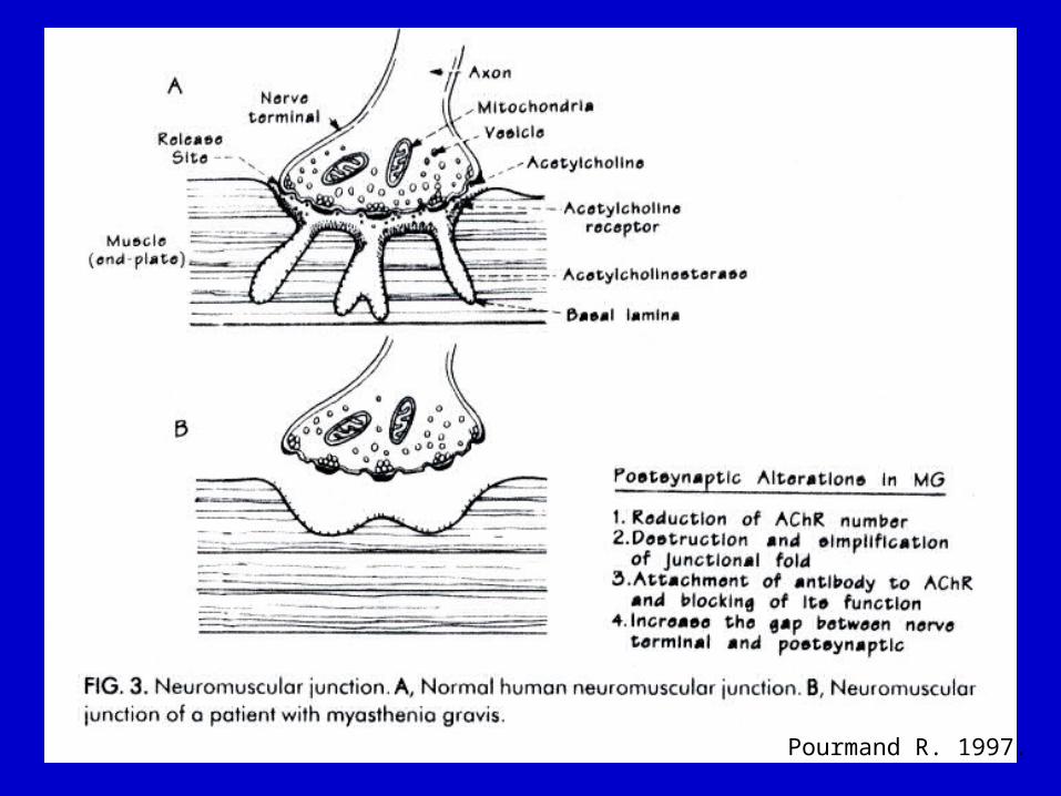
Pourmand R. 1997.

Graves’ Disease
• Self Ag: Thyroid-stimulating Hormone Receptor (TSHR)
• Hyperthyroidism• TSH receptor is activated
independent of the pituitary• Familial, HLA II, Ab titer:Disease
severity, neonatal hyperthyroidism from maternal IgG

Normal

Graves’Disease
Normal

Graves’ Diagnosis
• Blood test:– T3 and T4 are markedly elevated– TSH levels are low or absent. – Serum Ab detection– Goiter

Graves’ Symptoms
• Cardiac – arrhythmias (a-fib), tachycardias, widened pulse pressure
• Endocrine – weight loss, increased basal metabolic rate
• Dermatological – profuse sweating, clubbing of fingernails
• Neurological – tremor, weakness, proximal myopathy
• Ophthalmological – thyroid eye disease, proptosis.

Proptosis

Rheumatic Fever
• Self Ag: Cardiac proteins• Follows pharyngeal infection with group
A streptococci• Arthritis, carditis, chorea, s.c. nodules
– Chronic, progressive valve damage
• Major cause of acquired heart disease in many lesser developed countries
• Molecular mimicry

Diseases
1. Antibody Mediated 2. Immune-complex mediated
• Systemic Lupus Erythematosus (SLE)
3. T-cell mediated

SLE
• Self Ags: ssDNA, dsDNA, nucleohistones– Causes:
• Genetic predisposition• Environmental causes• Drug interactions
• Response to nuclear proteins• Normal: IgM; SLE: IgG• Cellular Ags exposed during apoptosis
incite an immune response– Increased Fas expression in B & T cells– Sunlight (keratinocytes)

SLE Incidence
• 1 / 1,000 white persons
• 1 / 250 black women• Concordance:
– 25% monozygotic twins
– 1-2% dizygotic twins
SLE Complications
• Musculoskeletal: osteoporosis, joint pain• Dermatologic : malar rash, photosensitivity• CNS : psychosis, seizures• Hematological: anemia• Renal failure (complex deposition)• CV: antiphospholipid Abs hypercoagulability
– Lupus anticoagulant, anticardiolipin Ab– Thrombosis, thrombocytopenia, pregnancy loss

Diseases
1. Antibody Mediated2. Immune-complex mediated3. T cell-mediated
• Multiple Sclerosis• Insulin-Dependent Diabetes Mellitus• Rheumatoid Arthritis

MS• Self Ag: Myelin sheath
– Demyelinating plaques• Optic nerve, periventricular and spinal cord white
matter, brain stem, cerebellum
• Symptoms:– changes in sensation in the arms, legs or
face (33%), complete or partial vision loss (optic neuritis) (16%), weakness (13%), double vision (7%), unsteadiness when walking (5%), and balance problems (3%)


MS
• Diagnosis– Clinical presentation– Cerebrospinal Fluid (CSF) abnormalities– Evoked potentials (visual,
somatosensory)– Neurological dysfunction becomes
“disseminated in space and time”– Magnetic Resonance Imaging (MRI)

An axial FLAIR (fluid-attenuated inversion recovery) image shows multiple ovoid and confluent hyperintense lesions in the periventricular white matter (Panel A). Nine months later, the number and size of the lesions have substantially increased (Panel B).
After the administration of gadolinium, many of the
lesions demonstrate enhancement, indicating the
breakdown of the BBB (Panel C). In Panel D, a
parasagittal T1-weighted MRI
scan shows "black holes" in the periventricular white
matter and corpus callosum (chronic lesions).
Noseworthy, et al. NEJM 2000.

MS• Relapsing-remitting (80%)
– characterized by unpredictable attacks (relapses) followed by periods of months to years of relative quiet (remission) with no new signs of disease activity
• Chronic Progressive (20%)– Primary, secondary progressive
• Quadriparesis; cognitive decline; visual loss; cerebellar, bowel, bladder, and sexual dysfunction

Insulin-Dependent Diabetes Mellitus (IDDM)
• Self Ag: insulin-secreting -islet cells of pancreas
• Type I, Juvenile-onset• HLA DR3 and DR4• Abs, T cells, adhesion molecules,
serum cytokine inhibitors, sustained expression of cytokines and Rs– TNF-, IFN-, IL-1

IDDM
• Symptoms: insulin abnormalities in glucose
metabolism ketoacidosis, thirst, polyuria, vision disturbances
• Late stages: progressive atherosclerotic vascular lesions arterial obstruction, renal failure, blindness

Rheumatoid Arthritis (RA)
• Self Ag: cartilage• HLA DR4, TNF genes, HSP gene
complex, infection (EBV)• Varied age (common in 20’s-40’s)• Women:Men 3:1

RA
synovial fluid with PMNs Cartilage replaced with fibrous tissue joint fusion
• Diagnosis:– >80%: Rheumatoid Factor (RF)– IgM anti-IgG Fc
• Immune involvement:– Ag-Ab complex, C’, PMNs, CD4+,
CD8+, MØ, NK cells, TNF-, IL-1

RA

Autoimmune Models
• Immunization– Self antigen
• Spontaneous– Genetically susceptible strains
• Induced– Virally, chemically, pharmacologically

Immunization Models
• Adjuvant = any substance that enhances the immune response to an antigen with which it is mixed– Does not form stable linkages w/ immunogen
• Mechanisms– Convert soluble protein Ags into particulate
material ingested by MØ– Bacterial products signal MØ or DC
cytokines

Experimental Autoimmune/Allergic Encephalomyelitis (EAE)
• Model of RR or chronic progressive MS
• Various mouse strains• Intradermal immunization with
myelin basic protein (MBP), proteolipid protein (PLP), myelin oligodendrocyte glycoprotein (MOG)
• Ascending paralyses of hind limbs, infiltration, demyelination
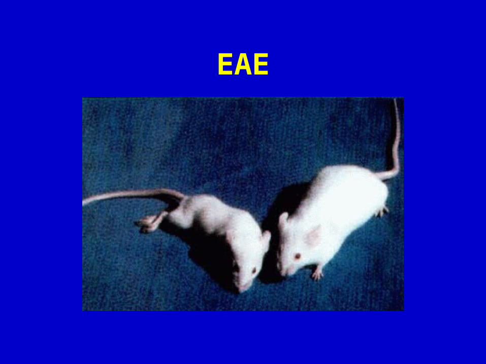
EAE

EAE
• Synthetic peptides– MBP 1-9: acute, no relapses
• single TCR
– PLP 43-64, MBP 89-101: chronic-relapsing• epitope spreading, large TCR repertoire

Experimental autoimmune arthritis (EAA)
• Collagen II Arthritis (CIA)• Joint swelling and edema• Heterologous collagen
– Active joint erosion; severe, self-limiting
• Autologous collagen– Cartilage erosion; chronic disease

EAA
• Mechanism– T-cell dependent B-cell activation Ab
production Fc-mediated C’ activation• IP injection of Abs synovial inflammation,
erosive arthritis• C’ depleted mice are resistant to EAA induction
• Adoptive transfer of T cells– Subclinical disease

Experimental Autoimmune Myasthenia Gravis (EAMG)
• Purified AChR• Rodents• Adoptive transfer to mice with Igs
from MG patients• C’-mediated lysis + rate of AChR
internalization by muscle cell

Spontaneous Models
• Murine Systemic Lupus Erythematosus– Defects in Fas/FasL lymphoproliferation– NZB/W mice: B cell differentiation defect– BXSB mice: defective Y-linked gene
– (NZBxNZW)F1 mice: mutations in TNF- gene decreased production
– 10 gene loci associated with murine SLE

Spontaneous Models
• Insulin-Dependent Diabetes Mellitus– Non-obese diabetic (NOD) mice– Initial epitope: Glutamic Acid Decarboxylase– 18-20 wks: overt diabetes, -islet cells
progressively destroyed– MØ, T and B cells, dendritic cells accumulate– Later: epitope spreading (other GAD, insulin,
HSP60)– CD8+ and CD4+ cells necessary for islet
destruction

Induced Models
• Infectious– Theiler’s murine encephalomyelitis virus
• Mice or rats, transferred with donor T cells
– Staphylococcus aureus strains• Arthritogenic in mice (septic arthritis)• Secretion of superantigenic endotoxin TSST 1
– V 11 T cell expansion in joints
– Bacterial cell wall fragments injected into rats• T cell-dependent chronic polyarthritis from MØ
activation

Questions?