auto-assembling detoxified staphylococcus aureus alpha … · na phosphate (ph 6.8) and was applied...
TRANSCRIPT

Auto-Assembling Detoxified Staphylococcus aureus Alpha-HemolysinMimicking the Wild-Type Cytolytic Toxin
Luigi Fiaschi,c Benedetta Di Palo,a Maria Scarselli,a Clarissa Pozzi,a Kelly Tomaszewski,b Bruno Galletti,a Vincenzo Nardi-Dei,a
Letizia Arcidiacono,a Ravi P. N. Mishra,c Elena Mori,a Michele Pallaoro,a Fabiana Falugi,c Antonina Torre,c Maria Rita Fontana,c
Marco Soriani,a Juliane Bubeck Wardenburg,b Guido Grandi,c Rino Rappuoli,a Ilaria Ferlenghi,a Fabio Bagnolia
GSK Vaccines, Research Center, Siena, Italya; Departments of Pediatrics and Microbiology, University of Chicago, Chicago, Illinois, USAb; Novartis Vaccines and Diagnostics,now GSK Vaccines, Siena, Italyc
Staphylococcus aureus alpha-hemolysin (Hla) assembles into heptameric pores on the host cell membrane, causing lysis, apopto-sis, and junction disruption. Herein, we present the design of a newly engineered S. aureus alpha-toxin, HlaPSGS, which lacksthe predicted membrane-spanning stem domain. This protein is able to form heptamers in aqueous solution in the absence oflipophilic substrata, and its structure, obtained by transmission electron microscopy and single-particle reconstruction analysis,resembles the cap of the wild-type cytolytic Hla pore. HlaPSGS was found to be impaired in binding to host cells and to its recep-tor ADAM10 and to lack hemolytic and cytotoxic activity. Immunological studies using human sera as well as sera from miceconvalescent from S. aureus infection suggested that the heptameric conformation of HlaPSGS mimics epitopes exposed by thecytolytic Hla pore during infection. Finally, immunization with this newly engineered Hla generated high protective immunityagainst staphylococcal infection in mice. Overall, this study provides unprecedented data on the natural immune responseagainst Hla and suggests that the heptameric HlaPSGS is a highly valuable vaccine candidate againstS. aureus.
Staphylococcus aureus is a major human pathogen, and currentantibiotics are not efficacious against emerging multidrug-re-
sistant strains. Therefore, there is an urgent need to develop vac-cines to target this pathogen. However, S. aureus vaccine develop-ment is hindered by the lack of known correlates of protection.Alpha-toxin, also known as alpha-hemolysin (Hla), is one of themajor virulence factors of S. aureus. The toxin is secreted as awater-soluble monomer and can bind to erythrocytes, platelets,monocytes, lymphocytes, and endothelial as well as epithelial cells(1, 2). Monomers assemble into a membrane-perforating homo-heptamer upon binding to its eukaryotic proteinaceous cellularreceptor, ADAM10 (1, 3–5). Hla-based vaccines have been previ-ously described (2, 6, 7). Nonetheless, all published studies on Hlahave so far used monomeric mutants of Hla without knowing ifthey were able to expose epitopes resembling the heptameric con-formation of the active toxin, which is assembled during infectionon host cells.
Herein, we present a study aimed at investigating the struc-tural, functional, and immunogenic characteristics of monomericversus heptameric Hla. We engineered the toxin by removing themembrane-spanning stem domain. This protein, HlaPSGS, is ableto form heptamers in aqueous solution in the absence of a host cellmembrane and resembles the cap of the wild-type cytolytic Hlapore. Therefore, we can use this mutant to understand if the im-mune response of the host during infection is more prominent tothe monomeric or the heptameric form of Hla. Immunologicalstudies using human sera as well as sera from mice convalescentfrom S. aureus infection suggested that the heptameric conforma-tion of HlaPSGS mimics epitopes exposed by the cytolytic Hlapore during infection.
Furthermore, we evaluated the molecule as a vaccine antigenagainst S. aureus. HlaPSGS was found to lack hemolytic and cyto-toxic activity and also to be impaired in binding to host cells. In
addition, immunization with HlaPSGS generated high protectiveimmunity against staphylococcal infection in mice.
Overall, these data suggest that host-pathogen interactions thatoccur during infection with S. aureus generate an immune re-sponse, in humans and mice, directed primarily against confor-mational epitopes that are present only in the heptameric Hlatoxin. Therefore, this study provides important observations forunderstanding the immunology and pathogenesis of S. aureus in-fections as well as for developing innovative vaccines.
MATERIALS AND METHODSBacterial strains, media, and growth conditions. For mouse infectionstudies, overnight cultures of the S. aureus LAC (USA300) strain werediluted 1:100 into fresh tryptic soy broth (TSB) and were grown at 37°Cwith shaking until reaching mid-log phase. Bacteria were centrifuged,washed with phosphate-buffered saline (PBS), and suspended in a volumeof PBS to yield the appropriate cell concentration, which varied according
Received 22 February 2016 Returned for modification 26 February 2016Accepted 16 March 2016
Accepted manuscript posted online 30 March 2016
Citation Fiaschi L, Di Palo B, Scarselli M, Pozzi C, Tomaszewski K, Galletti B, Nardi-Dei V, Arcidiacono L, Mishra RPN, Mori E, Pallaoro M, Falugi F, Torre A, Fontana MR,Soriani M, Bubeck Wardenburg J, Grandi G, Rappuoli R, Ferlenghi I, Bagnoli F. 2016.Auto-assembling detoxified Staphylococcus aureus alpha-hemolysin mimickingthe wild-type cytolytic toxin. Clin Vaccine Immunol 23:442– 450.doi:10.1128/CVI.00091-16.
Editor: D. L. Burns, Food and Drug Administration
Address correspondence to Ilaria Ferlenghi, [email protected], orFabio Bagnoli, [email protected].
Supplemental material for this article may be found at http://dx.doi.org/10.1128/CVI.00091-16.
Copyright © 2016 Fiaschi et al. This is an open-access article distributed under theterms of the Creative Commons Attribution 4.0 International license.
crossmark
442 cvi.asm.org June 2016 Volume 23 Number 6Clinical and Vaccine Immunology
on March 13, 2019 by guest
http://cvi.asm.org/
Dow
nloaded from

to the model used. The inoculum was verified experimentally by platingon tryptic soy agar (TSA) and colony enumeration.
For expression of Hla constructs, Escherichia coli was grown in LuriaBertani broth that contained 30 �g/ml kanamycin until an optical density(OD) of 0.4 and was then induced with 1 mM isopropyl-�-D-thiogalac-topyranoside (IPTG) and grown for 3 more hours at 25°C.
Cloning of Hla constructs. hla was amplified by PCR from the S.aureus NCTC8325 strain using oligonucleotides HlaF/HlaR. HlaPSGSwas amplified by splicing by overlapping extension PCR (SOE PCR) fromthe S. aureus NCTC8325 strain using oligonucleotide pairs HlaF/PSGSRand PSGSF/HlaR and then the pair HlaF/HlaR. HlaH35L was amplified bySOE PCR from the S. aureus NCTC8325 strain using oligonucleotide cou-ples HlaF/H35LR and H35LF/HlaR and then the couple HlaF/HlaR.
The HlaPSGS and HlaH35L constructs were cloned as N-terminal6His-tagged constructs into the pET-15b� vector using the polymeraseincomplete primer extension (PIPE) technique (8). hla and hlaPSGSgenes were also amplified by PCR using oligonucleotides HlanatF/HlanatR and were cloned as tagless constructs into the NdeI/XhoI sites ofthe pET-24b� vector. All of the constructs were transformed in E. coliBL21(DE3). For primers used in this study, see Table S1 in the supple-mental material.
Hla epitopes were generated as previously described (9).Purification of Hla recombinant proteins. Escherichia coli cells ex-
pressing HlaPSGS, HlaWt, or HlaH35L (Hla recombinant proteins) werecollected by centrifugation, suspended in a 50 mM potassium phosphatebuffer (pH 7.0), and disrupted by ultrasonic oscillation at 4°C for 20 minwith a Seiko Instruments ultrasonic disintegrator model 7500. Cell debriswas removed by centrifugation. The supernatant solution was suspendedin 50 ml of 30 mM Tris (pH 9.5) and was applied to a Q Sepharose HP ionexchange column (GE Healthcare) that was previously equilibrated in thesame cell suspension buffer. Flowthrough fractions containing Hla re-combinant proteins were collected and pooled. The pooled protein solu-tion (20 ml) was diluted 1:3 (i.e., to a final volume of 60 ml) with 10 mMNa phosphate (pH 6.8) and was applied to a 20-�m hydroxyapatite col-umn (Bio-Rad) that was previously equilibrated with 10 mM Na phos-phate (pH 6.8), and a 10 mM Na phosphate (pH 6.8) to 1 M Na phosphate(pH 6.8) gradient was applied. Hla recombinant proteins came out in theflowthrough fractions, which were pooled, while most impurities wereeluted during the gradient and discarded. A 500 mM Na phosphate solu-tion was added to the pooled protein solution in order to bring the phos-phate concentration to 50 mM, and pH was corrected to 6.3. This proteinsolution was loaded on an SP Sepharose HP ion exchange column (GEHealthcare) that was previously equilibrated in 50 mM Na phosphate (pH6.3). Flowthrough fractions that contained Hla recombinant proteinswere pooled. Fractions were concentrated by ultrafiltration and wereloaded on a Superdex 200 26/60 (GE Healthcare) size-exclusion chroma-tography column equilibrated in phosphate-buffered saline. Gel filtrationfractions were selected by SDS-PAGE and size-exclusion chromatographywith multiangle laser light scattering (SEC-MALLS) and were pooled.
Protein purity analysis. Purity and apparent molecular weight (MW)of HlaWt and HlaPSGS proteins were determined by an analytical size-exclusion high-pressure liquid chromatograph (HPLC) TSK G3000SWxl(7.8 by 300 mm; Tosoh Bioscience Corporation) with isocratic elution inPBS at a flow rate of 0.5 ml/min. UV absorbance was monitored at 280nm. For molecular mass determination, a gel filtration standard (670, 158,44, 17, and 1.35 kDa; Bio-Rad) was applied.
Erythrocyte ghost membrane preparation. Erythrocyte ghost mem-branes were prepared as previously described (10).
Molecular modeling. Structural models of monomeric HlaWt andHlaPSGS were obtained by computer modeling using the crystal structureof the monomeric Hla as a template. Heptameric HlaPSGS was built ontothe crystal structure of heptameric HlaWt (4). Swiss-PdbViewer (11) ver-sion 3.5b was used to generate all of the structural models by torsion anglemanipulation and energy minimization. Default parameters were used to
satisfy the spatial restraints. The stereochemical validity of the final mod-els was confirmed using ProCheck (12).
Electron microscopy. A 5-�l aliquot of purified protein with a con-centration of 0.10 �g/�l was incubated for 90 min at 37°C with 25 �lpurified rabbit erythrocyte ghost membrane, and then a 5-�l aliquot ofthe mixture was applied to 300-square mesh Formvar nickel grids coatedwith a thin carbon film and left to stand for 5 min. Excess solution wasblotted with Whatman filter paper (catalog no. 1001150). The grids werefirst washed by streaming several drops of PBS over the grids and werethen negatively stained with two drops of 1% buffered ammonium mo-lybdate (AMb), pH 7. The last drop was left on the grids for 40 s. Finally,the grids were washed with several drops of double-distilled water(ddH2O), the excess liquid was soaked off by Whatman filter paper, andthe grids were air dried. The grids were observed using a transmissionelectron microscope (TEM) FEI Tecnai G2 Spirit operating at 80 kV andequipped with a charge-coupled-device (CCD) camera Olympus SIS Mo-rada (2,000 � 2,000 pixels).
Image processing. Single particles were semiautomatically pickedfrom digitized images using the Boxer tool from the EMAN softwarepackage (13). Images were first cut into individual boxes of 128 by 128pixels, band-pass filtered with a Gaussian edge at 17 to 200 Å to removethe background, and then normalized using Imagic 5 version 2010 (14).The reconstruction was performed by using �10,000 particles that weretaken from 40 micrographs of HlaPSGS in the absence of ghost membranesubstrate at 80 keV at a nominal magnification of �220,000. Approxi-mately 5,000 particles were discarded from the initial set of particles. Theelimination process was performed manually during the reconstructionby comparing each particle to others in approximately the same orienta-tion and keeping only the most self-consistent data. Boxed particles wererotated, translated, and centered and were then classified by multivariatestatistical analysis (MSA) to sort images into class averages with similarfeatures. Euler angles were assigned to class averages that were used toreconstruct an initial three-dimensional (3D) map using Imagic 5 version2010. The initial 3D map was than refined by adding class averages of theside views as reprojections from the initial 3D map. Iterative cycles ofangular reconstitution and 3D reconstruction were applied until the Eulerangle values were stable. The final 3D map was refined at a 30-Å resolution(Fourier shell correlation [FSC], 0.5) according to EMAN or at a 28-Åresolution (FSC, ½ bit) according to Imagic 5 version 2010. Surface rep-resentations rendered in 3D were visualized in UCSF Chimera (15).
Transepithelial electric resistance measurements. To measure themaintenance of the integrity of epithelial monolayer permeability duringincubation with HlaWt and HlaPSGS, we used the xCELLigence system(Roche). To avoid possible interference of the His tag with the cell to celljunction dissociation activity of Hla, tagless versions of HlaWt andHlaPSGS were used. The instrument monitors cellular events in real timeand measures electrical impedance across interdigitated microelectrodesintegrated on the bottom of tissue culture E-plates. The impedance mea-surement provides quantitative information about the biological status ofthe cells, including cell number, viability, and morphology. Briefly, A549cells were plated on E-plates and grown to full confluence as monitored byreaching transepithelial resistance (TER) stability for 1 day. Cells werethen incubated with 100 �g/ml Hla and 100 �g/ml HlaPSGS. StreptolysinO (SLO), a well-known pore-forming toxin that is produced by Strepto-coccus pyogenes, was used as a positive control at a concentration of 20�g/ml, while PBS was used as a negative control. These concentrationswere selected during experimental setup on the basis of the ability of thetwo wild-type toxins to reach the plateau in terms of TER reduction.Measurements were taken in triplicate (mean � standard deviation [SD])and reported as an arbitrary cell index.
Binding assay. Binding of recombinant purified HlaWt and HlaPSGSto A549 cells (human lung carcinoma epithelial cells) was revealed withmouse polyclonal antibodies against the proteins and with R-phycoeryth-rin-conjugated goat anti-mouse IgG as secondary antibody. To avoid pos-sible interference of the His tag with the cell binding ability of Hla, tagless
Heptameric Hla and Immune Responses
June 2016 Volume 23 Number 6 cvi.asm.org 443Clinical and Vaccine Immunology
on March 13, 2019 by guest
http://cvi.asm.org/
Dow
nloaded from

versions of HlaWt and HlaPSGS were used. As a negative control, cellswere incubated with primary polyclonal antibodies that were detected byfluorescence-labeled secondary antibodies or by fluorescence-labeled sec-ondary antibodies alone. In order to quantify binding, fluorescent inten-sity was measured by a fluorescence-activated cell sorter (FACS), anddata were expressed as mean fluorescence intensity (subtracted from thefluorescence measured in the negative controls). The analysis was per-formed by CellQuest software and histograms relative to the intensity offluorescence based on a number of �10,000 events. Binding experimentswere performed at 4°C for 1 h.
Coimmunoprecipitation studies. Human alveolar epithelial cells(A549; 1.5 � 107 per condition) were resuspended in 1 ml of PBS and thentreated with 5 �g of recombinant toxin for 20 min at room temperature.Following incubation, cells were pelleted, supernatants were removed,and pellets were solubilized in 1 ml of radioimmunoprecipitation assay(RIPA) lysis buffer (50 mM Tris [pH 7.4], 150 mM NaCl, 0.1% SDS, 1%deoxycholate, 1% Triton X-100, complete EDTA-free protease inhibitor[Roche]) for 10 min on ice. Lysates were clarified by centrifugation(13,000 rpm for 10 min at 4°C), and immunoprecipitations were per-formed by incubating the lysates with 5 �l of anti-Hla rabbit polyclonalserum (6) for 2 h on ice followed by precipitation of immune complexeswith protein G agarose (Pierce) at 4°C for 1 h. Samples were washed threetimes in 1 ml of RIPA buffer and were then resuspended in nonreducingLaemmli sample buffer and separated by SDS-PAGE. For Western blot-ting analysis of ADAM10, the blot was blocked overnight at 4°C (5% milkin Tris-buffered saline containing 0.1% Tween 20) and probed with 1�g/ml of goat anti-ADAM (R&D Systems) and mouse anti-ADAM10monoclonal antibody (R&D Systems) for 1 h at room temperature. AlexaFluor 680-conjugated anti-goat and anti-mouse secondary antibodieswere utilized to facilitate protein detection in the Odyssey LI-COR infra-red imaging system. For Hla immunoblotting, blots were reblocked over-night (5% milk in Tris-buffered saline containing 0.1% Tween 20) andwere probed with anti-Hla polyclonal rabbit serum followed by goat anti-rabbit Alexa Fluor 680 prior to imaging.
Hemolysis assay. Erythrocytes derived from defibrinated rabbit bloodwere suspended in 5 ml PBS and placed on an orbital shaker at roomtemperature until used. Microplates were filled with 150 �l of recombi-nant antigens in PBS and 50 �l of erythrocytes. The concentration oferythrocytes in rabbit blood is roughly 5 � 1012 to 6 � 1012 per liter (16).To avoid possible interference of the His tag with the hemolytic activity ofHla, tagless versions of HlaWt and HlaPSGS were used. As a negativecontrol, erythrocytes were incubated with 10 mM K2H2PO4 and 150 mMNaCl plus 0.5% bovine serum albumin (BSA). Incubation with water plus1% Triton X-100, which causes osmotic lysis, was the positive control ofthe test. Proteins were diluted in 10 mM K2H2PO4 and 150 mM NaCl plus0.5% BSA. Plates were then incubated at 37°C for 30 min and centrifugedat 1,000 rpm for 5 min at 4°C. The supernatant was removed and analyzedspectrophotometrically by a SpectraMax 340PC384 absorbance micro-plate reader (Molecular Devices) at 540 nm. In experiments that tested theability of purified total IgG against HlaPSGS to prevent the lysis of eryth-rocytes, we incubated active Hla at a concentration of 100 nM with seriallydiluted purified total IgG against HlaPSGS or adjuvant alone. Total IgGfrom polyclonal mouse serum raised against HlaPSGS was purified by anIgG purification column and dialyzed against PBS. Before and after over-night dialysis, antibody concentration was determined by Bradford pro-tein assay. Incubation of purified Hla IgG with purified Hla toxin wasperformed at room temperature for 20 min before adding erythrocytes tothe samples. Plates were then incubated at 37°C for 30 min and centri-fuged at 1,000 rpm for 5 min at 4°C. Supernatant was then analyzedspectrophotometrically by a SpectraMax 340PC384 absorbance micro-plate reader (Molecular Devices) at 540 nm.
ADAM10 binding assay. The 35S-radiolabeled Hla toxin was synthe-sized by in vitro transcription and translation in E. coli S30 extract (Pro-mega) supplemented with T7 RNA polymerase, rifampin, and [35S]me-thionine according to the manufacturer’s instructions. Binding of Hla was
assessed using A549 cells at 5 � 105 cells/500 �l incubated with 15 �l ofserum collected from HlaPSGS-immunized mice or with serum collectedfrom sham-immunized mice (alum) for 20 min at room temperature.Then, 0.3 nM radiolabeled Hla was added to the cells for 5 min at roomtemperature. Following incubation of the toxin, 500 �l of cold PBS wasadded to the cells and then pelleted at 15,000 � g for 2 min and washedtwice in cold PBS. Cell pellets were then resuspended in 250 �l of PBS andadded to scintillation fluid for quantification of bound radioactivity(counts per minute) on a Beckman LS 6000 scintillation counter. Signif-icance was determined using Student’s t test in Prism.
Immunization protocol and pneumonia model. HlaPSGS was for-mulated in aluminum hydroxide adjuvant (alum, 2 mg/ml), and adjuvantalone was used for immunization of control mice. C57BL/6J mice receivedtwo intramuscular immunizations with a prime-booster injection 2 weeksapart and were infected 2 weeks after the second immunization. Eighteenmicrograms of HlaPSGS was administered to each mouse. For lung infec-tion, mice were inoculated with a 3 � 108 to 4 � 108 CFU per 30 �lsuspension of S. aureus strain LAC into the left naris as previously de-scribed (17). Animals were placed into the cage in a supine position forrecovery and observed. Survival curves were generated using GraphPadPrism 5 software.
Luminex assay. Antibody titers present in sera from immunized orinfected mice as well as in sera from human volunteers were measured byLuminex technology (Luminex 200TM). HlaPSGS, HlaWt, and glutathi-one S-transferase (GST) peptides were covalently conjugated to the freecarboxyl groups of microspheres using an N-hydroxysulfosuccinimide-enhanced carbodiimide-mediated conjugation chemistry. Antigen-spe-cific antibodies were revealed by phycoerythrin-labeled secondary anti-bodies. The assay read-out is a measure of fluorescence intensity at fixedserum dilution.
Statistical analysis. At least two independent experiments, run underthe same conditions, were performed for all studies. For the pneumoniamodels, survival curves were generated by the Kaplan-Meier analysismethod, and statistical significance was determined using the log-rank(Mantel-Cox) test. For the ADAM10-binding assay, significance was de-termined using Student’s t test in GraphPad Prism 5 software.
Ethics statement. Mice were monitored twice per day in order toevaluate early signs of pain and distress according to humane endpointsfor the experiment. These signs included respiration rate, posture, andloss of weight (more than 20%). Animals showing such conditions wereeuthanized in accordance with experimental protocols that were reviewedand approved by the Novartis Animal Welfare Body and the Italian Min-istry of Health (protocol no. 136/2010-B for mouse studies and no.201103 for rabbit studies).
Human sera from healthy subjects were purchased from 3H Biomed-ical (written informed consent was obtained from each subject in compli-ance with the world medical association declaration of Helsinki).
RESULTSDesigning a noncytolytic heptameric Hla. The hla gene encodesthe 293-amino-acid protein protomer, which forms completeheptameric beta-barrel pore structures on the cellular membrane(Fig. 1A). According to the structure of the Hla monomer (18), thestem � strands spanning from amino acid Thr135 to Val175 areoriented upward, which interfers with heptamer formation (Fig.1A). Upon contact with the host cell membrane, monomers un-dergo a conformational change, inserting the stem � strands in thecell membrane and forming heptameric pores (Fig. 1A). On thebasis of this structural model, the HlaPSGS mutant (Fig. 1B) wasdesigned to replace the 39-amino-acid stem with a short linkercomposed of the amino acid residues proline, serine, glycine, andserine (PSGS). We predicted two major consequences associatedwith this replacement: (i) prevention of transmembrane channelformation and therefore full detoxification of the protein and (ii)
Fiaschi et al.
444 cvi.asm.org June 2016 Volume 23 Number 6Clinical and Vaccine Immunology
on March 13, 2019 by guest
http://cvi.asm.org/
Dow
nloaded from
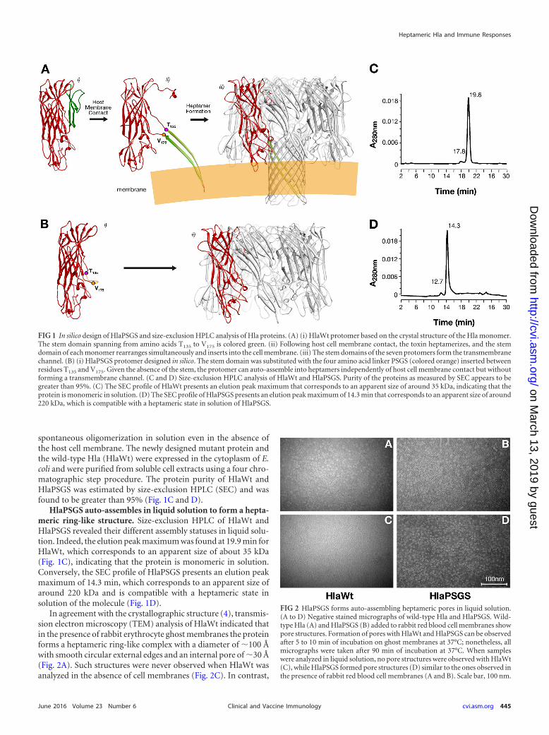
spontaneous oligomerization in solution even in the absence ofthe host cell membrane. The newly designed mutant protein andthe wild-type Hla (HlaWt) were expressed in the cytoplasm of E.coli and were purified from soluble cell extracts using a four chro-matographic step procedure. The protein purity of HlaWt andHlaPSGS was estimated by size-exclusion HPLC (SEC) and wasfound to be greater than 95% (Fig. 1C and D).
HlaPSGS auto-assembles in liquid solution to form a hepta-meric ring-like structure. Size-exclusion HPLC of HlaWt andHlaPSGS revealed their different assembly statuses in liquid solu-tion. Indeed, the elution peak maximum was found at 19.9 min forHlaWt, which corresponds to an apparent size of about 35 kDa(Fig. 1C), indicating that the protein is monomeric in solution.Conversely, the SEC profile of HlaPSGS presents an elution peakmaximum of 14.3 min, which corresponds to an apparent size ofaround 220 kDa and is compatible with a heptameric state insolution of the molecule (Fig. 1D).
In agreement with the crystallographic structure (4), transmis-sion electron microscopy (TEM) analysis of HlaWt indicated thatin the presence of rabbit erythrocyte ghost membranes the proteinforms a heptameric ring-like complex with a diameter of �100 Åwith smooth circular external edges and an internal pore of �30 Å(Fig. 2A). Such structures were never observed when HlaWt wasanalyzed in the absence of cell membranes (Fig. 2C). In contrast,
FIG 1 In silico design of HlaPSGS and size-exclusion HPLC analysis of Hla proteins. (A) (i) HlaWt protomer based on the crystal structure of the Hla monomer.The stem domain spanning from amino acids T135 to V175 is colored green. (ii) Following host cell membrane contact, the toxin heptamerizes, and the stemdomain of each monomer rearranges simultaneously and inserts into the cell membrane. (iii) The stem domains of the seven protomers form the transmembranechannel. (B) (i) HlaPSGS protomer designed in silico. The stem domain was substituted with the four amino acid linker PSGS (colored orange) inserted betweenresidues T135 and V175. Given the absence of the stem, the protomer can auto-assemble into heptamers independently of host cell membrane contact but withoutforming a transmembrane channel. (C and D) Size-exclusion HPLC analysis of HlaWt and HlaPSGS. Purity of the proteins as measured by SEC appears to begreater than 95%. (C) The SEC profile of HlaWt presents an elution peak maximum that corresponds to an apparent size of around 35 kDa, indicating that theprotein is monomeric in solution. (D) The SEC profile of HlaPSGS presents an elution peak maximum of 14.3 min that corresponds to an apparent size of around220 kDa, which is compatible with a heptameric state in solution of HlaPSGS.
FIG 2 HlaPSGS forms auto-assembling heptameric pores in liquid solution.(A to D) Negative stained micrographs of wild-type Hla and HlaPSGS. Wild-type Hla (A) and HlaPSGS (B) added to rabbit red blood cell membranes showpore structures. Formation of pores with HlaWt and HlaPSGS can be observedafter 5 to 10 min of incubation on ghost membranes at 37°C; nonetheless, allmicrographs were taken after 90 min of incubation at 37°C. When sampleswere analyzed in liquid solution, no pore structures were observed with HlaWt(C), while HlaPSGS formed pore structures (D) similar to the ones observed inthe presence of rabbit red blood cell membranes (A and B). Scale bar, 100 nm.
Heptameric Hla and Immune Responses
June 2016 Volume 23 Number 6 cvi.asm.org 445Clinical and Vaccine Immunology
on March 13, 2019 by guest
http://cvi.asm.org/
Dow
nloaded from

HlaPSGS formed ring-like structures regardless of the presence ofghost membranes (Fig. 2B and D). In order to trace the overallstructure of HlaPSGS and compare it with that of the wild-typeHla, the three-dimensional (3D) structure of the mutant proteinwas determined by using TEM combined with single-particle re-construction. About 5,000 particle images contributed to the finalreconstruction. Except for the absence of the stem region, themolecular features in the HlaPSGS electron density map werecomparable to those observed in the electron density map of thewild-type Hla. The heptameric form of the HlaPSGS ring-likestructure was clearly visible even at early stages of the 3D recon-struction (Fig. 3A). The final 3D model, obtained after 2 rounds ofiterative refinement (Fig. 3B), was generated at a 28-Å resolution(FSC, ½ bit). The model shows a seven-spike donut shape, with aclear 7-fold symmetry (C7-symmetric pore), a feature common toother toxins known in the literature (19–21). Seven thick arms,oriented clockwise, protrude from the bottom of the structure,which has an external diameter of �100 Å, is �70 Å in height, andhas an internal pore diameter of �35 Å (Fig. 3B).
Wild-type Hla, but not HlaPSGS, binds to ADAM10 and im-pairs human epithelial cell monolayer integrity. Hla is toxic forepithelial cells and has been recently demonstrated to promote celljunction dissolution by interacting with the cellular proteaseADAM10 (3, 22–24). Therefore, we wondered if the absence of thestem domain in HlaPSGS was sufficient to impair these activities.To that end, we measured the electrical impedance across a con-fluent monolayer of the human alveolar epithelial cell line A549using the xCELLigence system. Impedance reflects the status ofthe cell monolayer, including cell confluence, viability, and junc-tion functionality (25). A549 cells were grown to confluence onxCELLigence inserts, and the experiments were started whentransepithelial resistance (TER) was found to be constant betweentwo measures taken 24 h apart. Cells were then incubated for 24 hwith 100 �g/ml of HlaWt or HlaPSGS. Streptolysin O (SLO), awell-characterized pore-forming toxin produced by Streptococcuspyogenes, was used as a positive control at a concentration of 20
�g/ml, while PBS was used as a negative control. As shown in Fig.4A, SLO and HlaWt significantly affected epithelial monolayerintegrity as indicated by a decrease in TER (quantified by the de-crease in arbitrary cell index values), while cells treated withHlaPSGS maintained a constant TER throughout the duration ofthe experiment.
Furthermore, we observed a striking reduction in the capacityof HlaPSGS in binding to A549 cells compared to that in HlaWt(Fig. 4B). This suggested that removal of the stem domain affectsthe ability of the toxin to interact with the host cell and ADAM10.To assess this hypothesis, human A549 alveolar epithelial cells
FIG 3 Overall structure of the HlaPSGS heptamer. (A) Typical HlaPSGS classaverages clearly showing seven peaks corresponding to the seven identicalprotomers forming the ring-like HlaPSGS structure. (B) Top (i) and side (ii)surface views of the HlaPSGS electron density map obtained at a 28-Å resolu-tion (FSC, ½ bit). The ring has a seven-spike donut shape, with a clear 7-foldsymmetry (C7) and seven thick arms oriented clockwise and protruding fromthe bottom of the structure. It has an external diameter of 100 Å, a height of 70Å, and an internal pore diameter of 35 Å. (A) Scale bar, 100 Å.
FIG 4 HlaPSGS does not alter the integrity of human alveolar epithelial cellmonolayers, and its binding to the cells and the Hla host receptor ADAM10 isimpaired. (A) Integrity of the A549 monolayer following treatment withHlaWt and HlaPSGS. Graphs report the transepithelial electric resistance val-ues measured by xCELLigence. HlaWt and HlaPSGS were added at a concen-tration of 100 �g/ml and SLO at 20 �g/ml. The cell index is an arbitrary unit forelectric impedance measurement. Data are expressed as mean � SD of threeindependent wells. (B) The ability of HlaWt and HlaPSGS to bind to A549 cellswas assessed by an indirect immunofluorescence detection system and wasanalyzed by FACS. Columns in the plot indicate mean fluorescence intensity �SD of three independent experiments. (C) HlaPSGS does not interact withADAM10 on human alveolar epithelial cells. A549 alveolar epithelial cellstreated with HlaWt or HlaPSGS were subjected to Hla immunoprecipitationand ADAM10 immunoblotting, demonstrating an interaction of the wild typewith ADAM10 (left). In contrast, ADAM10 is not detected in HlaPSGS pre-cipitates (left). Equivalent amounts of HlaWt and the mutant variant werepresent in each immunoprecipitation (right).
Fiaschi et al.
446 cvi.asm.org June 2016 Volume 23 Number 6Clinical and Vaccine Immunology
on March 13, 2019 by guest
http://cvi.asm.org/
Dow
nloaded from

were treated with wild-type Hla or HlaPSGS, lysed, and subjectedto Hla immunoprecipitation. Precipitated proteins were probedfor ADAM10 and the toxin. Wild-type Hla interacted withADAM10, while the HlaPSGS mutant toxin did not precipitateADAM10 (Fig. 4C).
HlaPSGS is not hemolytic, and its antibodies neutralize theactivity of the wild-type toxin and binding to human epithelialcells. Hemolysis is usually considered the hallmark of Hla toxicityand of its pore-forming activity (1). Given that HlaPSGS lacks thestem domain responsible for the formation of the channel acrossthe host membrane, we assessed whether HlaPSGS was impairedin causing hemolysis. Rabbit erythrocytes were mixed with in-creasing concentrations of either HlaWt or HlaPSGS in a range of0.001 �g/ml to 50 �g/ml. As expected, HlaWt was able to inducelysis of erythrocytes in a dose-dependent fashion, reaching a pla-teau of hemolytic activity at a concentration of 0.5 �g/ml (Fig.5A). However, no lysis was observed in erythrocytes treated withHlaPSGS, even at 50 �g/ml.
Since anti-Hla antibodies neutralize Hla hemolytic activity(26), we then asked whether a similar inhibitory activity was alsoassociated with HlaPSGS antibodies. To this end, erythrocyteswere incubated for 20 min with 3.3 �g/ml recombinant HlaWttogether with different concentrations of purified rabbit poly-clonal IgG against HlaPSGS. A dose-dependent inhibition of he-molytic activity was observed using antibodies against HlaPSGS,reaching 100% inhibition at an antibody concentration of 0.45mg/ml (Fig. 5B).
Finally, we evaluated whether HlaPSGS-elicited antibodieswere also able to inhibit the binding of the wild-type toxin tohuman lung epithelial cells. The binding of [35S]methionine ra-diolabeled Hla was assessed using A549 cells at 5 � 105 cells/500 �lthat were incubated for 20 min at room temperature with 15 �l ofeither serum collected from HlaPSGS-immunized mice or serumcollected from sham-immunized mice (alum). Following a 5-minincubation with radiolabeled Hla, we observed that the presenceof serum raised against HlaPSGS (P � 0.0016) (Fig. 5C) effectivelyblocked active toxin binding.
Antibodies induced by HlaPSGS immunization and S. au-reus infection have comparable signatures. In order to under-stand whether S. aureus infection elicits antibodies that differen-tially recognize the monomeric and the heptameric form of Hla,we analyzed 94 serum samples from healthy donors (18 to 81 yearsold, females and males) for the ability to bind monomeric HlaWtand heptameric HlaPSGS. The analysis was carried out throughLuminex technology, immobilizing the two purified proteins ondifferently labeled microspheres. As shown in Fig. 6A, the serumsamples from most of the subjects recognized the heptamericform with higher efficiency than the monomer; the mean fluores-cence intensity (MFI) against HlaPSGS (10,375 � 698) was signif-icantly greater than that of HlaWt (6,192 � 400). A similar trendwas observed when serum samples from mice infected with sub-lethal doses of S. aureus Newman were analyzed (Fig. 6B).
The above data suggest that upon S. aureus infection, mouseand human immune systems preferentially recognize Hla when itis assembled in the heptameric form on the host cell membrane.This led us to hypothesize that immunization with the monomericor the heptameric Hla form may induce qualitatively differentantibody responses. To test this hypothesis, 7 overlapping 50-ami-no-acid-long fragments spanning the entire length of Hla (Fig. 6C[9]) were immobilized on microspheres and analyzed by Luminex
FIG 5 HlaPSGS lacks hemolytic activity, and its antibodies neutralize wild-type Hla-induced hemolysis and the binding of the toxin to A549 cells. (A)Rabbit blood hemolysis after treatment with active Hla toxin (HlaWt) orHlaPSGS. Graphs report the hemolysis (%) observed following the addi-tion of the three proteins at increasing concentrations. Each point is themean value of three independent experiments � SD. (B) Effect of purifiedanti-HlaPSGS IgG on blood hemolysis induced by wild-type Hla toxin (3.3�g/ml). Graphs report the hemolysis (%) observed using increasing con-centrations of IgG. Each point is the mean value of three independentexperiments � SD. (C) Binding inhibition assay of HlaWt toxin to A549cells. The binding of 0.3 nM 35S-radiolabeled Hla toxin on A549 cells at 5 �105 cells/500 �l was evaluated after incubation with 15 �l of HlaPSGSantiserum or from sham immunized mice (alum) for 20 min at roomtemperature. Hla antibody complexes were then incubated for 5 min withA549 cells. Finally, cells were pelleted and added to scintillation fluid forquantification of bound radioactivity (cpm). Significance was determinedusing Student’s t test.
Heptameric Hla and Immune Responses
June 2016 Volume 23 Number 6 cvi.asm.org 447Clinical and Vaccine Immunology
on March 13, 2019 by guest
http://cvi.asm.org/
Dow
nloaded from

technology using sera from mice immunized with monomeric Hlaand HlaPSGS. Data are expressed as the percentage of antibodytiters detected against each Hla peptide relative to the sum of an-tibody titers measured against the 7 peptides. Therefore, the anal-ysis provides a relative estimate of the titers against each fragmentand is independent of the total antibody titers present in the sera.As shown in Fig. 6C, immunization with the monomeric Hla in-duced antibodies that largely recognized the N-terminal region ofHla. In contrast, anti-HlaPSGS antibodies recognized all sevenfragments with similar levels of intensity. Interestingly, the frag-ment recognition profile of anti-HlaPSGS antibodies resembledthe profile observed with sera from S. aureus-infected mice andfrom human volunteers more closely than that associated withmonomeric Hla.
HlaPSGS vaccination protects mice against S. aureus infec-tion. In order to assess whether HlaPSGS is a suitable vaccinecandidate, we investigated whether immunization with this pro-tein provides protection in a mouse pneumonia model of S. aureusinfection. HlaPSGS was formulated with aluminum-hydroxideand was used to immunize C57BL/6J mice, while negative controlsreceived identical courses of PBS plus adjuvant (alum). Mice wereinfected intranasally with S. aureus LAC (USA300) strain, andsurvival was monitored for 7 days. The pneumonia model and thisS. aureus strain were selected because they are considered to be thegold standard for assessing the protective efficacy of Hla antigens(6). Immunization with HlaPSGS exhibited near complete protec-tion, while all sham-immunized animals succumbed on the firstday postinfection (Fig. 7).
DISCUSSION
Hla is a potent toxin produced by most S. aureus isolates and isknown to play a key role in S. aureus virulence. Since it has beenshown that anti-Hla antibodies neutralize Hla toxicity, it is notsurprising that Hla is considered one of the most promising S.aureus vaccine candidates. Over the past few decades, several non-toxigenic mutants have been generated, most of which containsingle point mutations that impair heptamer formation, such asthe well-characterized HlaH35L mutant (27). More recently, frag-
FIG 6 Reactivity profiling of sera from S. aureus-infected mice and human sera toward monomeric and heptameric Hla. Antibody titers against HlaWt andHlaPSGS present in 94 human serum samples (A) and serum samples from 12 S. aureus-infected mice (B). Data points indicate the fluorescence intensitymeasured by the Luminex assay in each serum, and the bar is the mean value � standard error of the mean (SEM). Statistical analysis was performed byMann-Whitney U test. (C) Seven serial 50-amino-acid segments of Hla were purified as GST fusion proteins and were used in the Luminex analysis to map theepitopes recognized by serum samples of different origin (top). Antibody titers against the 7 Hla peptides present in serum samples from 16 mice immunized withHlaPSGS or monomeric Hla (the mutant form HlaH35L, which is unable to form heptamers, was used to immunize mice to avoid toxicity associated with HlaWt)and in 20 human serum samples as well as in serum samples from 12 S. aureus-infected mice. Data are expressed as the median percentage of antibody titersdetected against each Hla peptide relative to the total antibody titers measured against the 7 peptides (bottom) (antibody titers are expressed as mean fluorescenceintensity [MFI]).
FIG 7 Protective efficacy of HlaPSGS in pneumonia model. Mice were immu-nized, 2 weeks apart, with HlaPSGS formulated with 2 mg/ml aluminum hy-droxide or with the adjuvant alone. Ten days after the second immunization,they were challenged with S. aureus. C57BL/6 mice were challenged intrana-sally with S. aureus LAC (n � 20 per group; 2 separate experiments), andsurvival was monitored for 1 week. Lines with circles indicate HlaPSGS, andthose with triangles indicate mice treated with alum alone. Statistical analysiswas performed by log-rank (Mantel-Cox) test.
Fiaschi et al.
448 cvi.asm.org June 2016 Volume 23 Number 6Clinical and Vaccine Immunology
on March 13, 2019 by guest
http://cvi.asm.org/
Dow
nloaded from

ments of the protein have been proposed as alternatives to pointmutation mutants (7). All of these engineered molecules remainmonomeric in aqueous solutions and when exposed to host cellmembranes. Therefore, they do not form lytic pores, they are notcytotoxic, and they are considered safe antigens for vaccine use. Toour knowledge, heptameric Hla mutants have never before beenpursued as vaccine candidates (28).
Since Hla is expected to rapidly form heptameric complexes onhost cells when released by S. aureus during infection, this confor-mation may trigger immune responses different from those elic-ited by the monomeric mutants used as vaccine candidates. Inorder to rationally design optimized Hla-based vaccines, it is crit-ical to understand whether there are differences in the anti-Hlaantibodies induced by monomeric and heptameric Hla during S.aureus infection.
We generated an Hla mutant that could spontaneously assem-ble into heptameric complexes in solution. Taking advantage ofthe availability of the 3D structure of the heptameric Hla wild-typetoxin, we created the HlaPSGS mutant, in which the 39-amino-acid stem, spanning from residue Thr135 to Val175, was removed.In the Hla monomer, this stem shields the surface area involved inmonomer-monomer interaction. As predicted, removal of thestem region led to the spontaneous formation of the heptamericcomplex, even in the absence of cell membrane. This was con-firmed by qualitative transmission electron microscopy (TEM)showing that HlaPSGS assembled into a ring-like shaped structureresembling the cap of the Hla wild-type pore. Further single-par-ticle reconstruction analysis applied to the TEM images clearlyshowed that HlaPSGS is composed of seven identical copies of thesame protomer arranged in a C7 symmetry, even in the absence ofthe stem region.
We then hypothesized that the heptameric conformation ofHlaPSGS was able to elicit antibodies with specificity comparableto that elicited by the Hla cytolytic pore. To confirm this hypoth-esis, antibody titers against wild-type monomeric Hla andHlaPSGS were measured in sera from S. aureus-infected mice.This analysis revealed higher IgG titers against the heptamericform than against the wild-type monomeric protein. A similarpattern was observed with the panel of human sera, suggestingthat in humans staphylococcal infection also generates antibodiesthat better recognize the toxin heptamer than the monomer. Ourepitope recognition data showed that antibodies induced byHlaPSGS immunization had an epitope recognition profile simi-lar to that of the sera from donors and S. aureus-infected mice. Inparticular, the seven Hla fragments were quite uniformly recog-nized by the sera. In contrast, the sera from animals vaccinatedwith HlaH35L, which cannot form heptameric complexes, had amore limited recognition profile and a remarkable preference forthe N-terminal region of the molecule.
These results demonstrate for the first time to our knowledgethat during infection the prevalent form of Hla exposed to theimmune system is the heptameric complex. The humoral re-sponse against Hla has recently been proposed as a correlate ofprotection against S. aureus human infections (29, 30). It will beimportant to understand whether protection is associated with aspecific Hla epitope recognition profile of antibodies present inhumans and their affinity toward monomeric versus the heptam-eric form.
At this point, we evaluated the molecule as a vaccine antigenagainst S. aureus. Interestingly, HlaPSGS was found to be im-
paired in binding to ADAM10 and human lung epithelial cells.This observation suggests that a preoligomerized toxin is physi-cally unable to bind to the receptor. The second feature representsanother significant novelty compared to previously published de-toxified mutants that still bind host cells (e.g., HlaH35L [22]).Removal of the stem domain effectively detoxifies Hla and impairsits ability to form the cytolytic pore and to exert hemolytic activity.We then demonstrated that HlaPSGS immunization elicits func-tional antibodies with neutralizing activity toward Hla-mediatederythrocyte lysis. In addition, HlaPSGS vaccination generatedhigh protective immunity against staphylococcal infection inmice.
In conclusion, the ability of HlaPSGS to self-assemble in liquidsolution, its safety profile as a non-pore-forming mutant, and itsability to mimic the immune response generated by infectionmake this newly engineered Hla molecule an important tool forstudying immune responses against the toxin as well as a promis-ing vaccine candidate.
ACKNOWLEDGMENTS
We are grateful to our colleagues Luca Moraschini, Fabio Rigat, and BeateBruske for scientific support of the study. We also thank our colleaguesMarco Tortoli and the animal care facility for technical assistance andGiorgio Corsi for art work. We thank Lauren Smith (Stanford University)for critically reading the paper. We also thank Giuseppe Mancuso andGiuseppe Teti (University of Messina) as well as Andrea DeDent (Univer-sity of Chicago) for assistance with animal experimentation. Finally, wethank Fabiola Giusti (University of Siena) for assistance with the trans-mission electron microscope.
All authors except Juliane Bubeck Wardenburg and Kelly Tomasze-wski were employees of the Novartis group of companies at the time thestudy was conducted. All authors except Juliane Bubeck Wardenburg,Kelly Tomaszewski, Fabiana Falugi, Guido Grandi, Ravi P. N. Mishra,Antonina Torre, Maria Rita Fontana, and Luigi Fiaschi are now employeesof GSK group companies. At the time the study was conducted, FabioBagnoli, Guido Grandi, and Rino Rappuoli owned Novartis stocks. At thetime the study was conducted, Juliane Bubeck Wardenburg had the po-tential to receive royalties from Novartis Vaccines and Diagnostics inrelation to patents owned by the University of Chicago.
FUNDING INFORMATIONAt the time the study was conducted, Novartis Vaccines and Diagnostics,Inc., was the funding source.
REFERENCES1. Bhakdi S, Tranum-Jensen J. 1991. Alpha-toxin of Staphylococcus aureus.
Microbiol Rev 55:733–751.2. Berube BJ, Bubeck Wardenburg J. 2013. Staphylococcus aureus alpha-
toxin: nearly a century of intrigue. Toxins (Basel) 5:1140 –1166. http://dx.doi.org/10.3390/toxins5061140.
3. Inoshima I, Inoshima N, Wilke GA, Powers ME, Frank KM, Wang Y,Bubeck Wardenburg J. 2011. A Staphylococcus aureus pore-forming toxinsubverts the activity of ADAM10 to cause lethal infection in mice. NatMed 17:1310 –1314. http://dx.doi.org/10.1038/nm.2451.
4. Song L, Hobaugh MR, Shustak C, Cheley S, Bayley H, Gouaux JE. 1996.Structure of staphylococcal alpha-hemolysin, a heptameric transmem-brane pore. Science 274:1859 –1866. http://dx.doi.org/10.1126/science.274.5294.1859.
5. Kawate T, Gouaux E. 2003. Arresting and releasing staphylococcalalpha-hemolysin at intermediate stages of pore formation by engi-neered disulfide bonds. Protein Sci 12:997–1006. http://dx.doi.org/10.1110/ps.0231203.
6. Bubeck Wardenburg J, Schneewind O. 2008. Vaccine protection againstStaphylococcus aureus pneumonia. J Exp Med 205:287–294. http://dx.doi.org/10.1084/jem.20072208.
Heptameric Hla and Immune Responses
June 2016 Volume 23 Number 6 cvi.asm.org 449Clinical and Vaccine Immunology
on March 13, 2019 by guest
http://cvi.asm.org/
Dow
nloaded from

7. Adhikari RP, Karauzum H, Sarwar J, Abaandou L, Mahmoudieh M,Boroun AR, Vu H, Nguyen T, Devi VS, Shulenin S, Warfield KL, AmanMJ. 2012. Novel structurally designed vaccine for S. aureus alpha-hemolysin: protection against bacteremia and pneumonia. PLoS One7(6):e38567. http://dx.doi.org/10.1371/journal.pone.0038567.
8. Klock HE, Lesley SA. 2009. The polymerase incomplete primer extension(PIPE) method applied to high-throughput cloning and site-directed mu-tagenesis. Methods Mol Biol 498:91–103. http://dx.doi.org/10.1007/978-1-59745-196-3_6.
9. Ragle BE, Bubeck Wardenburg J. 2009. Anti-alpha-hemolysin mono-clonal antibodies mediate protection against Staphylococcus aureuspneumonia. Infect Immun 77:2712–2718. http://dx.doi.org/10.1128/IAI.00115-09.
10. Burton GW, Ingold KU, Thompson KE. 1981. An improved procedurefor the isolation of ghost membranes from human red blood cells. Lipids16:946. http://dx.doi.org/10.1007/BF02535005.
11. Guex N, Peitsch MC. 1997. SWISS-MODEL and the Swiss-PdbViewer:an environment for comparative protein modeling. Electrophoresis 18:2714 –2723. http://dx.doi.org/10.1002/elps.1150181505.
12. Laskowski RA, MacArthur MW, Moss DS, Thornton JM. 1993.PROCHECK: a program to check the stereochemical quality of pro-tein structures. J Appl Cryst 26:283–291. http://dx.doi.org/10.1107/S0021889892009944.
13. Ludtke SJ, Baldwin PR, Chiu W. 1999. EMAN: semiautomated softwarefor high-resolution single-particle reconstructions. J Struct Biol 128:82–97. http://dx.doi.org/10.1006/jsbi.1999.4174.
14. van Heel M, Harauz G, Orlova EV, Schmidt R, Schatz M. 1996. A newgeneration of the IMAGIC image processing system. J Struct Biol 116:17–24. http://dx.doi.org/10.1006/jsbi.1996.0004.
15. Pettersen EF, Goddard TD, Huang CC, Couch GS, Greenblatt DM,Meng EC, Ferrin TE. 2004. UCSF Chimera–a visualization system forexploratory research and analysis. J Comput Chem 25:1605–1612. http://dx.doi.org/10.1002/jcc.20084.
16. Poljicak-Milas N, Kardum-Skelin I, Vudan M, Marenjak TS, Ballarin-Perharic A, Milas Z. 2009. Blood cell count analyses and erythrocytemorphometry in New Zealand white rabbits. Vet Arhiv 79:561–571.
17. Bubeck Wardenburg J, Patel RJ, Schneewind O. 2007. Surface proteinsand exotoxins are required for the pathogenesis of Staphylococcus aureuspneumonia. Infect Immun 75:1040 –1044. http://dx.doi.org/10.1128/IAI.01313-06.
18. Foletti D, Strop P, Shaughnessy L, Hasa-Moreno A, Casas MG, RussellM, Bee C, Wu S, Pham A, Zeng Z, Pons J, Rajpal A, Shelton D. 2013.Mechanism of action and in vivo efficacy of a human-derived antibodyagainst Staphylococcus aureus alpha-hemolysin. J Mol Biol 425:1641–1654.http://dx.doi.org/10.1016/j.jmb.2013.02.008.
19. De S, Olson R. 2011. Crystal structure of the Vibrio cholerae cytolysin
heptamer reveals common features among disparate pore-forming toxins.Proc Natl Acad Sci U S A 108:7385–7390. http://dx.doi.org/10.1073/pnas.1017442108.
20. Young JA, Collier RJ. 2007. Anthrax toxin: receptor binding, internaliza-tion, pore formation, and translocation. Annu Rev Biochem 76:243–265.http://dx.doi.org/10.1146/annurev.biochem.75.103004.142728.
21. Fivaz M, Abrami L, Tsitrin Y, van der Goot FG. 2001. Not as simple asjust punching a hole. Toxicon 39:1637–1645. http://dx.doi.org/10.1016/S0041-0101(01)00151-9.
22. Wilke GA, Bubeck Wardenburg J. 2010. Role of a disintegrin and met-alloprotease 10 in Staphylococcus aureus alpha-hemolysin-mediated cellu-lar injury. Proc Natl Acad Sci U S A 107:13473–13478. http://dx.doi.org/10.1073/pnas.1001815107.
23. Inoshima N, Wang Y, Bubeck Wardenburg J. 2012. Genetic requirementfor ADAM10 in severe Staphylococcus aureus skin infection. J Invest Der-matol 132:1513–1516. http://dx.doi.org/10.1038/jid.2011.462.
24. Powers ME, Kim HK, Wang Y, Bubeck Wardenburg J. 2012. ADAM10mediates vascular injury induced by Staphylococcus aureus alpha-hemolysin. J Infect Dis 206:352–356. http://dx.doi.org/10.1093/infdis/jis192.
25. Solly K, Wang X, Xu X, Strulovici B, Zheng W. 2004. Application ofreal-time cell electronic sensing (RT-CES) technology to cell-based assays.Assay Drug Dev Technol 2:363–372. http://dx.doi.org/10.1089/adt.2004.2.363.
26. Menzies BE, Kernodle DS. 1996. Passive immunization with antiserumto a nontoxic alpha-toxin mutant from Staphylococcus aureus is protectivein a murine model. Infect Immun 64:1839 –1841.
27. Menzies BE, Kernodle DS. 1994. Site-directed mutagenesis of the alpha-toxin gene of Staphylococcus aureus: role of histidines in toxin activity invitro and in a murine model. Infect Immun 62:1843–1847.
28. Cheley S, Malghani MS, Song L, Hobaugh M, Gouaux JE, Yang J,Bayley H. 1997. Spontaneous oligomerization of a staphylococcal alpha-hemolysin conformationally constrained by removal of residues that formthe transmembrane beta-barrel. Protein Eng 10:1433–1443. http://dx.doi.org/10.1093/protein/10.12.1433.
29. Fritz SA, Tiemann KM, Hogan PG, Epplin EK, Rodriguez M, Al-Zubeidi DN, Bubeck Wardenburg J, Hunstad DA. 2013. A serologiccorrelate of protective immunity against community-onset Staphylococcusaureus infection. Clin Infect Dis 56:1554 –1561. http://dx.doi.org/10.1093/cid/cit123.
30. Adhikari RP, Ajao AO, Aman MJ, Karauzum H, Sarwar J, Lydecker AD,Johnson JK, Nguyen C, Chen WH, Roghmann MC. 2012. Lower anti-body levels to Staphylococcus aureus exotoxins are associated with sepsis inhospitalized adults with invasive S. aureus infections. J Infect Dis 206:915–923. http://dx.doi.org/10.1093/infdis/jis462.
Fiaschi et al.
450 cvi.asm.org June 2016 Volume 23 Number 6Clinical and Vaccine Immunology
on March 13, 2019 by guest
http://cvi.asm.org/
Dow
nloaded from