author's personal copy - facm.ucl.ac.be · author's personal copy toxicology 290 (2011)...
TRANSCRIPT

This article appeared in a journal published by Elsevier. The attachedcopy is furnished to the author for internal non-commercial researchand education use, including for instruction at the authors institution
and sharing with colleagues.
Other uses, including reproduction and distribution, or selling orlicensing copies, or posting to personal, institutional or third party
websites are prohibited.
In most cases authors are permitted to post their version of thearticle (e.g. in Word or Tex form) to their personal website orinstitutional repository. Authors requiring further information
regarding Elsevier’s archiving and manuscript policies areencouraged to visit:
http://www.elsevier.com/copyright

Author's personal copy
Toxicology 290 (2011) 178–186
Contents lists available at SciVerse ScienceDirect
Toxicology
journa l homepage: www.e lsev ier .com/ locate / tox ico l
Modulation of the expression of ABC transporters in murine (J774) macrophagesexposed to large concentrations of the fluoroquinolone antibiotic moxifloxacin
Coralie M. Valleta,1, Béatrice Marqueza,1,2, Naïma Nhirib, Ahalieyah Anantharajaha,Marie-Paule Mingeot-Leclercqa, Paul M. Tulkensa, Jean-Yves Lallemandb, Eric Jacquetb,c,Francoise Van Bambekea,∗
a Université catholique de Louvain, Louvain Drug Research Institute, Pharmacologie cellulaire et moléculaire, B-1200 Brussels, Belgiumb Centre de recherche de Gif, Institut de Chimie des Substances naturelles, avenue de la Terrasse, 91198 Gif-sur-Yvette, Francec IMAGIF qPCR Platform, CNRS UPR2301, avenue de la Terrasse, 91198 Gif-sur-Yvette, France
a r t i c l e i n f o
Article history:Received 12 August 2011Received in revised form10 September 2011Accepted 12 September 2011Available online 17 September 2011
Keywords:MoxifloxacinCiprofloxacinEffluxMrp4P-gpBcrp
a b s t r a c t
Long-term exposure to pharmacological agents can select for cells that overexpress efflux transporters.We previously showed that mouse J774 macrophages cultivated for a prolonged period of time with toxicconcentrations of the fluoroquinolone ciprofloxacin overexpress the efflux transporter Mrp4 and displaya reduced accumulation of this antibiotic, but no change in the accumulation of moxifloxacin, a closelyrelated molecule (Antimicrob. Agents Chemother. [2006] 50, 1689–1695 and [2009] 53, 2410–2416).Because of this striking difference between the two fluoroquinolones, we have now examined themodifications in the expression of ABC efflux transporters induced by the prolonged exposure of J774macrophages to high concentrations of moxifloxacin. The resulting cell line showed (i) no difference inthe accumulation of moxifloxacin but an increased accumulation and decreased efflux of ciprofloxacin;(ii) an overexpression of the multidrug transporters Abcb1a (P-gp), Abcc2 (Mrp2) and Abcg2 (Bcrp1), anda decreased expression of Abcc4 (Mrp4). While P-gp and Bcrp1 were functional, they did not modify thecellular accumulation of fluoroquinolones. The data show that exposing cells to high concentrations ofa drug that is not affected by active efflux can trigger a pleiotropic response leading to a modulation inthe expression of several transporters. These changes, however, are not sufficient to protect cells againstthe toxicity that fluoroquinolones may exert at large concentrations. They could also cause unanticipateddrug interactions in vivo, should the drug exposure grossly exceed what is anticipated from its currentregistered use.
© 2011 Elsevier Ireland Ltd. All rights reserved.
1. Introduction
Exposure of eukaryotic cells to drugs can trigger modificationsin the expression of mechanisms susceptible to favor their elim-ination. In hepatocytes, this results in a global activation of thephases I, II, and III of drug elimination and transport, and is mostoften related to the transient induction of the transcriptional regu-lation of the corresponding genes by nuclear receptors (Fardel et al.,2001; Klaassen and Aleksunes, 2010; Scotto, 2003; Xu et al., 2005).In cancer cells, overexpression of efflux transporters is one of thebest known mechanisms of resistance to chemotherapy (Baguley,
∗ Corresponding author. Tel.: +32 2 764 73 78; fax: +32 2 764 73 73.E-mail address: [email protected] (F. Van Bambeke).
1 Both authors contributed equally to this study.2 Present address: Grupo Tumorigenesis endocrina y regulación hormonal del
cáncer, Instituto de Biomedicina de Sevilla–Hospitales Universitarios Virgen delRocío, Avenida Manuel Siurot s/n, 41013 Sevilla, Spain.
2010; Eckford and Sharom, 2009; Gillet et al., 2007). This resistancecan be reproduced in vitro by exposing cells for prolonged peri-ods of time to increasing concentrations of anticancer agents andoverexpression of drug transporters can result in this case from amultitude of mechanisms that are often slowly acquired and poorlyreversible (Gottesman et al., 1998; Scotto, 2003; Turk et al., 2009).Such stepwise selection approach can be applied to many otherdrugs to identify the transporter(s) responsible for their efflux, pro-vided they can exert sufficient toxicity at the initial concentrationused to trigger selection of a resistance mechanism.
Using this procedure, we were able to identify, in J774macrophages, the efflux transporter for the fluoroquinolone antibi-otic ciprofloxacin (see Fig. SP1 for structure) as being Mrp43
(Abcc4), a member of the C subfamily of ATP binding cassette
3 According to common conventions, genes and proteins of murine origin arewritten in low-case letters after the first initial, while genes and proteins of humanor non-specific origin are written in all-uppercase letters.
0300-483X/$ – see front matter © 2011 Elsevier Ireland Ltd. All rights reserved.doi:10.1016/j.tox.2011.09.003

Author's personal copy
C.M. Vallet et al. / Toxicology 290 (2011) 178–186 179
(ABC) transporters, which is expressed at high level even in wild-type macrophages (Marquez et al., 2009; Michot et al., 2006).Overexpression of this protein effectively reduces the cellularconcentration of ciprofloxacin in cells exposed to increasing con-centrations of this drug, providing a simple and straightforwardresistance mechanism. However, not all fluoroquinolones are sub-jected to efflux due to apparently minor but probably criticaldifferences in their structures (Michot et al., 2005; Vallet et al.,2011). Specifically, moxifloxacin (see Fig. SP1 for structure), isinsensitive to ATP-energized efflux in J774 macrophages, and accu-mulates to a similar level in wild-type or in ciprofloxacin-selectedmacrophages (Michot et al., 2005, 2006). We have now used J774macrophages to examine how cells would respond to stepwiseexposure to increasing concentrations of a drug in absence ofdetectable basal efflux. Much to our surprise, we observed majorchanges in the expression of several ABC transporters that did notaffect the accumulation or efflux of moxifloxacin itself, but causeda dramatic increase in ciprofloxacin accumulation, associated toa slowed-down efflux. This phenotype is thus strikingly differentfrom that observed in ciprofloxacin-selected cells (Michot et al.,2006).
2. Materials and methods
2.1. Chemicals
All cell culture reagents were purchased from Invitrogen (Carlsbad, CA). Mox-ifloxacin HCl and ciprofloxacin HCl (potency: 90.9% and 85%, respectively) werereceived from Bayer Schering Pharma AG (Berlin, Germany) as microbiological stan-dards. Fumitremorgin C (FTC) was kindly provided by Dr. R. Robey (NIH, Bethesda,MD). MK-571 was purchased from Enzo Life Sciences (Farmingdale, NY), and otherchemical products, from Sigma–Aldrich (St. Louis, MO).
2.2. Cell lines and culture conditions
J774 mouse macrophages (referred to as wild-type cells) were cultured andmaintained as already described (Michot et al., 2005). Moxifloxacin-selected cellswere obtained by serial culture in media containing increasing moxifloxacin con-centrations (37 mg/l [0.1 mM] for 3 months [10 passages], 55 mg/l [0.15 mM] for 3months [10 passages], and 74 mg/l [0.2 mM] for 12 months [50 passages]), followingthe general procedure used for selecting cells by ciprofloxacin (Michot et al., 2006).Cells were maintained thereafter in the continuous presence of 0.2 mM moxifloxacinand used for experiments between the 60th and the 80th passage.
2.3. Assessment of cell membrane intactness and mitochondrial metabolism
Trypan blue exclusion assay was used to detect alteration of membraneintactness. Cells detached by trypsinization and pelleted by low speed centrifu-gation (10 min, 100 × g), were incubated with 0.2% Trypan blue in phosphatebuffered saline (PBS) for 2 min and counted for stained and unstained cells.Mitochondrial metabolism was assessed by the MTT (3-(4,5-dimethylthiazol-2-yl)-2,5-diphenyltetrazolium) assay. In brief, cells were incubated for 48 h withciprofloxacin or moxifloxacin and rinsed with PBS. MTT was added at a concen-tration of 0.5 mg/ml to each well. After 1 h incubation at 37 ◦C, DMSO was added todissolve the formazan crystals and the absorbance was measured at 570 nm.
2.4. Accumulation and efflux of fluoroquinolones
Experiments were performed as previously described for wild-type andciprofloxacin-selected cells (Marquez et al., 2009; Michot et al., 2005, 2006), exceptthat moxifloxacin was removed from the culture medium of the moxifloxacin-selected cells for the last 48 h to eliminate cell-associated drug. Fluoroquinoloneswere assayed by fluorimetry (Michot et al., 2005). ATP-depletion was obtained bypre-incubating cells for 20 min in a medium containing 60 mM 2-d-deoxyglucoseand 5 mM NaN3, and maintaining them in the same medium during the wholeexperiment, as previously described (Michot et al., 2004). The cell drug contentwas expressed by reference to the total protein content measured by the Lowry’smethod (Lowry et al., 1951).
2.5. Genomic characterization of efflux transporters (real-time PCR and TaqManLow Density Array [TLDA])
Abcc4/Mrp4 mRNA levels were determined as previously described using real-time PCR experiments with SYBR Green detection and normalization with Ywhaz andRpl13a (Marquez et al., 2009). The complete transcriptional profile of 47 murine ABCtransporters was performed using TaqMan® Low Density custom Array on a 7900HT
Fast Real-Time PCR System (Applied Biosystems, Foster City, CA). For these experi-ments, total RNA was extracted from J774 mouse macrophage samples with TRIzol(Invitrogen) and treated with Turbo DNase (Ambion, Austin, TX) followed by RNAcleanup with RNeasy Mini Columns (QIAGEN GmbH, Hilden, Germany), accordingto the manufacturer’s instructions. Purified RNA concentration was measured witha Qubit fluorimeter, using the Quant-iT RNA BR assay kit (Invitrogen). High-capacitycDNA Reverse Transcription kit (Applied Biosystems) was used to synthesize cDNA,starting from 2 or 3 �g of total purified RNA in a final volume of 50 �l following themanufacturer’s instructions.
To identify stable genes to use for housekeeping in our study (Huggett et al.,2005), we first performed real-time PCR using the TaqMan® Mouse EndogenousControl Arrays (Applied Biosystems). 200 ng of cDNA and 50 �l of TaqMan® Univer-sal Master Mix were mixed in a final volume of 100 �l and each sample was used toload one port of the microfluidic card. 16 potential housekeeping genes were testedin triplicate for each sample. The most stable genes were then selected by analyz-ing the results with GeNorm and Normfinder functions in the Genex 4.3.8 software(MultiD, Göteborg, Sweden).
Gene expression quantification of the murine ABC transporters family wasdetermined using custom-designed TaqMan® Array cards format 96a (SupplementalTable SP1), pre-loaded with the TaqMan® assay for 47 murine ABC transportersand the 10 most stable housekeeping genes that have been selected previously.600 ng of cDNA and 100 �l of TaqMan® Universal Master Mix were mixed ina final volume of 200 �l and used to load 2 sample ports. Technical replicatesand biological replicates were performed for each biological sample. Real-timeq-PCR amplifications were carried out (10 min at 94.5 ◦C followed by 40 cyclesof 30 s at 97 ◦C and 1 min at 59.7 ◦C). Thermal cycling and fluorescence detectionwas performed with ABI SDS 2.3 and RQ manager Software. The thresholds andbaselines were set manually and cycle threshold (Ct) values were extracted. Theoptimal Ct value cut-off was limited to 35 cycles. According to Genex softwareanalyses, the best normalization was obtained with Gapdh and Gusb. The geometricmean of these two genes was then used to normalize gene expression levels ofABC transporter. Analysis of gene expression values was performed using relativequantification (Pfaffl, 2001), which determines target gene expression relative tohousekeeping genes expression levels and relative to the wild-type J774 controlsample. Variations in gene expression observed for Abcb1a, Abcc2 and Abcg2were also validated in microplate experiments, using different TaqMan® GeneExpression Assays. 25 ng of cDNA were mixed with 10 �l of TaqMan® Fast UniversalPCR Master Mix (Applied Biosystems) and the target gene assay (ABC transporteror housekeeping gene) in a final volume of 20 �l. Each sample was measured intriplicate with the following parameters: 95 ◦C for 20 s and 40 cycles of 95 ◦C for 1 sand 60 ◦C for 20 s. Gene expression analysis was performed as described previouslyfor TaqMan® Arrays experiments. mRNA levels are expressed as a variation ratiobetween the resistant (sample) and the wild-type (sample calibrator) cell lines,using Gapdh and Gusb as housekeeping (reference) genes, according to: RQ =2 ((Ct sample calibrator, target gene − Ct sample, target gene) − (Ct sample calibrator, reference gene − Ct sample,
referencegene)).
2.6. Western blot analysis of efflux transporters
Western-blots were performed on cell crude extracts for Abcc2/Mrp2 andAbcc4/Mrp4, and on membrane-enriched preparations for Abcb1a/P-gp andAbcg2/Bcrp1, using the NuPAGE electrophoresis system (Invitrogen) as previouslydescribed (Marquez et al., 2009). Crude extracts consisted of cells collected in ice-cold PBS and pelleted by low speed centrifugation, resuspended in 10 mM Tris–HClpH 7.4 and the subjected to heating for 10 min at 70 ◦C and to sonication, withgross debris eliminated by centrifugation for 30 min at 14,000 rpm (20,000 × g).Membrane-enriched preparations were prepared exactly as described previously(Marquez et al., 2009). The protein content of both preparations was measuredusing the bicinchoninic acid protein assay (Bradford assay; Pierce BCA Reagents,Pierce, Rockford, IL). Appropriate quantities of proteins were mixed to 4X NuPAGELDS Sample buffer and 10X NuPAGE reducing agent, then heated for 10 min at70 ◦C. Samples were loaded on acrylamide gels (NuPAGE 10% Bis-Tris Gel, Invit-rogen). After migration, proteins were electro-transferred onto a PVDF membrane(0.45 �m, Pierce), which was blocked by a 2 h incubation with 5% defatted milk inTris–buffered saline (TBS; 20 mM Tris–HCl, 500 mM NaCl pH 7.5) containing 0.05%tween-20. Membranes were then incubated overnight with the primary antibodiesM2III-5 (Alexis Biochemicals, Lausen, Switzerland), M4I-10 (Alexis Biochemicals),C219 (Signet, Covance Inc, Princeton, NJ), or BXP-53 (SantaCruz Biotechnology,Santa Cruz, CA) to detect Mrp2 (Abcc2), Mrp4 (Abcc4), P-gp (Abcb1), and Bcrp1(Abcg2), respectively (see figure caption for dilutions) and exposed to appropri-ate horseradish peroxidase-coupled secondary antibodies [diluted 1/300] for 5 h.Anti-actin (Sigma–Aldrich) or anti-prohibitin H-80 (SantaCruz Biotechnology) poly-clonal antibodies were used as loading control and treated the same way. Blots wererevealed by chemiluminescence (SuperSignal West Pico, Pierce).
2.7. Phenotypic characterization of efflux transporters
The activity of Mrp4 was measured using ciprofloxacin as substrate and gemfi-brozil and MK571 as broad spectrum and specific inhibitors, as described previously(Michot et al., 2004). The activity of P-gp and Bcrp1 was measured with rhodamine
Author's personal copy
180 C.M. Vallet et al. / Toxicology 290 (2011) 178–186
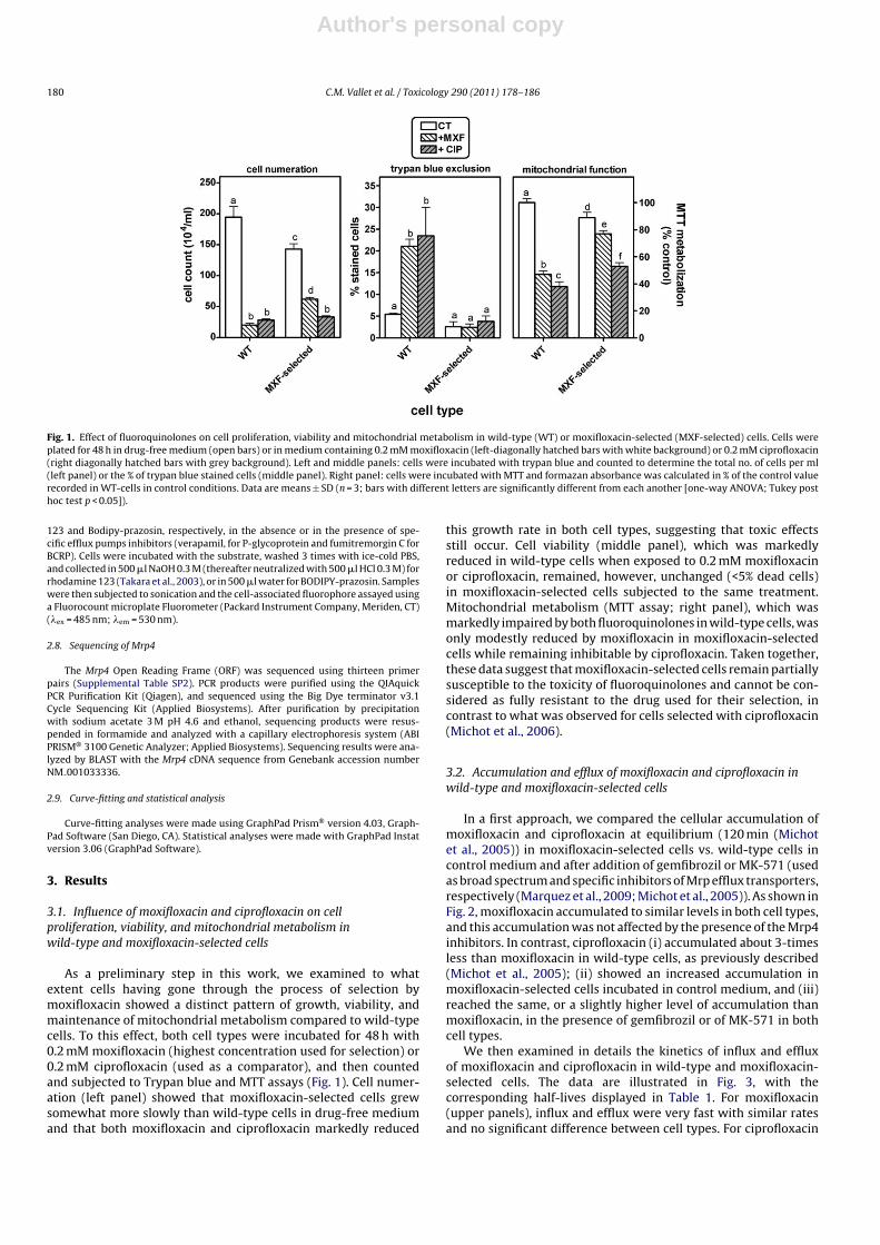
Fig. 1. Effect of fluoroquinolones on cell proliferation, viability and mitochondrial metabolism in wild-type (WT) or moxifloxacin-selected (MXF-selected) cells. Cells wereplated for 48 h in drug-free medium (open bars) or in medium containing 0.2 mM moxifloxacin (left-diagonally hatched bars with white background) or 0.2 mM ciprofloxacin(right diagonally hatched bars with grey background). Left and middle panels: cells were incubated with trypan blue and counted to determine the total no. of cells per ml(left panel) or the % of trypan blue stained cells (middle panel). Right panel: cells were incubated with MTT and formazan absorbance was calculated in % of the control valuerecorded in WT-cells in control conditions. Data are means ± SD (n = 3; bars with different letters are significantly different from each another [one-way ANOVA; Tukey posthoc test p < 0.05]).
123 and Bodipy-prazosin, respectively, in the absence or in the presence of spe-cific efflux pumps inhibitors (verapamil, for P-glycoprotein and fumitremorgin C forBCRP). Cells were incubated with the substrate, washed 3 times with ice-cold PBS,and collected in 500 �l NaOH 0.3 M (thereafter neutralized with 500 �l HCl 0.3 M) forrhodamine 123 (Takara et al., 2003), or in 500 �l water for BODIPY-prazosin. Sampleswere then subjected to sonication and the cell-associated fluorophore assayed usinga Fluorocount microplate Fluorometer (Packard Instrument Company, Meriden, CT)(�ex = 485 nm; �em = 530 nm).
2.8. Sequencing of Mrp4
The Mrp4 Open Reading Frame (ORF) was sequenced using thirteen primerpairs (Supplemental Table SP2). PCR products were purified using the QIAquickPCR Purification Kit (Qiagen), and sequenced using the Big Dye terminator v3.1Cycle Sequencing Kit (Applied Biosystems). After purification by precipitationwith sodium acetate 3 M pH 4.6 and ethanol, sequencing products were resus-pended in formamide and analyzed with a capillary electrophoresis system (ABIPRISM® 3100 Genetic Analyzer; Applied Biosystems). Sequencing results were ana-lyzed by BLAST with the Mrp4 cDNA sequence from Genebank accession numberNM 001033336.
2.9. Curve-fitting and statistical analysis
Curve-fitting analyses were made using GraphPad Prism® version 4.03, Graph-Pad Software (San Diego, CA). Statistical analyses were made with GraphPad Instatversion 3.06 (GraphPad Software).
3. Results
3.1. Influence of moxifloxacin and ciprofloxacin on cellproliferation, viability, and mitochondrial metabolism inwild-type and moxifloxacin-selected cells
As a preliminary step in this work, we examined to whatextent cells having gone through the process of selection bymoxifloxacin showed a distinct pattern of growth, viability, andmaintenance of mitochondrial metabolism compared to wild-typecells. To this effect, both cell types were incubated for 48 h with0.2 mM moxifloxacin (highest concentration used for selection) or0.2 mM ciprofloxacin (used as a comparator), and then countedand subjected to Trypan blue and MTT assays (Fig. 1). Cell numer-ation (left panel) showed that moxifloxacin-selected cells grewsomewhat more slowly than wild-type cells in drug-free mediumand that both moxifloxacin and ciprofloxacin markedly reduced
this growth rate in both cell types, suggesting that toxic effectsstill occur. Cell viability (middle panel), which was markedlyreduced in wild-type cells when exposed to 0.2 mM moxifloxacinor ciprofloxacin, remained, however, unchanged (<5% dead cells)in moxifloxacin-selected cells subjected to the same treatment.Mitochondrial metabolism (MTT assay; right panel), which wasmarkedly impaired by both fluoroquinolones in wild-type cells, wasonly modestly reduced by moxifloxacin in moxifloxacin-selectedcells while remaining inhibitable by ciprofloxacin. Taken together,these data suggest that moxifloxacin-selected cells remain partiallysusceptible to the toxicity of fluoroquinolones and cannot be con-sidered as fully resistant to the drug used for their selection, incontrast to what was observed for cells selected with ciprofloxacin(Michot et al., 2006).
3.2. Accumulation and efflux of moxifloxacin and ciprofloxacin inwild-type and moxifloxacin-selected cells
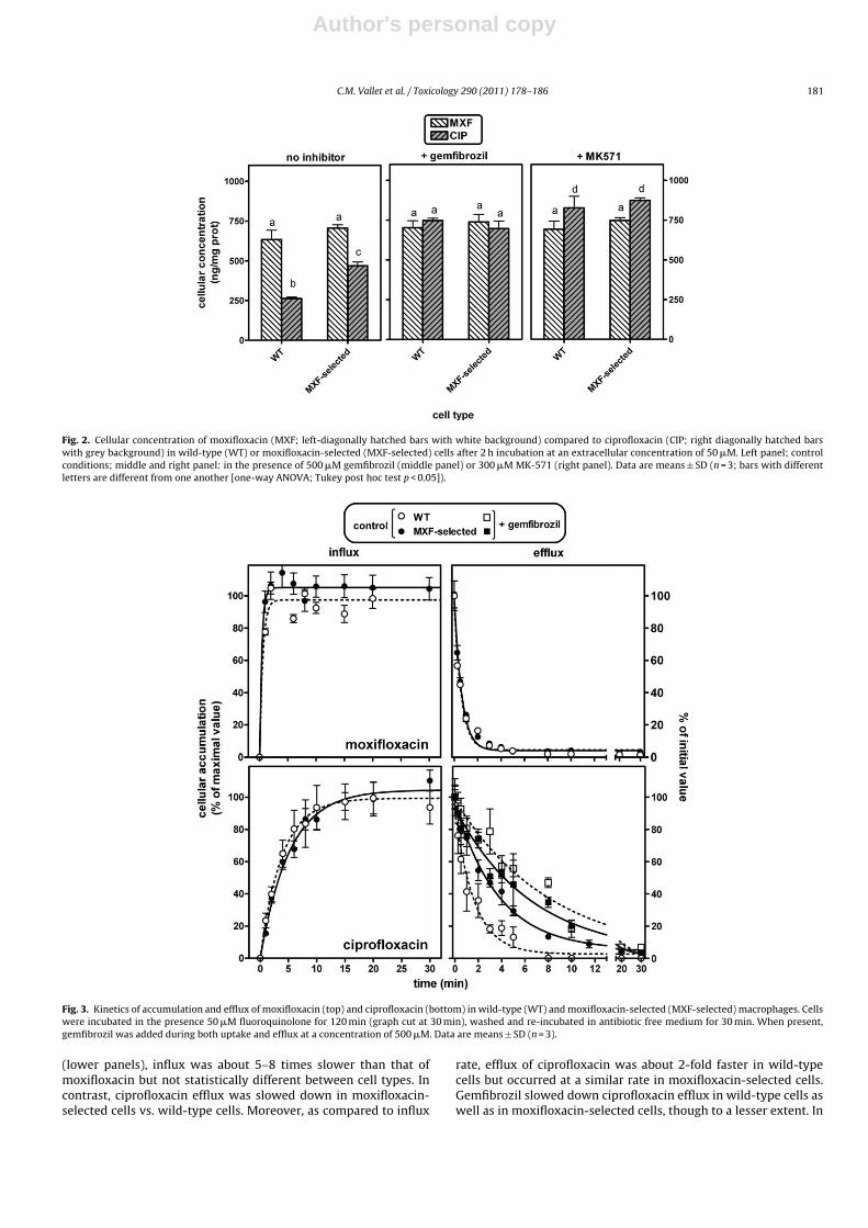
In a first approach, we compared the cellular accumulation ofmoxifloxacin and ciprofloxacin at equilibrium (120 min (Michotet al., 2005)) in moxifloxacin-selected cells vs. wild-type cells incontrol medium and after addition of gemfibrozil or MK-571 (usedas broad spectrum and specific inhibitors of Mrp efflux transporters,respectively (Marquez et al., 2009; Michot et al., 2005)). As shown inFig. 2, moxifloxacin accumulated to similar levels in both cell types,and this accumulation was not affected by the presence of the Mrp4inhibitors. In contrast, ciprofloxacin (i) accumulated about 3-timesless than moxifloxacin in wild-type cells, as previously described(Michot et al., 2005); (ii) showed an increased accumulation inmoxifloxacin-selected cells incubated in control medium, and (iii)reached the same, or a slightly higher level of accumulation thanmoxifloxacin, in the presence of gemfibrozil or of MK-571 in bothcell types.
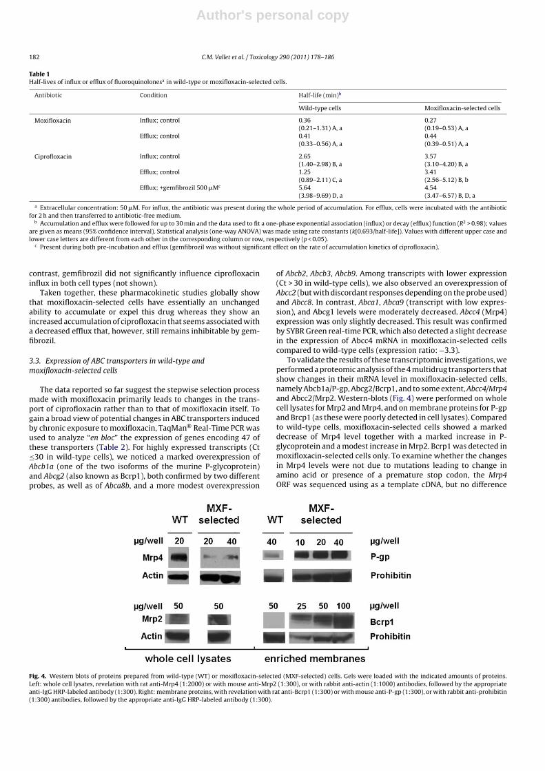
We then examined in details the kinetics of influx and effluxof moxifloxacin and ciprofloxacin in wild-type and moxifloxacin-selected cells. The data are illustrated in Fig. 3, with thecorresponding half-lives displayed in Table 1. For moxifloxacin(upper panels), influx and efflux were very fast with similar ratesand no significant difference between cell types. For ciprofloxacin
Author's personal copy
C.M. Vallet et al. / Toxicology 290 (2011) 178–186 181
Fig. 2. Cellular concentration of moxifloxacin (MXF; left-diagonally hatched bars with white background) compared to ciprofloxacin (CIP; right diagonally hatched barswith grey background) in wild-type (WT) or moxifloxacin-selected (MXF-selected) cells after 2 h incubation at an extracellular concentration of 50 �M. Left panel; controlconditions; middle and right panel: in the presence of 500 �M gemfibrozil (middle panel) or 300 �M MK-571 (right panel). Data are means ± SD (n = 3; bars with differentletters are different from one another [one-way ANOVA; Tukey post hoc test p < 0.05]).
Fig. 3. Kinetics of accumulation and efflux of moxifloxacin (top) and ciprofloxacin (bottom) in wild-type (WT) and moxifloxacin-selected (MXF-selected) macrophages. Cellswere incubated in the presence 50 �M fluoroquinolone for 120 min (graph cut at 30 min), washed and re-incubated in antibiotic free medium for 30 min. When present,gemfibrozil was added during both uptake and efflux at a concentration of 500 �M. Data are means ± SD (n = 3).
(lower panels), influx was about 5–8 times slower than that ofmoxifloxacin but not statistically different between cell types. Incontrast, ciprofloxacin efflux was slowed down in moxifloxacin-selected cells vs. wild-type cells. Moreover, as compared to influx
rate, efflux of ciprofloxacin was about 2-fold faster in wild-typecells but occurred at a similar rate in moxifloxacin-selected cells.Gemfibrozil slowed down ciprofloxacin efflux in wild-type cells aswell as in moxifloxacin-selected cells, though to a lesser extent. In
Author's personal copy
182 C.M. Vallet et al. / Toxicology 290 (2011) 178–186
Table 1Half-lives of influx or efflux of fluoroquinolonesa in wild-type or moxifloxacin-selected cells.
Antibiotic Condition Half-life (min)b
Wild-type cells Moxifloxacin-selected cells
Moxifloxacin Influx; control 0.36(0.21–1.31) A, a
0.27(0.19–0.53) A, a
Efflux; control 0.41(0.33–0.56) A, a
0.44(0.39–0.51) A, a
Ciprofloxacin Influx; control 2.65(1.40–2.98) B, a
3.57(3.10–4.20) B, a
Efflux; control 1.25(0.89–2.11) C, a
3.41(2.56–5.12) B, b
Efflux; +gemfibrozil 500 �Mc 5.64(3.98–9.69) D, a
4.54(3.47–6.57) B, D, a
a Extracellular concentration: 50 �M. For influx, the antibiotic was present during the whole period of accumulation. For efflux, cells were incubated with the antibioticfor 2 h and then transferred to antibiotic-free medium.
b Accumulation and efflux were followed for up to 30 min and the data used to fit a one-phase exponential association (influx) or decay (efflux) function (R2 > 0.98); valuesare given as means (95% confidence interval). Statistical analysis (one-way ANOVA) was made using rate constants (k[0.693/half-life]). Values with different upper case andlower case letters are different from each other in the corresponding column or row, respectively (p < 0.05).
c Present during both pre-incubation and efflux (gemfibrozil was without significant effect on the rate of accumulation kinetics of ciprofloxacin).
contrast, gemfibrozil did not significantly influence ciprofloxacininflux in both cell types (not shown).
Taken together, these pharmacokinetic studies globally showthat moxifloxacin-selected cells have essentially an unchangedability to accumulate or expel this drug whereas they show anincreased accumulation of ciprofloxacin that seems associated witha decreased efflux that, however, still remains inhibitable by gem-fibrozil.
3.3. Expression of ABC transporters in wild-type andmoxifloxacin-selected cells
The data reported so far suggest the stepwise selection processmade with moxifloxacin primarily leads to changes in the trans-port of ciprofloxacin rather than to that of moxifloxacin itself. Togain a broad view of potential changes in ABC transporters inducedby chronic exposure to moxifloxacin, TaqMan® Real-Time PCR wasused to analyze “en bloc” the expression of genes encoding 47 ofthese transporters (Table 2). For highly expressed transcripts (Ct≤30 in wild-type cells), we noticed a marked overexpression ofAbcb1a (one of the two isoforms of the murine P-glycoprotein)and Abcg2 (also known as Bcrp1), both confirmed by two differentprobes, as well as of Abca8b, and a more modest overexpression
of Abcb2, Abcb3, Abcb9. Among transcripts with lower expression(Ct > 30 in wild-type cells), we also observed an overexpression ofAbcc2 (but with discordant responses depending on the probe used)and Abcc8. In contrast, Abca1, Abca9 (transcript with low expres-sion), and Abcg1 levels were moderately decreased. Abcc4 (Mrp4)expression was only slightly decreased. This result was confirmedby SYBR Green real-time PCR, which also detected a slight decreasein the expression of Abcc4 mRNA in moxifloxacin-selected cellscompared to wild-type cells (expression ratio: −3.3).
To validate the results of these transcriptomic investigations, weperformed a proteomic analysis of the 4 multidrug transporters thatshow changes in their mRNA level in moxifloxacin-selected cells,namely Abcb1a/P-gp, Abcg2/Bcrp1, and to some extent, Abcc4/Mrp4and Abcc2/Mrp2. Western-blots (Fig. 4) were performed on wholecell lysates for Mrp2 and Mrp4, and on membrane proteins for P-gpand Brcp1 (as these were poorly detected in cell lysates). Comparedto wild-type cells, moxifloxacin-selected cells showed a markeddecrease of Mrp4 level together with a marked increase in P-glycoprotein and a modest increase in Mrp2. Bcrp1 was detected inmoxifloxacin-selected cells only. To examine whether the changesin Mrp4 levels were not due to mutations leading to change inamino acid or presence of a premature stop codon, the Mrp4ORF was sequenced using as a template cDNA, but no difference
Fig. 4. Western blots of proteins prepared from wild-type (WT) or moxifloxacin-selected (MXF-selected) cells. Gels were loaded with the indicated amounts of proteins.Left: whole cell lysates, revelation with rat anti-Mrp4 (1:2000) or with mouse anti-Mrp2 (1:300), or with rabbit anti-actin (1:1000) antibodies, followed by the appropriateanti-IgG HRP-labeled antibody (1:300). Right: membrane proteins, with revelation with rat anti-Bcrp1 (1:300) or with mouse anti-P-gp (1:300), or with rabbit anti-prohibitin(1:300) antibodies, followed by the appropriate anti-IgG HRP-labeled antibody (1:300).

Author's personal copy
C.M. Vallet et al. / Toxicology 290 (2011) 178–186 183
Table 2mRNA expression variations of ABC transporters in moxifloxacin-selected cells com-pared to wild-type macrophages.
Gene Expression ratio comparedto wild-type cellsa
Abca1 −3.51Abca2 −1.12Abca3 −1.56Abca4 ndAbca5 2.31Abca6 ndAbca7 −1.20Abca8a ndAbca8b 76.20Abca9 (−10.48)Abca13 2.02Abca14 ndAbca15 ndAbcb1a* 76.35
84.42Abcb1b 1.72Abcb2 4.30Abcb3 3.17Abcb4 2.48Abcb6 −1.22Abcb8 −1.25Abcb9 9.83Abcb10 1.43Abcb11 (1.19)Abcc1 −1.26Abcc2* (5.09)
(−1.26)Abcc3 1.09Abcc4 −1.82Abcc5 1.29Abcc6 ndAbcc7 ndAbcc8 (7.74)Abcc9 ndAbcc10 −1.40Abcc12 ndAbcd1 2.22Abcd2 1.79Abcd3 −1.51Abcd4 −2.03Abce1 −1.00Abcf2 1.21Abcf3 1.11Abcg1 −4.92Abcg2* 108.41
99.47Abcg3 ndAbcg4 (−1.07)
Data are the mean of duplicates from two biological samples. Genes with asteriskswere tested with two different probes (Table S2). Values with grey background:Ct < 30 (high expression) and significant variation (>3 or <−3); values with whitebackground: Ct < 30 (high expression but no significant variation); values withwhite background and in parenthesis: 30 < Ct < 35 (low expression); nd: Ct > 35 (nodetectable expression).
a See methods for calculations of this ratio; for a better understanding, a decreasein expression, corresponding to 0 < RQ < 1, is noted as a negative value, obtained by:−1/RQ.
was found between the sequence determined for wild-type andmoxifloxacin-selected cells.
3.4. Functionality of P-gp and Bcrp1 in wild-type andmoxifloxacin-selected cells
To test for the functionality of Abcb1a/P-gp and Abcg2/Bcrp1overexpressed in moxifloxacin-selected cells, we measured theaccumulation of their respective substrates (rhodamine 123and BODIPY-prazosin) in comparison with wild-type cells andin the absence and presence of their corresponding inhibitors
Fig. 5. Cellular accumulation of preferential substrates of P-glycoprotein or Bcrp1and influence of inhibitors in wild-type (WT) or moxifloxacin-selected (MXF-selected) cells. Left: cells were incubated during 2 h with 10 �M rhodamine 123in the absence (CT; control) or in the presence of 100 �M verapamil (+inhibitor).Right: cells were incubated during 1 h with 0.1 �M BODIPY-prazosin in the absence(CT; control) or in the presence of 10 �M Fumitremorgin C (+inhibitor). All valuesare expressed in percentage of the accumulation measured in control conditionsfor wild-type cells; they are the means of three independent determination ± SD.Statistical analysis (ANOVA, Tukey post hoc test): comparison between conditions:bars with different letters are different from one another (lower case, control condi-tions; upper case, +inhibitor; p < 0.05); comparison between control condition andinhibitor: *p < 0.05.
(verapamil and fumitremorgin C (Fontaine et al., 1996; Robey et al.,2001)). Fig. 5 shows that the accumulations of both rhodamine 123and BODIPY-prazosin (i) were significantly lower in moxifloxacin-selected cells compared to wild-type cells, and (ii) were increasedin the presence of the corresponding inhibitors in both cell types.We then examined whether P-gp and/or Bcrp1 could modulate theaccumulation of moxifloxacin or ciprofloxacin. As shown in Fig. 6,there was no significant increase in accumulation of either antibi-otic in moxifloxacin-selected cells upon addition of verapamil orfumitremorgin C. Combining these inhibitors with gemfibrozil didnot yield additional effect on ciprofloxacin accumulation comparedto what has been obtained with gemfibrozil alone. ATP depletionalso did not cause any significant change in moxifloxacin accu-mulation (whatever cell type tested) and did not yield any moreincrease in accumulation of ciprofloxacin than what was obtainedwith gemfibrozil.
4. Discussion
The present study demonstrates that the exposure of an eukary-otic cell line to increasing concentrations of moxifloxacin, a drugthat is apparently not effluxed by an ABC transporter, can never-theless cause profound modifications in the expression of severalof these transport proteins. These modifications have the potentialof affecting the transport of many drugs, and, as documented here,effectively increase the accumulation of another fluoroquinolone,ciprofloxacin. Most conspicuously, these changes do not seem tocontribute to the partial resistance of the cells to moxifloxacin itself.

Author's personal copy
184 C.M. Vallet et al. / Toxicology 290 (2011) 178–186
Fig. 6. Influence of preferential inhibitors of efflux transporters on ciprofloxacin and moxifloxacin accumulation. Wild-type (WT) or moxifloxacin-selected (MXF-selected)cells were incubated during 2 h with an extracellular concentration of 50 �M antibiotic alone (control, CT) or in the presence of 500 �M gemfibrozil (+GEM), 100 �M verapamil(+VER), 10 �M fumitremorgin C (+FTC) or combinations thereof, or in ATP-depleted cells. All values are expressed in percentage of the accumulation measured in controlconditions for wild-type cells; they are the means of three independent determination ± SD. Statistical analysis (ANOVA, Tukey post hoc test): comparison between conditions:bars with different letters are different from one another (p < 0.05).
The transport of fluoroquinolones in eukaryotic cells is highlydependent from the cell type, the species, and the moleculeexamined (Alvarez et al., 2008). Thus, P-glycoprotein has beenshown to transport grepafloxacin, sparfloxacin, danofloxacin, andmoxifloxacin (Brillault et al., 2009; Cormet-Boyaka et al., 1998;Lowes and Simmons, 2002; Schrickx and Fink-Gremmels, 2007)and MRP2, grepafloxacin and danofloxacin (Lowes and Simmons,2002; Schrickx and Fink-Gremmels, 2007) in human epithelialcells. BCRP/Bcrp1 is also involved in grepafloxacin, ciprofloxacin,norfloxacin, ofloxacin, and enrofloxacin transport in human andmice epithelial cells (Ando et al., 2007; Pulido et al., 2006). Inthe model used here (murine J774 macrophages), Mrp4 is themain transporter of ciprofloxacin, and to a lesser extent, of lev-ofloxacin and gemifloxacin (Marquez et al., 2009; Michot et al.,2005; Vallet et al., 2011) whereas the data reported earlier (andconfirmed here) indicate that moxifloxacin is not actively effluxed(no effect of ATP depletion or of the specific or broad spec-trum inhibitors used). We actually showed previously (Michotet al., 2005), and further document here, that uptake and effluxof moxifloxacin are much faster than those of ciprofloxacin, prob-ably because this molecule causes less disorder in the packingand ordering of lipid bilayers, as evidenced from model mem-branes made in comparison with ciprofloxacin (Bensikaddour et al.,2008).
As discussed earlier (Michot et al., 2005), and also observed forother drugs like quinidine and anthracyclines with P-glycoprotein(Eytan et al., 1996; Marbeuf-Gueye et al., 1999), rapid membranediffusion of a drug can make its active efflux functionally unde-tectable but does not rule out an interaction of the moleculewith the transporter. We, actually, know that moxifloxacin mayimpair the efflux of ciprofloxacin (Michot et al., 2005). Withthese considerations in mind, we may tentatively suggest thatthe drastic reduction in the expression of the Mrp4 ciprofloxacintransporter upon long-term exposure to moxifloxacin could berelated to the fact that moxifloxacin can interact with this
transporter. Because the effect is observed at the genomic level,however, we also need to postulate some sort of feed-back mech-anism. But, actually, such feed-back mechanism could be muchbroader, as we also see a pleiotropic modification in the expressionof several ABC transporters. First, 3 other multidrug transportersshown to play a role in fluoroquinolone transport in other models(Alvarez et al., 2008) are also overexpressed, namely P-gp/Abca1,Bcrp1/Abcg2, and Mrp2/Abcc2. In our model, this did not affectthe accumulation of ciprofloxacin probably because Mrp4 is largelypredominant in J774 macrophages (Marquez et al., 2009), so thatits markedly reduced expression in moxifloxacin-selected cellsincreases ciprofloxacin accumulation to a level that cannot becompensated by an increase in the expression of the other trans-porters. But many other ABC transporters were also affected intheir expression, which is reminiscent of what can be observedwith other toxic drugs. For instance, cells resistant to the P-gpsubstrate paclitaxel show a modulation of the expression of about700 genes, among which ABCB1 was the most upregulated (Yabukiet al., 2007). In the same line, doxorubicin-resistant cells over-express not only several genes directly implicated in their MDRphenotype (ABCB1, ABCC1 or ABCG2) but also many other genescoding for other ABC transporters (ABCA2, ABCC4, ABCC3, ABCC5,ABCB6, ABCF3, or ABCG1), depending on the cell line (Gillet etal., 2004). Beside multidrug transporters, many other ABC pro-teins show a modified expression level in moxifloxacin-selectedcells. The potential role of these changes needs to be furtherexplored. It is, nevertheless, tempting to speculate that these aresigns of global adaptation of the cells to what seems to be a majortoxic stress. The increased expression of Abcb2 (Tap1) and Abcb3(Tap2), involved in antigen presentation and immune response(Herget and Tampe, 2007), of Abcb9, a lysosomal ABC trans-porter associated with antigen processing-like processes (Zhanget al., 2000), of Abca8b (encoding a close homolog of the humanABCA8 (Annilo et al., 2003) able to transport leukotriene C4 andestradiol-beta-glucuronide (Tsuruoka et al., 2002), and of Abcc8

Author's personal copy
C.M. Vallet et al. / Toxicology 290 (2011) 178–186 185
that codes for a protein (SUR1) acting as a K+ selective pore(Bryan et al., 2007) may require attention. The reduced expres-sion of Abca9, Abca1, and Abcg1, involved in lipid homeostasisin macrophages (Piehler et al., 2002), and cholesterol and phos-pholipid transport (Wang et al., 2007) may also be a cause ofconcern.
We have no simple explanation for all these numerous changes,especially if considering that the toxicity of fluoroquinolonestowards eukaryotic cells is thought to be primarily due to theirability to impair topoisomerase activity (at larger concentrations,however, than in prokaryotic cells). There is no evidence for mox-ifloxacin being more potent than ciprofloxacin in this context(Perrone et al., 2002; Reuveni et al., 2008). Yet, as the methodused to select cells is prone to select multifactorial resistancemechanisms (Gottesman et al., 1998), further studies using globalapproaches to evaluate the proteome or the transcriptome wouldneed to be performed to further characterize the resistance pheno-type of moxifloxacin-selected cells. We can neither exclude that thediversity of changes observed arise from the fact that our mode ofselection was not clonal, so that the profile of alterations observedcould be the resultant of more targeted modifications occurringin individual cells. Although possibly complicating data interpre-tation, this mode of selection however better reflects what couldoccur in vivo under drug pressure. At this stage, our data thus clearlyillustrate that long-term exposure to close-to-toxic concentrationsof drug that is apparently not affected by efflux nevertheless causescomplexes changes in the expression of transporters that can trig-ger drug interactions and other potential undesired effects.
The pharmacological consequences of our observations alsoneed to be discussed. Because of their ability to accumulate insidecells and their bactericidal character, fluoroquinolones are amongthe most active antibiotics against intracellular infections (see forreview (Van Bambeke et al., 2006)). We previously showed thatthe activity of ciprofloxacin against intracellular L. monocytogeneswas concentration-dependent, and therefore affected by the levelof expression of Mrp4 (drastic reduction of activity in ciprofloxacin-selected J774 macrophages that are characterized by an increasedexpression of Mrp4 (Michot et al., 2006), but increased activityin wild-type J774 macrophages in the presence of inhibitors ofMrps (Seral et al., 2003)). In a broader context, this means that anycircumstance that could affect the cellular accumulation of fluoro-quinolones, including a change in the expression of the transportersresponsible for their efflux, could also modify their intracellu-lar activity. Yet moxifloxacin should escape this rule and remainequally active, being not affected by the level of expression of effluxtransporters.
These effects reported here were obtained for cells exposed tomoxifloxacin concentrations typically 5–20-fold larger than thosecommonly observed in the serum of patients (∼1.5 to ∼6 mg/l (Stassand Kubitza, 1999)) treated with the approved dose of 400 mg/dayfor its current indications (Anonymous, 2008), and these treat-ments are usually of short duration (5–21 days). However, (i)larger concentrations are reached in specific organs such as theurinary tract where concentrations may be 10-fold larger thanserum concentrations (Wagenlehner et al., 2008), and (ii) doses upto 800 mg/day and longer treatment durations are being consid-ered (but not registered) for treatment of tuberculosis (Ginsberg,2010). This may trigger further animal and human investigations toexamine whether the present in vitro data translate into significantmodifications in vivo that could limit the use of moxifloxacin forthis indication.
Conflict of interest
None.
Funding source
This study was supported by the Belgian Fonds de la RechercheScientifique Médicale [grants 3.4.597.06 and 3.4.583.08], the BelgianFonds de la Recherche Scientifique [grant 1.5.195.07], the BelgianFederal Science Policy Office [Research projects P5/33, P6/19], anda grant-in-aid from the French Association Mucoviscidose: ABCF2.C.M.V. is Boursier of the Belgian Fonds pour la Recherche dansl’Industrie et l’Agriculture (F.R.I.A.), B.M. was post-doctoral fellowof the First post-doc program of the Belgian Région wallonne, N.N.was supported by a Post-Doctoral Grant from ICSN, and F.V.B. isMaître de Recherches of the Belgian Fonds de la Recherche Scientifique(F.R.S.-FNRS).
Acknowledgments
We thank R. Robey (National Institutes of Health, Bethesda, MA)for the kind gift of fumitremorgin C and M.C. Cambier for skillfultechnical assistance.
Appendix A. Supplementary data
Supplementary data associated with this article can be found, inthe online version, at doi:10.1016/j.tox.2011.09.003.
References
Alvarez, A.I., Perez, M., Prieto, J.G., Molina, A.J., Real, R., Merino, G., 2008. Fluoro-quinolone efflux mediated by ABC transporters. J. Pharm. Sci. 97, 3483–3493.
Ando, T., Kusuhara, H., Merino, G., Alvarez, A.I., Schinkel, A.H., Sugiyama, Y., 2007.Involvement of breast cancer resistance protein (ABCG2) in the biliary excretionmechanism of fluoroquinolones. Drug Metab. Dispos. 35, 1873–1879.
Annilo, T., Chen, Z.Q., Shulenin, S., Dean, M., 2003. Evolutionary analysis of a clusterof ATP-binding cassette (ABC) genes. Mamm. Genome 14, 7–20.
Anonymous, 2008. Avelox Package insert. http://www.univgraph.com/bayer/inserts/avelox.pdf. 11.11.2008.
Baguley, B.C., 2010. Multiple drug resistance mechanisms in cancer. Mol. Biotechnol.46, 308–316.
Bensikaddour, H., Fa, N., Burton, I., Deleu, M., Lins, L., Schanck, A., Brasseur, R.,Dufrene, Y.F., Goormaghtigh, E., Mingeot-Leclercq, M.P., 2008. Characterizationof the interactions between fluoroquinolone antibiotics and lipids: a multitech-nique approach. Biophys. J. 94, 3035–3046.
Brillault, J., De Castro, W.V., Harnois, T., Kitzis, A., Olivier, J.C., Couet, W., 2009. P-glycoprotein-mediated transport of moxifloxacin in a Calu-3 lung epithelial cellmodel. Antimicrob. Agents Chemother. 53, 1457–1462.
Bryan, J., Munoz, A., Zhang, X., Dufer, M., Drews, G., Krippeit-Drews, P., Aguilar-Bryan,L., 2007. ABCC8 and ABCC9: ABC transporters that regulate K+ channels. PflugersArch. 453, 703–718.
Cormet-Boyaka, E., Huneau, J.F., Mordrelle, A., Boyaka, P.N., Carbon, C., Rubinstein,E., Tome, D., 1998. Secretion of sparfloxacin from the human intestinal Caco-2cell line is altered by P-glycoprotein inhibitors. Antimicrob. Agents Chemother.42, 2607–2611.
Eckford, P.D., Sharom, F.J., 2009. ABC efflux pump-based resistance to chemotherapydrugs. Chem. Rev. 109, 2989–3011.
Eytan, G.D., Regev, R., Oren, G., Assaraf, Y.G., 1996. The role of passive transbilayerdrug movement in multidrug resistance and its modulation. J. Biol. Chem. 271,12897–12902.
Fardel, O., Payen, L., Courtois, A., Vernhet, L., Lecureur, V., 2001. Regulation of bil-iary drug efflux pump expression by hormones and xenobiotics. Toxicology 167,37–46.
Fontaine, M., Elmquist, W.F., Miller, D.W., 1996. Use of rhodamine 123 to examinethe functional activity of P-glycoprotein in primary cultured brain microvesselendothelial cell monolayers. Life Sci. 59, 1521–1531.
Gillet, J.P., Efferth, T., Remacle, J., 2007. Chemotherapy-induced resistance by ATP-binding cassette transporter genes. Biochim. Biophys. Acta 1775, 237–262.
Gillet, J.P., Efferth, T., Steinbach, D., Hamels, J., de Longueville, F., Bertholet, V.,Remacle, J., 2004. Microarray-based detection of multidrug resistance in humantumor cells by expression profiling of ATP-binding cassette transporter genes.Cancer Res. 64, 8987–8993.
Ginsberg, A.M., 2010. Drugs in development for tuberculosis. Drugs 70, 2201–2214.Gottesman, M.M., Cardarelli, C., Goldenberg, S., Licht, T., Pastan, I., 1998. Selection
and maintenance of multidrug-resistant cells. Methods Enzymol. 292, 248–258.Herget, M., Tampe, R., 2007. Intracellular peptide transporters in
human–compartmentalization of the “peptidome”. Pflugers Arch. 453,591–600.
Huggett, J., Dheda, K., Bustin, S., Zumla, A., 2005. Real-time RT-PCR normalisation;strategies and considerations. Genes Immun. 6, 279–284.

Author's personal copy
186 C.M. Vallet et al. / Toxicology 290 (2011) 178–186
Klaassen, C.D., Aleksunes, L.M., 2010. Xenobiotic, bile acid, and cholesterol trans-porters: function and regulation. Pharmacol. Rev. 62, 1–96.
Lowes, S., Simmons, N.L., 2002. Multiple pathways for fluoroquinolone secretion byhuman intestinal epithelial (Caco-2) cells. Br. J. Pharmacol. 135, 1263–1275.
Lowry, O.H., Rosebrough, N.J., Farr, A.L., Randall, R.J., 1951. Protein measurementwith the Folin phenol reagent. J. Biol. Chem. 193, 265–275.
Marbeuf-Gueye, C., Ettori, D., Priebe, W., Kozlowski, H., Garnier-Suillerot, A., 1999.Correlation between the kinetics of anthracycline uptake and the resistancefactor in cancer cells expressing the multidrug resistance protein or the P-glycoprotein. Biochim. Biophys. Acta 1450, 374–384.
Marquez, B., Caceres, N.E., Mingeot-Leclercq, M.P., Tulkens, P.M., Van Bambeke, F.,2009. Identification of the efflux transporter of the fluoroquinolone antibioticciprofloxacin in murine macrophages: studies with ciprofloxacin-resistant cells.Antimicrob. Agents Chemother. 53, 2410–2416.
Michot, J.M., Heremans, M.F., Caceres, N.E., Mingeot-Leclercq, M.P., Tulkens, P.M.,Van Bambeke, F., 2006. Cellular accumulation and activity of quinolones inciprofloxacin-resistant J774 macrophages. Antimicrob. Agents Chemother. 50,1689–1695.
Michot, J.M., Seral, C., Van Bambeke, F., Mingeot-Leclercq, M.P., Tulkens, P.M.,2005. Influence of efflux transporters on the accumulation and efflux of fourquinolones (ciprofloxacin, levofloxacin, garenoxacin, and moxifloxacin) in J774macrophages. Antimicrob. Agents Chemother. 49, 2429–2437.
Michot, J.M., Van Bambeke, F., Mingeot-Leclercq, M.P., Tulkens, P.M., 2004. Activeefflux of ciprofloxacin from J774 macrophages through an MRP-like transporter.Antimicrob. Agents Chemother. 48, 2673–2682.
Perrone, C.E., Takahashi, K.C., Williams, G.M., 2002. Inhibition of human topoiso-merase IIalpha by fluoroquinolones and ultraviolet A irradiation. Toxicol. Sci.69, 16–22.
Pfaffl, M.W., 2001. A new mathematical model for relative quantification in real-timeRT-PCR. Nucleic Acids Res. 29, e45.
Piehler, A., Kaminski, W.E., Wenzel, J.J., Langmann, T., Schmitz, G., 2002. Molecularstructure of a novel cholesterol-responsive A subclass ABC transporter, ABCA9.Biochem. Biophys. Res. Commun. 295, 408–416.
Pulido, M.M., Molina, A.J., Merino, G., Mendoza, G., Prieto, J.G., Alvarez, A.I., 2006.Interaction of enrofloxacin with breast cancer resistance protein (BCRP/ABCG2):influence of flavonoids and role in milk secretion in sheep. J. Vet. Pharmacol.Ther. 29, 279–287.
Reuveni, D., Halperin, D., Shalit, I., Priel, E., Fabian, I., 2008. Quinolones as enhancersof camptothecin-induced cytotoxic and anti-topoisomerase I effects. Biochem.Pharmacol. 75, 1272–1281.
Robey, R.W., Honjo, Y., van de, L.A., Miyake, K., Regis, J.T., Litman, T., Bates, S.E., 2001.A functional assay for detection of the mitoxantrone resistance protein, MXR(ABCG2). Biochim. Biophys. Acta 1512, 171–182.
Schrickx, J.A., Fink-Gremmels, J., 2007. Danofloxacin-mesylate is a substrate for ATP-dependent efflux transporters. Br. J. Pharmacol. 150, 463–469.
Scotto, K.W., 2003. Transcriptional regulation of ABC drug transporters. Oncogene22, 7496–7511.
Seral, C., Carryn, S., Tulkens, P.M., Van Bambeke, F., 2003. Influence of P-glycoproteinand MRP efflux pump inhibitors on the intracellular activity of azithromycin andciprofloxacin in macrophages infected by Listeria monocytogenes or Staphylococ-cus aureus. J. Antimicrob. Chemother. 51, 1167–1173.
Stass, H., Kubitza, D., 1999. Pharmacokinetics and elimination of moxifloxacin afteroral and intravenous administration in man. J. Antimicrob. Chemother. 43(Suppl. B), 83–90.
Takara, K., Tsujimoto, M., Kokufu, M., Ohnishi, N., Yokoyama, T., 2003. Up-regulationof MDR1 function and expression by cisplatin in LLC-PK1 cells. Biol. Pharm. Bull.26, 205–209.
Tsuruoka, S., Ishibashi, K., Yamamoto, H., Wakaumi, M., Suzuki, M., Schwartz, G.J.,Imai, M., Fujimura, A., 2002. Functional analysis of ABCA8, a new drug trans-porter. Biochem. Biophys. Res. Commun. 298, 41–45.
Turk, D., Hall, M.D., Chu, B.F., Ludwig, J.A., Fales, H.M., Gottesman, M.M., Szakacs, G.,2009. Identification of compounds selectively killing multidrug-resistant cancercells. Cancer Res. 69, 8293–8301.
Vallet, C.M., Marquez, B., Ngabirano, E., Lemaire, S., Mingeot-Leclercq, M.P., Tulkens,P.M., Van Bambeke, F., 2011. Cellular accumulation of fluoroquinolones is notpredictive of their intracellular activity: studies with gemifloxacin, moxifloxacinand ciprofloxacin in a pharmacokinetic/pharmacodynamic model of uninfectedand infected macrophages. Int. J. Antimicrob. Agents 38, 249–256.
Van Bambeke, F., Barcia-Macay, M., Lemaire, S., Tulkens, P.M., 2006. Cellularpharmacodynamics and pharmacokinetics of antibiotics: current views and per-spectives. Curr. Opin. Drug Discov. Devel. 9, 218–230.
Wagenlehner, F.M., Kees, F., Weidner, W., Wagenlehner, C., Naber, K.G., 2008. Con-centrations of moxifloxacin in plasma and urine, and penetration into prostaticfluid and ejaculate, following single oral administration of 400 mg to healthyvolunteers. Int. J. Antimicrob. Agents 31, 21–26.
Wang, X., Collins, H.L., Ranalletta, M., Fuki, I.V., Billheimer, J.T., Rothblat, G.H., Tall,A.R., Rader, D.J., 2007. Macrophage ABCA1 and ABCG1, but not SR-BI, promotemacrophage reverse cholesterol transport in vivo. J. Clin. Invest. 117, 2216–2224.
Xu, C., Li, C.Y., Kong, A.N., 2005. Induction of phase I, II and III drugmetabolism/transport by xenobiotics. Arch. Pharm. Res. 28, 249–268.
Yabuki, N., Sakata, K., Yamasaki, T., Terashima, H., Mio, T., Miyazaki, Y., Fujii, T., Kitada,K., 2007. Gene amplification and expression in lung cancer cells with acquiredpaclitaxel resistance. Cancer Genet. Cytogenet. 173, 1–9.
Zhang, F., Zhang, W., Liu, L., Fisher, C.L., Hui, D., Childs, S., Dorovini-Zis, K., Ling,V., 2000. Characterization of ABCB9, an ATP binding cassette protein associatedwith lysosomes. J. Biol. Chem. 275, 23287–23294.

Vallet et al. Toxicology 290 (2011) 178–186 – Supplementary Material -- Page 1 of 3
Vallet et al. Modulation of the expression of ABC transporters in murine (J774) macrophages exposed to large concentrations of the fluoroquinolone antibiotic moxifloxacin Toxicology 290 (2011) 178–186
Supplementary Material
Table SP1: Sequence of the primers used for sequencing of the Mrp4 ORF, temperature
for PCR amplification and size of the corresponding amplicons
Amplicon Forward primer (5’-3’) Reverse primer (5’-3’) Ta (°C) for PCR reaction
Amplicon size (pb)
4-1 GTGCACACCGAGGTGAAAC TCGTCGGGGTCATACTTCTC 66 371
4-2 GGGCACTCGAGTAGTTCAGC GCCAGGCAGGAGATTCCTAT 66 408
4-3 GGTAACCGTCCTCCTCTGG CACAAACACGTGGCTAGCTG 60 388
4-4 TCGCAAACAAAGTCATCCTG ACCACGGCTAACAACTCACC 66 355
4-5 AACCCTGCAAGGTCTTTCCT GCGGATCATCAAGGAGGTAG 66 407
4-6 AGTGGAGGCCAGAAAGCTC CTGGATGACTGCTGAGACCA 66 392
4-7 CGACACTCAGGAAACGAACC CTCGCTATGCCAAAAAGGAC 60 406
4-8 CCAGAAATGCGAATGGAAAT AGCGGAACCAATGGTATGAG 60 370
4-9 TGCTCCTCGTCGTAAGTGTG GCCCAGCATTCAAAGTCTTC 60 393
4-10 CATCTGCGCCATCTTTGTAA CCAACCTTTTCCCTGGACTT 60 354
4-11 TGTCAGGAGCTGTTTGATGC CCCAATTTCGGTTGTCAAGA 60 561
4-12 GGGGAAAATCTGGATCGATAA TTGCAAGGCACACTAACTGTCT 60 260
4-13 GGATCCAATTTCAGTGTTGGA CGGGGCTGGTGAATGTAATA 60 397

Vallet et al. Toxicology 290 (2011) 178–186 – Supplementary Material -- Page 2 of 3
Table SP2: TaqMan® Gene Expression Assays references Gene Assay ID Gene Assay ID Abca1 Mm00442646_m1 Abcc10 Mm00467403_m1 Abca2 Mm00431553_m1 Abcc12 Mm00556685_m1 Abca3 Mm00550501_m1 Abcd1 Mm00431749_m1 Abca4 Mm00492004_m1 Abcd2 Mm00496455_m1 Abca5 Mm00461656_m1 Abcd3 Mm00436150_m1 Abca6 Mm00461636_m1 Abcd4 Mm00436180_m1 Abca7 Mm00497010_m1 Abce1 Mm00649858_m1 Abca8a Mm00462440_m1 Abcf2 Mm00457400_g1 Abca8b Mm00457361_m1 Abcf3 Mm00658695_m1 Abca9 Mm00461704_m1 Abcg1 Mm00437390_m1 Abca13 Mm00626523_m1 Abcg2 Mm00496364_m1 Abca14 Mm00509570_m1 Abcg2 Mm01181554_m1 Abca15 Mm00623451_m1 Abcg3 Mm00446072_m1 Abcb1a Mm00440761_m1 Abcg4 Mm00507250_m1 Abcb1a Mm01324136_m1 Abcg5 Mm00446249_m1 Abcb1b Mm00440736_m1 Abcg8 Mm00445970_m1 Abcb2 Mm00443188_m1 18S Hs99999901_s1 Abcb3 Mm00441668_m1 Aamp Mm00525080_m1 Abcb4 Mm00435630_m1 Actb Mm00607939_s1 Abcb6 Mm00470049_m1 B2m Mm00437762_m1 Abcb8 Mm00472410_m1 Gapdh Mm99999915_g1 Abcb9 Mm00498197_m1 Gusb Mm00446953_m1 Abcb10 Mm00497927_m1 Hmbs Mm00660262_g1 Abcb11 Mm00445168_m1 Hprt1 Mm00446968_m1 Abcc1 Mm00456156_m1 Ipo8 Mm01255158_m1 Abcc2 Mm00496883_m1 Pgk1 Mm00435617_m1 Abcc2 Mm00496899_m1 Polr2a Mm00839493_m1 Abcc3 Mm00551550_m1 Ppia Mm02342430_g1 Abcc4 Mm01226381_m1 Rplp2 Mm00782638_s1 Abcc5 Mm00443360_m1 Tbp Mm00446973_m1 Abcc6 Mm00497685_m1 Tfrc Mm00441941_m1 Abcc7 Mm00445197_m1 Ubc Mm01201237_m1 Abcc8 Mm00803450_m1 Ywhaz Mm01158417_g1 Abcc9 Mm00441638_m1

Vallet et al. Toxicology 290 (2011) 178–186 – Supplementary Material -- Page 3 of 3
Fig. SP1: Structural formulae of moxifloxacin and ciprofloxacin.
N N
F
OCH3
O
COOH
HN
H
H
HN
N N
F
O
COOH
Moxifloxacin:
7-[(4aS,7aS)-1,2,3,4,4a,5,7,7a-octahydropyrrolo[3,4-b]pyridin-6-yl]-1-cyclopropyl-6-fluoro-8-methoxy-4-oxoquinoline-3-carboxylic acid
Ciprofloxacin:
1-cyclopropyl-6-fluoro-4-oxo-7-piperazin-1-ylquinoline- 3-carboxylic acid