author: michael shea, m.d., 2008 license: unless otherwise noted, this material is made available...
TRANSCRIPT
Author: Michael Shea, M.D., 2008
License: Unless otherwise noted, this material is made available under the terms of the Creative Commons Attribution – Share Alike 3.0 License: http://creativecommons.org/licenses/by-sa/3.0/
We have reviewed this material in accordance with U.S. Copyright Law and have tried to maximize your ability to use, share, and adapt it. The citation key on the following slide provides information about how you may share and adapt this material.
Copyright holders of content included in this material should contact [email protected] with any questions, corrections, or clarification regarding the use of content.
For more information about how to cite these materials visit http://open.umich.edu/education/about/terms-of-use.
Any medical information in this material is intended to inform and educate and is not a tool for self-diagnosis or a replacement for medical evaluation, advice, diagnosis or treatment by a healthcare professional. Please speak to your physician if you have questions about your medical condition.
Viewer discretion is advised: Some medical content is graphic and may not be suitable for all viewers.

Citation Keyfor more information see: http://open.umich.edu/wiki/CitationPolicy
Use + Share + Adapt
Make Your Own Assessment
Creative Commons – Attribution License
Creative Commons – Attribution Share Alike License
Creative Commons – Attribution Noncommercial License
Creative Commons – Attribution Noncommercial Share Alike License
GNU – Free Documentation License
Creative Commons – Zero Waiver
Public Domain – Ineligible: Works that are ineligible for copyright protection in the U.S. (17 USC § 102(b)) *laws in your jurisdiction may differ
Public Domain – Expired: Works that are no longer protected due to an expired copyright term.
Public Domain – Government: Works that are produced by the U.S. Government. (17 USC §105)
Public Domain – Self Dedicated: Works that a copyright holder has dedicated to the public domain.
Fair Use: Use of works that is determined to be Fair consistent with the U.S. Copyright Act. (17 USC § 107) *laws in your jurisdiction may differ
Our determination DOES NOT mean that all uses of this 3rd-party content are Fair Uses and we DO NOT guarantee that your use of the content is Fair.
To use this content you should do your own independent analysis to determine whether or not your use will be Fair.
{ Content the copyright holder, author, or law permits you to use, share and adapt. }
{ Content Open.Michigan believes can be used, shared, and adapted because it is ineligible for copyright. }
{ Content Open.Michigan has used under a Fair Use determination. }
Mitral Valve Disease
Michael Shea, MD
Fall 2008
Lecture Outline
• Etiology
• Pathophysiology
• Clinical features
• Diagnostic testing
• Differential diagnosis
• Management
Mitral Stenosis Mitral Regurgitation

Mitral Stenosis: Pathophysiology
Etiology: rheumatic; female>male by 6:1
Mitral leaflets:
• Large anterior is contiguous to aorta
• Smaller posterior is contiguous to left atrial endocardium
• Normal area: 4-5cm2
Mitral Stenosis: Pathophysiology
• Fundamental problem: Inability to get blood from left atrium left ventricle
• Stenotic process:
– Scarring and fibrosis of leaflets and chordae tendineae
– Commissural fusion
– Leads to funnel-shaped orifice and pressure gradient across valve

Mitral Stenosis: Pathophysiology
Source Undetermined
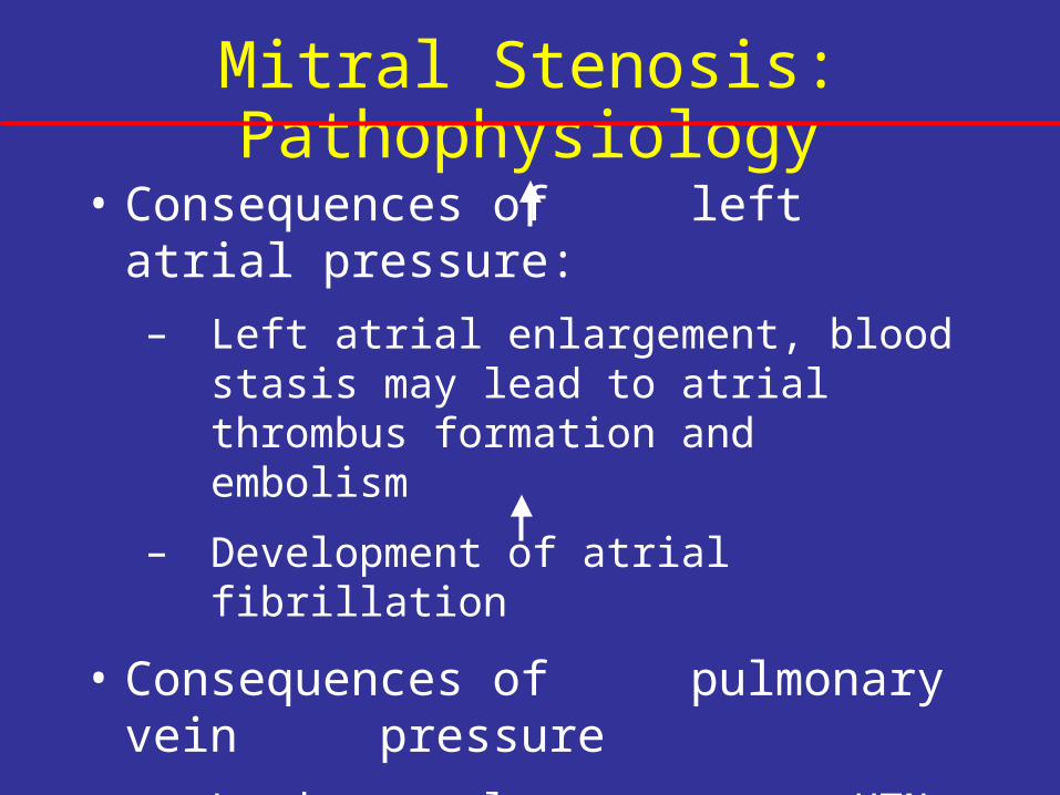
Mitral Stenosis: Pathophysiology
• Consequences of left atrial pressure:
– Left atrial enlargement, blood stasis may lead to atrial thrombus formation and embolism
– Development of atrial fibrillation
• Consequences of pulmonary vein pressure
– Leads to pulmonary artery HTN
– Then RV hypertrophy and dilation
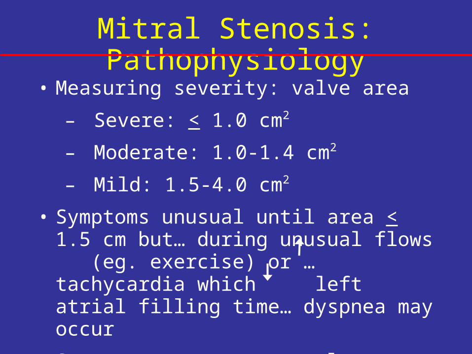
Mitral Stenosis: Pathophysiology
• Measuring severity: valve area
– Severe: < 1.0 cm2
– Moderate: 1.0-1.4 cm2
– Mild: 1.5-4.0 cm2
• Symptoms unusual until area < 1.5 cm but… during unusual flows (eg. exercise) or …tachycardia which left atrial filling time… dyspnea may occur
• Symptoms progress as valve narrows

Mitral Stenosis: Clinical FeaturesMitral Stenosis: Clinical Features
History
• Long course before sx onset
• Sx worsen with onset of atrial fibrillation
• Typically asx then dyspnea with marked effort then minimal effort then orthopnea, paroxysmal nocturnal dyspnea
Mitral Stenosis: Clinical FeaturesMitral Stenosis: Clinical Features
History
• Fatigue is common patient cannot
augment cardiac output
• Hemoptysis
• Embolic stroke…. usually when
patient is in atrial fibrillation
Mitral Stenosis: Clinical FeaturesMitral Stenosis: Clinical Features
Physical exam:
• Palpation – may be a parasternal lift (RV)
• Auscultation:
1. Accentuated first heart sound (S1)
2. Opening snap sudden stop in leaflet opening
3. Diastolic rumble
Higher left atrial Po, shorter S2 to OS interval
Mitral Stenosis: Clinical Features
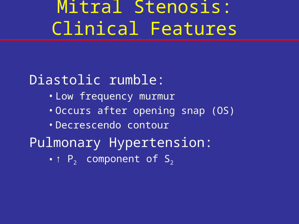
Diastolic rumble:• Low frequency murmur• Occurs after opening snap (OS)• Decrescendo contour
Pulmonary Hypertension:• ↑ P2 component of S2

Source Undetermined
Mitral Stenosis
Diagnostic testing
• Chest radiology
• Electrocardiography
• Echocardiography
• Cardiac catheterization
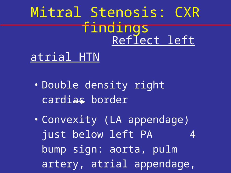
Mitral Stenosis: CXR findings
Reflect left atrial HTN
• Double density right cardiac border
• Convexity (LA appendage) just below
left PA 4 bump sign: aorta, pulm
artery, atrial appendage, left ventricle
• Elevated left main bronchus
• Kerley lines

Source Undetermined

Source Undetermined
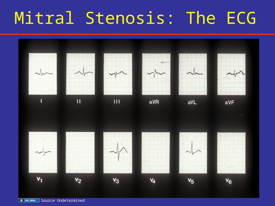
Mitral Stenosis: The ECG
Source Undetermined

Mitral Stenosis
Diagnostic testing
• Chest radiology
• Electrocardiography
• Echocardiography
• Cardiac catheterization

Echocardiography: Parasternal
Normal Mitral Stenosis
Source Undetermined Source Undetermined

Echocardiography: Short Axis
Normal Mitral Stenosis
Source Undetermined Source Undetermined

Mitral Stenosis: Clinical Manifestations and Diagnosis
Mitral Stenosis: Clinical Manifestations and Diagnosis
• Echo: 2D images
– Increased LA size
– Doming of valve leaflets
– Valve stenosis
– Valve area can be planimetered
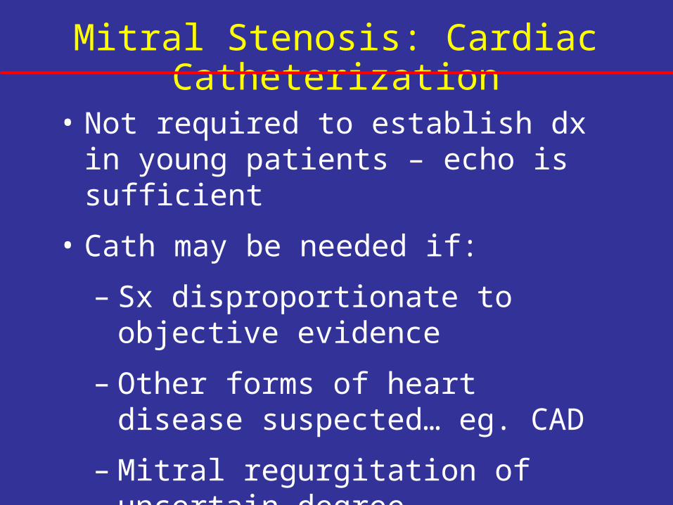
Mitral Stenosis: Cardiac Catheterization
• Not required to establish dx in young patients – echo is sufficient
• Cath may be needed if:
– Sx disproportionate to objective evidence
– Other forms of heart disease suspected… eg. CAD
– Mitral regurgitation of uncertain degree

Mitral Stenosis
• Atrial myxoma
• Cor triatriatum
• Congenital mitral stenosis
Differential Diagnosis
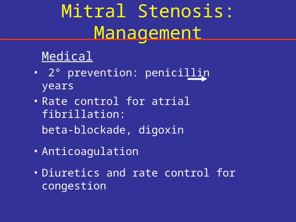
Mitral Stenosis: Management
Medical • 2° prevention: penicillin years
• Rate control for atrial fibrillation:
beta-blockade, digoxin
• Anticoagulation
• Diuretics and rate control for congestion

Mitral Stenosis
• Closed surgical commissurotomy
• Open surgical commissurotomy
• Valve replacement
• Balloon mitral commissurotomy
Mechanical Relief

Source Undetermined

Source Undetermined

Mitral Regurgitation

Mitral Regurgitation: Etiology
Mitral annulus
- Annular calcification
Leaflets
- Myxomatous degeneration
- Rheumatic disease
- Endocarditis
- SAM (hypertrophic cardiomyopathy)
Chordae tendineae
-Rupture (idiopathic)
- Endocarditis
Papillary muscles
- Dysfunction or rupture
Left ventricle
- Cavity dilatation
Schematic representation of
mitral valve pathologies
removed
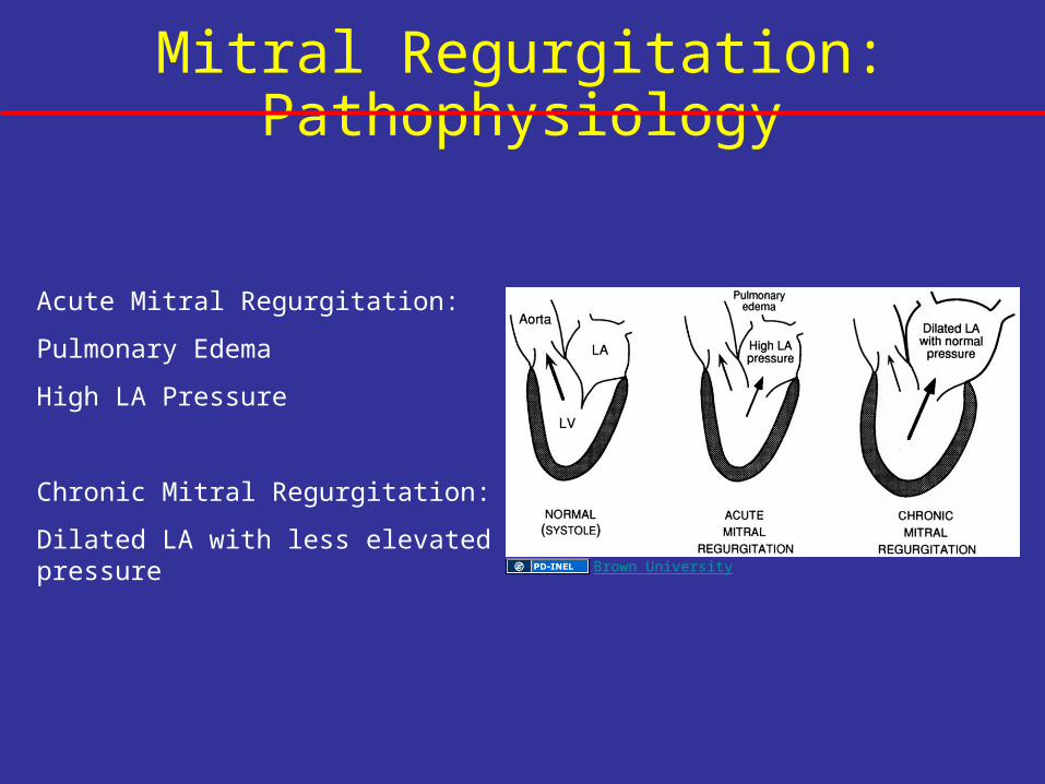
Mitral Regurgitation: Pathophysiology
Acute Mitral Regurgitation:
Pulmonary Edema
High LA Pressure
Chronic Mitral Regurgitation:
Dilated LA with less elevated pressureBrown University
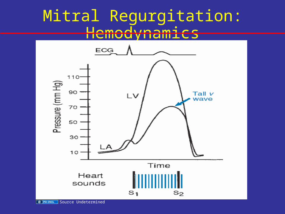
Mitral Regurgitation: Hemodynamics
Source Undetermined

Mitral Regurgitation: Pathophysiology
• May be acute or chronic
• Chronic MR:
– Total stroke volume increases
– Blood LA to offload LV
– LV enlarges (ventricular remodeling)

Mitral Regurgitation: Pathophysiology
Brown University

Mitral Regurgitation: Clinical FeaturesMitral Regurgitation: Clinical Features
• Mild MR no sx• When sx occur
– Fatigue – Dyspnea
• Physical Exam:• Lateral; dynamic LV apex beat• Often diminished S1 (leaflets don’t
coapt); S3 often present• Apical systolic murmur• Holosystolic murmur to axilla

Mitral Regurgitation: Auscultation
Source Undetermined

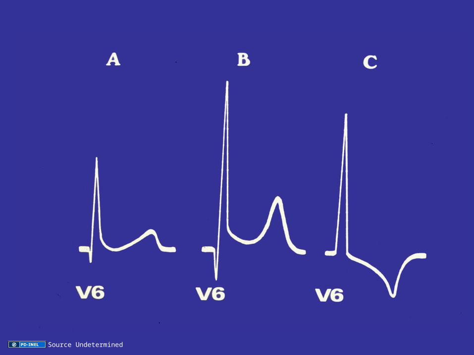
Mitral Regurgitation: Diagnostic TestsMitral Regurgitation: Diagnostic Tests
• CXR: LA and LV enlargement
• ECG: Normal initially…then shows LV hypertrophy
• Echo:
– LAE
– LV enlargement
– Doppler and color flow allow semi-quantitative estimate (1-4+)

Source Undetermined
Source Undetermined
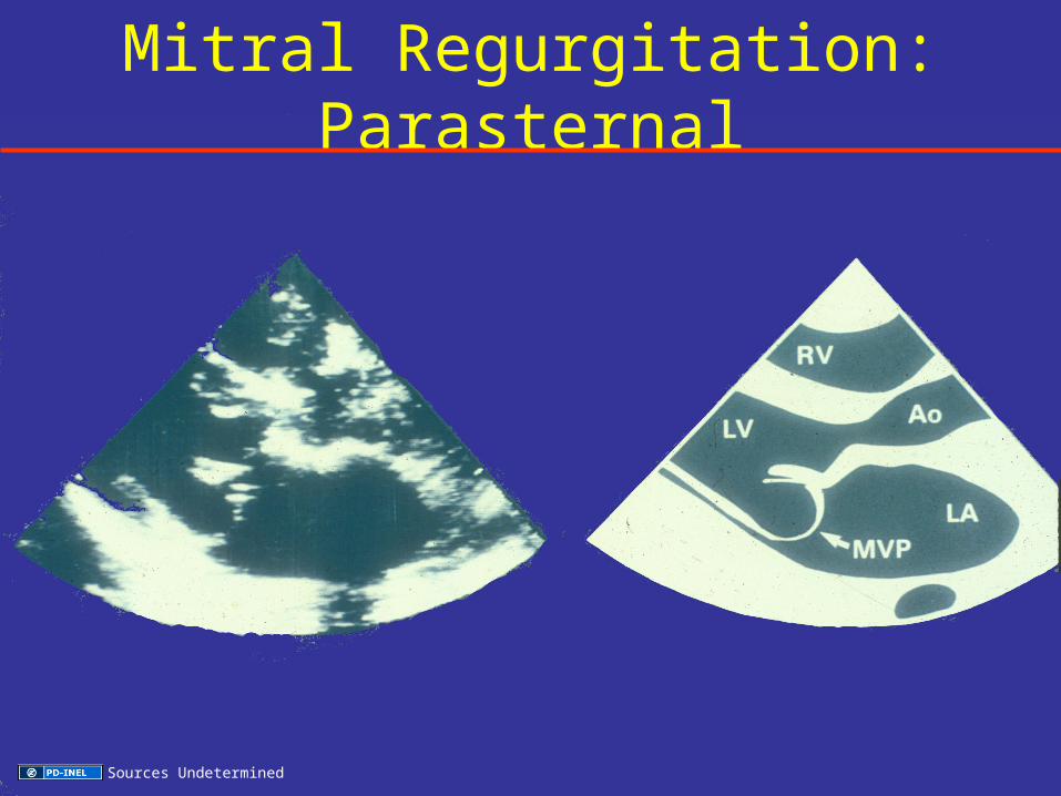
Mitral Regurgitation: Parasternal
Sources Undetermined
Severity of Mitral and Tricuspid Regurgitation
Schematic representation of
varying degrees of severity of
regurgitation removed

Mitral Regurgitation: Clinical FeaturesMitral Regurgitation: Clinical Features
Mitral Valve Prolapse:
• Protrusion of MV leaflets into LA during systole; more common in women
• Valve changes leaflets show…
- voluminous - thickened
- redundant - myxomatous
• Sx: palpitations, dyspnea if severe

Mitral Regurgitation: Mitral ProlapseMitral Regurgitation: Mitral Prolapse
Exam:
• Skeletal changes – scoliosis, pectus excavatum; Marfan’s in some
• Midsystolic click; may see late systolic murmur
• Echo: Mid to late systolic prolapse of posterior leaflet. Doppler or color echo reveals severity of MR

Mitral Regurgitation: Parasternal
Sources Undetermined

Mitral Regurgitation: Mitral ProlapseMitral Regurgitation: Mitral Prolapse
Complications:• Many patients go thru life without
problems• MR can progress• Chordal rupture can lead to sudden,
severe MR (esp. in men)• Endocarditis in those with murmur• TIA’s rare treat with ASA• Sudden death – very rare

Mitral Annulus
Schematic representation of heart beat stages
removed
Source Undetermined

Mitral Regurgitation: Clinical FeaturesMitral Regurgitation: Clinical Features
Papillary muscle dysfunction:
• Spectrum from intact but poorly functioning PM to acute rupture
• Frequently caused by:
– Ischemia or infarction of papillary muscle or underlying LV myocardium
• Less frequently by LV dilation or infiltrative process
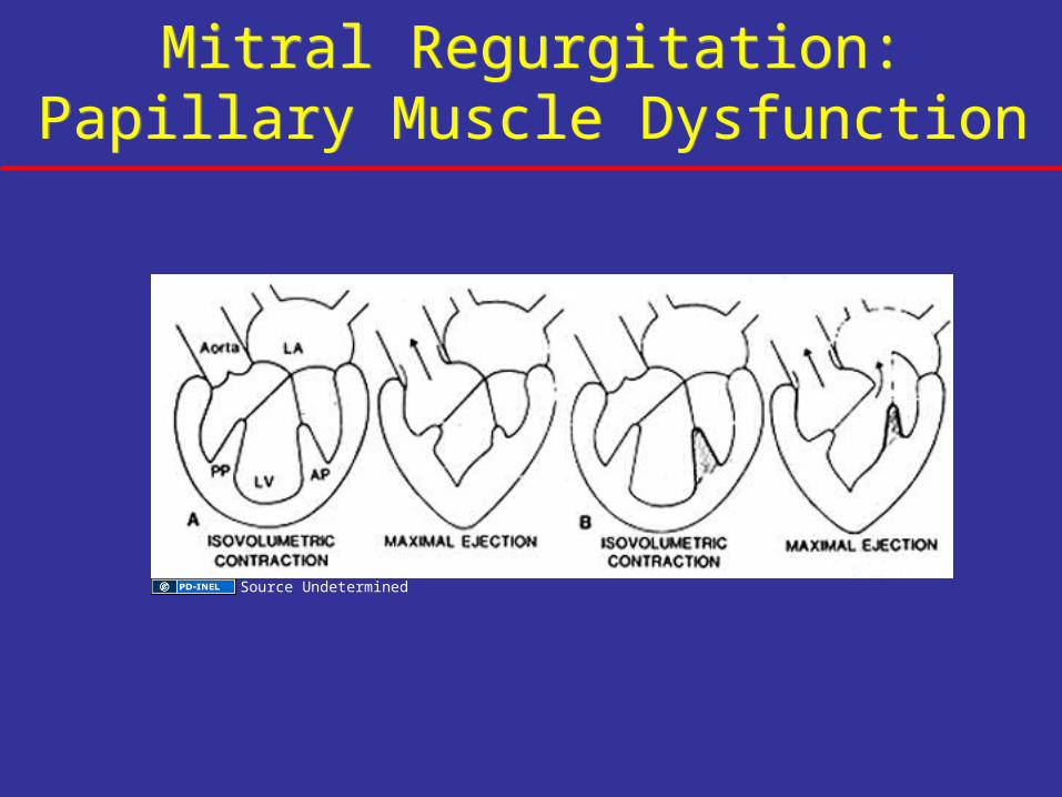
Mitral Regurgitation:Papillary Muscle Dysfunction
Mitral Regurgitation:Papillary Muscle Dysfunction
Source Undetermined

Mitral Regurgitation:Papillary Muscle Dysfunction
Mitral Regurgitation:Papillary Muscle Dysfunction
Source Undetermined

Mitral Regurgitation:Differential DiagnosisMitral Regurgitation:Differential Diagnosis
Conditions with systolic murmur:
• VSD
• Aortic stenosis
• Tricuspid regurgitation
• Hypertrophic cardiomyopathy

Mitral Regurgitation: Management
Asymptomatic
• Follow serially with visits and echo
• Recommend repair/replacement if:
– Clear sx develop
– LV ejection fraction falls < 60%

Mitral Regurgitation:Management and Prevention
Mitral Regurgitation:Management and Prevention
MR caused by LV dilation from poor LV:FXN
• Diuretics
• Vasodilators
Improves sx…
Symptomatic MR with preserved LV:
• Mitral repair or replacement before progressive LV dysfunction occurs
• B-Blockers
• Digitalis

Schematic representation of
mitral valve removed

Aortic Valve Disease

Lecture Outline
Aortic Stenosis Aortic Regurgitation
Etiology
Pathophysiology
Clinical Features
Diagnostic Testing
Differential Diagnosis
Management

Aortic Stenosis: PathologyNormal Congenital Acquired
Sources Undetermined

Aortic Stenosis
Pathophysiology
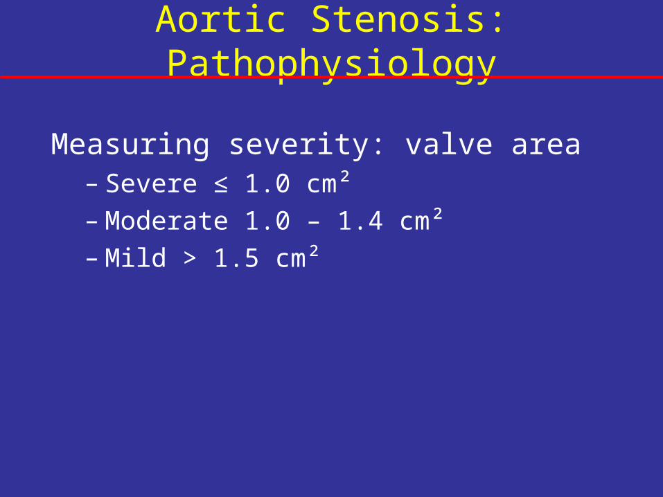
Aortic Stenosis: Pathophysiology
Measuring severity: valve area– Severe ≤ 1.0 cm²– Moderate 1.0 – 1.4 cm²– Mild > 1.5 cm²

Left Ventricular Pressure Overload
Global gene activation
Concentric hypertrophy
Gradient between LV and Aorta
Source Undetermined

Source Undetermined

Aortic Stenosis: Clinical Findings
• Dyspnea
• Angina pectoris
• Syncope
HeartRate
(bpm)
SystemicArterial
Pressure(mmHg)
PulmonaryArterial
Pressure(mmHg)
Pre
ssur
e
Aortic stenosis
Volume
Normal
Source Undetermined
M. Shea

Aortic Stenosis: Clinical Findings
• Dyspnea
• Angina pectoris
• Syncope
HeartRate
(bpm)
SystemicArterial
Pressure(mmHg)
PulmonaryArterial
Pressure(mmHg)
Pre
ssur
e
Aortic stenosis
Volume
Normal
Source Undetermined
M. Shea

Normal
Parvus et tardus pulse
Carotid Pulse
Sources Undetermined

Source Undetermined

Aortic Stenosis
Laboratory EvaluationChest radiology
Electrocardiography
Echocardiography
Stress testing
Catheterization
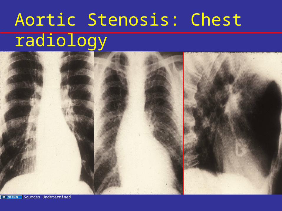
Aortic Stenosis: Chest radiology
Sources Undetermined

The Electrocardiogram
Source Undetermined
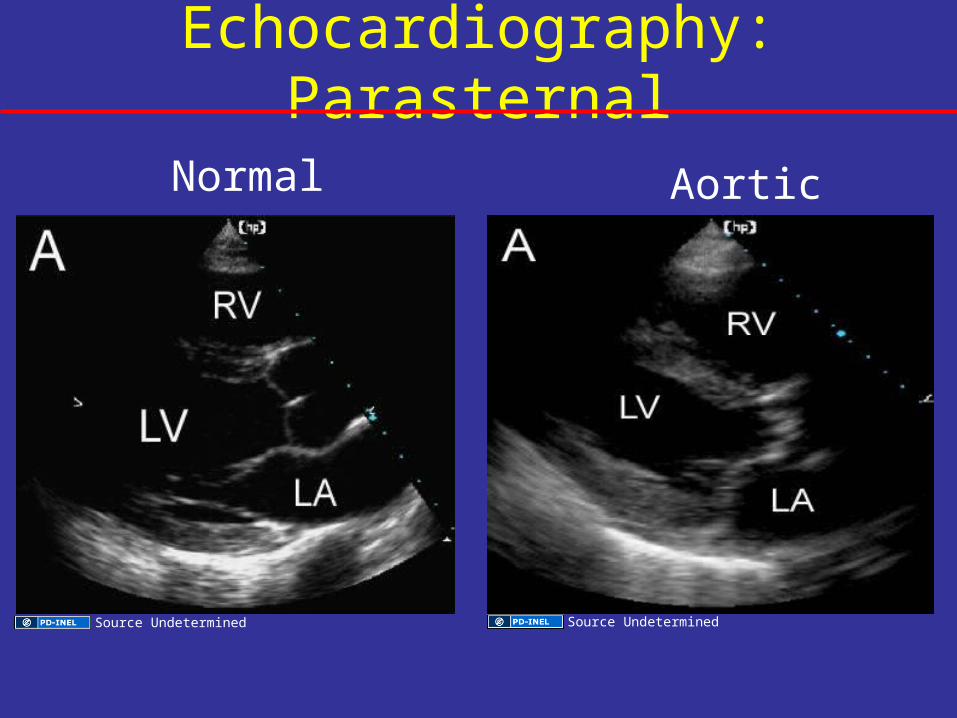
Echocardiography: Parasternal
Normal Aortic Stenosis
Source Undetermined Source Undetermined

Echocardiography: Short Axis
Normal: Aortic Stenosis
Source Undetermined Source Undetermined

Aortic Stenosis: Continuity Equation
Source Undetermined

Aortic Valve Stenosis: Echo Findings
Leaflet changes:
• Thickening
• Calcification
• Mobility
Ventricular changes:
• Left ventricular hypertrophy
Doppler changes:
• valve gradient / valve area

Aortic Stenosis
Laboratory EvaluationChest radiology
Electrocardiography
Echocardiography
Stress testing
Catheterization

Aortic Stenosis: Differential DiagnosisAny systolic murmur
Adapted by University of Michigan, Gray’s Anatomy, wikimedia commons

Natural History of Aortic Stenosis
Braunwald, Circulation, 1968
Source Undetermined

Schematic representation of
pulmonary autograph removed

Aortic Stenosis: Management
• Young patient– Balloon valvotomy– Ross procedure
• Adults– Valve replacement
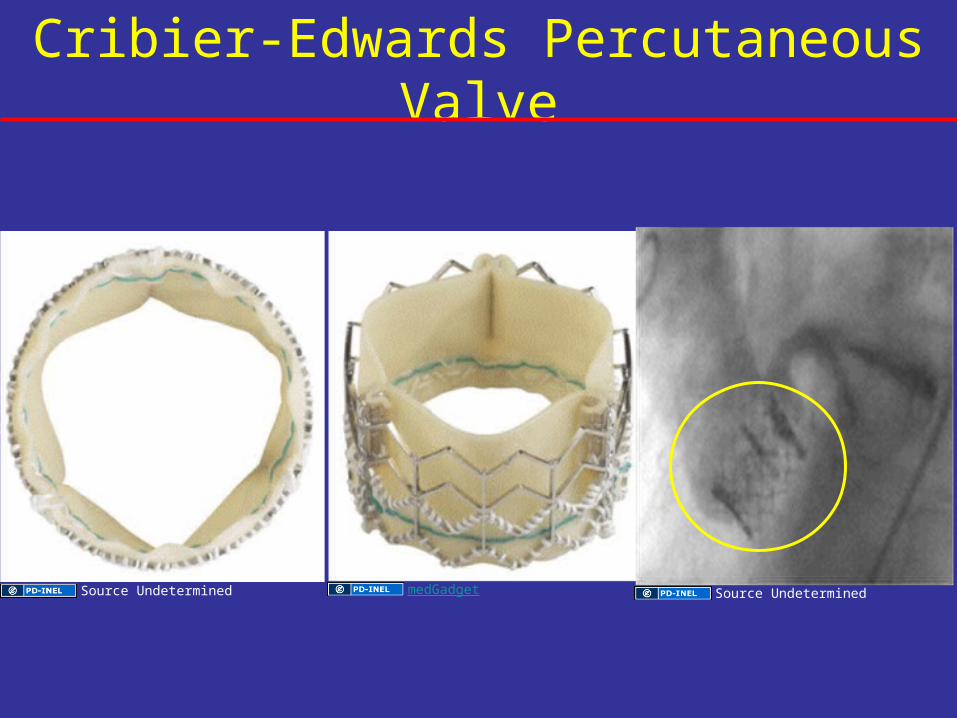
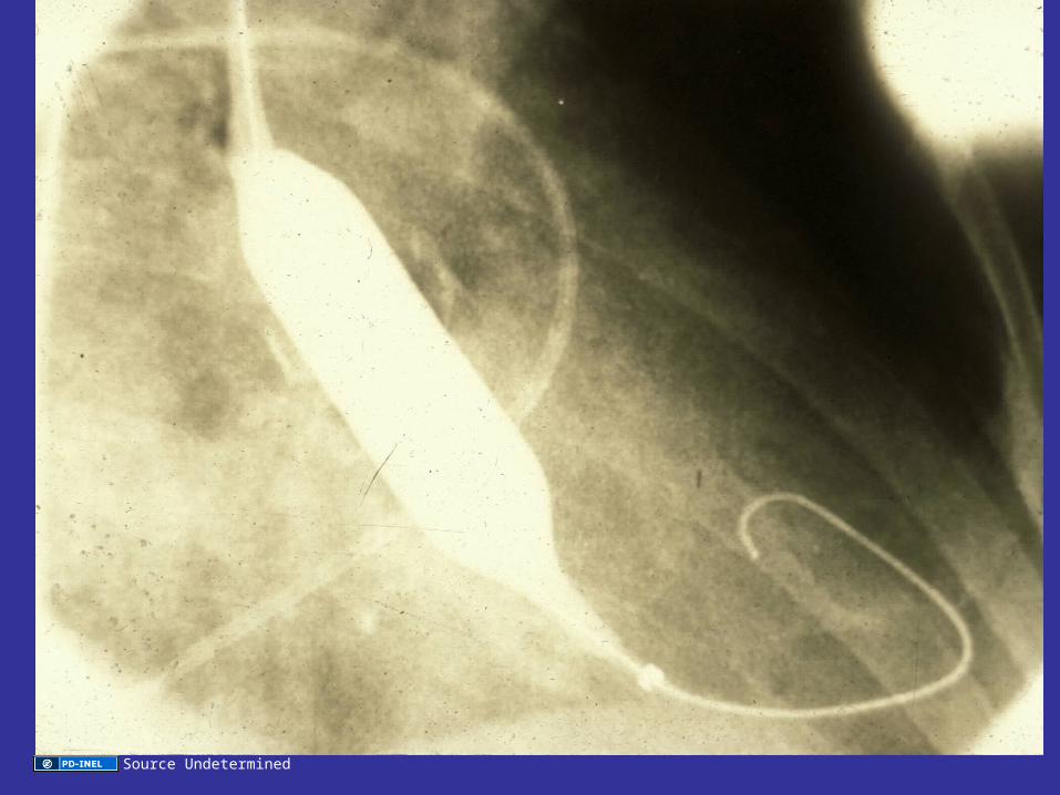
Cribier-Edwards Percutaneous Valve
medGadgetSource Undetermined Source Undetermined

Aortic Regurgitation

Aortic Regurgitation: Etiology
Abnormalities of valve leaflets
• Rheumatic• Endocarditis• Bicuspid valve
Dilatation of aortic root
• Aortic aneurysm/dissection• Annulo-aortic ectasia • Marfan syndrome• Syphilis

Normal Valve Function:• Total cusp area > aortic root area by 1.8 x• Allows leaflets to overlap/abut• Helps prevent prolapse in diastole
Impact of Diseases:• Rheumatic: Cusp area central defect• Endocarditis: Destroys cusp by tears• Aortic root: Dilation central defect
Aortic Valve Regurgitation: Pathophysiology

Dominant Hemodynamics: LV volume overload• Critical determinant of severity - area of
regurgitant orifice area• End diastolic volume increases & stroke
volume increases• Dilation and hypertrophy of LV• Diastolic burden reaches critical point
leading to heart failure• Low diastolic blood pressure: incomp. valve
and vasodilation
Aortic Valve Regurgitation: Pathophysiology

Aortic Valve Regurgitation: Pathophysiology - Acute vs. Chronic
Pulmonary
Congestion
Pressure
Pressure
N-
Pressure Pressure
N-
Brown University
Aortic Regurgitation: Clinical Features
• Long course
• Palpitations
• Dyspnea
• Fatigue
• Angina pectoris

The Arterial Pulse and Blood Pressures in Aortic Regurgitation
Mild Moderate Severe
132/76
144/67152/58
Blo
od P
ress
ure
(mm
/Hg
160
140
120
100
80
60
40
M. Shea

Hyperkinetic pulse
Carotid Pulse
Source Undetermined
Source Undetermined

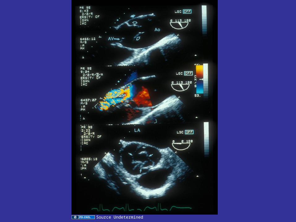
• LV apex impulse: displaced laterally,
downward, dynamic, enlarged
• Systolic murmur: may or may not imply valve
stenosis…rapid ejection of stroke volume
across aortic valve
• Diastolic murmur: decrescendo murmur;
valvular AR - louder LUSB. Aortic root
disease - louder RUSB
Aortic Valve Regurgitation: Physical Examination

Source Undetermined

Aortic Regurgitation
Laboratory Evaluation
• Chest radiology
• Electrocardiography
• Echocardiography
• Exercise testing
• Cardiac catheterization

Aortic Regurgitation: Chest X-ray
Source Undetermined
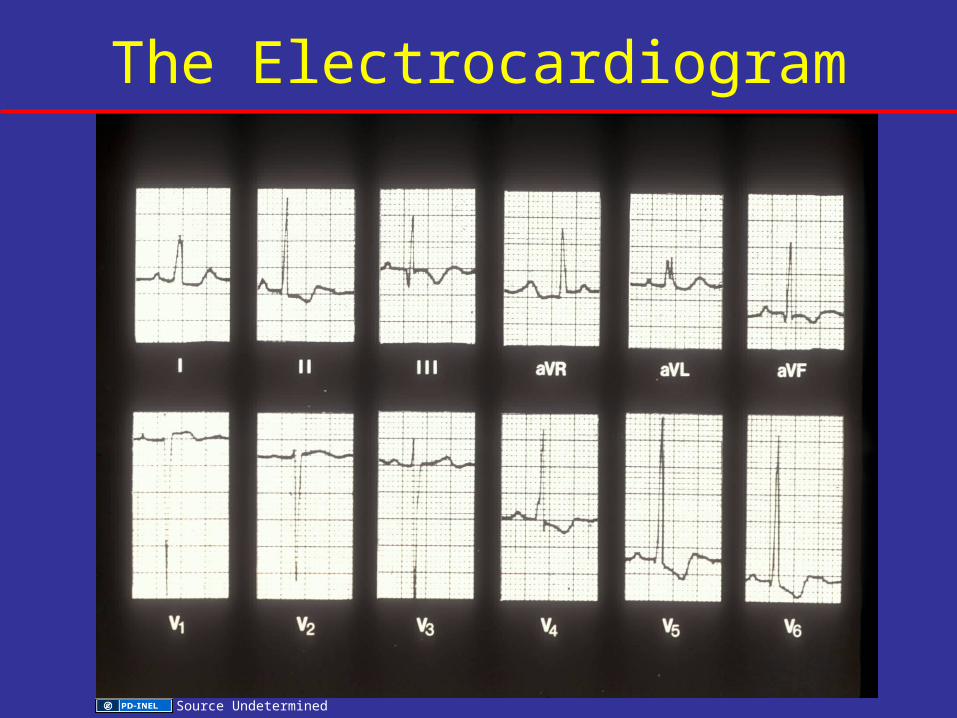
The Electrocardiogram
Source Undetermined
Source Undetermined

Aortic Regurgitation
Laboratory Evaluation
• Chest radiology
• Electrocardiography
• Echocardiography
• Exercise testing
• Cardiac catheterization

Aortic Regurgitation: Differential Diagnosis
• Mitral stenosis
• Pulmonic regurgitation
• Patent ductus arteriosus

Aortic Regurgitation
Management

Aortic Regurgitation: Management
Medical Therapy
• Noninvasive follow-up

100
80
60
40
20
0
Asy
mto
mat
ic p
atie
nts
with
no
rmal
LV
fun
ctio
n, %
0 2 4 6 8 10 12
Time, y
Sudden death
Onset of symptoms
Onset of asymptomatic left ventricular dysfunction
Severe Aortic Regurgitation:The Asymptomatic Patient
M. Shea

Aortic Regurgitation: Management
Surgical Therapy
Repair
• Aortic valve
Replacement
• Aortic root replacement

Slide 14: Source UndeterminedSlide 17: Source UndeterminedSlide 18: Source UndeterminedSlide 19: Source UndeterminedSlide 21: Sources UndeterminedSlide 22: Sources UndeterminedSlide 28: Source UndeterminedSlide 29: Source UndeterminedSlide 32: Brown University, http://www.brown.edu/Courses/Bio_281-cardio/cardio/handout2.html Slide 33: Source UndeterminedSlide 35: Brown University, http://www.brown.edu/Courses/Bio_281-cardio/cardio/handout2.html Slide 37: Source UndeterminedSlide 39: Source UndeterminedSlide 40: Source UndeterminedSlide 41: Sources UndeterminedSlide 45: Sources UndeterminedSlide 47: Source UndeterminedSlide 49: Source UndeterminedSlide 50: Source UndeterminedSlide 57: Sources UndeterminedSlide 60: Source UndeterminedSlide 61: Source UndeterminedSlide 62: Michael Shea; Source UndeterminedSlide 63: Michael Shea; Source UndeterminedSlide 64: Source UndeterminedSlide 65: Source UndeterminedSlide 67: Sources UndeterminedSlide 68: Source UndeterminedSlide 69: Sources UndeterminedSlide 70: Sources UndeterminedSlide 71: Source UndeterminedSlide 74: Adapted by University of Michigan, Gray’s Anatomy, Wikimedia Commons, http://commons.wikimedia.org/wiki/File:Heart-and-lungs.jpg Slide 75: Braunwald, Circulation, 1968Slide 76: Source UndeterminedSlide 79: Sources Undetermined; medGadget, http://medgadget.com/archives/2005/06/edwards_lifesci.html
Additional Source Informationfor more information see: http://open.umich.edu/wiki/CitationPolicy

Slide 84: Brown University, http://www.brown.edu/Courses/Bio_281-cardio/cardio/handout2.html Slide 86: Michael SheaSlide 87: Sources UndeterminedSlide 89: Source UndeterminedSlide 91: Source UndeterminedSlide 92: Source UndeterminedSlide 93: Source UndeterminedSlide 98: Michael Shea