author manuscript nih public access 1,*, bernd fritzsch...
TRANSCRIPT

Brn3a is a transcriptional regulator of soma size, target fieldinnervation and axon pathfinding of inner ear sensory neurons
Eric J. Huang1,*, Wei Liu2, Bernd Fritzsch3, Lynne M. Bianchi4, Louis F. Reichardt1,‡, andMengqing Xiang2,‡1 Program in Neuroscience, Department of Physiology, and Howard Hughes Medical Institute,University of California, San Francisco, CA 94143, USA2 Graduate Program in Molecular Genetics and Microbiology, Center for Advanced Biotechnologyand Medicine, and Department of Pediatrics, UMDNJ-Robert Wood Johnson Medical School,Piscataway, NJ 08854, USA3 Department of Biomedical Sciences, Creighton University, Omaha, NE 68178, USA4 Neuroscience Program, Oberlin College, Oberlin, OH, USA
SUMMARYThe POU domain transcription factors Brn3a, Brn3b and Brn3c are required for the properdevelopment of sensory ganglia, retinal ganglion cells, and inner ear hair cells, respectively. We haveinvestigated the roles of Brn3a in neuronal differentiation and target innervation in the facial-statoacoustic ganglion. We show that absence of Brn3a results in a substantial reduction in neuronalsize, abnormal neuronal migration and downregulation of gene expression, including that of theneurotrophin receptor TrkC, parvalbumin and Brn3b. Selective loss of TrkC neurons in the spiralganglion of Brn3a−/− cochlea leads to an innervation defect similar to that of TrkC−/− mice. Mostremarkably, our results uncover a novel role for Brn3a in regulating axon pathfinding and target fieldinnervation by spiral and vestibular ganglion neurons. Loss of Brn3a results in severe retardation indevelopment of the axon projections to the cochlea and the posterior vertical canal as early as E13.5.In addition, efferent axons that use the afferent fibers as a scaffold during pathfinding also showsevere misrouting. Interestingly, despite the well-established roles of ephrins and EphB receptors inaxon pathfinding, expression of these molecules does not appear to be affected in Brn3a−/− mice.Thus, Brn3a must control additional downstream genes that are required for axon pathfinding.
KeywordsBrn3a; POU domain; Transcription factor; Spiral ganglion; Vestibular ganglion; Innervation; Axonpathfinding; Mouse
INTRODUCTIONThe vertebrate sensory neurons convey different modalities of sensory information to thecentral nervous system. In the inner ear, sensory neurons in the spiral ganglion innervatecochlear hair cells that are auditory sensory receptors; those in the vestibular ganglion innervatevestibular hair cells that are sensory receptors for head position and movement. Similarly,neurons within the geniculate ganglion innervate taste buds in the tongue that transmit gustatory
‡Authors for correspondence (e-mail: E-mail: [email protected] and E-mail: [email protected]).*Present address: Pathology Service, VAMC and Department of Pathology, University of California, San Francisco, CA 94121, USA
NIH Public AccessAuthor ManuscriptDevelopment. Author manuscript; available in PMC 2009 July 14.
Published in final edited form as:Development. 2001 July ; 128(13): 2421–2432.
NIH
-PA Author Manuscript
NIH
-PA Author Manuscript
NIH
-PA Author Manuscript
information. During mouse embryogenesis, neurons in the spiral and vestibular ganglia arederived from the otic placode, while those in the geniculate ganglion arise from the firstepibranchial placode. The neuronal progenitors derived from these two placodes migrate andcoalesce around embryonic day 9 (E9) into a single sensory ganglion primordium – the facial-statoacoustic ganglion. By E12.5, the primordium differentiates into the geniculate ganglionand vestibulocochlear ganglion, which remain physically connected (Sher, 1971; Fig. 1K). ByE13.5- E14.5, the vestibulocochlear ganglion segregates into the vestibular and spiral ganglionprimordia. By E17.5, the spiral and vestibular ganglia are physically separated inside andoutside the otic capsule, respectively. The vestibulocochlear ganglion neurons start to innervatetheir peripheral targets as soon as the sensory epithelia are formed in the otocyst at E10.5(Sher, 1971; Morsli et al., 1998).
Although the ontogenies of sensory neurons within the inner ear and geniculate ganglia havebeen well described, much less is known about the molecular mechanisms controlling thisprocess. Several lines of evidence have indicated that transcription factors are criticallyinvolved in the development of these ganglia. For example, the bHLH factors neurogenin 1(Ngn1; Neurod3 – Mouse Genome Informatics) and neurogenin 2 (Ngn2; Atoh4 – MouseGenome Informatics) are expressed in the cranial ganglion placodes as early as E8.5. Targeteddisruption of Ngn1 or Ngn2 in mice, respectively, abolishes the development of the proximalcranial ganglia, which include the vestibular and cochlear ganglia, or the distal cranial ganglia,which include the geniculate ganglion (Fode et al., 1998; Ma et al., 1998). Consistent with therole of their Drosophila paralog atonal (Jarman et al., 1993), Ngn1 and Ngn2 have been shownto be essential for cell fate commitment (Fode et al., 1998; Ma et al., 1998; Ma et al., 1999;Ma et al., 2000a). In addition, the homeodomain-containing transcription factor Phox2a (Arix– Mouse Genome Informatics) has been shown to be required for differentiation andmaintenance of geniculate ganglion neurons (Morin et al., 1997). Gene targeting analyses havealso demonstrated essential roles for neurotrophins and their Trk receptors in supporting thesurvival of sensory neurons (reviewed in Farinas and Reichardt, 1997). In the inner ear sensoryganglia, brain-derived neurotrophic factor (BDNF), neurotrophin (NT) 3 (Ntf3 – MouseGenome Informatics), and their receptors TrkB and TrkC exert complementary roles inpromoting neuronal survival (Ernfors et al., 1995; Minichiello et al., 1995; Schimmang et al.,1995; Bianchi et al., 1996; Fritzsch et al., 1997a). For example, deletion of the genes for BDNFor NT3 in mice results in loss of more than 80% of neurons within the vestibular or spiralganglion, respectively. In a compound mutant lacking genes for both BDNF and NT3, there iscomplete loss of vestibular or spiral ganglion neurons. Similar analyses have shown that BDNF,NT4, NT5 and NT3 are survival factors for geniculate ganglion neurons (Farinas et al., 1994;Conover et al., 1995; Liu et al., 1995; Fritzsch et al., 1997b; Liebl et al., 1997).
The POU domain transcription factors Brn3a and Brn3b, also known as Brn3.0 and Brn3.2,respectively (Pou4f1 and Pou4f2 – Mouse Genome Informatics), have been shown to beexpressed in the trigeminal, dorsal root and inner ear sensory ganglia during mouseembryogenesis (Xiang et al., 1993; Xiang et al., 1995; Gerrero et al., 1993; Turner et al.,1994; Fedtsova and Turner, 1995; Ryan, 1997). Deletion of Brn3a results in a significant lossof spiral ganglion neurons and defects in their migration (McEvilly et al., 1996). In addition,consistent with the multiple sites of Brn3a expression, Brn3a−/− neonates show neuronal lossesin the somatosensory ganglia and in several brainstem nuclei, which eventually lead to perinatallethality (Xiang et al., 1996; McEvilly et al., 1996). For example, in the mutant trigeminalganglion, approximately 70% of the neurons are lost by postnatal day 0 (P0) as a result ofapoptotic cell death caused by a dramatic and specific downregulation of neurotrophinreceptors in the absence of Brn3a (Huang et al., 1999b). In contrast, absence of Brn3b doesnot have overt effects on the development of inner ear sensory ganglia. Instead, its absenceresults in improper differentiation and loss of a large set of retinal ganglion cells (Gan et al.,1996; Gan et al., 1999; Erkman et al., 1996; Xiang, 1998). Interestingly, expression of Brn3b
Huang et al. Page 2
Development. Author manuscript; available in PMC 2009 July 14.
NIH
-PA Author Manuscript
NIH
-PA Author Manuscript
NIH
-PA Author Manuscript

precedes that of Brn3a in the retina (Xiang, 1998). It remains unknown whether expression ofBrn3a precedes that of Brn3b in the spiral ganglion, where Brn3a appears to have a leadingrole.
Despite significant advances in our understanding of the molecular mechanisms governing thespecification and maintenance of auditory and gustatory sensory neurons, relatively little isknown about their differentiation. In this report, we show that Brn3a is expressed in the facial-statoacoustic ganglion prior to sensory neuron differentiation and innervation of the otocyst.Loss of Brn3a leads to downregulation of TrkC, Brn3b and the gene for parvalbumin in thespiral ganglion, indicating that these are downstream targets of Brn3a. More importantly, weprovide evidence that Brn3a is required for proper growth and migration of inner ear andgustatory sensory neurons, and is critically involved in target innervation and axon guidanceby spiral and vestibular ganglion neurons. Our data indicate that Brn3a controls survival anddifferentiation of sensory neurons by regulation of different downstream genes.
MATERIALS AND METHODSExperimental animals
Brn3a+/−, Brn3b+/− and Brn3b−/− mice were derived by targeted gene disruption as previouslydescribed (Gan et al., 1996; Xiang et al., 1996).
ImmunohistochemistryFor section immunohistochemistry using anti-Brn3a, anti-Brn3b, anti- NF 150, anti-p75NTR,and anti-parvalbumin, staged embryos and neonates were fixed in 4% paraformaldehyde, cryo-protected in 30% sucrose, embedded in OCT compound and sectioned at 10–14 μm using acryostat. Immunohistochemistry for TrkB and TrkC antibodies has been described previously(Huang et al., 1999a). The color reaction was developed using the NovaRed substrate kit whenHematoxylin counterstaining was applied. Antibodies were obtained from the followingsources: anti-Brn3a and anti-Brn3b (Xiang et al., 1993; Xiang et al., 1995); anti-TrkB and anti-TrkC (Huang et al., 1999a; Huang et al., 1999b); anti-NF 150 and anti-p75NTR (Chemicon);anti-parvalbumin (SWant); and anti-EphB antibodies (Santa Cruz Biotechnology).
Histochemistry and quantitationThe silver impregnation technique was performed as described by Ungewitter (Ungewitter,1951). Cresyl Violet labeling was performed as described by LaBossiere and Glickstein(LaBossiere and Glickstein, 1976). To determine the total number of neurons in each sensoryganglion, serial sections were stained with Cresyl Violet and scored every other section bylight microscopy. All neuronal profiles larger than glial cells were counted and counts werenot corrected for split neurons. All data were tested for significance using two sample Student’st-test with unequal variances. To compare neuronal size in wild-type and Brn3a−/− orBrn3b−/− sensory ganglia, cross-sectional areas of neuronal profiles were measured in a givenregion of the ganglion using the NIH Image program.
Analyses of afferent and efferent innervation patternsAnalyses of afferent innervations to the inner ear were performed as previously described(Fritzsch and Nichols, 1993). Briefly, lipophilic dye DiI (1,1′-diotadecyl-3,3,3′,3′-tetramethylindocarbocyanine percholate; Molecular Probes) -soaked filter strips were appliedto the alar plate rostral to the statoacoustic (octaval) nerve root in E13.5 and P0 Brn3a−/− miceand their wild-type littermates (n=4 for each genotype). After a diffusion time of 3–8 days at37°C, the ears with otic ganglia and facial nerve attached, were dissected from adjacentmesenchyme and photographed as whole mounts with an Olympus compound microscope.
Huang et al. Page 3
Development. Author manuscript; available in PMC 2009 July 14.
NIH
-PA Author Manuscript
NIH
-PA Author Manuscript
NIH
-PA Author Manuscript

After analyzing the DiI-labeling results, the ears were subsequently processed forimmunohistochemical staining using α-acetylated tubulin antibodies (Sigma, T9026). Briefly,cochleae were defatted, incubated with primary antibody for 2 days, washed and incubatedwith a secondary antibody conjugated to horseradish peroxidase.
RESULTSSequential expression of Brn3a and Brn3b during development of the facial-statoacousticganglion
To better understand the roles of Brn3a and Brn3b in development of the inner ear sensory andgeniculate ganglia, we first examined the expression patterns of these two transcription factorsin the developing mouse facial-statoacoustic ganglion, the precursor to these ganglia. A Brn3aantibody strongly labeled the facial-statoacoustic ganglion primordium located medioventrallyto the otic vesicle at E9.5 (Fig. 1A) and E10.5 (data not shown); by contrast, anti-Brn3b didnot label this ganglion at these early developmental stages (Fig. 1B). At E12.5, following partialseparation of the geniculate ganglion from the vestibulocochlear ganglion, Brn3a continues tobe expressed strongly in both ganglia, while Brn3b is now weakly expressed in a small fractionof cells in both ganglia (Fig. 1C,D). Thus, Brn3a is expressed in the facial-statoacousticganglion at least 3 days before the onset of Brn3b expression. At E16.5, however, both Brn3aand Brn3b are prominently expressed in perhaps all of the neurons in the spiral, vestibular andgeniculate ganglia (Fig. 1E,F). Expression of both transcription factors continues to be strongin all neurons at E17.5 (Fig. 1G–J). As elucidated in Fig. 1K, Brn3a is strongly expressed inthe facial-statoacoustic ganglion at E9 and continues to be strongly expressed in derivatives ofthis ganglion throughout embryogenesis. In contrast, weak expression of Brn3b is found in afew cells only at E12.5 and expression of this transcription factor does not become widespreaduntil E16.5.
As inner ear sensory neurons become postmitotic between E11.5 and E15.5 (Ruben, 1967),the onset of Brn3a expression at E9 in the primordium of these ganglia suggests that Brn3a isexpressed in dividing neuronal progenitors before neuronal differentiation. Previous work hasindeed detected Brn3a expression in proliferative cells of the vestibulocochlear ganglion(Fedtsova and Turner, 1995). Moreover, Brn3a expression significantly precedes peripheraltarget innervation by vestibulocochlear neurons and migration of cochlear neurons, both ofwhich begin at E10.5 (Sher, 1971). In contrast, Brn3b is present prevalently in the facial-statoacoustic ganglion only after initiation of these events.
Differential loss of neurons in Brn3a−/−, but not Brn3b−/−, spiral, vestibular and geniculateganglia
Given that expression of Brn3a is initiated in the facial-statoacoustic ganglion at E9 and persistsin all of its derivative ganglia throughout embryogenesis (Fig. 1), we investigated whether theabsence of this transcription factor affects neuronal survival. We determined the number ofneurons within the spiral, vestibular and geniculate ganglia in control and in Brn3a−/− mice byCresyl Violet staining. In the geniculate ganglion at P0, there are significantly fewer neuronsin Brn3a−/− mice than in the wild type (Fig. 2A,B). In sections of the Brn3a−/− vestibularganglia at P0, however, we were unable to visualize any obvious decrease in neuronal number(Fig. 2C,D). We next determined the total number of neurons within each sensory ganglion byscoring neurons on serial sections labeled with Cresyl Violet. As shown in Fig. 2E, at E18.5and P0, there is a 30–50% reduction of neurons in the geniculate ganglion, and anapproximately 30% decrease in the spiral ganglion in Brn3a−/− mice compared with controls.By contrast, there is no significant reduction in the number of neurons present in the vestibularganglion at E18.5 or P0. Therefore, absence of Brn3a affects the survival of sensory neuronsin the three ganglia to different extents. A similar quantitative analysis detected no significant
Huang et al. Page 4
Development. Author manuscript; available in PMC 2009 July 14.
NIH
-PA Author Manuscript
NIH
-PA Author Manuscript
NIH
-PA Author Manuscript
differences in the number of neurons within the spiral, vestibular and geniculate gangliabetween controls and mutant Brn3b mice at P0 (Fig. 2E). Thus, the presence of Brn3b is notessential for the maintenance of these sensory neurons.
Diminution of neuronal size in Brn3a−/− spiral, vestibular and geniculate gangliaTo investigate possible effects of Brn3a absence on differentiation of inner ear and geniculatesensory neurons, we compared these neurons using Cresyl Violet-labeled sections of wild-typeand mutant ganglia. Two notable phenotypes were detected in the mutant ganglia. First, incontrast to the wild-type spiral ganglion (in which neurons are tightly packed) the spiralganglion neurons in Brn3a−/− mice are scattered in the entire cochlear modiolus with manybarely separated from the vestibular ganglion (Fig. 2C,D). Second, although the vestibularganglion is well-formed and vestibular neurons appear to survive up to P0 in Brn3a−/− mice(Fig. 2E), we noticed a dramatic decrease in the volume of the vestibular ganglion and in thesize of its neurons (Fig. 2F and data not shown). To quantify changes in neuronal size, the areasof neuronal cell profiles were measured in vestibular, spiral and geniculate ganglia usingsections from both wild-type and mutant mice. In wild-type P0 neonates, the large majority ofneurons in the vestibular ganglion have areas between 30–200 μm2, with a peak at ~90 μm2.In addition, a significant number of these neurons are more than 200 μm2 with a few as largeas 390 μm2 in area (Fig. 2F). In Brn3a−/− vestibular ganglia at P0, however, the areas of mostof the neuronal profiles are distributed between 20–110 μm2, with a peak at only ~50 μm2 (Fig.2F). Similar decreases in the areas of neuronal profiles were seen at E16.5 and E18.5 in mutantvestibular ganglia (data not shown). The areas of neuronal profiles in control and mutant spiraland geniculate ganglia were also measured. In the wild-type spiral ganglion at P0, the areas ofmost neurons range between 25 and 85 μm2, with a peak at ~55 μm2 (Fig. 2G). In the mutantat P0, the range is 10–50 μm2, with a peak at ~25 μm2 (Fig. 2G). In the wild-type geniculateganglion at P0, most neurons have profiles between 35 and 175 μm2, with a peak at ~105μm2 (Fig. 2H). In the mutant, profile areas range from 25 to 135 μm2, with a peak at ~65μm2 (Fig. 2H). Therefore, in the absence of Brn3a, neuronal size is diminished in all threeganglia, including the vestibular ganglion (in which normal numbers of neurons survive). Incontrast, examination of Brn3b mutants revealed neither loss of neurons in these ganglia norany reduction in neuronal size in these ganglia (Fig. 2E and data not shown), indicating thatBrn3b plays little or no role in regulating the size of these neurons.
Altered gene expression in Brn3a−/− spiral, vestibular and geniculate gangliaAs members in the POU domain factor family are required for cellular differentiation duringdevelopment, it is possible that Brn3a may control downstream genes that are required for thesurvival and differentiation of sensory neurons. In the trigeminal ganglion, for example, Brn3ahas been shown to play a major role in the survival of sensory neurons by regulating theexpression of neurotrophin receptors TrkA, TrkB and TrkC (Ntrk1, Ntrk2 and Ntrk3 – MouseGenome Informatics; McEvilly et al., 1996; Huang et al., 1999b). To determine whether Brn3asimilarly regulates neurotrophin receptor expression in the spiral, vestibular and geniculateganglia, we examined by immunostaining the expression levels of TrkB, TrkC and p75NTR inthese ganglia in E13.5-E18.5 Brn3a−/− embryos. As early work has shown that TrkB and TrkCare the major neurotrophin receptors controlling neuronal survival in these ganglia, we wishedto determine the effect of Brn3a on the expression of TrkB and TrkC (Pirvola et al., 1994;Minichiello et al., 1995; Schimmang et al., 1995; Fritzsch et al., 1997b). In wild-type mice,both TrkB and TrkC immunoreactivities were detected in spiral ganglia and radial fibers of thecochlea (Fig. 3A,C,E). While the TrkB immunoreactivity in the Brn3a−/− was comparable withthat in the wild-type control (Fig. 3F), TrkC immunoreactivity was markedly reduced by E15.5and E18.5 (Fig. 3B,D). At E13.5, however, similar levels of TrkC immunoreactivity wereobserved in the vestibulocochlear ganglia and their projecting fibers between the wild-type andmutant (data not shown). Thus, the neuronal loss in Brn3a−/− cochleae is likely to result from
Huang et al. Page 5
Development. Author manuscript; available in PMC 2009 July 14.
NIH
-PA Author Manuscript
NIH
-PA Author Manuscript
NIH
-PA Author Manuscript

a failure to maintain proper TrkC expression in the spiral ganglion neurons. In Brn3a−/−
vestibular and geniculate ganglia, TrkB and TrkC were expressed at a level similar to that ofthe control (data not shown). Similar to the trigeminal ganglion (Huang et al., 1999b), we foundthat p75NTR was expressed at comparable levels in wild type and mutant in the fibers and cellbodies of spiral, vestibular and geniculate sensory neurons (data not shown). Thus, theexpression of TrkC in the spiral ganglion requires Brn3a, whereas the expression of TrkB andp75NTR is independent of Brn3a.
In addition to the Trk receptors, we also examined the expression of parvalbumin, Brn3b andneurofilament 150 (NF 150) in the spiral, vestibular and geniculate ganglia of E16.5 and E18.5Brn3a−/− embryos. In the control, the Ca2+-binding protein parvalbumin was prominentlylocalized in the cell bodies, as well as processes of spiral and vestibular ganglion neurons, butthis immunoreactivity was greatly diminished in the mutant (Fig. 3G,H). Similarly, the numberof neurons expressing Brn3b in the spiral ganglion was dramatically reduced in the mutant(Fig. 3I,J). This decrease was even more pronounced in the mutant vestibular ganglion, whereneurons positive for Brn3b could barely be seen (Fig. 3I–L). In the mutant geniculate ganglion,Brn3b expression was also greatly reduced (data not shown). However, no significantdifference in the level of NF 150 expression was observed in the neurons of spiral, vestibularand geniculate ganglia between the Brn3a wild type and mutant (data not shown). Thus, Brn3amay differentially control differentiation of sensory neurons by regulating the expression ofgenes common to all sensory ganglia as well as genes specific to different sensory ganglia.
Defects in target field innervation and axon pathfinding in vestibulocochlear ganglion ofBrn3a−/− mice
As expression of Brn3a is initiated in vestibulocochlear neurons before the beginning ofperipheral target innervation, it is possible that Brn3a helps regulate this important event. Totest this possibility, we examined the afferent and efferent innervation patterns of wild-typeand mutant inner ears at various developmental stages by DiI tracing andimmunohistochemistry.
In the P0 wild-type cochlea, DiI labeling reveals that the axons of spiral ganglion neurons inthe entire basal turn fasciculate into closely spaced radial fibers to centrifugally innervate haircells in the organ of Corti (Fig. 4A). In contrast, the afferent innervation by these sensoryneurons in the Brn3a−/− cochlea was sparse in the basal turn with an increased distance betweenfiber bundles and a paucity of radial fibers at the hook region (Fig. 4B). In addition, some radialfibers close to the base have shifted trajectories and innervate hair cells in the hook region.Consistent with the quantitation of neuronal numbers (Fig. 2E), spiral ganglion neurons aredepleted in the base and reduced in the basal turn of the mutant cochlea (Fig. 4B). Interestingly,the topographical losses of sensory neurons and deficiencies in afferent innervation present inthe Brn3a−/− cochlea resemble those found in TrkC−/− mutants (Fritzsch et al., 1998; Fig. 4C),consistent with the observation of a nearly complete absence of TrkC neurons in theBrn3a−/− spiral ganglion by P0 (Fig. 3A–D). In order to determine the timing of this deficitand its relation to the downregulation of TrkC expression, we performed similar experimentsin early embryonic stages before loss of TrkC expression occurs. At E13.5, while the cochlearsensory epithelium of wild-type animals is densely innervated by afferent fibers (Fig. 5A), onlya small number of afferent fibers are labeled in the Brn3a mutant cochlea (Fig. 5B). As theafferent innervation was eventually established in the P0 cochlea, the data from the embryonicstage indicate that afferent innervation to the cochlea is initially delayed in the absence ofBrn3a.
The overall patterns of cochlear innervation in Brn3a−/− mice were further investigated byimmunohistochemistry, using an anti-acetylated tubulin antibody that labels both afferents andefferents. In the P0 Brn3a−/− cochlea, tubulinimmunoreactive fibers display an innervation
Huang et al. Page 6
Development. Author manuscript; available in PMC 2009 July 14.
NIH
-PA Author Manuscript
NIH
-PA Author Manuscript
NIH
-PA Author Manuscript

pattern defect similar to afferent fibers as revealed by DiI labeling. More notably, there isexuberant outgrowth of misrouted fibers throughout the cochlea. Many of these fibers projectbeyond the boundary of hair cells, some reaching as far as the mesenchyme surrounding theorgan of Corti (Fig. 4E–G). None of these aberrant fibers are present in the control wild-typecochlea (Fig. 4D). As these abnormalities are not present in the DiI-labeled afferent pathways(Fig. 4B), it is most likely that these misrouted fibers represent cochlear efferent fibers in themutant.
In the P0 Brn3a−/− vestibular system, despite the marked reduction of vestibular ganglionneuronal size, DiI labeling shows grossly normal afferent innervation of the maculae of sacculeand utricle, and the cristae of anterior vertical and horizontal canals. However, there is acomplete absence of afferent fiber projection to the posterior vertical canal (Fig. 5C,D). Thisinnervation defect exists as early as E13.5, when the afferent fibers innervating the posteriorvertical canal in the mutant show a severe reduction in the fiber bundle size. More importantly,the few fibers that do innervate the posterior vertical canal appear to arise dorsally from theutricle, rather than by the more ventral and separate route (Fig. 5A,B). As there are no fibersinnervating the posterior vertical canal in the mutant by P0, this suggests that the originallymisrouted fibers have failed to maintain their projection.
We examined the overall patterns of vestibular innervation in Brn3a−/− mice byimmunostaining with antibodies against NF 150 and p75NTR, and by silver impregnation, allof which stain both afferent and efferent fibers (Fig. 6). In the E16.5 and E18.5 vestibularsystem, several innervation anomalies were observed in the mutant. For example, in at leasthalf of mutant embryos, a large fiber bundle splits from the bundle that innervates the crista ofthe anterior vertical canal, penetrates the bony labyrinth, and projects into the middle ear withor without further branching (Fig. 6B–D,F). In addition, the fiber bundle that innervates thesaccular macula often abnormally branches away to project into the cochlea of the mutant (Fig.6E). As these aberrant fibers are not found in DiI-labeled afferent projections (Fig. 5), theyprobably represent vestibular efferent fibers similar to those present in the mutant cochlea (Fig.4E–G). Therefore, these data together indicate that Brn3a is required for proper innervationand axon pathfinding of both afferent and efferent fibers in the cochlear and vestibular systems.
Loss of Brn3a does not affect expression of members in the Eph familyThe deficits in axon pathfinding in the inner ear of Brn3a−/− mutants are reminiscent of thephenotypes in mice with targeted deletion in EphB2 and raise the possibility that EphB2 couldbe a target gene whose expression is regulated by Brn3a. Because members in the ephrin andEph family have been implicated in axon pathfinding and cell migration, it is possible that lossof Brn3a may affect their expression. To test this hypothesis, we examined the expression ofEphB1, EphB2 and EphB6 in sensory ganglia and brainstem nuclei of Brn3a−/− mutants. Ourdata indicate that by immunohistochemistry there is no detectable difference in the amount ofEphB1 protein in the spiral ganglion neurons, trigeminal neurons and neurons in the inferiorolivary nucleus between Brn3a−/− mutants and their wild-type littermates (Fig. 7).Immunostaining of EphB2 and EphB6 shows less intense expression of both proteins in thespiral and vestibular neurons and there is no detectable difference between the wild type andBrn3a−/− at E12.5 and E16.5 (data not shown).
DISCUSSIONOur analyses of the development of facial-statoacoustic ganglion in the Brn3a−/− mice provideevidence to indicate novel functions of Brn3a in regulating neuronal survival, differentiation,migration and axon pathfinding of these ganglia (Fig. 8). We show that loss of Brn3a leads todownregulation of gene expression, most notably that of TrkC, Brn3b and parvalbumin (Fig.8B). In addition, our results uncover previously unidentified roles of Brn3a in regulating axon
Huang et al. Page 7
Development. Author manuscript; available in PMC 2009 July 14.
NIH
-PA Author Manuscript
NIH
-PA Author Manuscript
NIH
-PA Author Manuscript

pathfinding and target field innervation in spiral and vestibular ganglion neurons (Fig. 8A).The innervation defects appear earlier than the downregulation of TrkC expression, suggestingthat these two mutant phenotypes probably are independent of each other. This is furthersupported by the almost complete absence of innervation to the posterior vertical canal, aphenotype not identified in TrkC−/− mice. Interestingly, although the afferent axons eventuallyreach their targets, the selective loss of TrkC neurons in the spiral ganglion of Brn3a−/− cochlealeads to an innervation defect similar to that of TrkC−/− mice. In addition, efferent axons thatuse the afferent fibers as scaffold during pathfinding also show severe misrouting to the mutantinner ear. Despite similar axon pathfinding defect in Brn3a−/− mutants and EphB2 mutants,loss of Brn3a apparently does not result in dramatic loss of EphB1, EphB2 or EphB6. Thus,these data indicate that Brn3a controls additional factors that are required for axon pathfindingin the developing inner ear.
Sequential expression and crossregulation of Brn3a and Brn3b during sensorineuraldevelopment
During genesis of the inner ear and geniculate ganglia, the onset of Brn3a expression isobserved in neuronal progenitors in the facial-statoacoustic ganglion prior to fate commitmentand differentiation (Fig. 1). Thereafter, Brn3a continues to show robust expression indifferentiated neurons in these ganglia. Strong Brn3b expression, however, is not seen untilE16.5 when soma growth, axon targeting and migration of sensory neurons have beenestablished (Fig. 1; Sher, 1971). Interestingly, in contrast to the severe deficits in the trigeminaland facial-statoacoustic ganglia of Brn3a−/− mice, no obvious neuronal defects are present inBrn3b−/− mice (Fig. 2E;Gan et al., 1996;Xiang et al., 1996;McEvilly et al., 1996). In contrast,we have previously shown that Brn3b is turned on in retinal ganglion cells two days beforeBrn3a during retinogenesis (Xiang, 1998). Targeted deletion in Brn3b results in loss of ~70%of retinal ganglion cells whereas Brn3a deletion causes no overt defects in the retina (Gan etal., 1996;Erkman et al., 1996;Xiang et al., 1996).
Thus, these results indicate that the differential effects of Brn3a and Brn3b deletions ondevelopment of the sensory ganglia and retina correlate with the temporal expression ordersof Brn3a and Brn3b in these sensorineural structures. Although members in the Brn3 familyshow significant overlap in their expression patterns, the ones that demonstrate earlier andbroader expression always have more dominant roles in regulating cell differentiation inspecific sensory ganglia (Fig. 8B). Indeed, Brn3a and Brn3b have been shown to share thesame DNA-binding sites and both can activate gene expression (Turner et al., 1994;Gruber etal., 1997;Trieu et al., 1999;Liu et al., 2000a;Liu et al., 2000b), indicating that each may be ableto compensate for the other when co-expressed in the same cell. Such a model predicts thatforced expression of members of Brn3 family will compensate for the functions of the others.Consistent with this prediction, our recent data in the developing chick retinal ganglion cellsshow that all Brn3 factors are capable of promoting the differentiation of these neurons (Liuet al., 2000a).
The regulation by Brn3a of expression of Brn3b has added a layer of interesting complexityto the biological functions of these factors (Fig. 8B). In the sensory ganglia, for example, theabsence of Brn3a causes an almost complete loss of Brn3b expression (Xiang et al.,1996;McEvilly et al., 1996;Fig. 3IL), whereas elimination of Brn3b results in a dramaticreduction of Brn3a expression in the retina (Gan et al., 1996;Xiang, 1998). Thus, eliminationof Brn3a in the sensory ganglia and Brn3b in the retina effectively leads to a functional doubleknockout. Although the exact mechanism for the crossregulation between Brn3a and Brn3b isnot clear, one likely explanation is that there may be a direct transcriptional cross-activationof Brn3b by Brn3a and vice versa. Consistent with this model, Brn3a and Brn3b have been
Huang et al. Page 8
Development. Author manuscript; available in PMC 2009 July 14.
NIH
-PA Author Manuscript
NIH
-PA Author Manuscript
NIH
-PA Author Manuscript

shown to be capable of activating each other’s promoter in vitro (Trieu et al., 1999;Liu et al.,2001).
Role of Brn3a in the control of neuronal growth, axon pathfinding and innervation duringdevelopment of the facial-statoacoustic ganglion
In the nervous system, specific neuron types have a characteristic soma size. As neuronsprimarily grow to a particular size after exit from cell cycle, neuronal growth constitutes animportant part of neuronal differentiation which must be subject to tight developmentalregulation. However, there is little knowledge at present about what controls the size of neuronsand neural tissues (Conlon and Raff, 1999). Sensory neurons usually have a relatively largecell body, perhaps manifesting the requirement for a high biosynthetic capacity imposed bytheir long axons. The substantial reduction in soma size of neurons within Brn3a−/− spiral,vestibular and geniculate ganglia indicates a pivotal role for Brn3a in neuronal growth in thesesensory ganglia. The insulin signaling pathway has recently been shown to be conserved fromDrosophila to vertebrates in the regulation of cell size and number (Bohni et al., 1999;Montagne et al., 1999). It is therefore possible that Brn3a may control neuronal growth byregulating expression of components of the insulin signaling pathway or other growthregulators.
Axon pathfinding and innervation are other important aspects of neuron differentiationprogram that occur after terminal mitosis. During neurogenesis, the axons of inner ear sensoryneurons must navigate long distances to sort out and innervate their appropriate peripheral andcentral targets for proper hearing and balance. It is generally believed that this complicatedfeat is achieved in part by a chemotactic mechanism – the axonal growth cone integrates andresponds to a large number of guidance cues including both attractive and repulsive factors(Dodd and Jessell, 1988; Tessier-Lavigne and Goodman, 1996). Recent genetic analyses haveprovided new evidence that guidance of axon pathfinding can be controlled by transcriptionfactors. For example, the homeodomain transcription factor Hb9 and LIM homeodomaintranscription factors, Lim1 and Lmx1b, are required for specifying motoneuron identity andestablishing fidelity of trajectory for motoneuron axons (Arber et al., 1999; Thaler et al.,1999; Kania et al., 2000). Members of the class IV POU domain transcription factors havebeen shown to be important regulators in axon pathfinding and synaptic targeting. Deletion ofBrn3a or Brn3b in mice results in delay and misrouting in axon projection in thevestibulocochlear neurons and retinal ganglion cells, respectively (this study and Erkman etal., 2000). Most intriguingly, other class IV POU domain factors Unc-86 and Acj6 have alsobeen shown to be important in regulating motoneuron migration and axon projection inCaenorhabditis elegans and central neuron synaptic targeting in Drosophila (Sze et al.,1997; Certel et al., 2000). Thus, the functions of class IV POU domain transcription factorsappear to be conserved in evolution.
The innervation to the hair cells of cochlea and vestibular system provides an ideal system forinvestigating the molecular mechanisms that control this process. In the mouse otocyst, theearliest axons are detected by late E10.5 (Sher, 1971). The onset of Brn3a expression in thefacial-statoacoustic ganglion at E9 suggests a potential role for Brn3a in axon guidance andinnervation (Fig. 1). Indeed, multiple defects in axon pathfinding and innervation are observedin inner ears of mice that lack Brn3a, including afferent innervation defects in the cochlea andposterior vertical canal and axon misrouting of cochlear and vestibular efferents (Figs 4–6,8A). These axon innervation defects are present as early as E13.5 in the developing inner ear(Figs 4, 5), consistent with an early expression pattern of Brn3a in the vestibulocochlearganglia. Interestingly, although the final establishment of innervation by the afferent fibers isachieved at P0, there is a complete loss of innervation to the basal turn of the cochlea and theposterior vertical canal. Furthermore, significant gapping of afferent fibers is present in the
Huang et al. Page 9
Development. Author manuscript; available in PMC 2009 July 14.
NIH
-PA Author Manuscript
NIH
-PA Author Manuscript
NIH
-PA Author Manuscript

middle and apical turns of the cochlea (Fig. 4). Thus, our data strongly suggest that Brn3acontrols the initiation of axon pathfinding in the cochlea and posterior vertical canal. Thesedata also suggest that additional factors may be capable of compensating for the loss of Brn3ato establish the final innervation to middle and apical turns of the cochlea and to other verticalcanals.
The afferent innervation defects and neuron loss in Brn3a−/− cochleae are similar to those foundin TrkC−/− and Nft3−/− (NT3) mutants (Fritzsch et al., 1997a; Fritzsch et al., 1998). While ourdata indicate that this may be a result of severe downregulation of TrkC in Brn3a−/− spiralganglia by midgestation stage, it is possible that other mechanisms also contribute to thesedefects. In the inner ear, TrkC appears to be just one of the effector genes of Brn3a that arerequired to explain the multiple defects in Brn3a−/− mutants. Several innervation andpathfinding defects found in the Brn3a−/− inner ear are not observed in TrkC, TrkB, Ntf3 orBdnf mutants (reviewed by Fritzsch et al., 1999). These include the cochlear and vestibularafferent and/or efferent guidance defects, lack of innervation of the posterior vertical canal,and the developmental delay in cochlear afferent projection. Furthermore, the innervationdefects occur as early as E13.5, before the downregulation of TrkC, it is hence conceivablethat Brn3a may also regulate expression of other guidance molecules and signaling proteinsrequired for proper afferent and efferent pathfinding and innervation in the inner ear. Theabsence of EphB2 ephrin receptor has recently been reported to cause growth delay andabnormal navigation of inner ear efferents (Cowan et al., 2000). Although its expression doesnot appear to be affected by Brn3a, it would be interesting to examine whether other ephrinreceptors or ligands expressed in the ear (see Cowan et al., 2000 for a review) are altered inBrn3a-null mutants.
A cascade of transcriptional regulation in neuronal survival and differentiation in sensoryganglia
The development of sensory neurons in different ganglia appears to be controlled by a cascadeof transcriptional regulation (see Anderson, 1999 for a review). Our data and earlier work havedemonstrated an essential role for Brn3a in the differentiation and survival, but not fatecommitment of neurons in the spiral, vestibular, geniculate, trigeminal, and dorsal root ganglia(Xiang et al., 1996; McEvilly et al., 1996; Huang et al., 1999b). In contrast, expression of thebasic helix-loop- helix genes Ngn1 and Ngn2 precedes that of Brn3a in these sensory gangliaand they are required singly or in combination for generation of these neurons, indicating theyact upstream of Brn3a to determine sensory neuron progenitors (Fig. 8B; Fode et al., 1998;Ma et al., 1998; Ma et al., 1999). Indeed, ectopic expression of Ngn1 in non-neural tissue inchick induces expression of Brn3a and TrkC (Anderson, 1999; Perez et al., 1999). Interestingly,recent analyses on the inner ear sensory neurons of Ngn1 mutant mice showed a more severephenotype with a complete absence of both afferent and efferent fiber projections (Ma et al.,2000a). In contrast, compared with Brn3a mutant, mice with targeted deletion in NeuroD(Neurod1 – Mouse Genome Informatics) a downstream target of Ngn1, showed similar, albeitslightly more severe, defects in the axon projections to the cochlea and to the posterior verticalcanal (Liu et al., 2000b; Kim et al., 2001). Together these observations strongly suggest that acascade of transcription factors are required for survival and differentiation of inner ear sensoryneurons (Fig. 8B).
Sensory neurons have distinct morphology, innervation targets, survival requirements andphysiological properties. This diversity must be generated by differential patterns of geneexpression during development. Brn3a appears to regulate different sets of downstream genesin different sensory ganglia (Fig. 8B). In the Brn3a mutant, the expression levels of TrkA,TrkB and TrkC are markedly reduced in the trigeminal and dorsal root ganglia (McEvilly etal., 1996;Huang et al., 1999b); whereas in the vestibular and geniculate ganglia, the expression
Huang et al. Page 10
Development. Author manuscript; available in PMC 2009 July 14.
NIH
-PA Author Manuscript
NIH
-PA Author Manuscript
NIH
-PA Author Manuscript

of TrkB and TrkC is essentially normal (Fig. 3 and data not shown). In the Brn3a−/− spiralganglion, TrkB expression is similarly not affected, but expression of TrkC is found at a greatlyreduced level (Fig. 3A–D). The differential effects of Brn3a in controlling Trk receptorexpression in different sensory ganglia may in part explain why neuronal survival is affectedto different extents in these ganglia of Brn3a−/− animals. Although the exact mechanisms forthis disparity remain unclear, recent characterization of the TrkA enhancer has revealedmultiple binding sites for a wide variety of transcription factors (Ma et al., 2000b). Thus, it ispossible that different combinatorial arrays of transcription factors may be required for properTrk receptor expression in different sensory ganglion. Despite this downstream genespecificity, however, Brn3a appears to control expression of a common set of genes involvedin differentiation and survival of all the affected ganglia. For examples, Brn3b expression isdramatically reduced in all the ganglia affected in Brn3a−/− mice, including the trigeminal,inner ear, geniculate and dorsal root ganglia (Figs 3I–L,8B;Xiang et al., 1996;McEvilly et al.,1996).
Brn3a may also function in combination with other transcriptional regulators to generatediversity and specificity of sensory neurons, much as unique combinations of LIMhomeodomain proteins underlie the diversification of motor neuron subclasses (Tsuchida etal., 1994). In this regard, the homeodomain factor Phox2a begins to express in the geniculateganglion at the onset of Brn3a expression and genetically acts downstream of Ngn2 (Morin etal., 1997; Fode et al., 1998; Fig. 8B). The absence of Phox2a causes significant neuronal lossin the geniculate ganglion and prevents expression of dopamine-β-hydroxylase and c-Ret(Morin et al., 1997). Conceivably, Brn3a may act in concert with as yet unidentifiedtranscription factors in other sensory ganglia to control differentiation programs characteristicof each particular sensory ganglion.
AcknowledgmentsWe thank Esther Luo and Anne Hastings for their technical support, and Drs Cory Abate-Shen and Michael Shen forthoughtful comments on the manuscript. This work was supported by grants from the National Institutes of Health(EY12020 to M. X., MH482000 to L. F. R. and 2 P01 DC00215 to B. F.), March of Dimes Birth Defects Foundation(M. X.), Alexanderine and Alexander L. Sinsheimer Fund (M. X.), NSF IBN 9904566 (L. M. B.) and Howard HughesMedical Institute (L. F. R.). E. J. H. is supported in part by the Postdoctoral Fellowship for Physicians from HowardHughes Medical Institute and the Advanced Research Career Development Award from the Department of VeteransAffairs. L. F. R. is an investigator of the Howard Hughes Medical Institute and M. X. is a Basil O’Connor andSinsheimer scholar.
ReferencesAnderson DJ. Lineages and transcription factors in the specification of vertebrate primary sensory
neurons. Curr Opin Neurobiol 1999;9:517–524. [PubMed: 10508743]Arber S, Han B, Mendelsohn M, Smith M, Jessell TM, Sockanathan S. Requirement for the homeobox
gene Hb9 in the consolidation of motor neuron identity. Neuron 1999;23:659–674. [PubMed:10482234]
Bianchi LM, Conover JC, Fritzsch B, DeChiara T, Lindsay RM, Yancopoulos GD. Degeneration ofvestibular neurons in late embryogenesis of both heterozygous and homozygous BDNF null mutantmice. Development 1996;122:1965–1973. [PubMed: 8674435]
Bohni R, Riesgo-Escovar J, Oldham S, Brogiolo W, Stocker H, Andruss BF, Beckingham K, Hafen E.Autonomous control of cell and organ size by CHICO, a Drosophila homolog of vertebrate IRS1-4.Cell 1999;97:865–875. [PubMed: 10399915]
Certel SJ, Clyne PJ, Carlson JR, Johnson WA. Regulation of central neuron synaptic targeting by theDrosophila POU protein, Acj6. Development 2000;127:2395–2405. [PubMed: 10804181]
Conlon I, Raff M. Size control in animal development. Cell 1999;96:235–244. [PubMed: 9988218]
Huang et al. Page 11
Development. Author manuscript; available in PMC 2009 July 14.
NIH
-PA Author Manuscript
NIH
-PA Author Manuscript
NIH
-PA Author Manuscript

Conover JC, Erickson JT, Katz DM, Bianchi LM, Poueymirou WT, McClain J, Pan L, Helgren M, IpNY, Boland P, et al. Neuronal deficits, not involving motor neurons, in mice lacking BDNF and/orNT4. Nature 1995;375:235–238. [PubMed: 7746324]
Cowan CA, Yokoyama N, Bianchi LM, Henkemeyer M, Fritzsch B. EphB2 guides axons at the midlineand is necessary for normal vestibular function. Neuron 2000;26:417–430. [PubMed: 10839360]
Dodd J, Jessell TM. Axon guidance and the patterning of neuronal projections in vertebrates. Science1988;242:692–699. [PubMed: 3055291]
Erkman L, McEvilly RJ, Luo L, Ryan AK, Hooshmand F, O’Connell SM, Keithley EM, Rapaport DH,Ryan AF, Rosenfeld MG. Role of transcription factors Brn-3.1 and Brn-3.2 in auditory and visualsystem development. Nature 1996;381:603–606. [PubMed: 8637595]
Erkman L, Yates PA, McLaughlin T, McEvilly RJ, Whisenhunt T, O’Connell SM, Krones AI, KirbyMA, Rapaport DH, Bermingham JR, et al. A POU domain transcription factor-dependent programregulates axon pathfinding in the vertebrate visual system. Neuron 2000;28:779–792. [PubMed:11163266]
Ernfors P, Van De Water T, Loring J, Jaenisch R. Complementary roles of BDNF and NT-3 in vestibularand auditory development. Neuron 1995;14:1153–1164. [PubMed: 7605630]
Farinas I, Jones KR, Backus C, Wang XY, Reichardt LF. Severe sensory and sympathetic deficits in micelacking neurotrophin-3. Nature 1994;369:658–661. [PubMed: 8208292]
Farinas, I.; Reichardt, LF. Neurotrophic factors and their receptors, roles in neuronal development andfunction. In: Cowan, WM.; Jessell, TM.; Zipursky, SL., editors. Molecular and Cellular Approachesto Neural Development. New York: Oxford University Press; 1997. p. 220-263.
Fedtsova NG, Turner EE. Brn-3.0 expression identifies early post-mitotic CNS neurons and sensoryneural precursors. Mech Dev 1995;53:291–304. [PubMed: 8645597]
Fode C, Gradwohl G, Morin X, Dierich A, LeMeur M, Goridis C, Guillemot F. The bHLH proteinNEUROGENIN 2 is a determination factor for epibranchial placode-derived sensory neurons.Neuron 1998;20:483–494. [PubMed: 9539123]
Fritzsch B, Nichols DH. DiI reveals a prenatal arrival of efferents at the differentiating otocyst of mice.Hear Res 1993;65:51–60. [PubMed: 8458759]
Fritzsch B, Farinas I, Reichardt LF. Lack of neurotrophin 3 causes losses of both classes of spiral ganglionneurons in the cochlea in a region-specific fashion. J Neurosci 1997a;17:6213–6225. [PubMed:9236232]
Fritzsch B, Sarai PA, Barbacid M, Silos-Santiago I. Mice with a targeted disruption of the neurotrophinreceptor trkB lose their gustatory ganglion cells early but do develop taste buds. Int J Dev Neurosci1997b;15:563–576. [PubMed: 9263033]
Fritzsch B, Barbacid M, Silos-Santiago I. The combined effects of trkB and trkC mutations on theinnervation of the inner ear. Int J Dev Neurosci 1998;16:493–505. [PubMed: 9881298]
Fritzsch B, Pirvola U, Ylikoski J. Making and breaking the innervation of the ear: neurotrophic supportduring ear development and its clinical implications. Cell Tissue Res 1999;295:369–382. [PubMed:10022958]
Gan L, Xiang M, Zhou L, Wagner DS, Klein WH, Nathans J. POU domain factor Brn-3b is required forthe development of a large set of retinal ganglion cells. Proc Natl Acad Sci USA 1996;93:3920–3925.[PubMed: 8632990]
Gan L, Wang SW, Huang Z, Klein WH. POU domain factor Brn-3b is essential for retinal ganglion celldifferentiation and survival but not for initial cell fate specification. Dev Biol 1999;210:469–480.[PubMed: 10357904]
Gerrero MR, McEvilly RJ, Turner E, Lin CR, O’Connell S, Jenne KJ, Hobbs MV, Rosenfeld MG.Brn-3.0: a POU-domain protein expressed in the sensory, immune, and endocrine systems thatfunctions on elements distinct from known octamer motifs. Proc Natl Acad Sci USA 1993;90:10841–10845. [PubMed: 8248179]
Gruber CA, Rhee JM, Gleiberman A, Turner EE. POU domain factors of the Brn-3 class recognizefunctional DNA elements which are distinctive, symmetrical, and highly conserved in evolution. MolCell Biol 1997;17:2391–2400. [PubMed: 9111308]
Huang et al. Page 12
Development. Author manuscript; available in PMC 2009 July 14.
NIH
-PA Author Manuscript
NIH
-PA Author Manuscript
NIH
-PA Author Manuscript

Huang EJ, Wilkinson GA, Farinas I, Backus C, Zang K, Wong SL, Reichardt LF. Expression of Trkreceptors in the developing mouse trigeminal ganglion: in vivo evidence for NT-3 activation of TrkAand TrkB in addition to TrkC. Development 1999a;126:2191–2203. [PubMed: 10207144]
Huang EJ, Zang K, Schmidt A, Saulys A, Xiang M, Reichardt LF. POU domain factor Brn-3a controlsthe differentiation and survival of trigeminal neurons by regulating Trk receptor expression.Development 1999b;126:2869–2882. [PubMed: 10357931]
Jarman AP, Grau Y, Jan LY, Jan YN. atonal is a proneural gene that directs chordotonal organ formationin the Drosophila peripheral nervous system. Cell 1993;73:1307–1321. [PubMed: 8324823]
Kania A, Johnson RL, Jessell TM. Coordinate roles for LIM homeobox genes in directing the dorsoventraltrajectory of motor axons in the vertebrate limb. Cell 2000;102:161–173. [PubMed: 10943837]
Kim WY, Fritzsch B, Serls A, Bakel LA, Huang EJ, Reichardt LF, Barth DS, Lee JE. NeuroD-null miceare deaf due to a severe loss of the inner ear sensory neurons during development. Development2001;128:417–426. [PubMed: 11152640]
LaBossiere, E.; Glickstein, M. Histological Processing for the Neural Sciences. Springfield, IL: Thomas;1976. p. 39
Liebl DJ, Tessarollo L, Palko ME, Parada LF. Absence of sensory neurons before target innervation inbrain-derived neurotrophic factor-, neurotrophin 3-, and TrkC-deficient embryonic mice. J Neurosci1997;17:9113–9121. [PubMed: 9364058]
Liu X, Ernfors P, Wu H, Jaenisch R. Sensory but not motor neuron deficits in mice lacking NT4 andBDNF. Nature 1995;375:238–241. [PubMed: 7746325]
Liu W, Khare SL, Liang X, Peters MA, Liu X, Cepko CL, Xiang M. All Brn-3 genes can promote retinalganglion cell differentiation in the chick. Development 2000a;127:3237–3247. [PubMed: 10887080]
Liu M, Pereira FA, Price SD, Chu MJ, Shope C, Himes D, Eatock RA, Brownell WE, Lysakowski A,Tsai MJ. Essential role of BETA2/NeuroD1 in development of the vestibular and auditory systems.Genes Dev 2000b;14:2839–2854. [PubMed: 11090132]
Liu W, Mo Z, Xiang M. The Ath5 proneural genes function upstream of Brn3 POU domain transcriptionfactor genes to promote retinal ganglion cell development. Proc Natl Acad Sci USA 2001;98:1649–1654. [PubMed: 11172005]
Ma Q, Chen Z, del Barco Barrantes I, de la Pompa JL, Anderson DJ. Neurogenin1 is essential for thedetermination of neuronal precursors for proximal cranial sensory ganglia. Neuron 1998;20:469–482. [PubMed: 9539122]
Ma Q, Fode C, Guillemot F, Anderson DJ. Neurogenin1 and neurogenin2 control two distinct waves ofneurogenesis in developing dorsal root ganglia. Genes Dev 1999;13:1717–1728. [PubMed:10398684]
Ma Q, Anderson DJ, Fritzsch B. Neurogenin 1 null mutant ears develop fewer, morphologically normalhair cells in smaller sensory epithelia devoid of innervation. J Assoc Res Otolaryngol 2000a;1:129–143. [PubMed: 11545141]
Ma L, Merenmies J, Parada LF. Molecular characterization of the TrkA/NGF receptor minimal enhancerreveals regulation by multiple cis elements to drive embryonic neuron expression. Development2000b;127:3777– 3788. [PubMed: 10934022]
McEvilly RJ, Erkman L, Luo L, Sawchenko PE, Ryan AF, Rosenfeld MG. Requirement for Brn-3.0 indifferentiation and survival of sensory and motor neurons. Nature 1996;384:574–577. [PubMed:8955272]
Minichiello L, Piehl F, Vazquez E, Schimmang T, Hokfelt T, Represa J, Klein R. Differential effects ofcombined trk receptor mutations on dorsal root ganglion and inner ear sensory neurons. Development1995;121:4067–4075. [PubMed: 8575307]
Montagne J, Stewart MJ, Stocker H, Hafen E, Kozma SC, Thomas G. Drosophila S6 kinase: a regulatorof cell size. Science 1999;285:2126–2129. [PubMed: 10497130]
Morin X, Cremer H, Hirsch MR, Kapur RP, Goridis C, Brunet JF. Defects in sensory and autonomicganglia and absence of locus coeruleus in mice deficient for the homeobox gene Phox2a. Neuron1997;18:411–423. [PubMed: 9115735]
Morsli H, Choo D, Ryan A, Johnson R, Wu DK. Development of the mouse inner ear and origin of itssensory organs. J Neurosci 1998;18:3327–3335. [PubMed: 9547240]
Huang et al. Page 13
Development. Author manuscript; available in PMC 2009 July 14.
NIH
-PA Author Manuscript
NIH
-PA Author Manuscript
NIH
-PA Author Manuscript
Perez SE, Rebelo S, Anderson DJ. Early specification of sensory neuron fate revealed by expression andfunction of neurogenins in the chick embryo. Development 1999;126:1715–1728. [PubMed:10079233]
Pirvola U, Arumae U, Moshnyakov M, Palgi J, Saarma M, Ylikoski J. Coordinated expression andfunction of neurotrophins and their receptors in the rat inner ear during target innervation. Hear Res1994;75:131–144. [PubMed: 8071140]
Ruben RT. Development of the inner ear of the mouse: a radioautographic study of terminal mitoses.Acta Otolaryngol 1967;220(Suppl):4–44.
Ryan AF. Transcription factors and the control of inner ear development. Semin Cell Dev Biol1997;8:249–256. [PubMed: 10024487]
Schimmang T, Minichiello L, Vazquez E, San Jose I, Giraldez F, Klein R, Represa J. Developing innerear sensory neurons require TrkB and TrkC receptors for innervation of their peripheral targets.Development 1995;121:3381–3391. [PubMed: 7588071]
Sher AE. The embryonic and postnatal development of the inner ear of the mouse. Acta Otolaryngol1971;285(Suppl):1–77.
Sze JY, Liu Y, Ruvkun G. VP-16 activation of the C. elegans neural specification transcription factorUNC-86 suppresses mutations in downstream genes and causes defects in neural migration and axonoutgrowth. Development 1997;124:1159–1168. [PubMed: 9102303]
Tessier-Lavigne M, Goodman CS. The molecular biology of axon guidance. Science 1996;274:1123–1133. [PubMed: 8895455]
Thaler J, Harrison K, Sharma K, Lettieri K, Kehrl J, Pfaff SL. Active suppression of interneuron programswithin developing motor neurons revealed by analysis of homeodomain factor HB9. Neuron1999;23:675–687. [PubMed: 10482235]
Trieu M, Rhee JM, Fedtsova N, Turner EE. Autoregulatory sequences are revealed by complex stabilityscreening of the mouse brn-3.0 locus. J Neurosci 1999;19:6549–6558. [PubMed: 10414983]
Tsuchida T, Ensini M, Morton SB, Baldassare M, Edlund T, Jessell TM, Pfaff SL. Topographicorganization of embryonic motor neurons defined by expression of LIM homeobox genes. Cell1994;79:957– 970. [PubMed: 7528105]
Turner EE, Jenne KJ, Rosenfeld MG. Brn-3.2: a Brn-3- related transcription factor with distinctive centralnervous system expression and regulation by retinoic acid. Neuron 1994;12:205–218. [PubMed:7904822]
Ungewitter LH. A urea silver nitrate method for nerve fibers and nerve endings. Stain Technol1951;26:73–76. [PubMed: 14835020]
Xiang M, Zhou L, Peng YW, Eddy RL, Shows TB, Nathans J. Brn-3b: a POU domain gene expressedin a subset of retinal ganglion cells. Neuron 1993;11:689–701. [PubMed: 7691107]
Xiang M, Zhou L, Macke JP, Yoshioka T, Hendry SH, Eddy RL, Shows TB, Nathans J. The Brn-3 familyof POU-domain factors: primary structure, binding specificity, and expression in subsets of retinalganglion cells and somatosensory neurons. J Neurosci 1995;15:4762– 4785. [PubMed: 7623109]
Xiang M, Gan L, Zhou L, Klein WH, Nathans J. Targeted deletion of the mouse POU domain geneBrn-3a causes selective loss of neurons in the brainstem and trigeminal ganglion, uncoordinated limbmovement, and impaired suckling. Proc Natl Acad Sci USA 1996;93:11950– 11955. [PubMed:8876243]
Xiang M. Requirement for Brn-3b in early differentiation of postmitotic retinal ganglion cell precursors.Dev Biol 1998;197:155–169. [PubMed: 9630743]
Huang et al. Page 14
Development. Author manuscript; available in PMC 2009 July 14.
NIH
-PA Author Manuscript
NIH
-PA Author Manuscript
NIH
-PA Author Manuscript

Fig. 1.Expression of Brn3a and Brn3b during development of the mouse facial-statoacousticganglion. (A–J) Whole-mount (A,B) and inner ear sections (C–J) from embryos at the indicatedstages were immunostained with anti-Brn3a (A,C,E,G,I) and anti-Brn3b (B,D,F,H,J)antibodies. Prominent Brn3a expression is initially found in the facial-statoacoustic ganglion(FSAG) by E9-E9.5, and persists at later embryonic stages in all the ganglia derived from it,including the vestibulocochlear ganglion (VCG), spiral ganglion (SG), vestibular ganglion(VG) and geniculate ganglion (GG). Brn3b exhibits a much more delayed temporal expressionpattern than Brn3a with no expression in the FSAG at E9-E10.5, weak expression in some cellsof the VCG and GG by E12.5-E14.5, and prominent expression in the SG, VG and GG starting
Huang et al. Page 15
Development. Author manuscript; available in PMC 2009 July 14.
NIH
-PA Author Manuscript
NIH
-PA Author Manuscript
NIH
-PA Author Manuscript
only at E16.5. The location of the otic vesicle (OV) is indicated in A–D. (K) Schematicillustrating the derivation of inner ear and gustatory sensory ganglia during embryogenesis,and temporal expression patterns of Brn3a and Brn3b in these ganglia and their primordia.Scale bar: 25 μm in A–F,I,J; 100 μm in G,H.
Huang et al. Page 16
Development. Author manuscript; available in PMC 2009 July 14.
NIH
-PA Author Manuscript
NIH
-PA Author Manuscript
NIH
-PA Author Manuscript
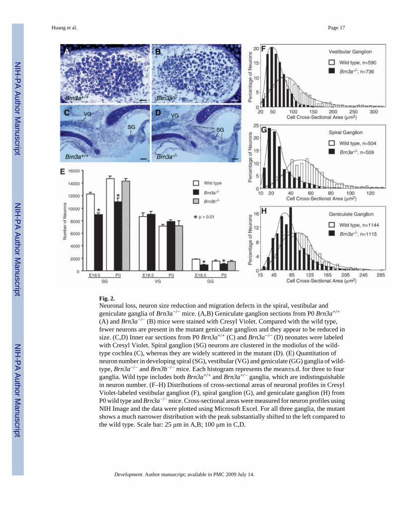
Fig. 2.Neuronal loss, neuron size reduction and migration defects in the spiral, vestibular andgeniculate ganglia of Brn3a−/− mice. (A,B) Geniculate ganglion sections from P0 Brn3a+/+
(A) and Brn3a−/− (B) mice were stained with Cresyl Violet. Compared with the wild type,fewer neurons are present in the mutant geniculate ganglion and they appear to be reduced insize. (C,D) Inner ear sections from P0 Brn3a+/+ (C) and Brn3a−/− (D) neonates were labeledwith Cresyl Violet. Spiral ganglion (SG) neurons are clustered in the modiolus of the wild-type cochlea (C), whereas they are widely scattered in the mutant (D). (E) Quantitation ofneuron number in developing spiral (SG), vestibular (VG) and geniculate (GG) ganglia of wild-type, Brn3a−/− and Brn3b−/− mice. Each histogram represents the mean±s.d. for three to fourganglia. Wild type includes both Brn3a+/+ and Brn3a+/− ganglia, which are indistinguishablein neuron number. (F–H) Distributions of cross-sectional areas of neuronal profiles in CresylViolet-labeled vestibular ganglion (F), spiral ganglion (G), and geniculate ganglion (H) fromP0 wild type and Brn3a−/− mice. Cross-sectional areas were measured for neuron profiles usingNIH Image and the data were plotted using Microsoft Excel. For all three ganglia, the mutantshows a much narrower distribution with the peak substantially shifted to the left compared tothe wild type. Scale bar: 25 μm in A,B; 100 μm in C,D.
Huang et al. Page 17
Development. Author manuscript; available in PMC 2009 July 14.
NIH
-PA Author Manuscript
NIH
-PA Author Manuscript
NIH
-PA Author Manuscript

Fig. 3.Reduction of TrkC, parvalbumin and Brn3b expression in the Brn3a−/− spiral ganglion. (A–F)Compared with the wild-type ganglia (A,C), TrkC immunoreactivity is greatly reduced in theE15.5 (B) and E18.5 (D) mutant spiral ganglia. By contrast, a similar level of TrkBimmunoreactivity is present in E18.5 spiral ganglion between wild-type (E) and Brn3a mutant(F). Arrows point to radial fibers. (G,H) Parvalbumin immunoreactivity shows substantialdecrease in the mutant spiral and vestibular ganglia and their nerve fibers. (I–L) In contrast tothe robust expression of Brn3b in wild-type ganglia, the intensity of Brn3b expression and thenumber of neurons positive for Brn3b is dramatically diminished in E18.5 mutant embryos.Cri, crista; Sac, saccule; SG, spiral ganglion; Utr, utricule; VG, vestibular ganglion. Scale bar:25 μm in A–D,K,L; 50 μm in E,F; 100 μm in G–J.
Huang et al. Page 18
Development. Author manuscript; available in PMC 2009 July 14.
NIH
-PA Author Manuscript
NIH
-PA Author Manuscript
NIH
-PA Author Manuscript

Fig. 4.Afferent and efferent projections to the cochlea in wild type, Brn3a−/− and TrkC−/− mice. (A–C) Afferent projection to the basal turn of cochlea at P0 is demonstrated by labeling the fiberswith DiI. This labeling highlights the modiolus (M), spiral ganglion (SG) and inner ear haircells (HC). In contrast to the densely organized afferent fibers in the wild-type cochlea (A),there is an overall reduction in fiber density in the basal turn of cochlea in Brn3a−/− mice,including an almost complete absence of the hook region of the ganglion in the basal turn(double arrowheads) and gapping between fiber bundles (B). These abnormalities in theBrn3a−/− cochlea are similar to those present in TrkC−/− mice (C). (D–G) The acetylated tubulinantibody labels both afferent (arrows) and efferent (arrowheads) fibers of the cochlea. Incontrast to the orderly arrangement of fibers in wild-type mice (D), there is disorderedoutgrowth of efferent fibers in the basal turn (BT), middle turn (MT) and apical turn (AT) ofthe Brn3a−/− cochlea (E–G). Scale bar: 200 μm in A–C; 100 μm in D–G.
Huang et al. Page 19
Development. Author manuscript; available in PMC 2009 July 14.
NIH
-PA Author Manuscript
NIH
-PA Author Manuscript
NIH
-PA Author Manuscript

Fig. 5.Abnormal afferent fiber projections in the semicircular canals of Brn3a−/− mice. (A,B) DiI-labeled flat mount of E13.5 inner ears shows a dramatic reduction in fiber projection to theposterior vertical canal (pvc) and the cochlea in Brn3a−/− embryos. Although slightly reduced,the innervation to the anterior vertical canal (avc), horizontal canal (hc) and utricle (u) inBrn3a−/− mice is comparable with that in the wild type. (C,D) In contrast to the denseinnervation in the wild type, the posterior vertical canal of Brn3a−/− mice shows essentially noinnervation at P0. Scale bars: 200 μm.
Huang et al. Page 20
Development. Author manuscript; available in PMC 2009 July 14.
NIH
-PA Author Manuscript
NIH
-PA Author Manuscript
NIH
-PA Author Manuscript

Fig. 6.Axon misrouting in the Brn3a−/− vestibular system. Inner ear sections from E16.5 (A–E) andE18.5 (F) Brn3a+/+ (A) and Brn3a−/− (B–F) embryos were immunostained with anti-NF 150(A–C) and anti-p75NTR (D,E) antibodies with counterstaining by Hematoxylin, or labeled bysilver impregnation (F). Although in the wild type (A), vestibular ganglion (VG)-derived nervefibers immunoreactive for NF 150 or p75NTR invariantly follow pathways leading toinnervation of only sensory epithelia of the saccule (Sac), utricule (Utr) and crista (Cri), theyoften form aberrant branches in the mutant that project into the outside of the inner ear (B–D),or into the cochlea (Co) (E). Tracing nerve fiber trajectories by silver impregnation revealssimilar anomalous fiber branches that penetrate the temporal bone and project away from theinner ear (F). Arrows indicate abnormal nerve fiber branches. Scale bar: 50 μm in C,F; 100μm in A,B,D,E.
Huang et al. Page 21
Development. Author manuscript; available in PMC 2009 July 14.
NIH
-PA Author Manuscript
NIH
-PA Author Manuscript
NIH
-PA Author Manuscript

Fig. 7.Expression of EphB1 in the nervous system of Brn3a mutant mice. Sections of E16.5 wild-type (A–C) and Brn3a−/− (D–F) embryos show no difference in the expression of EphB1.Neurons in the spiral ganglion, trigeminal ganglion and inferior olivary nucleus, which areaffected by Brn3a mutation, express similar level of EphB1 protein in both wild type andBrn3a mutants. Co, cochlea; ION, inferior olivary nucleus; SG, spiral ganglion; TG, trigeminalganglion. Scale bar: 100 μm in A,D; 50μm in B,C,E,F.
Huang et al. Page 22
Development. Author manuscript; available in PMC 2009 July 14.
NIH
-PA Author Manuscript
NIH
-PA Author Manuscript
NIH
-PA Author Manuscript

Fig. 8.Schematics illustrating inner ear defects and genetic interactions revealed by analysis ofBrn3a mutant mice. (A) Inner ear defects in Brn3a−/− mice. In the mutant, the spiral ganglion(SG) loses ~30% of neurons by P0, whereas the vestibular ganglion (VG) contains normalnumber of neurons. Substantial reduction in soma size of neurons is seen in both Brn3a−/− SGand VG. In addition, the mutant SG neurons are defective in migration and thus do not becomeclustered. In the mutant cochlea (Co), there is overall decrease in afferent fiber density in thebasal turn with a nearly complete loss of innervation at the base (broken green arrow).Moreover, efferent innervation displays profound pathfinding defects throughout the entirecochlea. In the mutant vestibular system, the saccule (S), utricle (U), and anterior vertical(AVC) and horizontal (HC) canals are all well innervated by afferent fibers. However, theposterior vertical canal (PVC) lacks afferent innervation (broken pink arrow). (B) Geneticinteractions between Brn3a and other transcriptional regulators during sensory gangliogenesis.Brn3a is required for differentiation and survival of sensory neurons in the trigeminal (TG),spiral, vestibular, geniculate (GG) and dorsal root (DRG) ganglia. For fate commitment ofneuron progenitors, the TG, SG and VG require Ngn1; the GG requires Ngn2; and the DRGrequires both Ngn1 and Ngn2. Therefore, Ngn1 and Ngn2 genetically act upstream of Brn3a.Brn3a regulates expression of TrkA, TrkB, TrkC, Brn3b and Brn3c in TG and DRG, and thatof parvalbumin (Parv) and Brn3b in SG and VG. In addition, it controls Brn3b expression inGG, and TrkC expression in SG. During GG development, Phox2a also genetically actsdownstream of Ngn2 as it is only required for differentiation and survival of GG neurons.Phox2a has been shown to control expression of c-Ret and dopamine-β-hydroxylase (DBH)in GG.
Huang et al. Page 23
Development. Author manuscript; available in PMC 2009 July 14.
NIH
-PA Author Manuscript
NIH
-PA Author Manuscript
NIH
-PA Author Manuscript