atypicalclinicalanddiagnosticfeaturesinmen´ etrier’s ... file2 case reports in...
TRANSCRIPT
Hindawi Publishing CorporationCase Reports in Gastrointestinal MedicineVolume 2011, Article ID 480610, 5 pagesdoi:10.1155/2011/480610
Case Report
Atypical Clinical and Diagnostic Features in Menetrier’sDisease in a Child
Michael Chung,1 Jaime Pittenger,2, 3 Deborah Flomenhoft,3, 4 Jeffrey Bennett,2, 3
Eun-Young Lee,5 and Harohalli Shashidhar6
1 University of Kentucky College of Medicine, 138 Leader Avenue, Lexington, KY 40506-9983, USA2 University of Kentucky Children’s Hospital, 800 Rose Street, Lexington, KY 40536-0298, USA3 Department of Pediatrics, Kentucky Clinic, J445 740 S. Limestone, Lexington, KY 40536-0284, USA4 Department of Internal Medicine, Kentucky Clinic, Room J525, 740 South Limestone, Lexington, KY 40536-0284, USA5 Department of Pathology, Medical Science Building, MS117, 800 Rose St., Lexington, KY 40536-0298, USA6 Division of Pediatric Gastroenterology and Nutrition, Department of Pediatrics, University of Kentucky College of Medicine,800 Rose Street, 109, Lexington, KY 40536-0298, USA
Correspondence should be addressed to Michael Chung, [email protected]
Received 13 June 2011; Accepted 7 July 2011
Academic Editors: K. Ina, S. Karoui, and M. Neri
Copyright © 2011 Michael Chung et al. This is an open access article distributed under the Creative Commons AttributionLicense, which permits unrestricted use, distribution, and reproduction in any medium, provided the original work is properlycited.
Menetrier’s disease is one of the rarest protein-losing gastropathies in childhood. It is characterized clinically by non-specificgastrointestinal symptoms and edema, biochemically by hypoalbuminemia, and pathologically by enlarged gastric folds. In adults,this disease can be devastating with significant morbidity and mortality. In childhood, it is a self-limiting, transient and benignillness. Its treatment is largely supportive with total parenteral nutrition (TPN) while oral intake is encouraged. Acute onset ofvomiting in healthy school age children can be initially explained by acute viral gastroenteritis. However, persistent vomitingassociated with hematemesis and severe abdominal pain should warrant further work-up. This case report illustrates a self-limitingand rare cause of protein-losing enteropathy called Menetrier’s disease that presented with several variant clinical features nottypically described in association with this entity.
1. Introduction
Menetrier’s disease is an uncommon cause of protein-losinggastropathy in children characterized by hypertrophy ofgastric folds affecting the gastric body [1]. Symptoms atonset include nausea and vomiting, with profound hypoal-buminemia developing within a few days. The clinical coursein children is distinct from adult-onset disease. The relativerarity of the diagnosis in combination with symptoms thatmimic nonspecific gastroenteritis at onset may delay earlyrecognition. This paper describes several variant clinicalfeatures not typically described in association with this entity.
2. Patient Presentation
A 7-year-old African-American female was readmitted toour institution 4 days after discharge from the hospital with
recurrent non-bloody, nonbilious emesis. She did not havediarrhea, fever, or skin rash. The family did not report anysick contacts.
She had spent ten days in the hospital during the firstadmission with an episode of hematemesis, abdominal pain,persistent nausea, and vomiting along with poor intakerequiring parenteral nutrition (PN) for 3 days. At the timeof discharge, she was tolerating a partial normal diet withno emesis. The hospital course had been complicated bydevelopment of gluteal abscess due to MRSA infection thatwas drained along with two days of clindamycin intravenousadministration.
An esophagogastroduodenoscopy (EGD) during the firstadmission had revealed severe gastropathy with thickenedand friable mucosal folds in the gastric body and numerousgastric erosions with evidence of recent bleeding. Tissue
2 Case Reports in Gastrointestinal Medicine
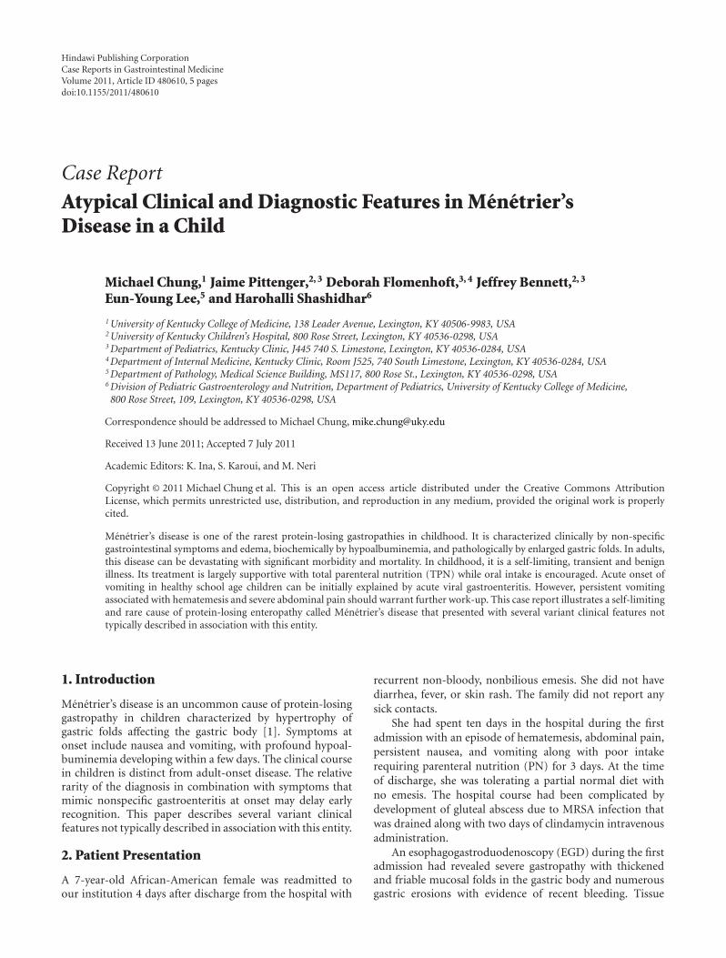
and stool viral cultures had not yielded any viral growth.The biopsy showed erosions and mild eosinophilia withabsent H. pylori. Despite the endoscopic appearance of severegastropathy, the biopsy did not reveal foveolar hyperplasia asis typical of Menetrier’s (Figure 1).
Laboratory evaluation during the first hospital admissionincluded a normal complete blood count and comprehensivemetabolic panel but two positive tests for stool occult blood.Urine analysis showed mild ketonuria. Hypoalbuminemia(1.8 mg/dL) was present by the third day of hospital stay.
The child had a long-standing history of frequent vom-iting and hypersensitivity reactions to certain foods and hadreceived several courses of immunotherapy. Mother reportedthat the child consciously restricted her intake to a diet thatconsisted of bland foods such as plain pasta and bread andshowed avoidance of dairy products. Despite recent weightloss, her BMI was at the 50th percentile.
Physical examination revealed a well-developed, but ill-appearing child. She was afebrile with stable vital signs. Gen-eral physical examination was notable for mild periorbitaledema and conjunctival pallor. Systemic examination wasunremarkable including absence of any abdominal findings.Peripheral edema was absent.
Laboratory evaluation at second admission revealednormal complete blood count, mild ketonuria, and a urine’sspecific gravity of >1030. A mild lipase elevation (62) waspresent, and serum albumin of 1.7 had further decreasedto 1.4 by the third day along with a normal prealbumin.Serum gastrin was not measured. Other studies over thecourse of the hospital stay included low immunoglobulinG and M levels (IgG < 200 mg/dL, IgM 27 mg/dL). Bythe time of discharge, immunoglobulins were normal withthe exception of improved but low IgG level (350 mg/dL).The serum albumin had remained low at 1.5. A fecal α-1-antitrypsin level was elevated at >1.33, consistent with aprotein-losing enteropathy. Additional testing included nor-mal serum cortisol level and negative tissue transglutaminaseassay.
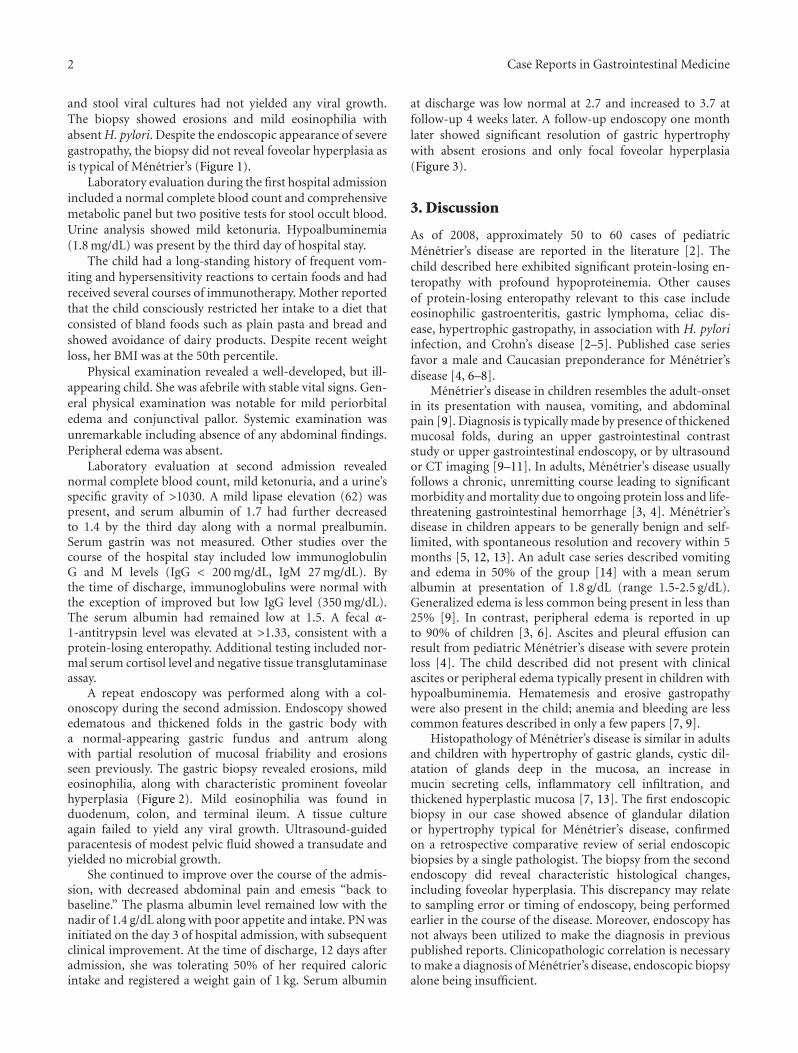
A repeat endoscopy was performed along with a col-onoscopy during the second admission. Endoscopy showededematous and thickened folds in the gastric body witha normal-appearing gastric fundus and antrum alongwith partial resolution of mucosal friability and erosionsseen previously. The gastric biopsy revealed erosions, mildeosinophilia, along with characteristic prominent foveolarhyperplasia (Figure 2). Mild eosinophilia was found induodenum, colon, and terminal ileum. A tissue cultureagain failed to yield any viral growth. Ultrasound-guidedparacentesis of modest pelvic fluid showed a transudate andyielded no microbial growth.
She continued to improve over the course of the admis-sion, with decreased abdominal pain and emesis “back tobaseline.” The plasma albumin level remained low with thenadir of 1.4 g/dL along with poor appetite and intake. PN wasinitiated on the day 3 of hospital admission, with subsequentclinical improvement. At the time of discharge, 12 days afteradmission, she was tolerating 50% of her required caloricintake and registered a weight gain of 1 kg. Serum albumin
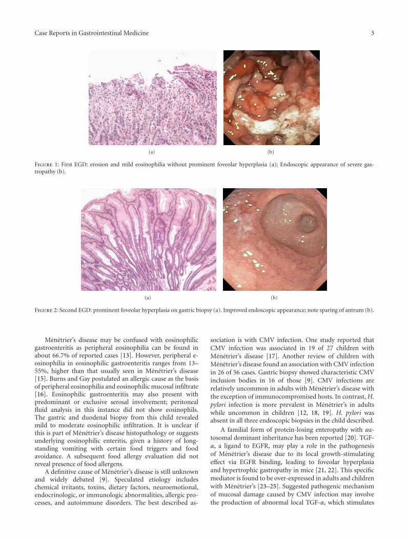
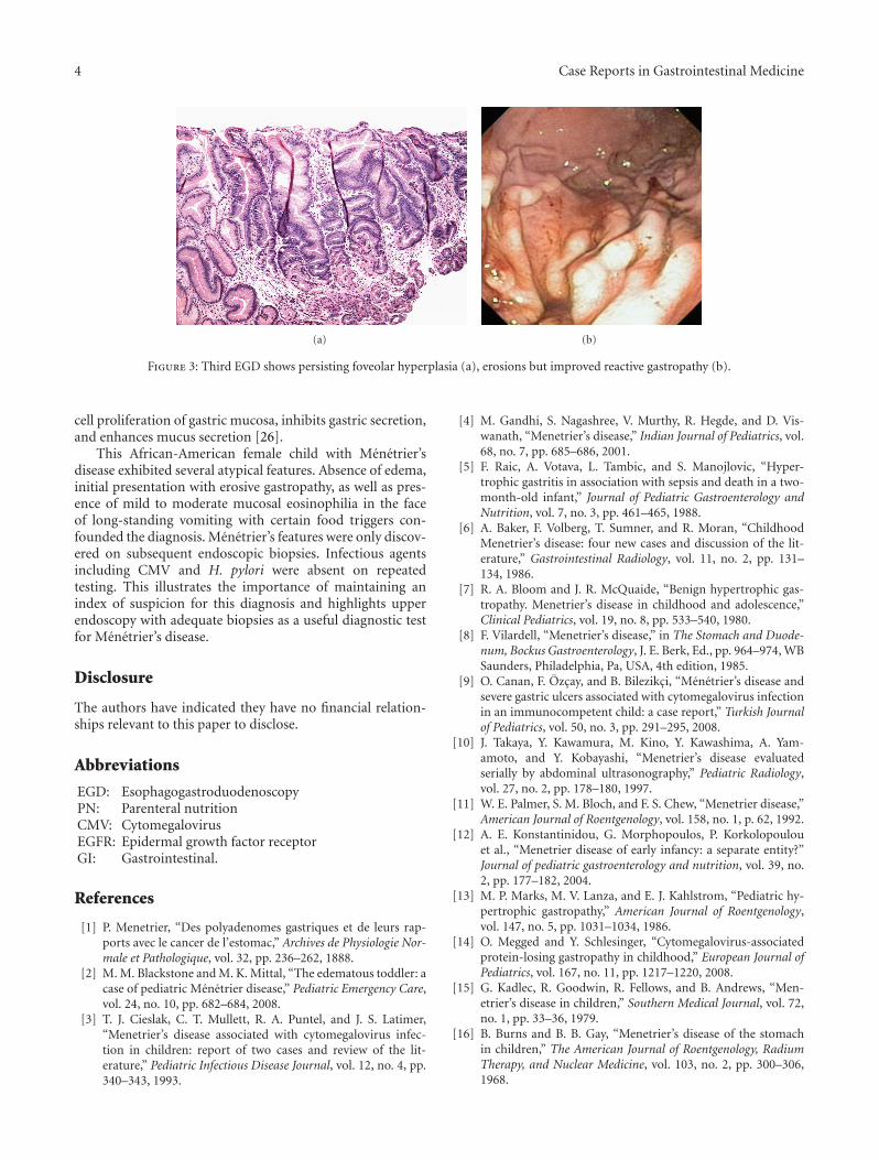
at discharge was low normal at 2.7 and increased to 3.7 atfollow-up 4 weeks later. A follow-up endoscopy one monthlater showed significant resolution of gastric hypertrophywith absent erosions and only focal foveolar hyperplasia(Figure 3).
3. Discussion
As of 2008, approximately 50 to 60 cases of pediatricMenetrier’s disease are reported in the literature [2]. Thechild described here exhibited significant protein-losing en-teropathy with profound hypoproteinemia. Other causesof protein-losing enteropathy relevant to this case includeeosinophilic gastroenteritis, gastric lymphoma, celiac dis-ease, hypertrophic gastropathy, in association with H. pyloriinfection, and Crohn’s disease [2–5]. Published case seriesfavor a male and Caucasian preponderance for Menetrier’sdisease [4, 6–8].
Menetrier’s disease in children resembles the adult-onsetin its presentation with nausea, vomiting, and abdominalpain [9]. Diagnosis is typically made by presence of thickenedmucosal folds, during an upper gastrointestinal contraststudy or upper gastrointestinal endoscopy, or by ultrasoundor CT imaging [9–11]. In adults, Menetrier’s disease usuallyfollows a chronic, unremitting course leading to significantmorbidity and mortality due to ongoing protein loss and life-threatening gastrointestinal hemorrhage [3, 4]. Menetrier’sdisease in children appears to be generally benign and self-limited, with spontaneous resolution and recovery within 5months [5, 12, 13]. An adult case series described vomitingand edema in 50% of the group [14] with a mean serumalbumin at presentation of 1.8 g/dL (range 1.5-2.5 g/dL).Generalized edema is less common being present in less than25% [9]. In contrast, peripheral edema is reported in upto 90% of children [3, 6]. Ascites and pleural effusion canresult from pediatric Menetrier’s disease with severe proteinloss [4]. The child described did not present with clinicalascites or peripheral edema typically present in children withhypoalbuminemia. Hematemesis and erosive gastropathywere also present in the child; anemia and bleeding are lesscommon features described in only a few papers [7, 9].
Histopathology of Menetrier’s disease is similar in adultsand children with hypertrophy of gastric glands, cystic dil-atation of glands deep in the mucosa, an increase inmucin secreting cells, inflammatory cell infiltration, andthickened hyperplastic mucosa [7, 13]. The first endoscopicbiopsy in our case showed absence of glandular dilationor hypertrophy typical for Menetrier’s disease, confirmedon a retrospective comparative review of serial endoscopicbiopsies by a single pathologist. The biopsy from the secondendoscopy did reveal characteristic histological changes,including foveolar hyperplasia. This discrepancy may relateto sampling error or timing of endoscopy, being performedearlier in the course of the disease. Moreover, endoscopy hasnot always been utilized to make the diagnosis in previouspublished reports. Clinicopathologic correlation is necessaryto make a diagnosis of Menetrier’s disease, endoscopic biopsyalone being insufficient.
Case Reports in Gastrointestinal Medicine 3
(a) (b)
Figure 1: First EGD: erosion and mild eosinophilia without prominent foveolar hyperplasia (a); Endoscopic appearance of severe gas-tropathy (b).
(a) (b)
Figure 2: Second EGD: prominent foveolar hyperplasia on gastric biopsy (a). Improved endoscopic appearance; note sparing of antrum (b).
Menetrier’s disease may be confused with eosinophilicgastroenteritis as peripheral eosinophilia can be found inabout 66.7% of reported cases [13]. However, peripheral e-osinophilia in eosinophilic gastroenteritis ranges from 13–55%, higher than that usually seen in Menetrier’s disease[15]. Burns and Gay postulated an allergic cause as the basisof peripheral eosinophilia and eosinophilic mucosal infiltrate[16]. Eosinophilic gastroenteritis may also present withpredominant or exclusive serosal involvement; peritonealfluid analysis in this instance did not show eosinophils.The gastric and duodenal biopsy from this child revealedmild to moderate eosinophilic infiltration. It is unclear ifthis is part of Menetrier’s disease histopathology or suggestsunderlying eosinophilic enteritis, given a history of long-standing vomiting with certain food triggers and foodavoidance. A subsequent food allergy evaluation did notreveal presence of food allergens.
A definitive cause of Menetrier’s disease is still unknownand widely debated [9]. Speculated etiology includeschemical irritants, toxins, dietary factors, neuroemotional,endocrinologic, or immunologic abnormalities, allergic pro-cesses, and autoimmune disorders. The best described as-
sociation is with CMV infection. One study reported thatCMV infection was associated in 19 of 27 children withMenetrier’s disease [17]. Another review of children withMenetrier’s disease found an association with CMV infectionin 26 of 56 cases. Gastric biopsy showed characteristic CMVinclusion bodies in 16 of those [9]. CMV infections arerelatively uncommon in adults with Menetrier’s disease withthe exception of immunocompromised hosts. In contrast, H.pylori infection is more prevalent in Menetrier’s in adultswhile uncommon in children [12, 18, 19]. H. pylori wasabsent in all three endoscopic biopsies in the child described.
A familial form of protein-losing enteropathy with au-tosomal dominant inheritance has been reported [20]. TGF-α, a ligand to EGFR, may play a role in the pathogenesisof Menetrier’s disease due to its local growth-stimulatingeffect via EGFR binding, leading to foveolar hyperplasiaand hypertrophic gastropathy in mice [21, 22]. This specificmediator is found to be over-expressed in adults and childrenwith Menetrier’s [23–25]. Suggested pathogenic mechanismof mucosal damage caused by CMV infection may involvethe production of abnormal local TGF-α, which stimulates
4 Case Reports in Gastrointestinal Medicine
(a) (b)
Figure 3: Third EGD shows persisting foveolar hyperplasia (a), erosions but improved reactive gastropathy (b).
cell proliferation of gastric mucosa, inhibits gastric secretion,and enhances mucus secretion [26].
This African-American female child with Menetrier’sdisease exhibited several atypical features. Absence of edema,initial presentation with erosive gastropathy, as well as pres-ence of mild to moderate mucosal eosinophilia in the faceof long-standing vomiting with certain food triggers con-founded the diagnosis. Menetrier’s features were only discov-ered on subsequent endoscopic biopsies. Infectious agentsincluding CMV and H. pylori were absent on repeatedtesting. This illustrates the importance of maintaining anindex of suspicion for this diagnosis and highlights upperendoscopy with adequate biopsies as a useful diagnostic testfor Menetrier’s disease.
Disclosure
The authors have indicated they have no financial relation-ships relevant to this paper to disclose.
Abbreviations
EGD: EsophagogastroduodenoscopyPN: Parenteral nutritionCMV: CytomegalovirusEGFR: Epidermal growth factor receptorGI: Gastrointestinal.
References
[1] P. Menetrier, “Des polyadenomes gastriques et de leurs rap-ports avec le cancer de l’estomac,” Archives de Physiologie Nor-male et Pathologique, vol. 32, pp. 236–262, 1888.
[2] M. M. Blackstone and M. K. Mittal, “The edematous toddler: acase of pediatric Menetrier disease,” Pediatric Emergency Care,vol. 24, no. 10, pp. 682–684, 2008.
[3] T. J. Cieslak, C. T. Mullett, R. A. Puntel, and J. S. Latimer,“Menetrier’s disease associated with cytomegalovirus infec-tion in children: report of two cases and review of the lit-erature,” Pediatric Infectious Disease Journal, vol. 12, no. 4, pp.340–343, 1993.
[4] M. Gandhi, S. Nagashree, V. Murthy, R. Hegde, and D. Vis-wanath, “Menetrier’s disease,” Indian Journal of Pediatrics, vol.68, no. 7, pp. 685–686, 2001.
[5] F. Raic, A. Votava, L. Tambic, and S. Manojlovic, “Hyper-trophic gastritis in association with sepsis and death in a two-month-old infant,” Journal of Pediatric Gastroenterology andNutrition, vol. 7, no. 3, pp. 461–465, 1988.
[6] A. Baker, F. Volberg, T. Sumner, and R. Moran, “ChildhoodMenetrier’s disease: four new cases and discussion of the lit-erature,” Gastrointestinal Radiology, vol. 11, no. 2, pp. 131–134, 1986.
[7] R. A. Bloom and J. R. McQuaide, “Benign hypertrophic gas-tropathy. Menetrier’s disease in childhood and adolescence,”Clinical Pediatrics, vol. 19, no. 8, pp. 533–540, 1980.
[8] F. Vilardell, “Menetrier’s disease,” in The Stomach and Duode-num, Bockus Gastroenterology, J. E. Berk, Ed., pp. 964–974, WBSaunders, Philadelphia, Pa, USA, 4th edition, 1985.
[9] O. Canan, F. Ozcay, and B. Bilezikci, “Menetrier’s disease andsevere gastric ulcers associated with cytomegalovirus infectionin an immunocompetent child: a case report,” Turkish Journalof Pediatrics, vol. 50, no. 3, pp. 291–295, 2008.
[10] J. Takaya, Y. Kawamura, M. Kino, Y. Kawashima, A. Yam-amoto, and Y. Kobayashi, “Menetrier’s disease evaluatedserially by abdominal ultrasonography,” Pediatric Radiology,vol. 27, no. 2, pp. 178–180, 1997.
[11] W. E. Palmer, S. M. Bloch, and F. S. Chew, “Menetrier disease,”American Journal of Roentgenology, vol. 158, no. 1, p. 62, 1992.
[12] A. E. Konstantinidou, G. Morphopoulos, P. Korkolopoulouet al., “Menetrier disease of early infancy: a separate entity?”Journal of pediatric gastroenterology and nutrition, vol. 39, no.2, pp. 177–182, 2004.
[13] M. P. Marks, M. V. Lanza, and E. J. Kahlstrom, “Pediatric hy-pertrophic gastropathy,” American Journal of Roentgenology,vol. 147, no. 5, pp. 1031–1034, 1986.
[14] O. Megged and Y. Schlesinger, “Cytomegalovirus-associatedprotein-losing gastropathy in childhood,” European Journal ofPediatrics, vol. 167, no. 11, pp. 1217–1220, 2008.
[15] G. Kadlec, R. Goodwin, R. Fellows, and B. Andrews, “Men-etrier’s disease in children,” Southern Medical Journal, vol. 72,no. 1, pp. 33–36, 1979.
[16] B. Burns and B. B. Gay, “Menetrier’s disease of the stomachin children,” The American Journal of Roentgenology, RadiumTherapy, and Nuclear Medicine, vol. 103, no. 2, pp. 300–306,1968.
Case Reports in Gastrointestinal Medicine 5
[17] R. O. Occena, S. F. Taylor, C. C. Robinson, and R. J. Sokol, “As-sociation of cytomegalovirus with Menetrier’s disease inchildhood: report of two new cases with a review of literature,”Journal of Pediatric Gastroenterology and Nutrition, vol. 17, no.2, pp. 217–224, 1993.
[18] E. Bayerdorffer, M. M. Ritter, R. Hatz, W. Brooks, G. Ruckde-schel, and M. Stolte, “Healing of protein losing hypertrophicgastropathy by eradication of Helicobacter pylori—is Heli-cobacter pylori a pathogenic factor in Menetrier’s disease?”Gut, vol. 35, no. 5, pp. 701–704, 1994.
[19] M. Yamada, R. Sumazaki, H. Adachi et al., “Resolution of pro-tein-losing hypertrophic gastropathy by eradication of Heli-cobacter pylori,” European Journal of Pediatrics, vol. 156, no. 3,pp. 182–185, 1997.
[20] B. Larsen, U. Tarp, and E. Kristensen, “Familial giant hyper-trophic gastritis (Menetrier’s disease),” Gut, vol. 28, no. 11, pp.1517–1521, 1987.
[21] P. J. Dempsey, J. R. Goldenring, C. J. Soroka et al., “Possiblerole of transforming growth factor α in the pathogenesis ofMenetrier’s disease: supportive evidence from humans andtransgenic mice,” Gastroenterology, vol. 103, no. 6, pp. 1950–1963, 1992.
[22] M. van den Berg, P. Stokkers, E. Rings, and H. Buller, “Trans-forming growth factor alpha in Menetrier’s disease,” Journalof Pediatric Gastroenterology and Nutrition, vol. 17, no. 2, pp.230–232, 1993.
[23] R. F. Bluth, H. A. Carpenter, M. R. Pittelkow, D. L. Page,and R. J. Coffey, “Immunolocalization of transforming growthfactor-α in normal and diseased human gastric mucosa,” Hu-man Pathology, vol. 26, no. 12, pp. 1333–1340, 1995.
[24] T. J. Sferra, B. R. Pawel, S. J. Qualman, and B. U. K. Li,“Menetrier disease of childhood: role of cytomegalovirus andtransforming growth factor alpha,” Journal of Pediatrics, vol.128, no. 2, pp. 213–219, 1996.
[25] R. M. Soetikno, “Menetrier’s disease: report of a transientcase associated with chylous ascites,” American Journal of Gas-troenterology, vol. 92, no. 8, pp. 1364–1367, 1997.
[26] C. Faure, M. Besnard, A. Hirsch et al., “Chronic hypertrophicgastropathy in a child resembling adult Menetrier’s disease,”Journal of Pediatric Gastroenterology and Nutrition, vol. 23, no.4, pp. 419–421, 1996.