attimarad s l et al / ijrap 3(3), may s l et al / ijrap 3(3), may – jun 2012 448 for their...
TRANSCRIPT
Attimarad S L et al / IJRAP 3(3), May – Jun 2012
447
Research Article www.ijrap.net
SCREENING, ISOLATION AND PURIFICATION OF ANTIBIOTIC(S) FROM MARINE ACTINOMYCETES Attimarad S L1*, Gaviraj E N2, Nagesh C1, Kugaji M S3, Sutar R S1 1Maratha Mandal’s College of Pharmacy, Belgaum, Karnataka, India 2B L D E A College of Pharmacy, Bijapur, Karnataka, India 3Maratha Mandal's NGH Institute of Dental Sciences and Research Centre, Belgaum, Karnataka, India Received on: 19/02/12 Revised on: 03/04/12 Accepted on: 25/04/12 *Corresponding author Sunil Attimarad, Maratha Mandal’s College of Pharmacy, Belgaum, Karnataka, India E-mail: [email protected] ABSTRACT As marine environmental conditions are extremely different from terrestrial ones, it is surmised that marine actinomycetes might produce novel bioactive compounds. Hence marine sediments, collected from the coastal areas of Gokharna and Muradeshwara of Karnataka state, were screened. Seventeen isolates were obtained on starch-casein agar media by soil dilution technique. However, only six isolates namely SUN-A2, SUN-A3, SUN-A4, SUN-A5, SUN-A7 and SUN-A15 showed significant antibacterial activity against both gram-positive and gram-negative bacteria. Further studies were carried out with the most active SUN-A2. Optimization of media, temperature and pH by shake flask fermentation indicated starch-casein, 28o C and pH 7 to be suitable for SUN-A2. The production of antibiotics began after 24 h reached maximum at 72 h and maintained at the same level up to 120 h. Ethyl acetate was used to extract antibacterial compounds from the culture filtrate. TLC was done on silica gel using ethyl acetate: methanol (6:4) and direct bioautography showed the presence of two active substance, one with Rf 0.8 more active than the other with Rf 0.4. Further purification is done by column chromatography using a mixture of dicholoromethane and ethyl acetate. The findings from this investigation reveal that the strain SUN-A2 in order exhibited superior antimicrobial activity to other sediment isolates of actinomycetes. Key words: Marine actinomycetes, Bioautography, Antibacterial, Fermentation.
INTRODUCTION Bioactive substances of low molecular compounds exhibit various activities. Microorganisms and plants have been important sources of natural medicinal substances1. There is a fast emergence of newer infections and the organisms developing resistance which render already existing antibiotics less effective. Therefore a constant search for new antibiotics to overcome these problems is a matter of necessity. Most of the antibiotics in clinical use are direct natural products or semisynthetic derivatives from actinomycetes or fungi. Actinomycetes are the most economically and biotechnologically valuable prokaryotes and are responsible for the production of about half of the discovered secondary metabolites. Because of the excellent track record of actinomycetes in regard a significant amount of effort has been focused on the successful isolation of novel actinomycetes from terrestrial sources for drug screening programmes in the past fifty years. Recently the rate of discovery of new compounds from terrestrial actinomycetes has decreased whereas the rate of reisolation of known compounds has increased. Thus, it is excited that new groups of actinomycetes from unexplored or under exploited habitals be persued as sources of novel bioactive secondary metabolites2. Although the diversity of life in the terrestrial environment is extraordinary, the greatest biodiversity is in the oceans3. More than 70% of our planet’s surface is covered by oceans and life on Earth originated from sea. In some marine ecosystems, such as the deep sea floor and coral reefs, experts estimate that the biological diversity is higher than in the tropical rainforests4. As marine environmental conditions are extremely different
from terrestrial ones, it is surmised that marine actinomycetes have different characteristics from those of terrestrial counterparts, and therefore, might produce different types of bioactive compounds5, 6. The present study deals with screening for the isolation of actinomycetes to produce antibiotics, isolated from the marine sediments in and around Gokharna and Murudeshwara coastal regions and determination of their antimicrobial activity. MATERIALS AND METHODS Collection of marine sediments The marine sediments were collected by using core sampler from the coastal regions of Gokharna and Murudeshwara of Karnataka, India. The sediment samples were brown to black in colour and of sandy texture. Isolation of actinomycetes Actinomycetes were isolated by soil dilution plate technique on starch casein agar medium, starch nitrate agar medium, glycerol glycine agar medium, and chitin agar medium7, 8. The plates were incubated at 28oc and the numbers of colonies were determined after 15 days. All the medium containing 50% sea water was supplemented with Nystatin 50μg/ml and Nalidixidic acid 20 μg/ml to inhibit the bacterial and fungal contamination respectively8. The selected colonies were picked up and further purified by streak plate technique over starch casein agar slants. Screening for antimicrobial activity The antimicrobial activity was studied preliminarily by cup plate method8 against bacteria and fungi. The test organisms were used are Bacillus subtilis, Staphylococcus aureus, Escherichia coli, Pseudomonas aeruginosa, Aspergillus niger. After preliminary testing of the isolates

Attimarad S L et al / IJRAP 3(3), May – Jun 2012
448
for their antimicrobial activities the most active isolates was selected for further study, Selection of suitable medium The test media selected were starch casein broth, nutrient broth and corn steep liquor broth. The seed inoculum was transferred at 10% level into 100 ml of medium contained in 250 ml conical flask. The seeded flasks were kept on gyratory shaker (200 rpm) at 280 C for 5days. Samples of 10 ml were withdrawn in sterile graduated centrifuge tubes immediately after seeding and at the interval of 24 h for 5 days. Samples were centrifuged at 4000 rpm for 20 minutes. Growth was determined by measuring the packed cell volume (PVC) and the supernatants were subjected to agar diffusion assay against Staphylococcus aureus for determining the production of the antibiotic9, 10.
Temperature and pH Optimum temperature for the productivity and growth of the strain SUN-A2 was determined by inoculating the organism with starch casein broth and incubation was done at 200, 240, 280, 320, and 380 C for 3 days on a gyratory shaker at 200 rpm9, 10. After incubation period, growth and production of the antibiotic were determined as described above. Similarly, optimum pH was determined by adjusting the pH of starch casein broth to values like 5, 6, 7, and 8. The pH values were adjusted by 0.1N NaOH and 0.1n HCl before sterilization9, 10. The incubation was done at 280C for 3 days at 200 rpm on a gyratory shaker. Growth and production of the antibiotic were determined as described above. Time course study of activity by shake flask fermentation Seed inoculum was prepared by using starch casein broth. Three 250 ml flasks each containing 100 ml starch casein broth of pH 7 were seeded with 48 h seed inoculum at the
concentration of 10%. The flasks were shaken (200 rpm) on a gyratory shaker at 28oC. The study was conducted for 6 days. Every day, 10 ml of culture was withdrawn for determining the packed cell volume (PCV) by centrifuging at 4000 rpm for 20 min. The supernatants were subjected to agar diffusion assay by cup plate technique against Staphylococcus aureus to study antibiotic production8, 11. Thin layer chromatography and bioautography The concentrated ethyl acetate extract was subjected to TLC using ethyl acetate:methanol (6:4). Direct bioautography using Staphylococcus aureus as marker organism was carried out to localize antimicrobial substances12-14. Column chromatography The concentrated ethyl acetate layer was loaded on to the column and eluted with dichloromethane and the polarity of the mobile phase was gradually increased by adding ethyl acetate15. All fractions collected were checked individually for purity by running thin layer chromatogram along with crude extract as reference in a different lane. RESULTS AND DISCUSSION A limited scale survey of bioactive actinomycetes from marine sediment collected from coastal areas of Gokharna and murudeshwara of Karnataka, India was made. During the course of our survey, a total of 17 actinomycetes were isolated and coaded as SUN-A1, SUN-A2, to SUN-A17 and analyzed for antimicrobial activities. Among these isolates, six isolates namely SUN-A2, SUN-A3, SUN-A4, SUN-A5, and SUN-A7 showed significant antibacterial activity (Table 1). However none of the isolates exhibited antifungal activity. Among these SUN-A2 showed maximum antibacterial activity, it was selected for detailed study.
Table 1: Sensitivity of different microorganisms against actinomycetes isolates
Isolates Zone of inhibition (in mm)
S. aureus E. coli B.subtilis P.aeruginosa A. niger C. albicans SUN-A1 - 7 - - - -
SUN-A2 18 15 12 14 - -
SUN-A3 10 8 7 7 - -
SUN-A4 15 14 9 8 - -
SUN-A5 13 10 7 7 - -
SUN-A6 8 - - 7 - -
SUN-A7 9 7 8 - - -
SUN-A8 7 7 - - - -
SUN-A9 - - - 8 - -
SUN-A10 - - - - - -
SUN-A11 7 - 9 - - -
SUN-A12 - - - - - -
SUN-A13 - - - - - -
SUN-A14 10 - - 9 - -
SUN-A15 7 7 9 - - -
SUN-A16 - 8 - - - -
SUN-A17 8 - - - -

Attimarad S L et al / IJRAP 3(3), May – Jun 2012
449
The growth of the organism was determined as packed cell volume (PCV). The culture supernatants were tested for activity against overnight culture of Staphylococcus aureus by cup plate technique. Zone of inhibition were measured after overnight incubation at 37o C. The growth of SUN-A2 calculated as PCV was found to be maximum after 72 h of fermentation in starch-casein broth (Graph 1). In case of nutrient and corn steep liquor broth, the growth was found to be maximal after 96 h of fermentation. The zone of inhibition was found to be maximum after 72 h of fermentation in seeded starch casein and nutrient broths. This indicated that the maximum production of antibiotic was occurring at the end of 3rd day of fermentation. In case of seeded corn steep liquor broth, the zone of inhibition was found to be maximum after 96 h of fermentation. The onset of antibiotic production started after 48 h in case of corn steep liquor medium while the onset of antibiotic production was much earlier in case of other two media. Among the media tested, seeded starch casein broth showed more zone of inhibition due to more antibiotic production than seeded nutrient and corn steep liquor broths. Hence, starch casein broth was selected as the suitable medium for shake flask fermentation of SUN-A2.
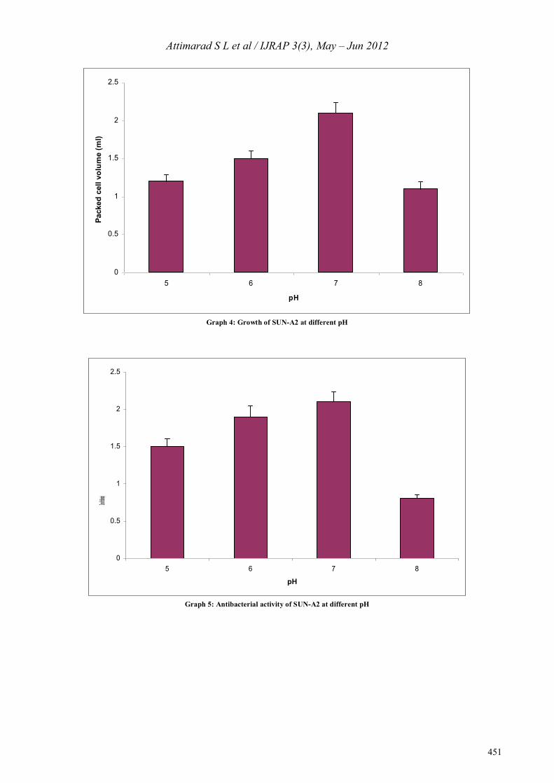
Optimum temperature for maximum growth and productivity of SUN-A2 was determined by studying their packed cell volume (PCV) and zone of inhibition (Graph 2&3). From the temperature optimization experiments, it was observed that the temperature adequate for growth is the same as that for antibiotic production. SUN-A2 strain has proved to show maximal growth and antibiotic production at 28° C. pH of the culture medium affects not only growth but also production of the antibiotic. The optimum pH for maximal growth and antibiotic production of SUN-A2 was studied by determining the packed cell volume (PCV) and zone of inhibition (Graph 4 and 5). From the results it was observed that pH 7 was found to be suitable for both growth and production of bioactive substances. Fermentation profile of SUN-A2 was carried out using starch-casein broth and is shown in (Graph 6). The production began after 24 h, reached maximum at 72 h and maintained at the same level up to 120 h. Later on the production was decreased. The biomass expressed in terms of packed cell volume (PCV) gradually increased and reached maximum at 72 h and then maintained at the same level indicating no autolysis even after 144 h.
0
0.5
1
1.5
2
2.5
0 24 48 72 96 120
Time of incubation (hr)
Pack
ed c
ell v
olum
e (m
l)
Starch-casein Nutrient Corn steep liquor
Graph 1: Growth and antibacterial activity of SUN-A2 on different media

Attimarad S L et al / IJRAP 3(3), May – Jun 2012
450
0
0.5
1
1.5
2
2.5
20 24 28 32 36Temperature (o C)
Pack
ed c
ell v
olum
e (m
l)
Graph 2: Growth of SUN-A2 at different temperature
0
0.5
1
1.5
2
2.5
20 24 28 32 36
Temperature (oC)
Zone of inhibition (cm)
Graph 3: Antibacterial activity of SUN-A2 different temperature
Attimarad S L et al / IJRAP 3(3), May – Jun 2012
451
0
0.5
1
1.5
2
2.5
5 6 7 8
pH
Pack
ed c
ell v
olum
e (m
l)
Graph 4: Growth of SUN-A2 at different pH
0
0.5
1
1.5
2
2.5
5 6 7 8
pH
Zone of inhibition (cm)
Graph 5: Antibacterial activity of SUN-A2 at different pH

Attimarad S L et al / IJRAP 3(3), May – Jun 2012
452
0
0. 5
1
1. 5
2
2. 5
0
0.5
1
1.5
2
2.5
0 24 48 72 96 120 144
Packed cell volume (ml)
Time of harvest (hr)
PCV Z one of inhibit io n
Graph 6: Shake flask fermentation profile of SUN-A2
The culture filtrate of SUN-A2 was subjected to solvent extraction using solvents of increasing polarity viz. dichloromethane, ethyl acetate and butanol. Ethyl acetate extract showed maximum activity compared to the other two extracts (Fig 1; Table 2). The spent broth obtained after ethyl acetate extraction when tested by cup plate technique showed negligible activity against Staphylococcus aureus. The result indicated that the antibiotic was almost extracted from the culture filtrate. Hence, the remaining culture filtrate was extracted with ethyl acetate.
Table 2: Antibacterial activity of the fractions of SUN-A2 culture
filtrate Sl No. Fraction Zone of inhibition (cm)*
1 Dichloromethane 0.8±0.05 2 Ethyl acetate 1.5±0.12 3 n-Butanol 1.1±0.1
The concentrated ethyl acetate extract was subjected to TLC analysis using three mobile phases namely dichloromethane: methanol (4:1), ethyl acetate: methanol (6:4) and n-butanol: acetic acid: water (4:1:5). After air-drying, the plates were visualized with iodine vapor. Of the three mobile phases, the plate developed with ethyl acetate: methanol (6:4) gave the best separation (Figure 2). Hence, bioautography was done for the plate developed with ethyl acetate: methanol (6:4) to localize antibacterial compounds.
Figure 1: Antimicrobial activity of ethyl acetate extract
Figure 2: TLC of ethyl acetate extract of culture filtrate of SUNA-2 Direct bioautography using Staphylococcus aureus as marker organism was used. The plate was incubated overnight at 37o C, sprayed with a 2% w/v aqueous solution of 2,3,5-triphenyl tetrazolium chloride and incubated for further 4 hr. The results indicated that presence of 2 active substances (Figure 3). The compound with Rf 0.8 showed more activity than the one with Rf 0.4.

Attimarad S L et al / IJRAP 3(3), May – Jun 2012
453
Figure 3: Bioautography of ethyl acetate extract of culture filterate
of SUN-A2
In order to isolate the most active compound, column chromatography was carried out. The ethyl acetate layer was concentrated, adsorbed over silica gel (100 to 120 mesh) and loaded onto the column. Elution was started with dichloromethane and the polarity of the mobile phase was gradually increased by adding ethyl acetate. All the fractions collected from column chromatography were checked individually for purity by running thin layer chromatogram along with crude extract as reference in a different lane. CONCLUSION The study has showed that Starch-casein agar supplemented with nystatin and nalidixic acid was found to be suitable for isolating actinomycetes from marine sediments, which were collected from the coastal areas of Gokharna and Muradeshwara of Karnataka state. Out of seventeen isolates only six exhibited significant antibacterial activity. Starch-casein broth, temperature of 28o C and pH of 7 were found to be suitable for the fermentation of SUN-A2, the most active isolate. Ethyl acetate was found to be suitable for extracting antibacterial substances from the culture filtrate. Bioautography revealed that the presence of two antibacterial substances. Dichloromethane: ethyl acetate (10: 90) was found to be suitable for isolating the antibacterial substance (Rf-0.8) from the silica gel column. The exploitation of marine actinomycetes as a source for novel secondary metabolites production is in its infancy. However, the discovery rate of novel secondary metabolites from marine actinomycetes has recently surpassed that of their terrestrial counterparts. In this context, ours is a small but an honest effort directed towards isolating antibiotic producing marine actinomycetes. But future success relies on the ability to isolate novel actinomycetes from the marine environments. Although isolation strategies directed towards new marine-derived actinomycetes have been
lacking, some progress has recently been made in this area. Further development work in improving isolation strategies in the recovery of marine actinomycetes is of utmost importance for ensuring success in this area. ACKNOWLEDGEMENT I sincerely thank management of Maratha Mandal’s College of Pharmacy and KLES’s College of Pharmacy Belgaum for providing facilities to conduct this research work. REFERENCES 1. Vandamme EJ. Biotechnology of antibiotics, New York: Marcel
Dekker; 1984. 2. Fenical W, Baden D, Burg M, de Goyet CV, Grimes JD, Katz M,
et al. Marine derived pharmaceuticals and related bioactive compounds, Understanding the Ocean’s role in human health, 1999:71-86.
3. Donia M, Humann MT. Marine natural products and their potential applications as anti infective agents. Lancet infect dis, 2003; 3:338-348.
4. Haefner B: Drugs from the deep marine natural products as drug candidates, Drug discov today, 2003; 8:536-544.
5. Bull AT, Stach JEM, Ward AC, Goodfellow M. Marine actinobacteria perspectives, challenges, future directions, Antonie van leeuwenhook, 2005; 87:65-79.
6. Starch JEM, Maldonado LA, Ward AC, Goodfellow M, Bull AT. New primers for the class Actinobacteria: Applications to marine and terrestrial environments, Environ microbial, 2003; 5: 824-841.
7. Williams ST, Cross T. Actinomycetes isolation from soil, Methods in microbiology, Academic press, London, New York, 1971; 4: 295-334.
8. Dubey RC, Maheshwari DK. Practical Microbiology, New Delhi: S Chand and Company Ltd; 2005.
9. James PDA, Edwards C, Dawson M. The effects of temperature, pH and growth rate on secondary metabolism in Streptomyces thermoviolaceus grown in a chemostat. J Gen Microbiol, 1991; 137:1715-1720.
10. Gesheva V, Ivanova V, Gesheva R. Effects of nutrients on the production of AK-111-81 macrolide antibiotic by Streptomyces hygroscopicus. Microbiol Res, 2005; 160: 243-248.
11. Shomura T, Gomi S, Ito M, Yoshida J, Tanaka E, Amono S et al. Studies on new antibiotics SF2415 I. taxonomy, fermentation, isolation, physico-chemical properties and biological activities. J Antibiot, 1987; 11: 732-739.
12. Homans AL, Fuchs A. Direct bioautography on thin-layer chromatograms as a method for detecting fungitoxic substances. J Chromatogr, 1970; 51: 327-329.
13. Pandey B, Ghimire P, Agrawal VP. Studies on the antibacterial activity of the actinomycetes isolated from the Khumbu Region of Mount Everest. The Great Himalayas Climate, Health, Ecology, Management and Conservation, Kathmandu, 2004.
14. Staneck JL, Roberts GD. Simplified approach to identification of aerobic actinomycetes by thin layer chromatography. Appl Microbiol, 1994; 28: 226-231.
15. Ohshima T, Takada H, Yoshimura T, Esaki N, Soda K. Distribution, Purification, and Characterization of Thermostable Phenyalanine Dehydrogenase from Thermophillic actinomycetes. J Bacteriol, 1991; 173: 3943-3948
Source of support: Nil, Conflict of interest: None Declared