atomic-resolution structure of the cap-gly domain of ... · atomic-resolution structure of the...
TRANSCRIPT
Atomic-resolution structure of the CAP-Gly domain ofdynactin on polymeric microtubules determined bymagic angle spinning NMR spectroscopySi Yana,1, Changmiao Guoa,1, Guangjin Houa, Huilan Zhanga, Xingyu Lua, John Charles Williamsb,and Tatyana Polenovaa,2
aDepartment of Chemistry and Biochemistry, University of Delaware, Newark, DE 19716; and bDepartment of Molecular Medicine, Beckman ResearchInstitute of City of Hope, Duarte, CA 91010
Edited by Robert Tycko, National Institutes of Health, Bethesda, MD, and accepted by the Editorial Board October 20, 2015 (received for review May 19, 2015)
Microtubules and their associated proteins perform a broad arrayof essential physiological functions, including mitosis, polarizationand differentiation, cell migration, and vesicle and organelle trans-port. As such, they have been extensively studied at multiple levelsof resolution (e.g., from structural biology to cell biology). Despitethese efforts, there remain significant gaps in our knowledge con-cerning how microtubule-binding proteins bind to microtubules,how dynamics connect different conformational states, and howthese interactions and dynamics affect cellular processes. Struc-tures of microtubule-associated proteins assembled on polymericmicrotubules are not known at atomic resolution. Here, we reporta structure of the cytoskeleton-associated protein glycine-rich(CAP-Gly) domain of dynactin motor on polymeric microtubules,solved by magic angle spinning NMR spectroscopy. We present theintermolecular interface of CAP-Gly with microtubules, derived byrecording direct dipolar contacts between CAP-Gly and tubulin usingdouble rotational echo double resonance (dREDOR)-filtered experi-ments. Our results indicate that the structure adopted by CAP-Glyvaries, particularly around its loop regions, permitting its interac-tion with multiple binding partners and with the microtubules. Toour knowledge, this study reports the first atomic-resolution struc-ture of a microtubule-associated protein on polymeric microtu-bules. Our approach lays the foundation for atomic-resolutionstructural analysis of other microtubule-associated motors.
magic angle spinning NMR | microtubules | dynactin’s CAP-Gly domain |structure determination | intermolecular interface determination
Microtubules (MTs), polymeric assemblies of α/β tubulin,and their associated proteins are central to most cellular
functions, including maintenance of the cytoskeleton, mitosis,differentiation, and intracellular transport of various cargos, in-cluding signaling molecules (1). Due to their critical role in celldivision, MTs are targets of potent cancer therapeutics, includingtaxol, auristatin, and other MT-stabilizing/destabilizing drugs.Their clinical success, however, is also associated with dose-lim-iting toxicities, which has led to the development of new thera-peutics against specific MT-associated proteins (2). On the otherhand, mutations in, and/or dysfunction of, MTs and their associ-ated proteins are invariably associated with disease. Specifically,point mutations in the CAP-Gly domain of the p150Glued subunitof the dynactin complex (the structure of the complex is shown inFig. 1), which binds to MTs and the associated protein EB1, havebeen described in patients with Perry syndrome, distal spinalbulbar muscular atrophy, and amyotrophic lateral sclerosis (ALS)(3–6). Mutations in the cargo-binding domain and other regions ofthe dynein–dynactin motor complex lead to developmental defectsand neurological diseases, such as ALS, Charcot–Marie–Toothdisease, and Huntington’s disease (7–10). Despite their prevalentand critical roles in cellular processes, there remains a significantgap, particularly at the atomic level, in understanding how MTsand their associated proteins function (generate directional forceand maintain processivity), both in healthy and in diseased states.
Although impressive advances have been made using X-ray dif-fraction, electron microscopy, biochemistry, and cell biologymethods (7, 11–20), including the recent 4.0-Å cryo-EM structureof the dynactin complex (21), essential information concerningthe mechanisms of microtubule-based transport is lacking. Thereare no atomic-resolution structures of motor proteins assembled onpolymerized microtubules. Structure determination of these assem-blies is challenging due to their insolubility, large size, and lack oflong-range order, precluding their analysis by the traditional atomic-resolution techniques, X-ray diffraction and solution NMR. Moreimportantly, diffraction and EMmethods do not report on dynamics,perhaps the most fundamental missing link needed to understandhow MTs and MT motor protein produce force, how “information”is transmitted (allostery), and how mutations related to disease manifestthemselves in terms of function. Magic angle spinning (MAS) NMRspectroscopy is uniquely poised to bridge this gap and provide atomic-resolution insights on microtubule-associated proteins bound topolymeric MTs (22–24). Recently, using a hybrid MAS NMR/mo-lecular dynamics (MD) approach, we have gained a comprehensiveview of the residue-specific conformational dynamics of CAP-Gly
Significance
Microtubules and their associated proteins are central to mostcellular functions. They have been extensively studied at mul-tiple levels of resolution; however, significant knowledge gapsremain. Structures of microtubule-associated proteins bound tomicrotubules are not known at atomic resolution. We usedmagic angle spinning NMR to solve a structure of dynactin’scytoskeleton-associated protein glycine-rich (CAP-Gly) domainbound to microtubules and to determine the intermolecularinterface, the first example, to our knowledge, of the atomic-resolution structure of a microtubule-associated protein onpolymeric microtubules. The results reveal remarkable struc-tural plasticity of CAP-Gly, which enables CAP-Gly’s binding tomicrotubules and other binding partners. This approach offersatomic-resolution information of microtubule-binding proteinson microtubules and opens up the possibility to study criticalparameters such as protonation states, strain, and dynamics onmultiple time scales.
Author contributions: G.H., J.C.W., and T.P. designed research; S.Y., C.G., H.Z., and X.L.performed research; S.Y., C.G., G.H., H.Z., and T.P. analyzed data; and S.Y., C.G., and T.P.wrote the paper.
The authors declare no conflict of interest.
This article is a PNAS Direct Submission. R.T. is a guest editor invited by the Editorial Board.
Data deposition: The atomic coordinates, chemical shifts, and restraints have been de-posited in the Protein Data Bank, www.pdb.org (PDB ID code 2MPX) and the BiologicalMagnetic Resonance Data Bank (BMRB code 25005).1S.Y. and C.G. contributed equally to this work.2To whom correspondence should be addressed. Email: [email protected].
This article contains supporting information online at www.pnas.org/lookup/suppl/doi:10.1073/pnas.1509852112/-/DCSupplemental.
www.pnas.org/cgi/doi/10.1073/pnas.1509852112 PNAS | November 24, 2015 | vol. 112 | no. 47 | 14611–14616
BIOPH
YSICSAND
COMPU
TATIONALBIOLO
GY
Dow
nloa
ded
by g
uest
on
Feb
ruar
y 16
, 202
0
occurring over six decades of motional timescales (nano- to milli-seconds), in its free form, assembled on MTs, and bound to EB1(24). We discovered that loop regions of CAP-Gly are dynamic whenboth free and bound to MTs whereas their mobility is attenuated incomplex with EB1.In this report, we present a structure of CAP-Gly assembled with
polymerized MTs, determined de novo by MAS NMR spectroscopy.We demonstrate that, by using three CAP-Gly/MT samples withthree kinds of isotopic labels and numerous 13C-13C and 13C-15Ndistance restraints recorded from homo- and heteronuclear corre-lation spectra, in conjunction with torsion angle and hydrogenbonding restraints, the structure of CAP-Gly bound to MTs is solvedto 1.9–2.5 Å equivalent resolution, with very tight NMR structuralensembles. To our knowledge, this study is the first atomic-resolutionstructure of any microtubule-associated protein bound to polymericmicrotubules. For determination of intermolecular interfaces formedby CAP-Gly and MTs and/or other binding partners, we incorporatedrotational echo double resonance (REDOR) and double-REDOR(dREDOR) filters into homo- and heteronuclear correlation experi-ments. This approach is critical when one of the binding partnerscannot be isotopically labeled with magnetically active nuclei, such asis the case with mammalian microtubules. Our results reveal that thebinding interfaces of CAP-Gly with MTs and with EB1 partly overlap.Finally, our study underscores the remarkable structural plasticity ofCAP-Gly, permitting the protein to adopt different conformationsdepending on its binding partner.More broadly, the MAS NMR approaches introduced in this work
can be applied to determination of atomic-resolution structures ofother proteins and their complexes with polymeric microtubules, actin,and other polymeric cytoskeleton structures.
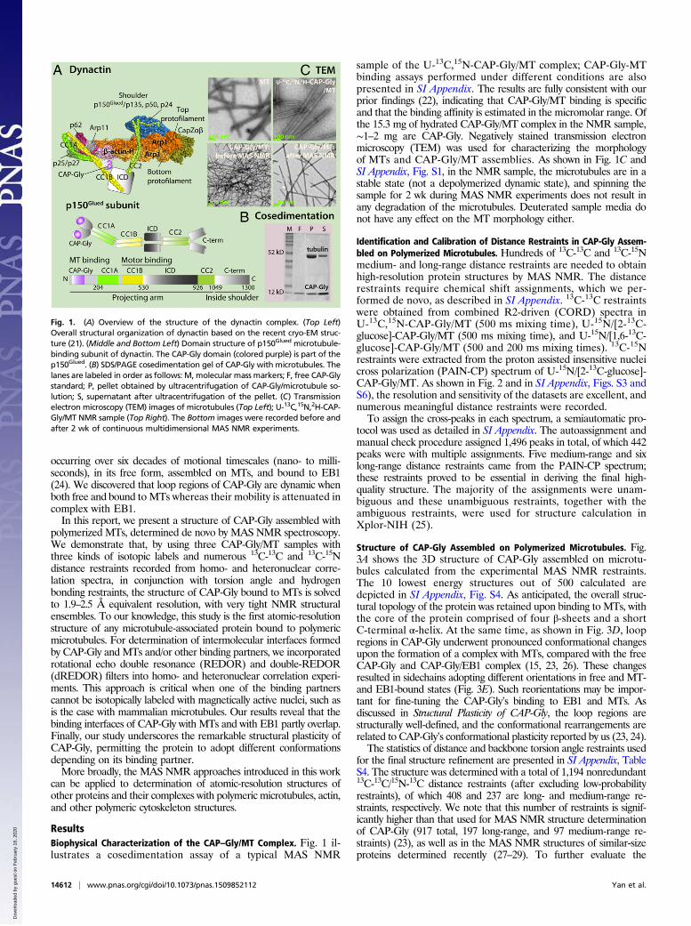
ResultsBiophysical Characterization of the CAP–Gly/MT Complex. Fig. 1 il-lustrates a cosedimentation assay of a typical MAS NMR
sample of the U-13C,15N-CAP-Gly/MT complex; CAP-Gly-MTbinding assays performed under different conditions are alsopresented in SI Appendix. The results are fully consistent with ourprior findings (22), indicating that CAP-Gly/MT binding is specificand that the binding affinity is estimated in the micromolar range. Ofthe 15.3 mg of hydrated CAP-Gly/MT complex in the NMR sample,∼1–2 mg are CAP-Gly. Negatively stained transmission electronmicroscopy (TEM) was used for characterizing the morphologyof MTs and CAP-Gly/MT assemblies. As shown in Fig. 1C andSI Appendix, Fig. S1, in the NMR sample, the microtubules are in astable state (not a depolymerized dynamic state), and spinning thesample for 2 wk during MAS NMR experiments does not result inany degradation of the microtubules. Deuterated sample media donot have any effect on the MT morphology either.
Identification and Calibration of Distance Restraints in CAP-Gly Assem-bled on Polymerized Microtubules. Hundreds of 13C-13C and 13C-15Nmedium- and long-range distance restraints are needed to obtainhigh-resolution protein structures by MAS NMR. The distancerestraints require chemical shift assignments, which we per-formed de novo, as described in SI Appendix. 13C-13C restraintswere obtained from combined R2-driven (CORD) spectra inU-13C,15N-CAP-Gly/MT (500 ms mixing time), U-15N/[2-13C-glucose]-CAP-Gly/MT (500 ms mixing time), and U-15N/[1,6-13C-glucose]-CAP-Gly/MT (500 and 200 ms mixing times). 13C-15Nrestraints were extracted from the proton assisted insensitive nucleicross polarization (PAIN-CP) spectrum of U-15N/[2-13C-glucose]-CAP-Gly/MT. As shown in Fig. 2 and in SI Appendix, Figs. S3 andS6), the resolution and sensitivity of the datasets are excellent, andnumerous meaningful distance restraints were recorded.To assign the cross-peaks in each spectrum, a semiautomatic pro-
tocol was used as detailed in SI Appendix. The autoassignment andmanual check procedure assigned 1,496 peaks in total, of which 442peaks were with multiple assignments. Five medium-range and sixlong-range distance restraints came from the PAIN-CP spectrum;these restraints proved to be essential in deriving the final high-quality structure. The majority of the assignments were unam-biguous and these unambiguous restraints, together with theambiguous restraints, were used for structure calculation inXplor-NIH (25).
Structure of CAP-Gly Assembled on Polymerized Microtubules. Fig.3A shows the 3D structure of CAP-Gly assembled on microtu-bules calculated from the experimental MAS NMR restraints.The 10 lowest energy structures out of 500 calculated aredepicted in SI Appendix, Fig. S4. As anticipated, the overall struc-tural topology of the protein was retained upon binding to MTs, withthe core of the protein comprised of four β-sheets and a shortC-terminal α-helix. At the same time, as shown in Fig. 3D, loopregions in CAP-Gly underwent pronounced conformational changesupon the formation of a complex with MTs, compared with the freeCAP-Gly and CAP-Gly/EB1 complex (15, 23, 26). These changesresulted in sidechains adopting different orientations in free and MT-and EB1-bound states (Fig. 3E). Such reorientations may be impor-tant for fine-tuning the CAP-Gly’s binding to EB1 and MTs. Asdiscussed in Structural Plasticity of CAP-Gly, the loop regions arestructurally well-defined, and the conformational rearrangements arerelated to CAP-Gly’s conformational plasticity reported by us (23, 24).The statistics of distance and backbone torsion angle restraints used
for the final structure refinement are presented in SI Appendix, TableS4. The structure was determined with a total of 1,194 nonredundant13C-13C/15N-13C distance restraints (after excluding low-probabilityrestraints), of which 408 and 237 are long- and medium-range re-straints, respectively. We note that this number of restraints is signif-icantly higher than that used for MAS NMR structure determinationof CAP-Gly (917 total, 197 long-range, and 97 medium-range re-straints) (23), as well as in the MAS NMR structures of similar-sizeproteins determined recently (27–29). To further evaluate the
Fig. 1. (A) Overview of the structure of the dynactin complex. (Top Left)Overall structural organization of dynactin based on the recent cryo-EM struc-ture (21). (Middle and Bottom Left) Domain structure of p150Glued microtubule-binding subunit of dynactin. The CAP-Gly domain (colored purple) is part of thep150Glued. (B) SDS/PAGE cosedimentation gel of CAP-Gly with microtubules. Thelanes are labeled in order as follows: M, molecular mass markers; F, free CAP-Glystandard; P, pellet obtained by ultracentrifugation of CAP-Gly/microtubule so-lution; S, supernatant after ultracentrifugation of the pellet. (C) Transmissionelectron microscopy (TEM) images of microtubules (Top Left); U-13C,15N,2H-CAP-Gly/MT NMR sample (Top Right). The Bottom images were recorded before andafter 2 wk of continuous multidimensional MAS NMR experiments.
14612 | www.pnas.org/cgi/doi/10.1073/pnas.1509852112 Yan et al.
Dow
nloa
ded
by g
uest
on
Feb
ruar
y 16
, 202
0
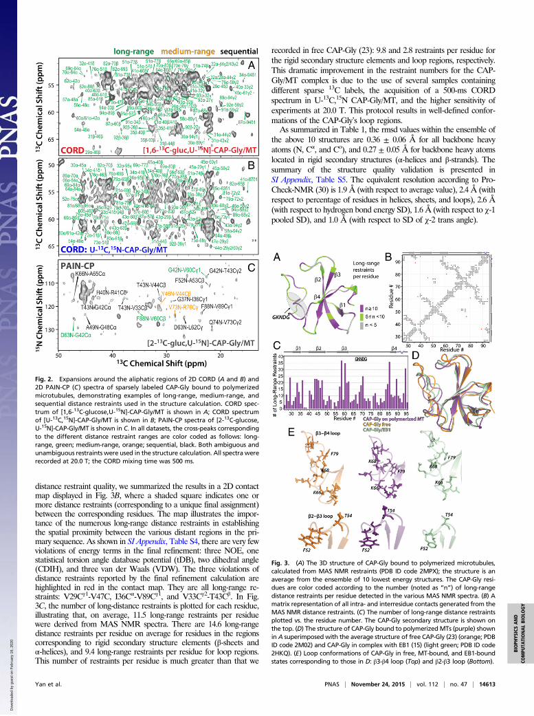
distance restraint quality, we summarized the results in a 2D contactmap displayed in Fig. 3B, where a shaded square indicates one ormore distance restraints (corresponding to a unique final assignment)between the corresponding residues. The map illustrates the impor-tance of the numerous long-range distance restraints in establishingthe spatial proximity between the various distant regions in the pri-mary sequence. As shown in SI Appendix, Table S4, there are very fewviolations of energy terms in the final refinement: three NOE, onestatistical torsion angle database potential (tDB), two dihedral angle(CDIH), and three van der Waals (VDW). The three violations ofdistance restraints reported by the final refinement calculation arehighlighted in red in the contact map. They are all long-range re-straints: V29Cγ1-V47C, I36Cα-V89Cγ1, and V33Cγ2-T43Cβ. In Fig.3C, the number of long-distance restraints is plotted for each residue,illustrating that, on average, 11.5 long-range restraints per residuewere derived from MAS NMR spectra. There are 14.6 long-rangedistance restraints per residue on average for residues in the regionscorresponding to rigid secondary structure elements (β-sheets andα-helices), and 9.4 long-range restraints per residue for loop regions.This number of restraints per residue is much greater than that we
recorded in free CAP-Gly (23): 9.8 and 2.8 restraints per residue forthe rigid secondary structure elements and loop regions, respectively.This dramatic improvement in the restraint numbers for the CAP-Gly/MT complex is due to the use of several samples containingdifferent sparse 13C labels, the acquisition of a 500-ms CORDspectrum in U-13C,15N CAP-Gly/MT, and the higher sensitivity ofexperiments at 20.0 T. This protocol results in well-defined confor-mations of the CAP-Gly’s loop regions.As summarized in Table 1, the rmsd values within the ensemble of
the above 10 structures are 0.36 ± 0.06 Å for all backbone heavyatoms (N, Cα, and Co), and 0.27 ± 0.05 Å for backbone heavy atomslocated in rigid secondary structures (α-helices and β-strands). Thesummary of the structure quality validation is presented inSI Appendix, Table S5. The equivalent resolution according to Pro-Check-NMR (30) is 1.9 Å (with respect to average value), 2.4 Å (withrespect to percentage of residues in helices, sheets, and loops), 2.6 Å(with respect to hydrogen bond energy SD), 1.6 Å (with respect to χ-1pooled SD), and 1.0 Å (with respect to SD of χ-2 trans angle).
Fig. 2. Expansions around the aliphatic regions of 2D CORD (A and B) and2D PAIN-CP (C) spectra of sparsely labeled CAP-Gly bound to polymerizedmicrotubules, demonstrating examples of long-range, medium-range, andsequential distance restraints used in the structure calculation. CORD spec-trum of [1,6-13C-glucose,U-15N]-CAP-Gly/MT is shown in A; CORD spectrumof [U-13C,15N]-CAP-Gly/MT is shown in B; PAIN-CP spectra of [2-13C-glucose,U-15N]-CAP-Gly/MT is shown in C. In all datasets, the cross-peaks correspondingto the different distance restraint ranges are color coded as follows: long-range, green; medium-range, orange; sequential, black. Both ambiguous andunambiguous restraints were used in the structure calculation. All spectra wererecorded at 20.0 T; the CORD mixing time was 500 ms.
Fig. 3. (A) The 3D structure of CAP-Gly bound to polymerized microtubules,calculated from MAS NMR restraints (PDB ID code 2MPX); the structure is anaverage from the ensemble of 10 lowest energy structures. The CAP-Gly resi-dues are color coded according to the number (noted as “n”) of long-rangedistance restraints per residue detected in the various MAS NMR spectra. (B) Amatrix representation of all intra- and interresidue contacts generated from theMAS NMR distance restraints. (C) The number of long-range distance restraintsplotted vs. the residue number. The CAP-Gly secondary structure is shown onthe top. (D) The structure of CAP-Gly bound to polymerizedMTs (purple) shownin A superimposed with the average structure of free CAP-Gly (23) (orange; PDBID code 2M02) and CAP-Gly in complex with EB1 (15) (light green; PDB ID code2HKQ). (E) Loop conformations of CAP-Gly in free, MT-bound, and EB1-boundstates corresponding to those in D: β3-β4 loop (Top) and β2-β3 loop (Bottom).
Yan et al. PNAS | November 24, 2015 | vol. 112 | no. 47 | 14613
BIOPH
YSICSAND
COMPU
TATIONALBIOLO
GY
Dow
nloa
ded
by g
uest
on
Feb
ruar
y 16
, 202
0
Intermolecular Interface of CAP-Gly with Microtubules. Determina-tion of intermolecular interfaces formed by a biomolecule inter-acting with its binding partner by MAS NMR generally relies on (i)analysis of chemical shift perturbations or intensity changes observedfor the biomolecule in the complex with respect to the free bio-molecule (31, 32) and (ii) analysis of intermolecular dipolar-basedheteronuclear correlations in differentially isotopically enriched sam-ples, where one biomolecule contains a certain set of isotope labels(e.g., 13C) and its binding partner a different kind of labels (e.g., 15N)(33–35). For analysis of the CAP-Gly/MT interface, we could not useeither approach because (i) the chemical shift perturbations are rel-atively small and present throughout the entire protein and (ii) iso-topic labeling of MTs is a challenge. One approach is to prepare theU-13C,15N,2H-CAP-Gly/MT complex and use the nonexchangeablealiphatic 1H sites on MTs for selective magnetization transfers acrossthe interface to establish correlations with heteronuclei (13C/15N) onCAP-Gly. However, this approach leads to artifacts associated with
intramolecular transfers due to residual protonation in aliphaticsidechains (SI Appendix, Figs. S7 and S8).We therefore turned our attention to the REDOR filter-based ex-
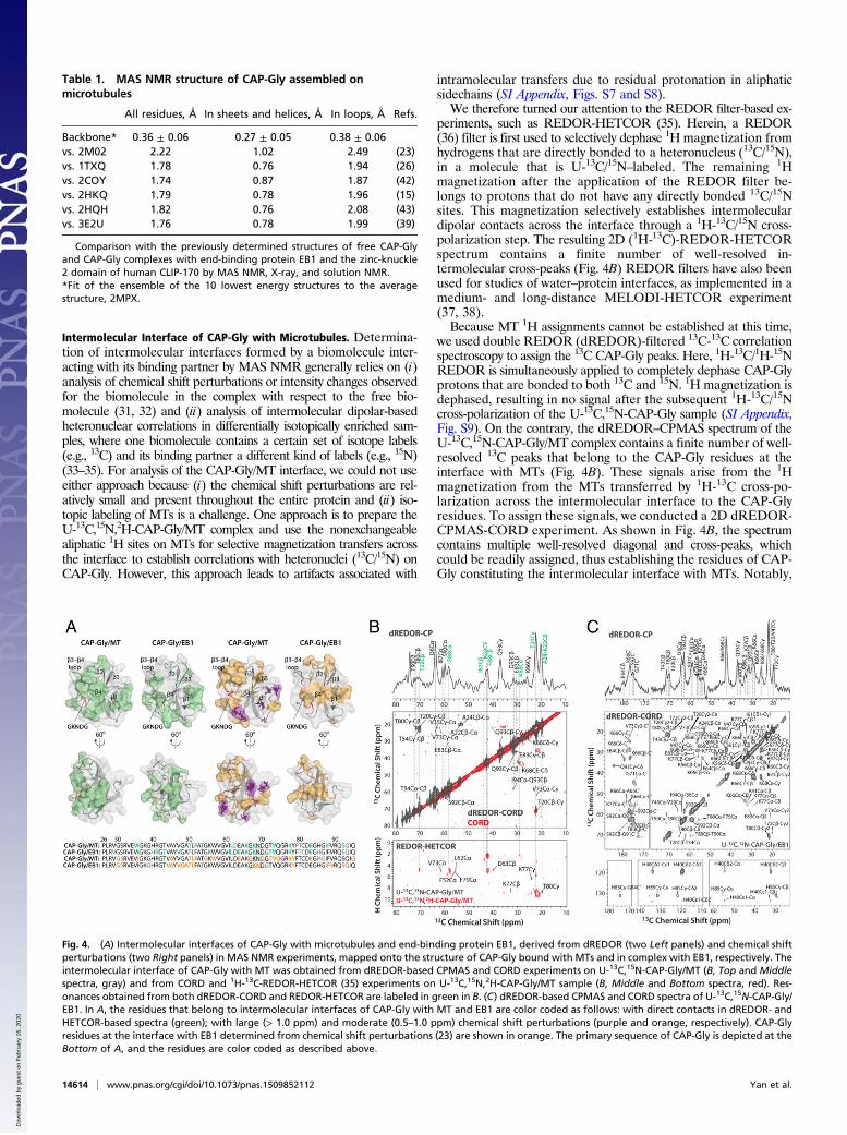
periments, such as REDOR-HETCOR (35). Herein, a REDOR(36) filter is first used to selectively dephase 1H magnetization fromhydrogens that are directly bonded to a heteronucleus (13C/15N),in a molecule that is U-13C/15N–labeled. The remaining 1Hmagnetization after the application of the REDOR filter be-longs to protons that do not have any directly bonded 13C/15Nsites. This magnetization selectively establishes intermoleculardipolar contacts across the interface through a 1H-13C/15N cross-polarization step. The resulting 2D (1H-13C)-REDOR-HETCORspectrum contains a finite number of well-resolved in-termolecular cross-peaks (Fig. 4B) REDOR filters have also beenused for studies of water–protein interfaces, as implemented in amedium- and long-distance MELODI-HETCOR experiment(37, 38).Because MT 1H assignments cannot be established at this time,
we used double REDOR (dREDOR)-filtered 13C-13C correlationspectroscopy to assign the 13C CAP-Gly peaks. Here, 1H-13C/1H-15NREDOR is simultaneously applied to completely dephase CAP-Glyprotons that are bonded to both 13C and 15N. 1H magnetization isdephased, resulting in no signal after the subsequent 1H-13C/15Ncross-polarization of the U-13C,15N-CAP-Gly sample (SI Appendix,Fig. S9). On the contrary, the dREDOR–CPMAS spectrum of theU-13C,15N-CAP-Gly/MT complex contains a finite number of well-resolved 13C peaks that belong to the CAP-Gly residues at theinterface with MTs (Fig. 4B). These signals arise from the 1Hmagnetization from the MTs transferred by 1H-13C cross-po-larization across the intermolecular interface to the CAP-Glyresidues. To assign these signals, we conducted a 2D dREDOR-CPMAS-CORD experiment. As shown in Fig. 4B, the spectrumcontains multiple well-resolved diagonal and cross-peaks, whichcould be readily assigned, thus establishing the residues of CAP-Gly constituting the intermolecular interface with MTs. Notably,
Table 1. MAS NMR structure of CAP-Gly assembled onmicrotubules
All residues, Å In sheets and helices, Å In loops, Å Refs.
Backbone* 0.36 ± 0.06 0.27 ± 0.05 0.38 ± 0.06vs. 2M02 2.22 1.02 2.49 (23)vs. 1TXQ 1.78 0.76 1.94 (26)vs. 2COY 1.74 0.87 1.87 (42)vs. 2HKQ 1.79 0.78 1.96 (15)vs. 2HQH 1.82 0.76 2.08 (43)vs. 3E2U 1.76 0.78 1.99 (39)
Comparison with the previously determined structures of free CAP-Glyand CAP-Gly complexes with end-binding protein EB1 and the zinc-knuckle2 domain of human CLIP-170 by MAS NMR, X-ray, and solution NMR.*Fit of the ensemble of the 10 lowest energy structures to the averagestructure, 2MPX.
Fig. 4. (A) Intermolecular interfaces of CAP-Gly with microtubules and end-binding protein EB1, derived from dREDOR (two Left panels) and chemical shiftperturbations (two Right panels) in MAS NMR experiments, mapped onto the structure of CAP-Gly bound with MTs and in complex with EB1, respectively. Theintermolecular interface of CAP-Gly with MT was obtained from dREDOR-based CPMAS and CORD experiments on U-13C,15N-CAP-Gly/MT (B, Top and Middlespectra, gray) and from CORD and 1H-13C-REDOR-HETCOR (35) experiments on U-13C,15N,2H-CAP-Gly/MT sample (B, Middle and Bottom spectra, red). Res-onances obtained from both dREDOR-CORD and REDOR-HETCOR are labeled in green in B. (C) dREDOR-based CPMAS and CORD spectra of U-13C,15N-CAP-Gly/EB1. In A, the residues that belong to intermolecular interfaces of CAP-Gly with MT and EB1 are color coded as follows: with direct contacts in dREDOR- andHETCOR-based spectra (green); with large (> 1.0 ppm) and moderate (0.5–1.0 ppm) chemical shift perturbations (purple and orange, respectively). CAP-Glyresidues at the interface with EB1 determined from chemical shift perturbations (23) are shown in orange. The primary sequence of CAP-Gly is depicted at theBottom of A, and the residues are color coded as described above.
14614 | www.pnas.org/cgi/doi/10.1073/pnas.1509852112 Yan et al.
Dow
nloa
ded
by g
uest
on
Feb
ruar
y 16
, 202
0
the same residues also gave rise to the cross-peaks in theREDOR-HETCOR spectrum of the U-13C,15N,2H-CAP-Gly/MTcomplex (Fig. 4B). Furthermore, in the REDOR-HETCOR spec-trum, the K68 Ce, T20 Cγ, and K77 Cγ resonances correlated tomultiple protons, which is a further indication that these residuesare in spatial proximity to tubulin. In contrast, the CORD spectrumof U-13C,15N,2H-CAP-Gly/MT complex contained additional cross-peaks that belong to intramolecular correlations among CAP-Glyresidues arising from residual protonation of the correspondingnonexchangeable sites. The residual proton signals were verifiedby the solution NMR spectra of the free U-13C,15N,2H-CAP-Gly(SI Appendix, Fig. S7).The intermolecular interface of CAP-Gly with microtubules, de-
rived on the basis of the above experiments, is illustrated in Fig. 4Aand SI Appendix, Fig. S4B. The β3-β4 loop and the GKNDG motifwithin this loop constitute the primary interface with microtubules.This finding corroborates prior hypotheses, made on the basis ofstructural analysis of CAP-Gly complexes with EB1 and CLIP170,that the GKNDG motif likely constitutes a binding interface, withMT being a specific recognition sequence for EEY/F motifs ubiq-uitously present in CLIP170 and EB proteins, as well as in α-tubulin(26, 39). Our current results also clearly indicate that the bean-shapedCAP-Gly molecule binds to the microtubules with its flat surfacecomprised by its loops, the C-terminal β4-strand, and the shortα-helix. These findings are in agreement with the recent cryo-EMresults on the CAP-Gly(1-105)/MT and CAP-Gly(25-144)/MT com-plexes (40), which revealed the same flat side of CAP-Gly that formsthe interface, including the proposed interaction of the GKNDGmotifwith the C terminus of tubulin. Of note, most of the surface-exposed,hydrophobic residues are on the flat side of CAP-Gly and are likelyimportant for promoting the binding interaction.Our results reveal that the intermolecular interfaces of CAP-Gly
with microtubules and EB1 partly overlap (β3-β4 loop and S92-Q93 athelix turn). The binding interface of CAP-Gly with EB1 is known fromprevious studies (15, 26). The dREDOR-based spectra of the U-13C,15
N-CAP-Gly/EB1 complex shown in Fig. 4C corroborate the previousfindings and validate the dREDOR approach as applied to the CAP-Gly/MT complex.As illustrated in Fig. 4A, chemical shift perturbations detected in
CAP-Gly upon formation of the complex with MTs encompassmultiple residues not found in the REDOR-based experiments.These residues are located at the N terminus, on the opposite side ofthe interface. Such perturbations are due to conformational changesin the protein residues upon binding to MTs but are not associatedwith the residues participating in the formation of the intermolecularinterface with MTs. This finding underscores the fact that, whenbinding affinity is moderate, such as in CAP-Gly/MT, binding interfaceidentification should not rely solely on chemical shift perturbations.The CAP-Gly/MT interface contacts determined by MAS NMR
are consistent with those predicted by us using molecular docking inClusPro (41). We used the recent cryo-EM structure of MTs (12)and our MAS NMR structure of CAP-Gly bound to MTs. In the fivetop-ranked electrostatically driven models, CAP-Gly binds MT in thesame orientation, at the C terminus of tubulin: The flat side (with β4at the center) faces MT, and the GKNDG motif is oriented towardthe minus end of MT (SI Appendix, Fig. S11). This predicted ori-entation and the binding contacts are in overall agreement with theinterface determined from MAS NMR. We note that K77, F79, andT80 residues that, according to dREDOR, form direct contacts withMTs are on the opposite side of CAP-Gly and do not interact withtubulin in the docking model. This result is not surprising because thenegatively charged C-terminal tail of tubulin (the E-hook), which isknown to directly interact with CAP-Gly, is missing in the cryo-EMstructure of MT, and the corresponding interactions are not capturedin the model. Our MAS NMR experiments thus detect states in-visible in cryo-EM due to conformational heterogeneity. On the basisof our results, we propose that the E-hook passes through the
hydrophobic patch at the flat binding side and stretches to the β3-β4loop of CAP-Gly to directly bind with the GKNDG motif.
Structural Plasticity of CAP-Gly.CAP-Gly possesses conformationalplasticity that is important for dynactin’s interactions with mul-tiple binding partners (23). This conformational plasticity is in-timately connected to CAP-Gly’s inherent mobility that spansmany decades of motional timescales, from nano- to milliseconds(24). CAP-Gly’s motional profile is modulated by its environ-ment, and, remarkably, both free and MT-bound proteins aredynamic in loops whereas CAP-Gly in complex with EB1 is con-siderably more rigid (24).The atomic resolution of CAP-Gly bound to polymerized mi-
crotubules presented here provides an opportunity to gain addi-tional insights into the conformational plasticity of the protein.To this end, we compared the MAS NMR structures of CAP-Glyassembled on MTs and free with the solution NMR structure of freeCAP-Gly, and the X-ray structures of CAP-Gly/EB1 and CAP-Gly/ZnCLIP complexes. Table 1 contains the summary of the results inthe form of rmsds between the different structures. The results in-dicate that, interestingly, the backbone rmsd between the MASNMR-derived structures of CAP-Gly/MT complex and free CAP-Gly is the largest. The backbone rmsd between the MAS NMRstructure of CAP-Gly/MTs and the solution NMR structure of freeCAP-Gly (1.74 Å for all residues) has similar overall value as thermsd for CAP-Gly complexes with EB1 or ZnCLIP (1.76–1.82 Åfor all residues), but significantly larger rmsd in rigid secondarystructure elements (0.87 Å vs. 0.76–0.78 Å). It is important to notethat the overall structure of the core β2-β4 sheets superimposes verywell in the different structures: free CAP-Gly, CAP-Gly assembledmicrotubules, and CAP-Gly in complex with EB1, as illustrated in Fig.3D. At the same time, the β1 conformation shows relatively largedifferences in the structures of free CAP-Gly and CAP-Gly assem-bled on MTs. Not surprisingly, the largest differences among thesestructures are found for the loop regions spanning residues D63 toG86 and containing the GKNDG motif. In the free CAP-Glystructure, the long loop extends farther away from the core formedby β2-β4. The conformation of free CAP-Gly is more open. Incontrast, upon binding to the microtubules or EB1, CAP-Gly adoptsa more closed conformation. This result is also consistent with thefact that CAP-Gly is locked into a single conformation upon bindingto EB1 or microtubules (23). Taken together, the results of this in-vestigation and our prior studies indicate that the CAP-Gly loopsreorganize for optimal interactions with microtubules and the mul-tiple dynactin’s binding partners.
DiscussionRecently, a cryo-EM study reported the structure of severalCAP-Gly constructs on polymerized MTs at 9.7–10.2 Å resolution(40). Although atomic resolution was not achieved in this study,several important observations were made: (i) The interface ofCAP-Gly with MT includes the GKNDG motif, which interactswith the C terminus of tubulin; (ii) the tubulin E-hooks interactwith the basic patches 1–25 and 106–144 of the protein, possiblypermitting it to diffuse laterally to the next binding site; (iii) theorientation of CAP-Gly on MTs depends on the length of theconstruct used; and (iv) the recognition sites for CAP-Gly andEB1 on tubulin are nonoverlapping. Interestingly, electron den-sity could not be detected for CAP-Gly(25-105), presumablydue to the weaker association with MTs compared with CAP-Gly(1-105) and CAP-Gly(25-144) and the dynamic nature of theresulting CAP-Gly(25-105)/MT complex. Furthermore, owing toits flexibility the CAP-Gly domain and the entire dynactin’sshoulder projection are not visible in the high-resolution EM mapsof the dynactin complex (21), precluding their structural analysis.Indeed, as we reported recently (24), CAP-Gly(19-107) bound topolymeric MTs is dynamic on the timescales spanning nano- tomilliseconds, and the lack of electron density in the cryo-EM
Yan et al. PNAS | November 24, 2015 | vol. 112 | no. 47 | 14615
BIOPH
YSICSAND
COMPU
TATIONALBIOLO
GY
Dow
nloa
ded
by g
uest
on
Feb
ruar
y 16
, 202
0
studies is consistent with our results. In contrast to the EM-basedstudies, MAS NMR experiments permit detection of dynamiccomplexes, as shown in our current work. Despite the dynamicnature of CAP-Gly(19-107)/MT, the atomic-resolution structure ofCAP-Gly in this complex, including the intermolecular interface,could be determined through MAS NMR.Our study revealed that CAP-Gly interacts with MTs through
its loops (including the GKNDG motif), as well as residues onthe C-terminal β4-strand and α-helix. Although we could notassign the tubulin residues interacting with CAP-Gly because atpresent we have not isotopically labeled mammalian tubulin,the topology of CAP-Gly’s interface derived on the basis of ourMAS NMR data suggests that there needs to be a flexibleregion of tubulin “wrapping around” the CAP-Gly, indirectlycorroborating the cryo-EM conclusion that tubulin’s E-hookmust be involved in the formation of the intermolecular in-terface. In the future, it will be advantageous to use a hybridapproach combining MAS NMR with cryo-EM and compu-tational analysis to gain atomic-resolution structural in-formation on both the MT-associated proteins and theirorientations on the MTs.
Concluding RemarksThe atomic-resolution structure of CAP-Gly on polymerized mi-crotubules solved by MAS NMR establishes the foundations for our
understanding of the biological function of microtubule-as-sociated proteins assembled with polymerized microtubules. Asdemonstrated in this work, MAS NMR enables investigations ofdynamic complexes between motor proteins and MTs, whichcannot be addressed by other structural biology techniques. Thestructure of dynactin’s CAP-Gly on polymerized MTs revealedstructural plasticity of the protein that is necessary for dynactin’sinteraction with multiple binding partners. In the future, we en-vision that hybrid MAS NMR/cryo-EM/computational approacheswill become essential to gain atomic-level understanding on themotor protein assemblies with microtubules.
Materials and MethodsSamples of isotopically labeled CAP-Gly(19-107) bound to polymerized MTswere prepared as reported previously (22, 24). Cosedimentation assays wereperformed, and negatively stained transmission electron micrographs wereacquired (SI Appendix). Details of MAS NMR experiments, spectral analysis,and structural calculation are described in SI Appendix.
ACKNOWLEDGMENTS. This work was supported by the National Institutesof Health (NIH) Grant R01GM085306 from the National Institute of Gen-eral Medical Sciences. We acknowledge the support of the National Sci-ence Foundation Grant CHE0959496 for the acquisition of the 850-MHzNMR spectrometer and of NIH Grants P30GM103519 and P30GM110758for the support of core instrumentation infrastructure at the Universityof Delaware.
1. Vale RD (2003) The molecular motor toolbox for intracellular transport. Cell 112(4):467–480.
2. Huszar D, Theoclitou ME, Skolnik J, Herbst R (2009) Kinesin motor proteins as targetsfor cancer therapy. Cancer Metastasis Rev 28(1-2):197–208.
3. Farrer MJ, et al. (2009) DCTN1 mutations in Perry syndrome. Nat Genet 41(2):163–165.4. Puls I, et al. (2003) Mutant dynactin in motor neuron disease. Nat Genet 33(4):455–456.5. Puls I, et al. (2005) Distal spinal and bulbar muscular atrophy caused by dynactin
mutation. Ann Neurol 57(5):687–694.6. Tanaka F, Ikenaka K, Yamamoto M, Sobue G (2012) Neuropathology and omics in
motor neuron diseases. Neuropathology 32(4):458–462.7. Ahmed S, Sun S, Siglin AE, Polenova T, Williams JC (2010) Disease-associated muta-
tions in the p150(Glued) subunit destabilize the CAP-gly domain. Biochemistry 49(25):5083–5085.
8. Chen XJ, Xu H, Cooper HM, Liu Y (2014) Cytoplasmic dynein: A key player in neuro-degenerative and neurodevelopmental diseases. Sci China Life Sci 57(4):372–377.
9. Eschbach J, et al. (2011) Mutations in cytoplasmic dynein lead to a Huntington’s dis-ease-like defect in energy metabolism of brown and white adipose tissues. BiochimBiophys Acta 1812(1):59–69.
10. Eschbach J, et al. (2013) Dynein mutations associated with hereditary motor neu-ropathies impair mitochondrial morphology and function with age. Neurobiol Dis 58:220–230.
11. Aldaz H, Rice LM, Stearns T, Agard DA (2005) Insights into microtubule nucleationfrom the crystal structure of human gamma-tubulin. Nature 435(7041):523–527.
12. Alushin GM, et al. (2014) High-resolution microtubule structures reveal the structuraltransitions in αβ-tubulin upon GTP hydrolysis. Cell 157(5):1117–1129.
13. Berezuk MA, Schroer TA (2007) Dynactin enhances the processivity of kinesin-2. Traffic8(2):124–129.
14. Fletcher DA, Mullins RD (2010) Cell mechanics and the cytoskeleton. Nature 463(7280):485–492.
15. Honnappa S, et al. (2006) Key interaction modes of dynamic +TIP networks. Mol Cell23(5):663–671.
16. Roberts AJ, Kon T, Knight PJ, Sutoh K, Burgess SA (2013) Functions and mechanics ofdynein motor proteins. Nat Rev Mol Cell Biol 14(11):713–726.
17. Roll-Mecak A, Vale RD (2006) Making more microtubules by severing: A commontheme of noncentrosomal microtubule arrays? J Cell Biol 175(6):849–851.
18. Siglin AE, et al. (2013) Dynein and dynactin leverage their bivalent character to form ahigh-affinity interaction. PLoS One 8(4):e59453.
19. Vallee RB, Williams JC, Varma D, Barnhart LE (2004) Dynein: An ancient motor proteininvolved in multiple modes of transport. J Neurobiol 58(2):189–200.
20. Yildiz A, Tomishige M, Vale RD, Selvin PR (2004) Kinesin walks hand-over-hand.Science 303(5658):676–678.
21. Urnavicius L, et al. (2015) The structure of the dynactin complex and its interactionwith dynein. Science 347(6229):1441–1446.
22. Sun S, Siglin A, Williams JC, Polenova T (2009) Solid-state and solution NMR studies ofthe CAP-Gly domain of mammalian dynactin and its interaction with microtubules.J Am Chem Soc 131(29):10113–10126.
23. Yan S, et al. (2013) Three-dimensional structure of CAP-gly domain of mammalian dy-nactin determined by magic angle spinning NMR spectroscopy: Conformational plas-ticity and interactions with end-binding protein EB1. J Mol Biol 425(22):4249–4266.
24. Yan S, et al. (2015) Internal dynamics of dynactin CAP-Gly is regulated by microtu-bules and plus end tracking protein EB1. J Biol Chem 290(3):1607–1622.
25. Schwieters CD, Kuszewski JJ, Tjandra N, Clore GM (2003) The Xplor-NIH NMR mo-lecular structure determination package. J Magn Reson 160(1):65–73.
26. Hayashi I, Wilde A, Mal TK, Ikura M (2005) Structural basis for the activation of mi-crotubule assembly by the EB1 and p150Glued complex. Mol Cell 19(4):449–460.
27. Fasshuber HK, et al. (2015) Structural heterogeneity in microcrystalline ubiquitinstudied by solid-state NMR. Protein Sci 24(5):592–598.
28. Morag O, Sgourakis NG, Baker D, Goldbourt A (2015) The NMR-Rosetta capsid modelof M13 bacteriophage reveals a quadrupled hydrophobic packing epitope. Proc NatlAcad Sci USA 112(4):971–976.
29. Demers J-P, et al. (2014) High-resolution structure of the Shigella type-III secretionneedle by solid-state NMR and cryo-electron microscopy. Nat Commun 5:4976.
30. Laskowski RA, Rullmannn JA, MacArthur MW, Kaptein R, Thornton JM (1996) AQUAand PROCHECK-NMR: Programs for checking the quality of protein structures solvedby NMR. J Biomol NMR 8(4):477–486.
31. Zech SG, Olejniczak E, Hajduk P, Mack J, McDermott AE (2004) Characterization ofprotein-ligand interactions by high-resolution solid-state NMR spectroscopy. J AmChem Soc 126(43):13948–13953.
32. Schütz AK, et al. (2011) The amyloid-Congo red interface at atomic resolution. AngewChem Int Ed Engl 50(26):5956–5960.
33. Asami S, Rakwalska-Bange M, Carlomagno T, Reif B (2013) Protein-RNA interfacesprobed by 1H-detected MAS solid-state NMR spectroscopy. Angew Chem Int Ed Engl52(8):2345–2349.
34. Weingarth M, Baldus M (2013) Solid-state NMR-based approaches for supramolecularstructure elucidation. Acc Chem Res 46(9):2037–2046.
35. Yang J, Tasayco ML, Polenova T (2008) Magic angle spinning NMR experiments forstructural studies of differentially enriched protein interfaces and protein assemblies.J Am Chem Soc 130(17):5798–5807.
36. Gullion T, Schaefer J (1989) Rotational-echo double-resonance NMR. J Magn Reson81(1):196–200.
37. Yao XL, Schmidt-Rohr K, Hong M (2001) Medium- and long-distance 1H–13C hetero-nuclear correlation NMR in solids. J Magn Reson 149(1):139–143.
38. Li S, Su Y, Luo W, Hong M (2010) Water-protein interactions of an arginine-richmembrane peptide in lipid bilayers investigated by solid-state nuclear magnetic res-onance spectroscopy. J Phys Chem B 114(11):4063–4069.
39. Weisbrich A, et al. (2007) Structure-function relationship of CAP-Gly domains. NatStruct Mol Biol 14(10):959–967.
40. Wang Q, Crevenna AH, Kunze I, Mizuno N (2014) Structural basis for the extendedCAP-Gly domains of p150(glued) binding to microtubules and the implication fortubulin dynamics. Proc Natl Acad Sci USA 111(31):11347–11352.
41. Comeau SR, Gatchell DW, Vajda S, Camacho CJ (2004) ClusPro: An automated dockingand discrimination method for the prediction of protein complexes. Bioinformatics20(1):45–50.
42. Saito K, et al. (2004) The CAP-Gly domain of CYLD associates with the proline-richsequence in NEMO/IKKgamma. Structure 12(9):1719–1728.
43. Hayashi I, Plevin MJ, Ikura M (2007) CLIP170 autoinhibition mimics intermolecularinteractions with p150Glued or EB1. Nat Struct Mol Biol 14(10):980–981.
14616 | www.pnas.org/cgi/doi/10.1073/pnas.1509852112 Yan et al.
Dow
nloa
ded
by g
uest
on
Feb
ruar
y 16
, 202
0