atlas of microscopic anatomy inter
TRANSCRIPT
8/6/2019 Atlas of Microscopic Anatomy Inter
http://slidepdf.com/reader/full/atlas-of-microscopic-anatomy-inter 1/28
ATLAS OF MICROSCOPIC ANATOMY

8/6/2019 Atlas of Microscopic Anatomy Inter
http://slidepdf.com/reader/full/atlas-of-microscopic-anatomy-inter 2/28
SPINAL CORD-Cervical region
Dorsal root: The dorsal root carries both myelinated and unmyelinated afferent fibers to thespinal cord. Each fiber is the central process of a dorsal root ganglion cell.
Posterior gray column: Long and narrow column of gray matter reaches almost to the surface
of the spinal cord. Primarily concerned with sensory input.
Anterior gray column: Short and broad column of gray matter. Concerned with motor function.
Both posterior and anterior gray columns are sites where sensory and motor cell bodies,respectively, are found.
Ventral root: Bundle of somatic motor fibers (axons of somatic motor neurons) and
preganglionic fibers (axons of autonomic motor neurons). Constitute the efferent outflow of thespinal cord.
Anterior median fissure: About 3 mm deep. Contains blood vessels (anterior spinal artery)
supplying the anterior two thirds of the cord.
Anterior lateral sulcus: Site of exit of ventral root. Hardly distinguishable in this preparation.
Anterior funiculus: Between the anterior median fissure and anterolateral sulcus (ventral root).
Merges with the lateral funiculus. Contains ascending and descending tracts.
Lateral funiculus: Between the dorsal and ventral roots. Merges with the anterior funiculus.Contains ascending and descending tracts.
Posterior lateral sulcus: Site of entry of dorsal root.
8/6/2019 Atlas of Microscopic Anatomy Inter
http://slidepdf.com/reader/full/atlas-of-microscopic-anatomy-inter 3/28
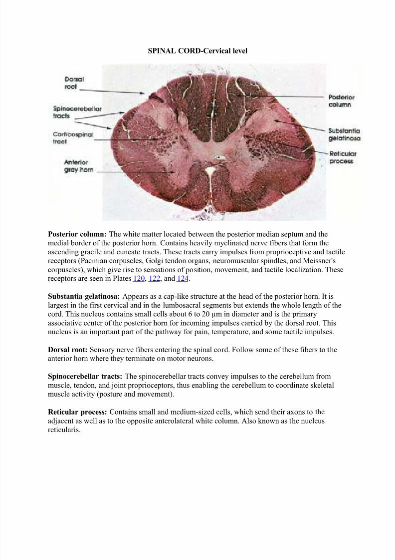
SPINAL CORD-Cervical level
Posterior column: The white matter located between the posterior median septum and themedial border of the posterior horn. Contains heavily myelinated nerve fibers that form the
ascending gracile and cuneate tracts. These tracts carry impulses from proprioceptive and tactilereceptors (Pacinian corpuscles, Golgi tendon organs, neuromuscular spindles, and Meissner's
corpuscles), which give rise to sensations of position, movement, and tactile localization. Thesereceptors are seen in Plates 120, 122, and 124.
Substantia gelatinosa: Appears as a cap-like structure at the head of the posterior horn. It islargest in the first cervical and in the lumbosacral segments but extends the whole length of thecord. This nucleus contains small cells about 6 to 20 µm in diameter and is the primary
associative center of the posterior horn for incoming impulses carried by the dorsal root. Thisnucleus is an important part of the pathway for pain, temperature, and some tactile impulses.
Dorsal root: Sensory nerve fibers entering the spinal cord. Follow some of these fibers to theanterior horn where they terminate on motor neurons.
Spinocerebellar tracts: The spinocerebellar tracts convey impulses to the cerebellum from
muscle, tendon, and joint proprioceptors, thus enabling the cerebellum to coordinate skeletal
muscle activity (posture and movement).
Reticular process: Contains small and medium-sized cells, which send their axons to the
adjacent as well as to the opposite anterolateral white column. Also known as the nucleusreticularis.

8/6/2019 Atlas of Microscopic Anatomy Inter
http://slidepdf.com/reader/full/atlas-of-microscopic-anatomy-inter 4/28
SPINAL CORD-Thoracic region
Dorsal root fibers: Central processes of dorsal root ganglia.
Reticular process: Characteristic of cervical levels of the spinal cord but also located at other
spinal levels. Located between the posterior and anterior horns and produced by an extension of gray matter into the adjacent white substance. Constitutes the lateral zone of lamina V of Rexed.
Nucleus dorsallis: Distinct nuclear mass located in the medial part of the base of the posterior
horn. In this nucleus, dorsal root fibers synapse with neurons destined to form the dorsal(posterior) spinocerebellar tract. The nucleus extends between C8 and L2 spinal segments. Also
known as the column of Clarke*.
Posterior funiculus: The white matter of the cord located between the posterior central (median)
septum and the medial border of the posterior horn. Contains heavily myelinated fibers that formthe gracile and cuneate tracts. Note the large size of this funiculus at this level compared to lower
levels of the spinal cord.
Posterior gray horn: A mass of neurons in the posterolateral part of the spinal cord. Receivecollaterals or terminals of dorsal root fibers. Sends axons to anterior horn cells, interneurons, or
to ascending tracts.

8/6/2019 Atlas of Microscopic Anatomy Inter
http://slidepdf.com/reader/full/atlas-of-microscopic-anatomy-inter 5/28
SPINAL CORD-Lumbar region
Dorsal roots: Central processes of dorsal root ganglion cells. Convey afferent (sensory)impulses to the spinal cord from peripheral receptor organs.
Substantia gelatinosa: Cap-like structure at the head of the posterior horn. Extends the whole
length of the cord. Contains small neurons about 6 to 20 µm in diameter and is the primaryassociative center of the posterior horn for incoming impulses carried by the dorsal root. This
nuclear mass is an important part of the pathway for pain, temperature, and some tactileimpulses.
Dorsal root fiber collaterals: Heavily myelinated dorsal root fiber collaterals that enter the
spinal cord to modify pain transmission or establish segmental reflexes.
Anterior funiculus: Between the anterior median fissure and anterolateral sulcus (ventral roots).
Merges with the lateral funiculus. Contains several ascending and descending tracts.
Ventral roots: Bundles of somatic motor fibers (axons of somatic motor neurons in the anterior horn). Constitute the efferent outflow of the spinal cord.
Anterior gray horn: A mass of large multipolar motor neurons and interneurons. Axons of
motor neurons form the ventral roots. Compare size of anterior gray horn at this level with thoseseen at higher and lower levels.
Lateral funiculus: Between the dorsal and ventral roots. Merges with the anterior funiculus.Contains major ascending and descending fiber tracts, including the lateral corticospinal,
spinothalamic, and spinocerebellar tracts.

8/6/2019 Atlas of Microscopic Anatomy Inter
http://slidepdf.com/reader/full/atlas-of-microscopic-anatomy-inter 6/28
SPINAL CORD-Sacral region
Dorsal root fibers: Bundles of heavily myelinated nerve fibers entering the spinal cord.Represent central processes of dorsal root ganglion neurons. Convey afferent impulses from
peripheral organs to the spinal cord. Some of these fibers go directly to form tracts (fasciculusgracilis), others give collaterals or terminate on neurons in the spinal cord.
Zone of Lissauer*: Also known as fasciculus dorsolateralis. Composed of fine myelinated andnon- myelinated fibers that carry pain, thermal, and light touch impulses or that interconnect
different levels of the substantia gelatinosa.
Ventral root fibers: Axons of somatic, fusimotor, and visceral motor neurons in the anterior (ventral) and lateral gray columns. Heavily myelinated.
Fasciculus gracilis: Heavily myelinated ascending fiber system. Conveys kinesthetic sense anddiscriminative touch. Note the absence of the fasciculus cuneatus, which appears at spinal cord
levels above T6.
Substantia gelatinosa: An expanded cell mass that forms the cap of the posterior gray horn of the spinal cord. Its size is related to that of the dorsal root. This area functions as an association
region for incoming impulses. This region corresponds to lamina II of Rexed.
Central canal: Runs throughout the length of the cord. Partially obliterated in the adult.
8/6/2019 Atlas of Microscopic Anatomy Inter
http://slidepdf.com/reader/full/atlas-of-microscopic-anatomy-inter 7/28
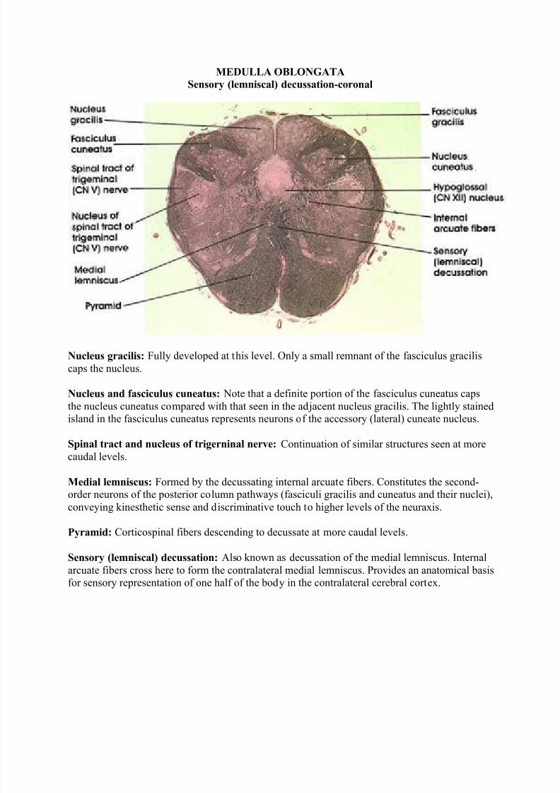
MEDULLA OBLONGATA
Sensory (lemniscal) decussation-coronal
Nucleus gracilis: Fully developed at this level. Only a small remnant of the fasciculus gracilis
caps the nucleus.
Nucleus and fasciculus cuneatus: Note that a definite portion of the fasciculus cuneatus caps
the nucleus cuneatus compared with that seen in the adjacent nucleus gracilis. The lightly stained
island in the fasciculus cuneatus represents neurons of the accessory (lateral) cuneate nucleus.
Spinal tract and nucleus of trigerninal nerve: Continuation of similar structures seen at more
caudal levels.
Medial lemniscus: Formed by the decussating internal arcuate fibers. Constitutes the second-order neurons of the posterior column pathways (fasciculi gracilis and cuneatus and their nuclei),
conveying kinesthetic sense and discriminative touch to higher levels of the neuraxis.
Pyramid: Corticospinal fibers descending to decussate at more caudal levels.
Sensory (lemniscal) decussation: Also known as decussation of the medial lemniscus. Internalarcuate fibers cross here to form the contralateral medial lemniscus. Provides an anatomical basisfor sensory representation of one half of the body in the contralateral cerebral cortex.

8/6/2019 Atlas of Microscopic Anatomy Inter
http://slidepdf.com/reader/full/atlas-of-microscopic-anatomy-inter 8/28
MEDULLA OBLONGATA
Inferior olive-coronal
Nucleus gracilis: Fully developed at this level. Only a small remnant of the fasciculus graciliscaps the nucleus.
Cuneate nucleus and tract: Note that a definite portion of the fasciculus cuneatus caps thenucleus cuneatus as compared to that seen on the adjacent nucleus gracilis. The lightly stained
island in the fasciculus cuneatus represents neurons of the accessory cuneate nucleus.
Spinal tract and nucleus of nerve V: Continuation of similar structures seen at more caudal
Medial longitudinal fasciculus: Note the change of position of the fasciculus in this figure ascompared to a more caudal level (see Plate 328 ). This is a result of the formation of the medial
lemniscus, which displaces the medial longitudinal fasciculus to a more dorsal location.
Principal and medial accessory inferior olive: This nuclear group distinguishes sections of the
medulla at this level. The principal olive is the larger component with its hilum directedmedially. The medial accessory olive is found along the border of the medial lemniscus. Inferior
olive neurons give rise to olivocerebellar fibers that project into the cerebellum.
Pyramid: See the same structure at more caudal levels. Plates 324, 325, 326, 328, and 329.
Internal arcuate fibers: Axons of gracile and cuneate neurons.
Spinocerebellar tract: Continuation of the same tract seen in the spinal cord.

8/6/2019 Atlas of Microscopic Anatomy Inter
http://slidepdf.com/reader/full/atlas-of-microscopic-anatomy-inter 9/28
MEDULLA OBLONGATA
Inferior olive-coronal
Choroid plexus: Located in the caudal part of the roof of the fourth ventricle.
Inferior vestibular nucleus: One of four vestibular nuclei. Characteristically located medial anddorsal to the restiform body, and traversed by myelinated bundles.
Tractus solitarius: Contains general visceral as well as special visceral (taste) fibers from the
vagus, glossopharyngeal, and facial nerves. Fibers project onto neurons in the nucleus solitariuslocated in close proximity to the tract.
Nucleus solitarius: Located in close proximity to the tractus solitarius, from which it receivesfibers.
Hypoglossal (CN XII) nucleus: A group of large neurons located dorsal to the medial
longitudinal fasciculus in the floor of the fourth ventricle in a paramedian position. Rootlets of hypoglossal nerve course in tegmentum of medulla between the medial lemniscus and inferior
olive.
Hypoglossal (CN XII) rootlets: Coursing in the tegmentum of the medulla oblongata betweenthe medial lemniscus and the inferior olive. Exit from the ventral surface of the medulla between
the pyramid and inferior olive.

8/6/2019 Atlas of Microscopic Anatomy Inter
http://slidepdf.com/reader/full/atlas-of-microscopic-anatomy-inter 10/28
Dorsal accessory olive: A component of the inferior olivary complex located dorsal to the principal olive.
Medial longitudinal fasciculus: Descending portion of a fiber system with ascending and
descending components. Neurons of origin are from various brain stem nuclei, but with a major
vestibular component. The fibers descending in this fasciculus are destined to synapse on motor neurons in the cervical region of the spinal cord, which supply neck musculature.
Accessory cuneate nucleus: Receives fibers of the dorsal spinocerebellar tract entering thespinal cord above the eighth cervical segment. Projects to the cerebellum via the restiform body.
Restiform body: Also known as the inferior cerebellar peduncle. A compact bundle of nervefibers connecting the medulla with the cerebellum. Described first in 1695 and named by
Humphrey Ridley, an English anatomist. Tracts and fibers forming this bundle originate in themedulla and the spinal cord.
Nucleus of spinal tract of trigeminal (CN V) nerve: Receives exteroceptive fibers from theipsilateral side of the face via the spinal tract of the trigeminal nerve. Lesions result in loss of pain sensation in the ipsilateral face.
Medial lemniscus: Axons of gracile and cuneate nuclei. Forward continuation of the same
structure seen in more caudal sections.
Principal inferior olive: Located dorsal and lateral to the pyramid. Note the characteristic
convoluted appearance. The principal inferior olive is the largest component of the inferior olivary complex, which includes, in addition, the dorsal accessory inferior olive and the medial
accessory inferior olive.
Pyramid: Heavily myelinated motor fiber system. Represents descending fibers from thecerebral cortex that pass through the internal capsule, cerebral peduncle, and pons before
reaching the medullary pyramids. Fibers in the pyramid undergo partial crossing in the motor decussation to give rise to the lateral corticospinal tract. It is estimated that, in man, about one
million fibers are present in each pyramid.

8/6/2019 Atlas of Microscopic Anatomy Inter
http://slidepdf.com/reader/full/atlas-of-microscopic-anatomy-inter 11/28
MEDULLA OBLONGATA
Cochleovestibular and
Glossopharyngeal nerves
Medial longitudinal fasciculus: Descending portion of a fiber system with ascending anddescending components. Neurons of origin are from various brain stem nuclei, but with a major
vestibular component. This system is concerned with eye and neck movements. The fibers in thedescending component are destined to synapse on motor neurons in the cervical spinal cord that
supply neck musculature.
Medial lemniscus: Continuation of the same system noted in caudal sections.
Glossopharyngeal (CN IX) nerve: A mixed nerve. Characteristically enters the medulla,
inferior and medial to the restiform body.
Amiculurn olivae: A bundle of fibers surrounding the inferior olivary complex. Contains fibersthat terminate on neurons of the olivary complex.
Inferior olive: Convoluted laminae of gray matter dorsal to the pyramid. Receives fibers fromcortical and subcortical sites and projects fibers primarily to the contralateral but also to the
ipsilateral cerebellum via the restiform body. Concerned with motor control.
Pyramid: Heavily myelinated motor fiber system. Contains descending fibers from the cerebralcortex that pass through the internal capsule, cerebral peduncle, and pons before reaching the
pyramids. Fibers in the pyramid undergo partial crossing in the motor decussation caudal to thislevel.

8/6/2019 Atlas of Microscopic Anatomy Inter
http://slidepdf.com/reader/full/atlas-of-microscopic-anatomy-inter 12/28
Arcuate nucleus: Motor neurons ventral to the pyramid. Receives cortical input and projects tothe cerebellum via the stria medullaris and restiform body. Homologous to pontine nuclei.
Olivocerebellar tract: Axons of neurons in the inferior olivary complex. Fibers arise from both
olivary complexes but primarily from the contralateral complex. Destined for the cerebellum via
inferior cerebellar peduncle (restiform body). Olivocerebellar fibers constitute the major component of the restiform body.
Cochlear (CN VIII) nerve: Central processes of bipolar neurons in the spiral ganglion. Entersthe lateral surface of the pons lateral and dorsal to the restiform body. Projects upon the dorsal
and ventral cochlear nuclei. Lesions in the cochlear nerve result in ipsilateral loss of hearing.
Ventral cochlear nucleus: Located ventral and lateral to the restiform body. Receives axons of
the cochlear nerve originating in the upper turns of the cochlea.
Restiform body: Also known as inferior cerebellar peduncle. A compact bundle of nerve fibers
connecting the medulla with the cerebellum. Tracts and fibers forming this bundle originate inthe medulla and the spinal cord.
Dorsal cochlear nucleus: Characteristically located dorsal and lateral to the restiform body.Receives axons of the cochlear nerve originating in the lower turns of the cochlea.
Inferior vestibular nucleus: One of four vestibular nuclei. Characteristically located medial tothe restiform body and traversed by myelinated bundles.
Stria medullaris: Axons of arcuate neurons. Courses in the floor of the fourth ventricle. Joins
the restiform body to reach the cerebellum.
Medial vestibular nucleus: One of four vestibular nuclei. Characteristically located medial tothe inferior vestibular nucleus in the floor of the fourth ventricle. Axons of neurons in this
nucleus form the medial vestibulospinal tract.

8/6/2019 Atlas of Microscopic Anatomy Inter
http://slidepdf.com/reader/full/atlas-of-microscopic-anatomy-inter 13/28
PONS
Trapezoid body coronal
Vermis of the cerebellum: Overlying the fourth ventricle. Midline portion of cerebellum.
Medial longitudinal fasciculus: Continuation of the same structure seen at more caudal levels.Concerned with ocular movement in response to vestibular stimulation.
Central tegmental tract: Compact fiber bundle located dorsal to the medial lemniscus. Carriesfibers from midbrain tegmenturn, red nucleus, and periaqueductal gray matter to the inferior
olivary complex.
Superior olivary nucleus: One of the tegmental nuclei that belong to the cochlear system.Receives fibers from the trapezoid body and contributes to the formation of the lateral lemniscus.
Trapezoid body: Also known as the inferior acoustic stria. Axons of neurons in the inferior
cochlear nucleus form the trapezoid body.
Pontocerebellar tract: Axons of pontine nuclei on their way to the cerebellum via the brachiurn
pontis.
Pontine nuclei: Scattered between the descending corticospinal, corticopontine, andcorticobulbar fibers and the horizontally oriented pontocerebellar fibers. Receive input from the

8/6/2019 Atlas of Microscopic Anatomy Inter
http://slidepdf.com/reader/full/atlas-of-microscopic-anatomy-inter 14/28
cerebral cortex via the corticopontine tract and project to cerebellum via the pontocerebellar tract.
Brachium conjunctivum: Also known as the superior cerebellar peduncle. Most important
efferent fiber system of the deep cerebellar nuclei. Located dorsolateral to the fourth ventricle.
Later in its course, it dips into the tegmenturn of the pons and midbrain. Nerve fibers in this bundle are destined to reach the contralateral red nucleus and ventrolateral nucleus of thethalamus.
Restiform body: Also known as the inferior cerebellar peduncle. Continuation of the same fiber
system seen at more caudal levels. The restiform body is shown here entering the cerebellum.
Brachium pontis: A massive bundle of fibers connecting the basal portion of the pons with the
cerebellum. Also known as the middle cerebellar peduncle. Contains pontocerebellar tract.
Spinal tract and nucleus of trigerninal nerve: Continuation of the same structures described at
caudal levels.
Medial lemniscus: Continuation of the same fiber system described at more caudal levels. Note
change in orientation of fibers from (previously) vertical in medulla oblongata to horizontal herein the pons.
Corticospinal, corticopontine, corticobulbar tracts: Descending fiber system sectionedtransversely. Destined for the pontine nuclei, cranial nerve nuclei, and the spinal cord
motoneurons.

8/6/2019 Atlas of Microscopic Anatomy Inter
http://slidepdf.com/reader/full/atlas-of-microscopic-anatomy-inter 15/28
PONSFacial and abducens
Genu of facial (CN VII) nerve: A bundle of facial nerve fibers in the floor of the fourth
ventricle.
Brachium conjunctivum: Also known as the superior cerebellar peduncle. Most important
efferent fiber system of the deep cerebellar nuclei. Located clorsolateral to the fourth ventricle.
Superior vestibular nucleus: Located dorsal and medial to the restiform body. One of four
vestibular nuclei. Receives fibers from the vestibular component of the vestibulocochlear (CNVIII) nerve and projects fibers to the cerebellum via the restiform body and to nuclei of
extraocular movement via the medial longitudinal fasciculus.
Facial (CN VII) nerve: Coursing ventrolaterally to emerge at the lateral border of the pons.
Facial nucleus: Located medial to the facial nerve. Axons of neurons in the facial nucleuscourse medially and dorsally to reach the floor of the fourth ventricle (genu of facial nerve)
before turning laterally and ventrally to exit from the lateral surface of the pons.
Abducens nerve: Rootlets of the abducens nerve are seen coursing in the tegmenturn of the
pons. They arise from the medial aspect of the nucleus and exit from the ventral surface at thecaudal border of the pons. Supply the lateral rectus muscle of the eye.

8/6/2019 Atlas of Microscopic Anatomy Inter
http://slidepdf.com/reader/full/atlas-of-microscopic-anatomy-inter 16/28
Medial lemniscus: Continuation of the same structure seen at more caudal and more rostrallevels.
Pontocerebellar tract: Continuation of the same structure seen at more caudal levels. Axons of
pontine nuclei destined for the cerebellum.
Pontine nuclei: Scattered between pontocerebellar fibers and the corticospinal, corticopontine,and corticobulbar fibers. Relay station between the cerebral cortex and cerebellum.
Corticospinal, corticopontine, corticobulbar tracts: Long descending fiber system originatingin the cerebral cortex. Sectioned transversely as it passes through the basal portion of the pons.
Abducens nucleus: Located in a paramedian position in the floor of the fourth ventricle. Axons
of neurons in this nucleus emerge from the medial aspect of the nucleus to form the abducensnerve. Lesions of the abducens nucleus result in ipsilateral paralysis of lateral gaze. The
abducens nucleus and the adjacent genu of facial nerve together form the facial colliculus, a
paramedian elevation in the floor of the fourth ventricle.
Brachium pontis: A massive bundle of fibers connecting the basal portion of the pons with the
cerebellum. Also known as the middle cerebellar peduncle.
Restiform body: Continuation of the same structure seen at more caudal levels. Seen entering
the cerebellum.
Dentate nucleus: The largest of the deep cerebellar nuclei. Axons of this nucleus are major components of the brachium conjunctivum.
Facial colliculus: A paramedian elevation in the floor of the fourth ventricle overlying theabducens nucleus and the genu of the facial nerve.

8/6/2019 Atlas of Microscopic Anatomy Inter
http://slidepdf.com/reader/full/atlas-of-microscopic-anatomy-inter 17/28
PONS
Trigeminal nerve coronal
Fourth ventricle: The anterior part of the fourth ventricle overlying the pons.
Brachium conjunctivum: Massive outflow tract from the cerebellum seen at this level prior to
decussation. Lesions in this area will result in a disorder of coordinated movement. Note thechange in position of this structure in more rostral sections.
Principal (main) sensory nucleus of trigerninal (CN V) nerve: Located lateral to the motor
nucleus of the trigeminal. Receives touch sensations from the ipsilateral face via the trigeminalnerve.
Motor nucleus of trigerninal (CN V) nerve: Located in the dorsal part of the tegmentum.
Axons form the motor root of the trigerninal nerve and supply muscles of mastication, and thetensor tympani, tensor palati, mylohyoid, and the anterior belly of the digastric muscles.
Brachium pontis: Also known as the middle cerebellar peduncle. A massive bundle of fibersconnecting the basal portion of the pons with the cerebellum. Contains pontocerebellar fibers.
Pontocerebellar tract: Axons of pontine nuclei destined for the cerebellum via the brachium
pontis.

8/6/2019 Atlas of Microscopic Anatomy Inter
http://slidepdf.com/reader/full/atlas-of-microscopic-anatomy-inter 18/28
Corticospinal, corticopontine, corticobulbar tracts: Long descending fiber system originatingin the cerebral cortex. Sectioned transversely as it passes through the basal portion of the pons.
Note the horizontally oriented pontocerebellar tract.
Medial lemniscus: Continuation of the same system seen in more caudal levels.
Trigeminal nerve: Sensory-motor cranial nerve. Seen coursing in the lateral part of the pons.
Central tegmental tract: Compact fiber bundle located in the tegmenturn of the pons. Carries
fibers from midbrain tegmentum, red nucleus, and periaqueductal gray matter to the inferior olivary complex.
Medial longitudinal fasciculus: Ascending component of a fiber system originating in
vestibular nuclei and destined to synapse with neurons in nuclei of extraocular movement (CNIII, IV, and VI). Concerned with control of eye movement.
Superior medullary velum: Forms the anterior (superior) part of the roof of the fourth ventricle.

8/6/2019 Atlas of Microscopic Anatomy Inter
http://slidepdf.com/reader/full/atlas-of-microscopic-anatomy-inter 19/28
PONS
Trigeminal nerve coronal
Brachium conjunctivum: Also known as superior cerebellar peduncle. Contains axons of deepcerebellar nuclei destined for the red nucleus and thalamus. Forms part of the lateral wall of the
fourth ventricle.
Central tegmental tract: A compact fiber bundle located dorsal to the lateral part of the medial
lemniscus. Carries fibers from midbrain tegmentum, red nucleus, and periaqueductal gray matter to the inferior olivary complex.
Brachium pontis: Also known as the middle cerebellar peduncle. Massive bundle of fibers
connecting the basal portion of the pons with the cerebellum. Contains pontocerebellar fibersfrom the contralateral half of the pons. Some pontocerebellar fibers from the ipsilateral half of
the pons are also contained in the brachium pontis.
Trigeminal (CN V) nerve: A mixed nerve with a larger sensory component (portio major) and a
smaller motor component (portio minor).
Pontine nuclei: Located in the basal part of the pons. Continuous caudally with arcuate nuclei inthe medulla oblongata. Receive corticofugal fibers and project (pontocerebellar tract) mainly to
the contralateral cerebellum.
Corticospinal, corticopontine, corticobulbar tracts: A long descending fiber system sectionedtransversely as it courses through the basal part of the pons.

8/6/2019 Atlas of Microscopic Anatomy Inter
http://slidepdf.com/reader/full/atlas-of-microscopic-anatomy-inter 20/28
Pontocerebellar tract: Axons of pontine nuclei destined for the cerebellum. Constitutes themajor component of the middle cerebellar peduncle (brachium pontis).
Medial lemniscus: Continuation of the same fiber system noted in several more caudal levels.
Note the change of orientation of this fiber bundle from a vertical orientation in the medulla to a
horizontal orientation at this level.
Lateral lemniscus: Continuation of trapezoid body. Conveys auditory impulses.
Medial longitudinal fasciculus: The ascending component of this fasciculus. Contains fibersfrom the vestibular nuclei destined for the nuclei of extraocular movement. Lesions of the medial
longitudinal fasciculus at this level will result in a characteristic clinical picture known asinternuclear ophthalmoplegia.

8/6/2019 Atlas of Microscopic Anatomy Inter
http://slidepdf.com/reader/full/atlas-of-microscopic-anatomy-inter 21/28
PONS-MESENCEPHALIC JUNCTION
Trochlear nerve coronal
Brachium conjunctivum: Massive outflow tract of the cerebellum. Fibers are seen just prior toand beginning decussation. Fibers project, after decussation, into the red nucleus and
ventrolateral nucleus of the thalamus. Lesions in this tract result in a disorder of coordinatedmovement.
Pontocerebellar tract: The same structure seen at more caudal levels.
Corticospinal, corticopontine, corticobulbar tracts: The same structures seen at more caudal
levels. Cut in cross section as they descend to lower caudal levels.
Pontine nuclei: Scattered between pontocerebellar fibers and the corticospinal, corticopontine,
and corticobulbar tracts.
Brachium pontis: Axons of pontine nuclei on their way to the cerebellum.
Medial lemniscus: Continuation of the same structure seen at more caudal levels.
Spinal lemniscus: Continuation of the same structure seen at more caudal levels. Contains
spinothalamic and spinotectal fibers.
Lateral lemniscus: Contains cochlear fibers. Located laterally and dorsally on its way to theinferior colliculus and medial geniculate body. Concerned with audition.
8/6/2019 Atlas of Microscopic Anatomy Inter
http://slidepdf.com/reader/full/atlas-of-microscopic-anatomy-inter 22/28
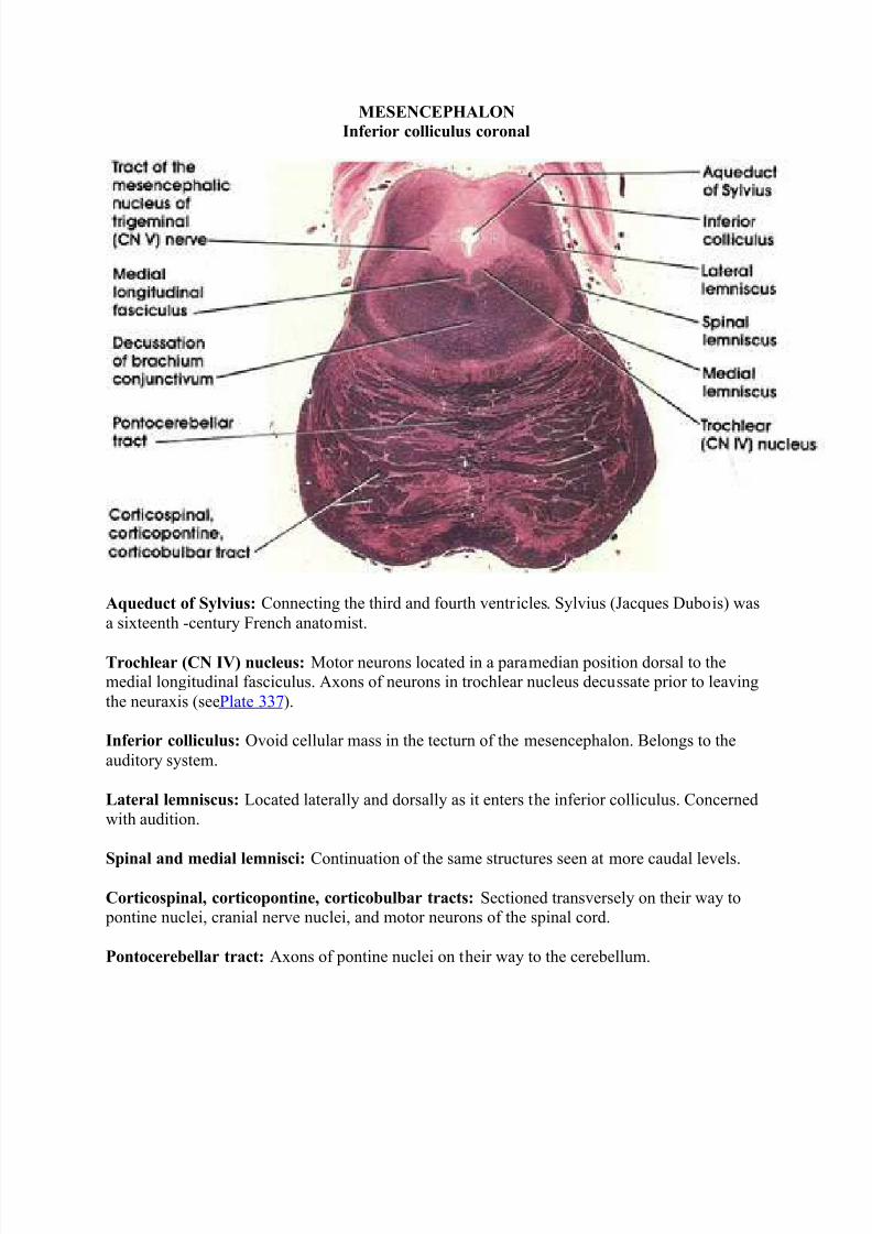
MESENCEPHALON
Inferior colliculus coronal
Aqueduct of Sylvius: Connecting the third and fourth ventricles. Sylvius (Jacques Dubois) was
a sixteenth -century French anatomist.
Trochlear (CN IV) nucleus: Motor neurons located in a paramedian position dorsal to themedial longitudinal fasciculus. Axons of neurons in trochlear nucleus decussate prior to leaving
the neuraxis (seePlate 337).
Inferior colliculus: Ovoid cellular mass in the tecturn of the mesencephalon. Belongs to the
auditory system.
Lateral lemniscus: Located laterally and dorsally as it enters the inferior colliculus. Concernedwith audition.
Spinal and medial lemnisci: Continuation of the same structures seen at more caudal levels.
Corticospinal, corticopontine, corticobulbar tracts: Sectioned transversely on their way to pontine nuclei, cranial nerve nuclei, and motor neurons of the spinal cord.
Pontocerebellar tract: Axons of pontine nuclei on their way to the cerebellum.

8/6/2019 Atlas of Microscopic Anatomy Inter
http://slidepdf.com/reader/full/atlas-of-microscopic-anatomy-inter 23/28
MESENCEPHALON-Superior colliculus
Aqueduct of Sylvius: Connecting the third and fourth ventricles.
Periaqueductal (central) gray: Surrounds the aqueduct of Sylvius. Contains neurons related to
both pain inhibition and stimulation.
Superior colliculus: Laminated cellular mass in the tecturn of the mesencephalon. Related to thevisual system.
Brachium of inferior colliculus: Also known as inferior quadrigeminal brachium. Bundle of nerve fibers from the lateral lemniscus and the inferior colliculus to the medial geniculate body.
Conveys auditory impulses to the thalamus.
Medial longitudinal fasciculus: Continuation of the same structure seen at more rostral andmore caudal levels.
Substantia nigra: Largest nuclear mass in mesencephalon. Sandwiched between the mediallemniscus and the cerebral peduncle. A mass of pigmented cells (melanin) connected with basalganglia and thalamus. Important in motor control. This area is invariably the site of pathological
changes associated with Parkinson's disease. Parkinson was an eighteenth-century English physician.
Cerebral peduncle: Descending corticofugal fiber system. Lesions here result in weakness or paralysis of the contralateral half of the body, including the face.

8/6/2019 Atlas of Microscopic Anatomy Inter
http://slidepdf.com/reader/full/atlas-of-microscopic-anatomy-inter 24/28
MESENCEPHALON-DIENCEPHALON JUNCTION
Pulvinar: Belongs to the lateral group of thalamic nuclei. Has reciprocal connections with themedial and lateral geniculate bodies caudally and the association parietal, temporal, and occipital
cortices rostrally. Plays a role in several neural functions, including vision, audition, speech, and
pain.
Lateral geniculate body: A thalamic relay nucleus concerned with vision. Receives fibers from
the optic tract and projects to the primary visual cortex.
Substantia nigra: A mass of pigmented cells containing melanin located dorsal to the cerebral peduncle. This area is invariably the site of pathologic changes associated with Parkinson's
disease.
Cerebral peduncle: Descending corticofugal fiber system. Lesion results in contralateral muscle
weakness or paralysis.
Oculomotor (CN III) nerve: Coursing in the tegmentum of the midbrain medial to thesubstantia nigra and cerebral peduncle.
Red nucleus: So-called because of a pinkish color in the fresh state owing to its high vascularity.
Links the cerebellum, cerebral cortex, and spinal cord.

8/6/2019 Atlas of Microscopic Anatomy Inter
http://slidepdf.com/reader/full/atlas-of-microscopic-anatomy-inter 25/28
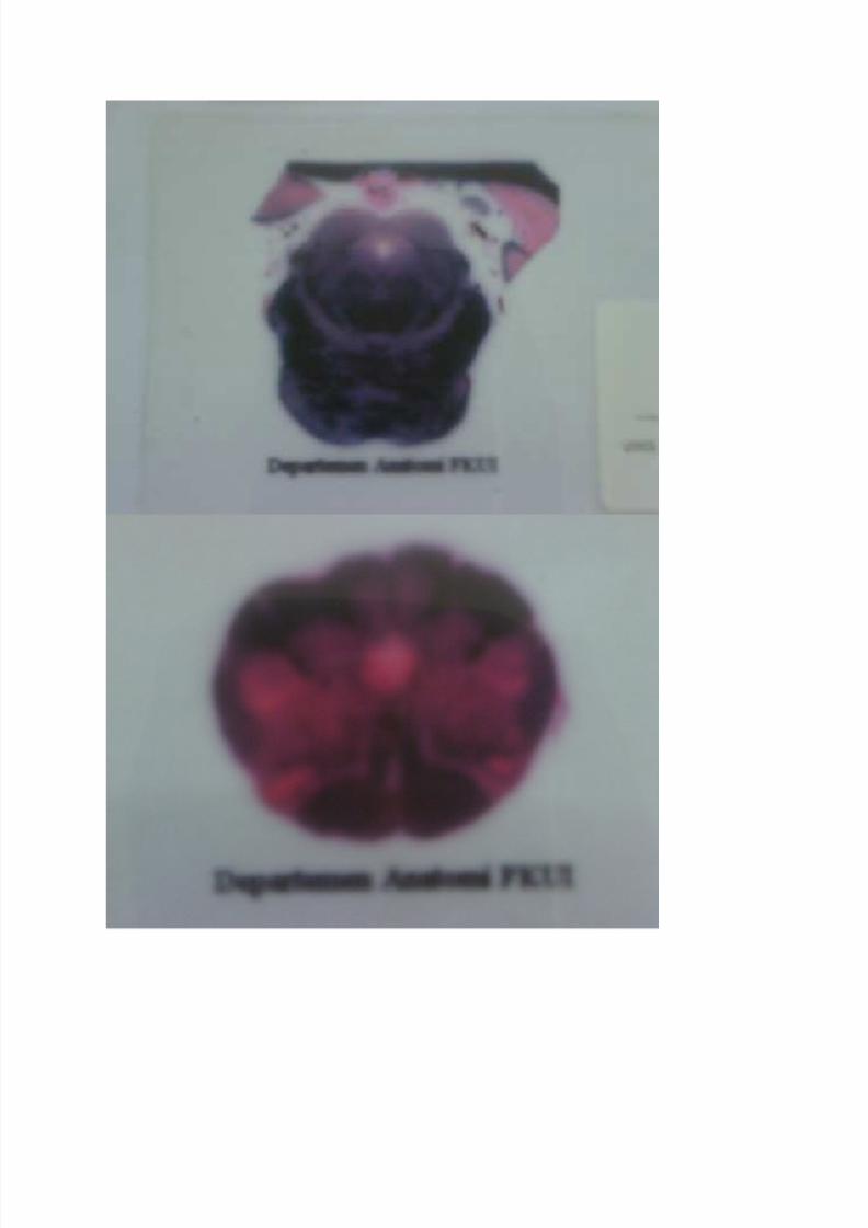
Nah, ini gw dapet weigert slides dari xxxxx..
Silahkan cocokkan ya!!

8/6/2019 Atlas of Microscopic Anatomy Inter
http://slidepdf.com/reader/full/atlas-of-microscopic-anatomy-inter 26/28
8/6/2019 Atlas of Microscopic Anatomy Inter
http://slidepdf.com/reader/full/atlas-of-microscopic-anatomy-inter 27/28

8/6/2019 Atlas of Microscopic Anatomy Inter
http://slidepdf.com/reader/full/atlas-of-microscopic-anatomy-inter 28/28