at low power – vascular relationships are preserved, there
TRANSCRIPT

1

At low power – vascular relationships are preserved, there is some sinusoidal dilatation
in perivenular areas.
2
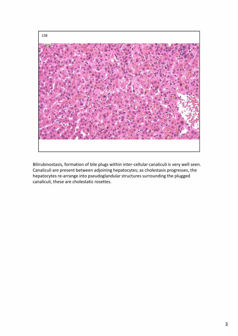
Bilirubinostasis, formation of bile plugs within inter-cellular canaliculi is very well seen.
Canaliculi are present between adjoining hepatocytes; as cholestasis progresses, the
hepatocytes re-arrange into pseudoglandular structures surrounding the plugged
canaliculi, these are cholestatic rosettes.
3

Despite the prominence of the bile stasis, there is very little evidence of hepatocyte
injury. Hepatocytes are relatively uniform in size, without swelling/ballooning, acidophil
body formation or inflammatory infiltrate. With time, PASD positive scavenger
macrophages may become more prominent.
4
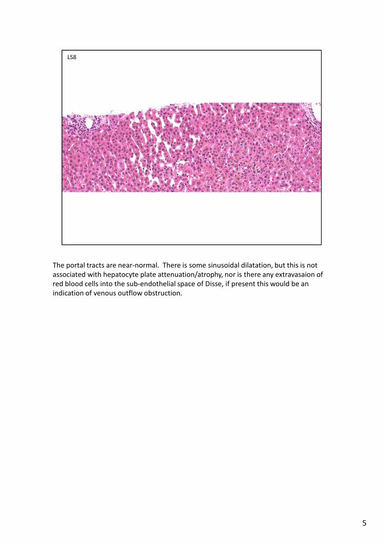
The portal tracts are near-normal. There is some sinusoidal dilatation, but this is not
associated with hepatocyte plate attenuation/atrophy, nor is there any extravasaion of
red blood cells into the sub-endothelial space of Disse, if present this would be an
indication of venous outflow obstruction.
5

6

There is no inflammatory infiltrate in the small portal tracts.
7

There is a single area of inflammation at the edge of one portal area – but this is not a
generalised feature and so is of uncertain significance and not an indication of a
‘hepatitis’.
8
9
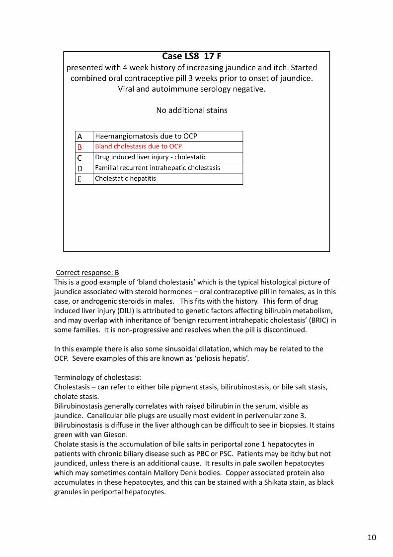
Correct response: B
This is a good example of ‘bland cholestasis’ which is the typical histological picture of
jaundice associated with steroid hormones – oral contraceptive pill in females, as in this
case, or androgenic steroids in males. This fits with the history. This form of drug
induced liver injury (DILI) is attributed to genetic factors affecting bilirubin metabolism,
and may overlap with inheritance of ‘benign recurrent intrahepatic cholestasis’ (BRIC) in
some families. It is non-progressive and resolves when the pill is discontinued.
In this example there is also some sinusoidal dilatation, which may be related to the
OCP. Severe examples of this are known as ‘peliosis hepatis’.
Terminology of cholestasis:
Cholestasis – can refer to either bile pigment stasis, bilirubinostasis, or bile salt stasis,
cholate stasis.
Bilirubinostasis generally correlates with raised bilirubin in the serum, visible as
jaundice. Canalicular bile plugs are usually most evident in perivenular zone 3.
Bilirubinostasis is diffuse in the liver although can be difficult to see in biopsies. It stains
green with van Gieson.
Cholate stasis is the accumulation of bile salts in periportal zone 1 hepatocytes in
patients with chronic biliary disease such as PBC or PSC. Patients may be itchy but not
jaundiced, unless there is an additional cause. It results in pale swollen hepatocytes
which may sometimes contain Mallory Denk bodies. Copper associated protein also
accumulates in these hepatocytes, and this can be stained with a Shikata stain, as black
granules in periportal hepatocytes.
10

Comments on other options
A Haemangiomatosis due to OCP. Haemangiomatosis is a condition where capillary
vessels have an infiltrative pattern within the liver, often also with areas of more
circumscribed haemangioma. More commonly, ‘peliosis hepatis’ can be seen in
association with the OCP – areas of wider dilatation of the sinusoidal spaces between
hepatocytes, with or without a lining endothelium.
C Drug induced liver injury – cholestatic. This is also correct, but less precise than bland
cholestasis. Most DILI have a pattern of cholestatic hepatitis, where there is clearly
lobular disarray and hepatocyte injury associated with the bilirubinostasis, suggesting
that the cholestasis is a consequence of the hepatitis injury. Some drugs can cause a
chronic cholestatic syndrome, sometimes with ductopenia. Therefore DILI – cholestatic
alone is a less complete response.
D. Familial recurrent intrahepatic cholestasis. This could have the same histological
features. But in this case the clinical history clearly indicates that the cause of the
cholestasis is the OCP.
E. Cholestatic hepatitis. A morphological description of hepatitis with cholestasis. This
usually is accompanied clinically by mixed hepatitis/cholestatic liver enzymes and of the
causes of hepatitis (autoimmune, viral, drug induced) is suggestive of DILI.
10